HAL Id: hal-01787876
https://hal.archives-ouvertes.fr/hal-01787876
Submitted on 14 May 2018HAL is a multi-disciplinary open access archive for the deposit and dissemination of sci-entific research documents, whether they are pub-lished or not. The documents may come from teaching and research institutions in France or abroad, or from public or private research centers.
L’archive ouverte pluridisciplinaire HAL, est destinée au dépôt et à la diffusion de documents scientifiques de niveau recherche, publiés ou non, émanant des établissements d’enseignement et de recherche français ou étrangers, des laboratoires publics ou privés.
REG3β modifies cell tumor function by impairing
extracellular vesicle uptake
Laia Bonjoch, Meritxell Gironella, Juan Lucio Iovanna, Daniel Closa
To cite this version:
Laia Bonjoch, Meritxell Gironella, Juan Lucio Iovanna, Daniel Closa. REG3β modifies cell tumor function by impairing extracellular vesicle uptake. Scientific Reports, Nature Publishing Group, 2017, 7 (1), �10.1038/s41598-017-03244-4�. �hal-01787876�
Reg3β modifies cell tumor function by impairing extracellular vesicle uptake
Laia Bonjoch1, Meritxell Gironella2, Juan Iovanna3, Daniel Closa1 1
Department of Experimental pathology, Institut d'Investigacions Biomèdiques de Barcelona-Consejo Superior de Investigaciones científicas (IIBB-CSIC), Institut d'Investigacions Biomèdiques August Pi i Sunyer (IDIBAPS), Barcelona, Spain; 2Department of Gastroenterology, Hospital Clínic de Barcelona, Centro de Investigación Biomédica en Red de Enfermedades Hepáticas y Digestivas (CIBEREHD), Institut d'Investigacions Biomèdiques August Pi i Sunyer (IDIBAPS), Barcelona, Spain; 3Centre de Recherche en Cancérologie de Marseille (CRCM), Institut National De La Santé Et De La Recherche Médicale (INSERM) Unit 1068, Centre National De La Recherche Scientifique (CNRS) Unit 7258, Aix-Marseille Université and Institut Paoli-Calmettes, Aix-Marseille, France
Extracellular vesicles (EV), including exosomes and microvesicles, are nano-sized membrane vesicles containing proteins and nucleic acids which act as intercellular messengers1. These EV are released most probably by all cell types and are uptaken by target cells through different mechanisms, some of them dependent on glycoproteins present in the membrane2,3. They play an important role in a variety of physiological processes as well as in pathological situations such as inflammation or cancer4. In the case of pancreatic ductal adenocarcinoma, one of the most lethal cancers, there is an increase in the concentration of circulating EV in plasma5 that has been linked to the formation of liver metastases6. The origin of these EV is not well defined, but it is supposed to be caused by the increase in the synthesis by cancer cells and other cells of the tumor stroma. Here we show that the healthy pancreatic tissue surrounding the tumor releases Reg3β, a lectin7 that binds to the glycoproteins present in the surface of EV, thus interfering with their uptake and internalization by target cells. Consequently, Reg3β prevents the changes induced by EV in the phenotype acquired by target cells and results in an increase of circulating Reg3β+ EV in the plasma of pancreatic cancer patients. Our results indicate that a higher amount of circulating EV in pancreatic cancer is not only
due to an increased production but also a lower uptake in the tumor microenvironment. This work also highlights the importance of the distant microenvironment, constituted by the healthy pancreatic tissue surrounding the tumor, in modulating the EV-mediated interactions between different cell types presents in the tumor.
Main
Extracellular vesicles (EV) play a major role in the interactions between tumor cells and extratumoral cells, including inflammatory cells. EV have been shown to regulate the tumor immune response by inducing immune tolerance to tumors8,9 but also generating anti-tumor immune responses10. Although the common explanation for these opposite results is differences in experimental designs, the interfering effect of soluble mediators must be also considered. Uptake of EV involves different protein interactions, having the lectin/glycoprotein interactions a prominent role11. This could be particularly important in pancreatic cancer since acinar cells are a well-known source of Reg3β, a soluble c-type lectin also known as PAP or HIP, released in situations of pancreatic inflammation and cell injury12.
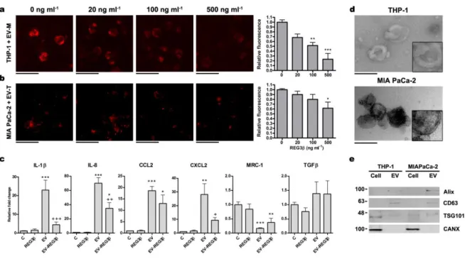
We measured the uptake of EV and found that, in macrophage-differentiated THP-1 cells, Reg3β inhibited in a dose-dependent manner the uptake of EV purified from the pancreatic cancer cell line MIAPaCa-2 (Fig. 1a). A similar result was observed on the MIAPaCa-2 uptake of EV purified from THP-1 cells (Fig. THP-1b). In the case of macrophages, this inhibition prevented the induction of the inflammatory phenotype triggered by these EV (Fig. 1c). Electron microscopy analysis of these EV revealed a size ranging from 50 to 100 nm of diameter and the presence of a lipid bilayer (Fig. 1d). The presence of exosome markers CD63, Alix and TSG101 evidenced by Western blot (Fig. 1e) indicated that this vesicular population contained exosomes.
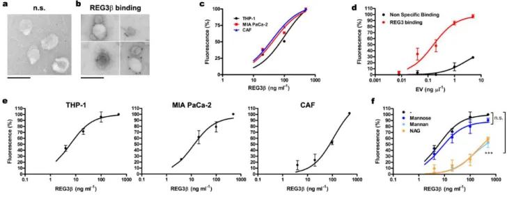
On the basis of these results we hypothesized that Reg3β binds glycoproteins that are present on the membrane of EV. This was verified by immunoelectron microscopy using a specific anti-Reg3β antibody and a 12nm gold-conjugated secondary antibody. In the absence of Reg3β very little
labelling was observed (Fig. 2a), similar to the samples from which the primary antibody had been omitted. By contrast, specific immunogold labelling was apparent on the surface of EV treated with Reg3β (Fig. 2b). This was corroborated using magnetic beads coated with anti-Reg3β antibody to purify EV stained with PKH26 dye (Fig. 2c). Notably, recovery of EV was dependent on the amount of Reg3β added to the system.
To further confirm the binding of Reg3β to EV, we designed an immunoassay experiment, coating a multiwell plate with rabbit anti-human Reg3β antibody. In a first experiment, we saturated the plate with Reg3β and increasing amounts of EV-PKH26 were added. After washing, fluorescence revealed a dose-dependent binding that achieved a maximum when the amount of EV corresponding to 1 ng µl-1 of EV protein was added (Fig. 2d). Thus, this concentration of EV was selected in the subsequent assays. In a second experiment, the amount of Reg3β added to the plate was increased from 0 to 500 ng ml-1 and fixed amounts (1 ng protein µl-1) of EV-PKH26 were added. After washing the unbound EV, fluorescence measurement showed a clear dose-response dependent on the amount of Reg3β added to the plate, indicating that the binding of EV was effectively mediated by its interaction with Reg3β (Fig. 2e). This binding was observed in EV purified from MIAPaCa-2 and THP-1, but also in EV from CAF, suggesting the unspecific nature of Reg3β-EV interaction.
The lectin nature of the interaction with Reg3β was verified by evaluating the interfering effect of different sugars on the EV-Reg3β binding. It has been reported that although Reg3β binds to complex sugars, as N-Acetyl-D-glucosamine (NAG) or mannan (a polysaccharide composed of polymerized mannose), it does not bind to monomeric mannose13. In this line, we found that binding of Reg3β to EV was significantly blocked by the presence of mannan and NAG, while mannose did not modify the binding activity (Fig. 2e).
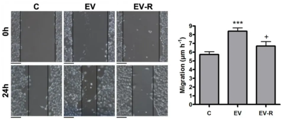
The importance of the interfering effect on EV is not restricted to the blocking effect on the activation of inflammatory cells. Since tumor cells also respond to EV, we evaluated the effect of Reg3β on the EV-induced migration of MIAPaCa-2 cells. Using a scratch-wound healing assay, we
found that Reg3β binding to EV purified from THP-1 cells prevented their stimulatory effect on the migration of tumor cells (Fig. 3).
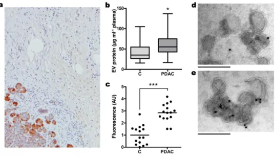
Trafficking of EV in pancreatic cancer could be affected by the presence of Reg3β, but this effect can also be conditioned by changes in the production of this protein along the progression of the disease. Although high levels of Reg3β are generated by pancreatic acinar cells, it is known that this synthesis is progressively inhibited during the progression of the pre-cancerous lesions along the PanIN stages, being completely absent in the tumor cells of pancreatic ductal adenocarcinoma14. Nevertheless, high expression of Reg3β occurs in healthy pancreatic acinar cells surrounding the tumor, being this distant microenvironment the major source of Reg3β in pancreatic cancer (Fig. 4a). It would be important to take these changes into account when interpreting or extrapolating the role of EV in pancreatic cancer.
The release of Reg3β around the tumor suggests that the uptake of EV generated inside the tumor could be impaired due to the blocking effect of this secreted lectin. Consequently, it could be expected a relevant release of Reg3β+ EV to bloodstream in pancreatic cancer. We checked the amount of circulating EV in a cohort of healthy donors and pancreatic cancer patients matched by sex and age and we effectively observed a significant increase in the number of EV released to the plasma (Fig. 4b) as expected. Then, when the same amount of EV was added to anti-Reg3β coated plates, we confirmed that the most part of Reg3β+ EV were associated with pancreatic cancer (Fig. 4c). This observation was further supported by immunogold labelling and electron microscopy image acquisition of EV from healthy donors (Fig. 4d) or pancreatic cancer patients (Fig. 4e).
Altogether indicates that, in pancreatic cancer, the physiological importance of EV could be markedly conditioned by the presence of Reg3β. It also seems reasonable to suggest that in diseases in which EV have a significant role, their effects could be modulated by using Reg3β or antibodies against Reg3β.
Methods Cells
Cell culture conditions for THP-1 and MIAPaCa-2 cells are described in Supplementary Methods.
Extracellular Vesicles isolation
The EV isolation was performed as previously described15 (see Supplementary Methods).
SDS-PAGE and Western blot
Protein extracts from EV and donor cells were run on 12% SDS-PGE gels, transferred to PVDF membranes and probed with antibodies against several EV specific markers (see Supplementary Methods for details).
Extracellular Vesicles staining
For internalization and binding assays, EV were labeled with PKH26 Red Fluorescent Cell Linker Dye (Sigma Aldrich). The staining reaction was stopped with 3% BSA for 1 min, and labeled EV (EV-PKH26) were washed three times with PBS in order to remove the unbound dye, using 300 KDa Nanosep centrifugal devices (Pall Corporation).
Extracellular Vesicles uptake
To monitor EV uptake, 3 µg ml-1 of EV-PKH26 from MIAPaCa-2 cells were added to THP-1 macrophages in the presence of different concentrations of Reg3β (0, 20, 100 and 500 ng ml-1) for 45 min. The same experiment was performed with MIAPaCa-2 cells, which were incubated with EV-PKH26 from THP-1 cells for 2 h. EV internalization was analyzed by reading the amount of fluorescence with a fluorometric plate reader (Spectramax Gemini XS) and by fluorescence microscopy.
In a second experiment, THP-1 macrophages were incubated with 3 µg ml-1 of MIAPaCa-2 EV with or without Reg3β (500 ng ml-1). After 24 h, RNA was extracted using TRizol reagent (Life Technologies).
RT-PCR and qPCR
RNA samples were quantified using a Nanodrop ND-1000 device. iScript cDNA synthesis kit (Bio Rad) and iTaq Universal SYBR Green Supermix (Bio Rad) were used for the cDNA synthesis and the qPCR reaction, respectively. Protocols and all primer sequences are included in Supplementary Methods.
Binding assay
96-well black FLUOTRAC-600 plates (Greiner Bio-One) were coated with a specific anti-human Reg3β antibody overnight at 4°C. The next day, plates were blocked with 3% BSA for 2 h at room temperature. Then, Reg3β was diluted in the blocking solution and added for 1 h, followed by three washing steps. After that, EV-PKH26 were added for 1 h in the presence of 1 mg ml-1 (5,5 mM) of mannose, 1 mg ml-1 of mannan or 5 mM of NAG, when needed. Finally, plates were washed to remove the unbound EV and read on a Spectramax Gemini XS fluorimeter. All the steps were performed with mild agitation.
SureBeads™ Protein G Magnetic Beads (Bio-Rad) were used following the supplier’s specifications. Beads were coated with 50 µg ml-1 of anti-human Reg3β antibody. Then, different concentrations of Reg3β were added and the beads were placed in a rotator for 1 h. After washing, they were resuspended with EV-PKH26 and further incubated for 1 h in the same conditions. The EV were dissociated from the complex with an elution buffer (glycine 20 mM pH 2.0), and the amount of fluorescence of each condition was read on a fluorimeter.
Migration assay
The scratch-wound healing assay was performed as described in Supplementary Methods.
Isolated EV were fixed in 2% paraformaldehyde, adsorbed in formvar-coated nickel grids and negative stained with uranyl oxalate.
For the immunogold labeling, see Supplementary Methods.
Plasma from pancreatic ductal adenocarcinoma patients and healthy donors
Plasma samples obtained from patients diagnosed of pancreatic ductal adenocarcinoma (n=15) or healthy volunteers without personal history of any cancer (n=15) were used to separate exosomes. Patients were recruited from Hospital Clinic of Barcelona (Catalonia, Spain) and blood samples were obtained before any treatment was applied to the patients. The study was approved by the Institutional Ethics Committee of this Institution, and written informed consent was obtained from all participants in accordance with the Declaration of Helsinki.
10 ml of whole blood from each participant were collected in EDTA tubes. Blood samples were placed at 4°C until plasma separation, and plasma was frozen within 6 h of the blood draw. Briefly, samples were centrifuged at 1,600 xg for 10 min at 4°C to spin down blood cells, and plasma was transferred into new tubes, followed by further centrifugation at 16,000 x g for 10 minutes at 4°C to completely remove cellular components. Plasma was then aliquoted and stored at -80°C until use.
Immunolocalization of Reg3β in human pancreatic ductal adenocarcinoma
Pancreatic sections were fixed in 4% paraformaldehyde and embedded in paraffin. Sections were probed with the primary antibody against Reg3β and revealed by goat anti-rabbit IgG secondary antibody horseradish peroxidase (HRP)-conjugate. Samples were examined with a Nikon Eclipse 90i microscope.
Statistics
Statistical analysis was performed with Graphpad Prism software. Data are presented as mean ± s.e.m. Differences between groups were analysed by One-way analysis of variance (ANOVA) followed
by Tukey’s post-test, and were considered statistically significant when p<0.05. For experiments performed with human plasma samples, the two-tailed Mann–Whitney U test was used.
References
1. Valadi, H. et al. Exosome-mediated transfer of mRNAs and microRNAs is a novel mechanism of genetic exchange between cells. Nat. Cell Biol. 9, 654–9 (2007).
2. Batista, B. S., Eng, W. S., Pilobello, K. T., Hendricks-Muñoz, K. D. & Mahal, L. K. Identification of a conserved glycan signature for microvesicles. J. Proteome Res. 10, 4624–33 (2011).
3. Gerlach, J. Q. et al. Surface glycosylation profiles of urine extracellular vesicles. PLoS One 8, e74801 (2013).
4. Tickner, J. A., Urquhart, A. J., Stephenson, S.-A., Richard, D. J. & O’Byrne, K. J. Functions and therapeutic roles of exosomes in cancer. Front. Oncol. 4, 127 (2014).
5. Melo, S. A. et al. Glypican-1 identifies cancer exosomes and detects early pancreatic cancer.
Nature 523, 177–182 (2015).
6. Costa-Silva, B. et al. Pancreatic cancer exosomes initiate pre-metastatic niche formation in the liver. Nat. Cell Biol. 17, 816–826 (2015).
7. Closa, D., Motoo, Y. & Iovanna, J. L. Pancreatitis-associated protein: from a lectin to an anti-inflammatory cytokine. World J Gastroenterol 13, 170–174 (2007).
8. Whiteside, T. L. Immune modulation of T-cell and NK (natural killer) cell activities by TEXs (tumour-derived exosomes). Biochem. Soc. Trans. 41, 245–51 (2013).
9. Liu, C. et al. Murine Mammary Carcinoma Exosomes Promote Tumor Growth by Suppression of NK Cell Function. J. Immunol. 176, 1375–1385 (2006).
10. Gastpar, R. Heat Shock Protein 70 Surface-Positive Tumor Exosomes Stimulate Migratory and Cytolytic Activity of Natural Killer Cells. Cancer Res. 65, 5238–5247 (2005).
11. Hao, S. et al. Mature dendritic cells pulsed with exosomes stimulate efficient cytotoxic T-lymphocyte responses and antitumour immunity. Immunology 120, 90–102 (2007). 12. Dusetti, N. J., Ortiz, E. M., Mallo, G. V., Dagorn, J.-C. & Iovanna, J. L. Pancreatitis-associated
Protein I (PAP I), an Acute Phase Protein Induced by Cytokines: Identification of two functional interleukin-6 response elements in the rat PAP I promoter region. J. Biol. Chem. 270, 22417– 22421 (1995).
13. Cash, H. L., Whitham, C. V & Hooper, L. V. Refolding, purification, and characterization of human and murine RegIII proteins expressed in Escherichia coli. Protein Expr. Purif. 48, 151–9 (2006).
14. Loncle, C. et al. IL-17 functions through the novel REG3β-JAK2-STAT3 inflammatory pathway to promote the transition from chronic pancreatitis to pancreatic cancer. Cancer Res. in press, (2015).
15. Théry, C., Amigorena, S., Raposo, G. & Clayton, A. Isolation and characterization of exosomes from cell culture supernatants and biological fluids. Curr. Protoc. Cell Biol. Chapter 3, Unit 3.22 (2006).
Acknowledgements This work was supported by the projects SAF 2009-07605 from Ministerio de Ciencia e Innovación, and FIS PI13/00019 from Instituto de Salud Carlos III. L. Bonjoch is supported by a predoctoral fellowship from Generalitat de Catalunya (AGAUR, FI DGR 2013).
Author Contributions Author Information
Figure 1. Reg3β inhibits the uptake of EV.
a, b, Fluorescence microscopy of THP-1 macrophages (a) and MIA PaCa-2 cells (b) incubated with 3 µg µl-1 of EV-PKH26 from MIA PaCa-2 (EV-M) or THP-1 (EV-T) and increasing concentrations of Reg3β. On the right, quantification of the amount of EV internalization by fluorimetric reading (n=3). Data represent mean ± s.e.m. *P < 0.05, **P < 0.01, *** P < 0.001 compared to 0 ng ml-1 Reg3β (One-way analysis of variance (ANOVA) with Tukey’s post-test). Scale bars, 50 µm. c, qPCR analysis of different pro-inflammatory (IL-1β, IL-8, CCL2, CXCL2) and anti-inflammatory/regulatory (MRC-1, TGFβ) markers on THP-1 macrophages treated with 500 ng ml-1 of Reg3β and 3 µg ml-1 of MIA PaCa-2 EV. (n = 6). Data are depicted as relative expression to GAPDH housekeeping gene, mean ± s.e.m. *P < 0.05, ** P < 0.01, *** P < 0.001 compared to Control (C), +P < 0.05, ++ P < 0.01, +++ P < 0.001 compared to EV (ANOVA with Tukey’s post-test). d, Transmission electron microscopy images of 120,000 xg pelleted EV from THP-1 macrophages (up) and MIA PaCa-2 cells (down). 2x magnification in the lower-right corner to appreciate the double membrane. Scale bars, 200 nm. e, Representative western blot to confirm the presence of classical exosome markers (CD63, Alix ,TSG101) and the absence of endoplasmic reticulum contamination (CANX).
Figure 2. Reg3β interacts with EV through its lectin domain
a, b, Immunogold labeling of THP-1 macrophage EV incubated with Reg3β. Non-specific (n.s) binding of human anti-Reg3β antibody was tested in the absence of Reg3β (a). For EV incubated with 500 ng ml-1 of Reg3β, images from four different samples are shown (b). Scale bars, 200 nm. c, EV-PKH26 binding to magnetic beads coated with human anti- Reg3β antibody and incubated with increasing concentrations of Reg3β (20, 100 and 500 ng ml-1). Data are depicted as relative expression to
saturation, observed at 500 ng ml-1 of Reg3β for each EV population. One site binding fitting values of
R² = 0,928 (THP-1), R² = 0,933 (MIA PaCa-2) and R² = 0,990 (CAF). d, THP-1 EV-PKH26 binding to
96-well FLUOTRAC 600 plates coated with anti-Reg3β antibody and saturated with 500 ng ml-1 of Reg3β. EV were quantified by Bradford assay, and increasing concentrations were added to the plate (0.008, 0.04, 0.2, 1 and 5 µg µl-1). Non-specific binding was assessed without Reg3β saturation. The dose corresponding to 1 ng µl-1 of EV protein was selected in the subsequent assays (e, f). One site binding fitting values of R² = 0,826 (Non-specific) and R² = 0,918 (Reg3β binding). Data are depicted as relative expression to saturation (500 ng ml-1 of Reg3β), mean ± s.e.m of four independent assays. e, f, EV-PKH26 binding to 96-well FLUOTRAC 600 plates coated with anti-Reg3β antibody and incubated with increasing concentrations of Reg3β (4, 20, 100 and 500 ng ml-1). A competitive assay with different sugars (1 mg ml-1 Mannose, 1 mg ml-1 Mannan or 5 mM of NAG) was also performed with
THP-1 EV-PKH26 (f). Data are depicted as relative expression to saturation (500 ng ml-1 of Reg3β), mean ± s.e.m of four independent assays. One site binding fitting values of R² = 0,941 (THP-1), R² = 0,898 (MIA PaCa-2), R² = 0,923 (CAF), R² = 0,698 (THP-1 + Mannose), R² = 0,795 (THP-1 + Mannan), R² = 0,692 (THP-1 + NAG). *** P < 0.001 compared to non-sugar binding (F test).
Figure 3. Reg3β blockage of THP-1 EV prevents their stimulatory role on MIAPaCa-2 cell migration.
Scratch-wound healing assay of MIAPaCa-2 cells incubated with 3 µg ml-1 of non-treated THP-1 EV (EV) or Reg3β-blocked EV (EV-R) for 24 h (n=6). On the right, quantification of cell migration. Data represent mean ± s.e.m. *** P < 0.001 compared to Control (C), + P < 0.05 compared to EV (ANOVA with Tukey’s post-test). Scale bars, 200 µm.
Figure 4. Reg3β-mediated EV blockage triggers Reg3β+ EV release to bloodstream in PDAC patients.
a, Immunolocalization by immunohistochemistry of Reg3β in human PDAC. Reg3β+ is expressed by
the healthy acinar cells surrounding the tumor. b, c, d, e, Circulating EV were isolated from samples of healthy donors (C) (n=15) and PDAC patients (n=15). Quantification of EV levels in plasma was assessed by an indirect measurement of their protein content through a Bradford assay (b). The presence of Reg3β on the EV membrane was confirmed by PKH26-staining and binding to 96-well FLUOTRAC 600 plates coated with anti-Reg3β antibody (c). Binding levels are expressed relative to control samples. To further confirm the mechanism, immunogold labeling was also performed on samples from healthy donors (d) or PDAC patients (e) with human anti-Reg3β antibody. Scale bars, 200 nm. Significance determined by two-tailed Mann–Whitney U test. * P < 0.05, *** P < 0.001.