Defining the Contributors to Mammalian Cell Mass
by Aaron M. Hosios A.B. Molecular Biology Princeton University, 2011
Submitted to the Department of Biology
in Partial Fulfillment of the Requirements for the Degree of DOCTOR OF PHILOSOPHY
at the
MASSACHUSETTS INSTITUTE OF TECHNOLOGY June 2017
0 2017 Aaron M. Hosios. All rights reserved.
The author hereby grants to MIT permission to reproduce and to distribute publicly paper and electronic copies of this thesis document in whole or in part in any medium now known or hereafter created.
Signature of the author
Signature redacted
Accepted by
Department of Biology
April 7, 2017
Signature redacted
Matthew G. Vander Heiden Associate Professor of Biology Thesis Supervisorredacted'
V Amy E. Keating Professor of BiologySignature
Certified by MASSACHUSETTS INSTITUTE OF TECHNOLOGYDefining the Contributors to Mammalian Cell Mass
by Aaron M. Hosios
Submitted to the Department of Biology on May 26, 2017 in Partial Fulfillment of the Requirements for the Degree of
Doctor of Philosophy in Biology
Abstract
Proliferation can be thought of as the sum of many biosynthetic processes. To proliferate, a cell must not only physically divide but must also newly synthesize each of its components as it progresses through the cell cycle. Metabolism allows a cell to meet these demands. Metabolic alterations associated with
proliferating cells have been characterized, and increasing research interest seeks to provide mechanistic and teleological insight into these metabolic alterations. The following dissertation provides a framework for understanding how proliferating mammalian cells use the nutrients available to them to synthesize macromolecule precursors that are ultimately used to synthesize new cell mass. Substantial research efforts have focussed on the abilities of glucose and glutamine to serve as sources of biosynthetic material for cell growth, especially since proliferating cells avidly consume these nutrients. Many other nutrients are consumed at much lower rates, and we have quantified how each contributes to biosynthesis, demonstrating that amino acids are the primary contributors to mammalian cell mass. Although glucose consumption does not directly relate to its contribution to cell mass, glycolytic flux is important to sustain cell growth, and activation of this pathway is thought to promote biosynthesis. To better understand regulation of this pathway, we have explored the biochemical properties of two glycolytic enzymes, pyruvate kinase and enolase. Although pyruvate kinase isoform M2 (PKM2) expression enables
proliferation in some contexts, we demonstrate that this is not because of its putative activity as a protein kinase. We additionally characterize a novel modification of enolase by its substrate,
phosphoenolpyruvate, which can covalently modify a catalytic residue and inhibit enzyme activity. These studies collectively contribute to an understanding of how metabolism can support rapid proliferation in mammalian cells, and lay the foundation for future studies to understand proliferative metabolism.
Thesis supervisor: Matthew G. Vander Heiden Title: Associate Professor of Biology
Biographical Note
Education:
Ph.D. (Biology), Massachusetts Institute of Technology, Cambridge MA 2011-2017
A.B. (Molecular Biology), Princeton University, Princeton NJ 2007-2011
Research Experience:
2012-2017: Graduate Studies
Laboratory of Dr. Matthew Vander Heiden, Massachusetts Institute of Technology
2010-2011: Undergraduate Research
Laboratory of Dr. Joshua Rabinowitz, Princeton University, Princeton NJ
Summer 2009: Summer Undergraduate Research
Laboratory of Dr. Charles Boone, University of Toronto, Toronto, Ont.
Awards:
HHMI International Student Research Fellowship, Howard Hughes Medical Institute 2014-2016
Vertex Scholarship, Vertex Pharmaceuticals 2013-2014
Gene Brown-Merck Teaching Award 2013
Magna cum laude graduate in Molecular Biology, Princeton University 2011
Publications:
Hosios, A.M., and Vander Heiden, M.G. (2017). Endothelial Cells Get
p-ox-ed
In to Support Lymphangiogenesis. Developmental Cell 40, 118-119.Mayers, J.R., Torrence, M.E., Danai, L.V., Papagiannakopoulos, T., Davidson, S.M., Bauer, M.R., Lau, A.N., Ji, B.W., Dixit, P.D., Hosios, A.M., et al. (2016). Tissue of origin dictates branched-chain amino acid metabolism in mutant Kras-driven cancers. Science 353, 1161-1165.
Gui, D.Y., Sullivan, L.B., Luengo, A., Hosios, A.M., Bush, L.N., Gitego, N., Davidson, S.M., Freinkman, E., Thomas, C.J., and Vander Heiden, M.G. (2016). Environment Dictates Dependence on Mitochondrial Complex I for NAD+ and Aspartate Production and Determines Cancer Cell Sensitivity to Metformin. Cell Metabolism 24, 716-727.
Hosios, A.M., Hecht, V.C., Danai, L.V., Johnson, M.O., Rathmell, J.C., Steinhauser, M.L., Manalis, S.R., and Vander Heiden, M.G. (2016). Amino Acids Rather than Glucose Account for the Majority of Cell Mass in Proliferating Mammalian Cells. Developmental Cell 36, 540-549.
Hecht, V.C., Sullivan, L.B., Kimmerling, R.J., Kim, D.H., Hosios, A.M., Stockslager, M.A., Stevens, M.M., Kang, J.H., Wirtz, D., Vander Heiden, M.G., et al. (2016). Biophysical changes reduce energetic
demand in growth factor-deprived lymphocytes. J Cell Biol 212, 439-447.
Hosios, A.M., Fiske, B.P., Gui, D.Y., and Vander Heiden, M.G. (2015). Lack of Evidence for PKM2 Protein Kinase Activity. Molecular Cell 59, 850-857.
Sullivan, L.B., Gui, D.Y., Hosios, A.M., Bush, L.N., Freinkman, E., and Vander Heiden, M.G. (2015). Supporting Aspartate Biosynthesis Is an Essential Function of Respiration in Proliferating Cells. Cell 162,
552-563.
Lunt, S.Y., Muralidhar, V., Hosios, A.M., Israelsen, W.J., Gui, D.Y., Newhouse, L., Ogrodzinski, M., Hecht, V., Xu, K., Acevedo, P.N., et al. (2015). Pyruvate kinase isoform expression alters nucleotide synthesis to impact cell proliferation. Molecular Cell 57, 95-107.
Son, S., Stevens, M.M., Chao, H.X., Thoreen, C., Hosios, A.M., Schweitzer, L.D., Weng, Y., Wood, K., Sabatini, D., Vander Heiden, M.G., et al. (2015). Cooperative nutrient accumulation sustains growth of mammalian cells. Sci Rep 5, 17401.
Hosios, A.M., and Vander Heiden, M.G. (2014). Acetate metabolism in cancer cells. Cancer & Metabolism 2, 27.
Israelsen, W.J., Dayton, T.L., Davidson, S.M., Fiske, B.P., Hosios, A.M., Bellinger, G., Li, J., Yu, Y., Sasaki, M., Homer, J.W., et al. (2013). PKM2 isoform-specific deletion reveals a differential requirement for pyruvate kinase in tumor cells. Cell 155, 397-409.
Reaves, M.L., Young, B.D., Hosios, A.M., Xu, Y.F., and Rabinowitz, J.D. (2013). Pyrimidine homeostasis is accomplished by directed overflow metabolism. Nature 500, 237-241.
Teaching Experience:
Head Teaching Assistant, General Biochemistry (7.05), MIT Spring 2015 Teaching Assistant, Principles of Biochemical Analysis (7.51), MIT Fall 2012
Acknowledgements
The work described in this dissertation would not have possible to accomplish without the contributions, support, dedication, and companionship of many people. I owe a tremendous debt of gratitude to each of the following individuals for the roles that they played during my time in graduate school at MIT. At MIT, I have grown as a scientist, as a student, and as a person, and this is due in large part to the mentorship and example set by my advisor, Dr. Matthew Vander Heiden. His leadership and instruction have been instrumental to my progress and development as a graduate student, and I have learned a tremendous amount from him. Matt's constant positivity has been a crucial source of encouragement and support throughout my time at MIT, and I am grateful for his prioritizing my personal happiness and success both in and out of lab. Matt is an upstanding colleague and mentor; he is both an incredibly imaginative and a careful scientist, and I will strive to emulate the example he has set. No scientific problem is too large or too small to discuss with him, and his eagerness to engage with all scientists and their work has set a standard I hope to follow.
I am also deeply grateful for the roles that Dr. Angelika Amon and Dr. David Sabatini played as members of my thesis committee. My graduate research has benefited immensely from their input, and I am thankful for their advice and suggestions as I have considered my next steps after graduating.
I have been extremely lucky to have had the opportunity to work with a great number of talented graduate students, post-doctoral fellows, undergraduate students, and technicians in the Vander Heiden lab. The lab environment has always been a pleasure to work in, and I am appreciative of the friendship and
camaraderie I have experienced there. I am particularly indebted to Dr. Caroline Lewis, Alba Luengo, and Dan Gui who have been close friends throughout my time at MIT. The lab has not only been a supportive and fun environment but has also helped to shape me as a scientist, and I know that I have benefited intellectually from my peers. I have been fortunate to share my time in the lab with: Dr. Laura Danai, Dr.
Shawn Davidson, Emily Dennstedt, Frances Diehl, Dr. Brian Fiske, Dan Gui, Dr. William Israelsen, Zachary Johnson, Mitali Kini, Dr. Allison Lau, Dr. Caroline Lewis, Zhaoqi Li, Dr. Evan Lien, Alba Luengo, Dr. Sophia Lunt, Dr. Katherine Mattaini, Dr. Jared Mayers, Dr. Alexander Muir, Dr. Vinayak Muralidhar, Anna Nguyen, Dr. Benjamin Olenchock, Dr. Daniel Schmidt, Dr. Lucas Sullivan, Mark
Sullivan, Sara Tollestrup, Meghan Torrence, Marit van Gorsel, Dr. Natalie Vokes, and Amelia Yu. My research experiences have also been greatly enriched through collaborations both within MIT and outside of it, and I am thankful to have been able to work with Dr. Vivian Hecht from Dr. Scott Manalis's Lab at MIT; Brian Ji and Dr. Purutosham Dixit from Dr. Dennis Vitkup's Lab at Columbia University;
Dr. Matthew Steinhauser at Harvard Medical School; and Marc Johnson from Dr. Jeff Rathmell's Lab at Vanderbilt University. Vivian Hecht played a role integral to the project described in Chapter 2, which would not have been possible without her enthusiastic efforts.
I am fortunate to have made truly wonderful friends during my time in graduate school. They have been a constant source of support, compassion, and fun, and I couldn't imagine my experience at MIT without them. Bridget Begg, Forrest Brookes, Alex Godfrey, Sean McGeary, and Cory Pender have been
exceptional friends and my cheerleaders throughout, and I cherish the memories we've shared over the past several years. These friends have been great role models as we've grown together, and I have learned from and admire each of them. Audra Amasino, Dr. Douglas Cattie, and Steven Sando have been
Sachs, and Marc Tyndel have all been tremendous sources of inspiration and encouragement during my time in graduate school.
The support and love of my family has been critical to my successes in and out of graduate school, and I am indebted to them especially for their kind words and encouragement as I've worked to complete this dissertation. I owe so much of who I am to my family: to my parents, Arthur and Louise, and to my sister, Ilana. My parents have always been wonderful role models and great sources of wisdom and support as I've pursued my education. I have learned so much from them regarding both how to excel academically and personally. They have taught me how to be the best person I can be, a mensch, and I've carried their lessons with me over the years as I have grown. Ilana has supported me every step along the way and serves as an example to emulate. I am constantly inspired by her work ethic, resilience, and dedication to everything she does. My family has been an unwavering source of encouragement and love over the past years, and completing this degree would not have been possible without them. Thank you!
Table of Contents
A bstract ... 3
Biographical Note ... 5
A cknow ledgem ents ... 7
Table of Contents...9
C hapter 1: The m etabolic requirem ents of proliferating cells ... 11
Introduction to proliferative metabolism ...
11
The m am malian m etabolic network ... 12
A erobic glycolysis ... 14
G lutam inolysis ... 16
D efining the macrom olecular components of a cell... 18
Cell com position ... 18
Coordinating growth and proliferation ... 20
N utrient sources contributing to macromolecule precursors... 22
A m ino acids ... 22
Lipids ... 27
N ucleic acids...32
Nutrient consumption ... 35
Regulation of biosynthesis and metabolism ... 38
Coordination w ith the cell cycle ... 38
Growth factors and signalling pathways... 40
M etabolite sensing ... 41
Pyruvate kinase M 2 ... 43
Biochem istry ... 43
Role in cellular m etabolism ... 46
Role in cancer ... 47
Energy and redox reactions impotantfor biosynthesis ... 49
S u m m a ry ... 5 0 R ef e ren c es ... 5 2 Chapter 2: Amino acids rather than glucose account for the majority of cell mass in proliferating m am m alian cells...67
A b s tra c t ... 6 8 In tro d u c tio n ... 6 8 R e s u lts ... 7 0 The contribution of glucose to cell m ass ... 70
A m ino acids contribute the m ajority of m am m alian cell m ass... 76
Cell m ass partially turns over in non-proliferating m am m alian cells... 77
Fatty acids and acetate are m inor sources contributing to cell m ass ... 82
Serum provides the m ajority of cellular lipids...83
The fate of carbon incorporated into cell m ass ... 86
N utrient deprivation increases the contribution of glucose to cell m ass... 87
D
isc
u ssio
n ... 9 1 M aterials and M ethods...93References ... 102
Chapter 3: Lack of evidence for PKM2 protein kinase activity...109
A b str a c t ... 1 1 0 Introduction...110
R e s u lts ... 1 1 3 PEP-dependent phosphorylation events in cell lysates are independent of PKM2...113
PKM2-dependent regeneration of ATP can account for PEP-dependent phosphorylation...118
Lack of evidence for an ATP-dependent PKM2 protein kinase activity ... 125
D isc u ss io n ... 1 2 9 M aterials and M ethods...131
Acknowledgements ... 135
Author Contributions...135
References ... 135
Chapter 4: Enolase is covalently inhibited by phosphoenolpyruvate...139
A b stra c t ... 1 4 0 Introduction...140
R e s u lts ... 1 4 2 [32 ]-Phosphoenolpyruvate covalently labels a 47 kDa protein ... 142
Identification of enolase as the protein m odified by PEP...144
Enolase is inhibited by PEP-ylation...146
D isc u ss io n ... 1 4 7 M aterials and M ethods...152
Author Contributions...155
References ... 155
Chapter 5: Discussion and future directions...157
S u m m a ry ... 1 5 7 D iscussion ... 158
How does the in vivo environm ent influence cellular m etabolism ? ... 158
W hat is lim iting for proliferating cells?...162
W hy do cells have high glycolytic flux?...164
Is pyruvate kinase an essential enzym e?...166
Conclusion...167
References ... 168
Appendix A : Concentrations of key m etabolites in the body ... 173
Appendix B: Proliferating cells are sensitive to cystine deprivation ... 177
Appendix C: Biochemical properties of phosphoserine phosphatase...189
Appendix D: Evidence for pyruvate kinase-independent glycolysis...197
Chapter 1: The metabolic requirements of proliferating cells
Introduction to proliferative metabolism
Proliferation, the growth and division of cells, imposes a substantial biosynthetic requirement upon them. In order to replicate, a cell must acquire or newly synthesize each of its components, effectively doubling its mass throughout the cell cycle. All cells, whether they are proliferating or not, require a source of energy and reducing power to maintain homeostasis and combat cellular stress. Proliferating cells must also synthesize their macromolecular contents: DNA, RNA, proteins, and lipids. To meet this demand, proliferating cells carry out diverse metabolic reactions to generate the precursors for these molecules (Lunt and Vander Heiden, 2011). The reactions that compose the metabolic network have been biochemically characterized and a large number of metabolites and enzymes are known. Nevertheless, mammalian cells grow in complex environments and are exposed to many nutrients, so
which nutrients are used and how these contribute to macromolecule precursors cannot be discerned from the metabolic network alone. Current studies seek to understand how metabolism supports proliferation in diverse cell types with the goal of using this knowledge to alter proliferation.
The majority of cells in an adult individual are non-proliferating, but mammalian cells can proliferate in a variety of contexts. For example, proliferation is regulated in developing embryos, stem cells, and immune cells. By contrast, cancer is a disease of uncontrolled proliferation. These examples of proliferating cells have many metabolic requirements in common but exhibit differences as well, and there is growing interest in exploiting these differences for cancer therapy (Tennant et al., 2010; Vander Heiden, 2011; Weinberg and Chandel, 2015). Several drugs currently used to treat diverse cancers have metabolic targets and many metabolic enzymes are emerging as potential drug targets. Inhibition of crucial metabolic processes has the potential to stop proliferation, and therefore an understanding of how metabolism supports proliferation is required.
In this chapter, I will describe the metabolic requirements of proliferating cells and give examples of how they are met, discussing in particular which nutrients and which metabolic pathways support macromolecule synthesis. This will provide a framework to understand how metabolism is altered to support proliferation in a variety of contexts, including cancer.
The mammalian metabolic network
The metabolic network is composed of reactions that enable cells to derive energy, reducing power, and biosynthetic precursors from the nutrients they consume. Many metabolic reactions are important to mammalian physiology but are dispensable for the growth and proliferation of individual cells. The following discussion pertains to those reactions required to synthesize macromolecule
precursors necessary for proliferation of individual cells. The core component of this metabolic network is central carbon metabolism, composed of glycolysis and the tricarboxylic acid (TCA) cycle. Glycolytic and TCA cycle intermediates serve as substrates for anabolic pathways that synthesize macromolecule precursors (Figure 1). Both of these pathways, in particular the TCA cycle when it is coupled to oxidative phosphorylation, also provide cells with energy in the form of adenosine triphosphate (ATP). Catabolic reactions break down macromolecules to replenish glycolysis and the TCA cycle, allowing cells to derive energy from the degradation of macromolecule precursors.
Glucose is a critical source of energy and biosynthetic material for many cell types, and it is primarily catabolized by glycolysis to yield ATP and pyruvate. Pyruvate can undergo additional oxidation to acetyl-CoA, which is ultimately oxidized to carbon dioxide in the TCA cycle, enabling cells to generate substantial additional ATP via oxidative phosphorylation. The TCA cycle has two metabolic roles: to oxidize acetyl-CoA and to supply four- and five-carbon molecules for biosynthesis. Importantly, these molecules, which are depleted by biosynthesis, facilitate the oxidation of acetyl-CoA, and sustained TCA-cycle activity therefore requires that they be replaced. Anaplerotic reactions replenish these intermediates
Glucose PRPP - Ribose-5-P : Glucose-6-P Fructose-6-P * FBP Purine Nucleotides GAP Glycine
4,
k- o Serine +- 3PG One-Carbon Units Thymidine Nucleotides Cysteine PEP Alanine - Pyruvate Acetyl-CoA Pyrimidine Oxaloacetate C '-E Nucleotides Aspartate Asparagine itrat Phospholipids & Triglycerides : DHAP * Head Groups Glycerol-3-PFatty Acyl-CoA + Fatty Acid
- Lactate
e Acetyl-CoA : Acetate
Cholesterol aKG +-+ Glutamate
Glutamine
Figure 1: The mammalian metabolic network synthesizes macromolecule precursors from glucose and other sources.
Major metabolic pathways that enable synthesis of amino acids (blue), lipids (purple), and nucleotides (green) are outlined, indicating the starting materials that supply carbon atoms to synthesize
macromolecule precursors. Central carbon metabolism (glycolysis and the TCA cycle) is highlighted in orange. 3PG, 3-phosphoglycerate; aKG, alpha-ketoglutarate; CoA, co-enzyme A; DHAP,
dihydroxyacetone-phosphate; FBP, fructose-1,6-bisphosphate; GAP, glyceraldehyde-3-phosphate; P, phosphate; PEP, phosphoenolpyruvate; PRPP, phosphoribosyl pyrophosphate.
r
by amino acid catabolism or pyruvate carboxylation. Glutamine catabolism (glutaminolysis) is the primary anaplerotic reaction for mammalian cells proliferating in vitro, and glutamine is the most consumed amino acid by these cells (DeBerardinis et al., 2007; Jain et al., 2012). Glucose and glutamine are the most consumed nutrients by cells in culture and are among the most abundant nutrients in human plasma. While they do not supply the majority of biosynthetic material to cells (Hosios et al., 2016), they are important sources of energy and biosynthetic material for cells, and substantial work has focussed on how their metabolism supports proliferation.
Aerobic glycolysis
Pyruvate has two primary fates in mammals: it can be fermented to lactate and excreted or oxidized to carbon dioxide in the TCA cycle. The latter process requires oxygen to serve as an electron acceptor, and when oxygen is scarce, fermentation is favored. As a result, in many cell types the fate of pyruvate is dependent on the presence of oxygen (Wheaton and Chandel, 2011). Proliferating cells, however, carry out fermentation regardless of oxygen availability. This phenomenon, termed aerobic glycolysis, was first described by Otto Warburg (Warburg, 1924; Warburg, 1956). Warburg studied fermentation using tissue slices and observed that, unlike normal tissue, tumors fermented glucose to
lactate in the presence of oxygen. His finding was subsequently confirmed in vivo by Carl and Gerty Cori. They studied blood that had passed through tumor-bearing tissue and noted a decrease in glucose and a corresponding increase in lactate relative to normal blood (Cori and Cori, 1925). In his work, Warburg noted that fermentation in the presence of oxygen is not unique to tumors. Proliferating yeast, for example, ferment glucose to ethanol in the presence of oxygen and only oxidize ethanol when glucose is exhausted (Lemoigne et al., 1954). Non-transformed mammalian cells also carry out aerobic glycolysis when rapidly proliferating. Compared to their resting state, lymphocytes and thymocytes stimulated to proliferate in vitro consume increased amounts of glucose and convert a greater portion to lactate (Brand,
1985; Brand et al., 1986; Hume et al., 1978). Similar findings were obtained in fibroblasts from several species and in proliferating cells derived from chicken embryos (Steck et al., 1968; Wang et al., 1976).
Transformation with Rous sarcoma virus did not enhance glycolytic rate in these embryonic cells, indicating that aerobic glycolysis is a feature of proliferating cells independent of oncogenic
transformation. Warburg postulated that aerobic glycolysis was necessitated by mitochondrial defects, but work since then has demonstrated intact mitochondrial metabolism in many tumor cells (Zu and Guppy, 2004). In fact, mitochondrial metabolism is thought to be essential for mammalian cell proliferation and tumor formation (Sullivan et al., 2015; Tan et al., 2015), and the mitochondrial inhibitor metformin has
antitumor effects in some contexts (Luengo et al., 2014; Wheaton et al., 2014).
Aerobic glycolysis may be beneficial to cells. Fermentation is seemingly wasteful, as complete oxidation of pyruvate generates more ATP, but producing lactate may allow cells to maximize their glycolytic rate. Glycolysis converts NAD+ to NADH and cannot proceed without the regeneration of NAD+. In the cytosol, this is accomplished by lactate dehydrogenase (LDH), a highly expressed and very
efficient enzyme (Wuntch et al., 1970). Oxidative phosphorylation in the mitochondria also serves to regenerate NAD+; however, multiple enzymatic steps are required to transport cytosolic NADH into the mitochondria. Notably, oxidative phosphorylation often does not proceed at its maximal rate and may be limited by ATP turnover (Krebs, 1959). LDH could, therefore, provide an efficient means of regenerating NAD+ when cells are not ATP-limited or when mitochondrial regeneration of NAD* is limiting (Sullivan et al., 2015).
A high glycolytic rate can provide several advantages to proliferating cells. Glycolysis can be an important source of energy for cells, generating two molecules of ATP per molecule of glucose.
Nonetheless, glycolysis supplies a minority of cellular ATP for a wide variety of cells in vitro (Fan et al., 2013; Zu and Guppy, 2004), but may play a greater role when oxidative phosphorylation is impaired. Furthermore, ATP generation is hypothesized to not be rate limiting for proliferation (Racker, 1976). An alternative hypothesis is that high glycolytic flux is necessary to sustain the biosynthetic reactions
required for proliferation (Hsu and Sabatini, 2008; Hume and Weidemann, 1979; Pavlova and Thompson, 2016; Vander Heiden et al., 2009). Glycolytic intermediates serve as precursors for amino acids,
Weidemann (1979) speculated that aerobic glycolysis maintains increased concentrations of glycolytic intermediates necessary to drive synthesis of macromolecule precursors, and they observed that mitogen-activated thymocytes increase incorporation of glucose carbon into macromolecules. Newsholme et al. postulated that rapid glycolytic flux was important to the regulation of biosynthetic pathways that branch from glycolysis (Newsholme et al., 1985b). In their model of metabolic control, low flux pathways (biosynthesis) branch from a high flux pathway (glycolysis), and as a result, the cells can activate biosynthetic pathways when needed. Since biosynthetic reactions are slow relative to glycolysis, they cannot substantially deplete glycolytic intermediates, which would ultimately reduce biosynthesis.
Aerobic glycolysis is a feature of proliferative metabolism, but how rapid flux through this pathway supports proliferation remains subject to debate. Understanding the effect of glycolytic rate on the rates of biosynthesis, ATP production, and lactate excretion will be critical to provide a definitive explanation.
Glutaminolysis
In addition to enhanced glycolysis, many proliferating cells catabolize glutamine. In the studies described above, thymocytes and lymphocytes stimulated to proliferate in vitro also consume more glutamine than non-proliferating cells (Brand, 1985; Brand et al., 1986; Newsholme et al., 1985b). These proliferating cells displayed elevated activity of enzymes that facilitate entry of glutamine carbon into the TCA cycle (glutaminolysis). Newsholme et al. hypothesized that high glutaminolytic flux is required for the same purpose as high glycolytic flux, to supply material to low-flux biosynthetic pathways
(Newsholme et al., 1985a, b). Indeed, glutamine can be used to make several other amino acids, as well as other macromolecule precursors (discussed in detail below). Glutamine can serve as a source of both carbon and nitrogen for biosynthesis (Hosios et al., 2016), but its use exceeds the cellular demand for nitrogen in culture, indicating its importance for anaplerosis and energy generation (Altman et al., 2016; DeBerardinis and Cheng, 2010; DeBerardinis et al., 2007). Consistent with this, a large fraction of ATP is generated by oxidation of glutamine (Fan et al., 2013). Importantly, anaplerosis must be balanced by
cataplerosis (withdrawal of TCA cycle intermediates), and glutamine carbon leaves the TCA cycle through amino acid biosynthesis and malic enzyme activity (Coloff et al., 2016; DeBerardinis et al., 2007). Collectively, these findings underscore the importance of glutamine utilization for biosynthesis and energy generation.
Glutaminolysis has consequently become an appealing pathway to target for cancer therapy (Altman et al., 2016; Daye and Wellen, 2012; DeBerardinis and Cheng, 2010; Vander Heiden, 2011); however, recent work has called into question how effective this may be in vivo. Glutamine can be catabolized in some Myc-driven cancer models and inhibiting glutaminolysis in this context impairs growth (Xiang et al., 2015; Yuneva et al., 2012). However, metabolic tracer studies of non-small cell lung cancer (NSCLC) metabolism in vivo detected minimal glutamine entry into the TCA cycle, contrary to what has been observed for cells in culture (Davidson et al., 2016). NSCLC in vivo and in some contexts
in vitro predominantly uses glucose a source of anaplerotic carbon, supplied by pyruvate carboxylase (Hensley et al., 2016; Sellers et al., 2015), and similar results have been obtained in glioblastoma models (Marin-Valencia et al., 2012; Mashimo et al., 2014; Tardito et al., 2015). A definitive explanation for the discrepancy between glutamine metabolism in vivo and in vitro is lacking, although nutrient environment is an intriguing possibility, given that lung cancer cell lines use glutamine differently if grown in vitro or as tumors (Davidson et al., 2016).
Glutamine is a significant nutrient source at least in some contexts, and glutaminolysis can fuel energetic reactions and supply carbon and nitrogen for biosynthesis. In addition to aerobic glycolysis, this pathway can be another means by which proliferating cells meet their metabolic requirements. Although in many contexts in vivo glutamine is not a major source of TCA cycle anaplerosis, studies of
glutaminolysis have revealed the importance of anaplerotic reactions to biosynthesis and proliferation. Anaplerosis, therefore, appears to be a vulnerability of proliferating cells, and future studies should seek to identify anaplerotic reactions used by cells that rely less on glutamine.
Defining the macromolecular components of a cell
Cell composition
Regardless of which nutrient sources contribute to the mass acquired by proliferating cells, all cells must synthesize DNA, RNA, protein, and lipids, duplicating each component as they grow. These macromolecule classes are present in different quantities within a cell, and a cell's composition dictates its biosynthetic demands. An assessment of the metabolic requirements of proliferation therefore requires an understanding of cell composition.
Data regarding the composition of mammalian cells is minimal and has been calculated for only a small number of cell lines. Two approaches have been used to determine mammalian cell composition. In the first, cells are lysed and macromolecules are quantified using assays specific to each macromolecule (for example, Biuret assay to quantify protein) (Bonarius et al., 1996; Xie and Wang, 1994).
Alternatively, cells are assayed by infrared spectroscopy and the relative contributions of macromolecule classes to the spectrum is calculated (Mourant et al., 2005). While there is some variability among these techniques, it is agreed that protein comprises the majority of cell mass, followed by lipids, RNA, and DNA (Figure 2). In rat liver and in early mouse embryos, ribosomes were found to comprise 60-80% of total cellular RNA, which is similar to what has been observed in E. coli (Blobel and Potter, 1967; Ecker and Kokaisl, 1969; Piko and Clegg, 1982). Ribosome content partly determines protein synthesis rate, and together these are major determinants of cell size.
Importantly, cell composition is not fixed and can vary across conditions and among cell types (Dolfi et al., 2013). Cell composition and growth rate are critical determinants of the metabolic demands of macromolecule synthesis. Although nutrient consumption rates correlate poorly with cell proliferation rate alone (Jain et al., 2012), they correlate more closely with the acquisition of new cell mass when composition is taken into account (Dolfi et al., 2013).
RNA
Glycogen
DNA
Lipids
Protein
Coordinating growth and proliferation
As a cell proliferates, it must coordinate the acquisition of new cell mass with cell division, and the mechanism by which these processes are linked has been the subject of extensive research. The ability to sense size would be advantageous to a cell, preventing division until sufficient new mass is
accumulated. The cell cycle can be thought of as two distinct, but coordinated, cycles: the growth cycle (acquisition of new cell mass) and the DNA division cycle (DNA replication and cell division). Hartwell and colleagues provided evidence for this model by decoupling the cycles in the budding yeast
Saccharomyces cerevisiae (Johnston et al., 1977). They noted that abnormally small cells took longer to initiate DNA replication and cell division, waiting until they reached a critical size. This suggests that a mechanism exists to control cell size, and that growth, and not DNA replication or cell division, is rate limiting for proliferation. Killander and Zetterberg (1965) obtained similar results with mouse fibroblasts, where time to initiation of DNA replication was inversely related to size following division. Fibroblast sizes were more variable immediately following cell division than by the time DNA replication began, suggesting that DNA replication is initiated at a constant cell size. This implies that sensors exist to couple growth to initiation of DNA replication; in other words, cells possess a mechanism to ensure that a new cell cycle does not occur until they have acquired the components required to generate a new cell. The existence of means for size control is intuitive. Small random asymmetry in cell division can create variability in cell size throughout a population. If no mechanism for size control existed, over time increasingly small and large cells would be generated by chance (i.e. the variance of cell sizes would increase over time). However, this is not observed, suggesting that size is regulated.
Genetic studies in yeast have implicated several proteins in size sensing. In the fission yeast Schizosaccharomycespombe, cell size is sensed physically. As the cell elongates, membrane bound Pomi kinase is separated in space from its cytosolic substrates, resulting in the inhibition of Weel, a protein that prevents the G2/M transition (Moseley et al., 2009). In S. cerevisiae, biosynthesis itself is sensed and cells must acquire sufficient mass to pass Start in the cell cycle. One size-determining mechanism senses protein synthesis rate (Turner et al., 2012); the short-lived cyclin Cln3 is synthesized at rates closely
linked to cell size, allowing its synthesis to serve as a readout of cell size. Cln3 acts to relieve inhibition of the G 1/S transition by Whi5, allowing cells to proceed with DNA replication and cell division once a minimum size is reached. Recent work, however, has argued that the more relevant parameter is dilution of Whi5 as cell size increases (Schmoller et at., 2015). Protein synthesis appears to be an important input to cell size control in mammalian cells as well. The mammalian target of rapamycin complex 1
(mTORC I) has been shown to control cell size through its dual effects on protein synthesis rate (Fingar et al., 2002).Ribosome biogenesis itself plays also a role in determining the size of both yeast and
mammalian cells (reviewed in Jorgensen and Tyers, 2004).
Whether mammalian cell size is sensed as strictly as in yeast has been the subject of intense debate (Conlon and Raff, 2003; Cooper, 2004; Sveiczer et al., 2004; reviewed in Jorgensen and Tyers, 2004). Central to this debate is the question of whether the mass of a mammalian cell increases linearly or exponentially with time. Exponential mass increase requires that larger cells gain mass more rapidly, which might be expected if newly synthesized mass itself has the ability to produce new mass (i.e. contains ribosomes). An understanding of size sensing in mammalian cells is complicated by the fact that different cell types could acquire mass differently and that this can also be growth-factor controlled. Hepatocytes in re-fed mice, for example, acquire mass at a linear rate independent of their size (Conlon and Raff, 2003). Conversely, Dolznig et al. demonstrated that for chick erythroblasts and mouse
fibroblasts whose size could be genetically manipulated, larger cells synthesize protein more quickly and spend less time in GJ (Dolznig et al., 2004). Any model to explain mammalian cell size control must also account for differences in cell size, addressing why the size threshold differs between cell types.
Collectively, size control mechanisms allow proliferating cells to sense their composition by providing a readout of macromolecule synthesis. This allows the macromolecular requirements for proliferation to be met before a cell commits to dividing. Mammalian cells use a variety of biosynthetic pathways to meet this demand by making use of the available nutrients, and regulatory mechanisms link this biosynthesis to cues promoting growth and division.
Nutrient sources contributing to macromolecule precursors
Nutrient environment, cell type, and growth signals can each influence how a cell generates the macromolecule precursors it requires to proliferate. Both in vivo and in vitro, mammalian cells are exposed to complex nutrient environments (Mayers and Vander Heiden, 2015), and multiple pathways exist to generate amino acids, nucleotides, and lipids. As a result, the nutrients that contribute to synthesis of a given macromolecule can vary substantially. The pathways proliferating cells commonly use to generate these precursors are described below.
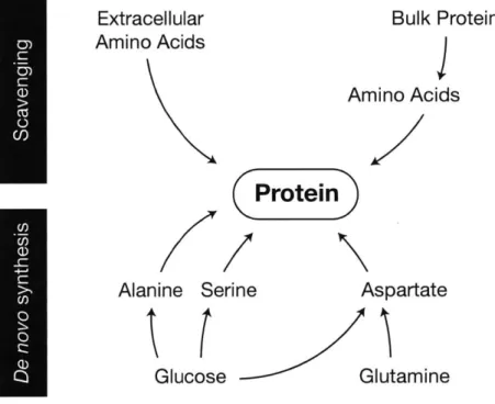
Amino acids
Protein represents the largest fraction of cell mass, and acquisition of amino acids is therefore the primary biosynthetic requirement of cells. Cells can acquire amino acids through a variety of means, and cell type, environment, and genetics determine which pathways are used. Mammalian cells can synthesize non-essential amino acids de novo but must obtain essential amino acids from the extracellular
environment (Figure 3). In nutrient rich conditions, uptake of essential amino acids is correlated with protein synthesis rates, indicating that these amino acids are not catabolized and only supply material for protein synthesis (Dolfi et al., 2013; Hosios et al., 2016). Human plasma contains each of the twenty amino acids at concentrations ranging from 10-600 tM (see Appendix A). Some amino acid
concentrations are much less in other bodily fluids, such as cerebrospinal fluid (McGale et al., 1977), and the availability of essential amino acids may limit proliferation of cells in these contexts. Free amino acids supplied by plasma can be derived from the diet as well as protein breakdown or synthesis in the liver (Mayers et al., 2014), and cells in the tumor microenvironment can be exposed to free amino acids generated by local protein breakdown (Hirayama et al., 2009; Kamphorst et al., 2015).
In some contexts, cells synthesize amino acids to fulfill their requirement for protein synthesis, and glucose is the source for several of these non-essential amino acids (Figure 3). Mammalian cells with high glycolytic flux excrete alanine in addition to lactate, albeit at lower rates (DeBerardinis et al., 2007;
Extracellular
Bulk Protein
Amino Acids
j
Amino Acids
Protein
Alanine Serine
Aspartate
Glucose
Glutamine
Jain et al., 2012). Proliferating cells are therefore rarely limited for alanine, and most media formulations
exclude this amino acid. Alanine is not always synthesized in excess of a cell's needs, however, and
Chuang et al. (1990) observed enhanced lymphocyte proliferation when alanine was added to their
growth medium. Sousa et al. (2016) found that pancreatic cancer cells consume alanine produced by
pancreatic stellate cells when grown in a minimal medium in vitro, indicating the potential for a metabolic
interaction in vivo. Another pathway supported by rapid glycolytic flux is the serine biosynthesis
pathway. Serine is generated from glycolytic intermediates through several reactions, the first of which is
catalyzed by phosphoglycerate dehydrogenase (PHGDH). Even though serine can be acquired from
exogenous sources, de novo synthesis is advantageous in some contexts. For example, PHGDH
expression is elevated in some cancers (Locasale et al., 2011; Pollari et al., 2011; Possemato et al., 2011)
and this correlates with an increased rate of serine synthesis. Overexpression of this enzyme is growth
promoting, although the mechanism remains debated (DeNicola et al., 2015; Locasale et al., 2011;
Possemato et al., 2011). Intracellular serine is derived from both de novo synthesis and consumed serine, and whether the source influences serine's fate is unclear. Serine can provide carbon for nucleotide
biosynthesis (discussed further below) and is catabolized to produce glycine. Glycine can also be derived
from exogenous sources, but many cells in culture rapidly consume serine to produce glycine and only
use exogenous glycine when their serine supply is exhausted (Labuschagne et al., 2014). Some cells are
sensitive to deprivation of serine and glycine (Maddocks et al., 2013), indicating that both amino acid
consumption and de novo synthesis are required to fulfill the biosynthetic requirement for these amino
acids and maximize proliferation.
Serine serves a further role in protein synthesis as it can also be used to synthesize cysteine via
the transsulfuration pathway. Cysteine can therefore be obtained exogenously or by de novo synthesis
from glucose or exogenous serine. The transsulfuration pathway combines serine with homocysteine to
produce cystathionine, which is cleaved by cystathionase to generate cysteine. Not all proliferating cells
are capable of de novo cysteine synthesis, and some are auxotrophic for this amino acid, requiring an
after birth in mammals (Gaull et al., 1972). Cells in culture exhibit variable dependence on this pathway, and in some cases cysteine deprivation can be rescued with cystathionine or homocysteine, depending on which enzymes they express (Eagle et al., 1966). As a result, cell type and gene expression play
significant roles in determining how a cell obtains the cysteine it requires for protein synthesis.
When glutamine is used by proliferating cells, it can supply carbon and nitrogen for synthesis of glutamate, aspartate, asparagine, and proline, fulfilling a substantial amino acid requirement in these cells (Hosios et al., 2016). Glutaminolysis is sufficient to sustain biosynthesis of these amino acids, and they are often excluded from culture media (DeBerardinis and Cheng, 2010; Eagle et al., 1956b). Glutaminase activity produces glutamate, which can serve as a nitrogen donor for transamination reactions in the biosynthesis of other amino acids, such as serine and alanine. In their study of pancreatic cancer cell metabolism, Sousa et al. (2016) observed that providing cells with alanine promoted serine biosynthesis, and this is likely because alanine production as a major fate of glutamate nitrogen was blocked. Amino acid synthesis downstream of glutamine has other advantages as well. For example, proline synthesis from glutamine is upregulated by oncogenic signalling to promote proliferation of lymphoma cells (Liu et al., 2015).
Unlike other amino acids, which can be used by cells when supplied exogenously, aspartate is necessarily synthesized de novo: most cells (except in the prostate and nervous system) lack high-affinity transporters for aspartate, and plasma aspartate concentrations (typically 20 tM or less) are insufficient to enable its consumption (Birsoy et al., 2015; Franklin et al., 2006). Supraphysiological concentrations of aspartate are needed to rescue proliferation of cells incapable of synthesizing it de novo (Sullivan et al., 2015). Aspartate can be synthesized from glutamine carbon, but as discussed above, glutaminolysis is not observed in all proliferating cells. In cases where glutamine is not used, glucose provides carbon to the TCA cycle via pyruvate carboxylase and pyruvate dehydrogenase (Davidson et al., 2016; Hensley et al., 2016; Sellers et al., 2015). In this context, amino acids other than glutamine can provide nitrogen atoms to glutamate to support transamination reactions (Mayers et al., 2016).
While an exogenous supply of many non-essential amino acids is dispensable for proliferation, proliferating cells can become auxotrophic for asparagine or arginine. In some contexts, cells are unable to synthesize sufficient quantities of these amino acids to meet their biosynthetic demands, and depletion of these amino acids can impair proliferation. Asparaginase is a bacterial enzyme used to treat patients with acute lymphoblastic leukemia (Masetti and Pession, 2009). The enzyme hydrolyzes plasma asparagine, thereby starving cells of this amino acid, arguing that cells sensitive to this treatment are asparagine auxotrophs. Why cells have impaired asparagine synthesis is unclear, but this may help to preserve intracellular aspartate pools. Studies have also identified a variety of tumors that are arginine auxotrophs. This amino acid is non-essential for adult humans, but some cells are unable to synthesize arginine de novo. As with cysteine, some cells auxotrophic for arginine can proliferate in its absence if provided with either of its precursors, citrulline and omithine, which are present in plasma at levels comparable to other amino acids (see Appendix A) (Morgan et al., 1958). Some cancers are unable to synthesize arginine because they have lost expression of argininosuccinate synthase (ASS), an enzyme required for arginine synthesis. ASS consumes aspartate, and loss of its expression may serve as a means of preserving cellular aspartate pools (Rabinovich et al., 2015). Cancers lacking fumarate hydratase have high levels of fumarate, which can drive ASS in reverse, consuming arginine and effectively making cells arginine auxotrophs. Arginine deiminase, an enzyme that can deplete extracellular arginine, is being tested for efficacy against these cancers (Phillips et al., 2013).
In addition to de novo synthesis and consumption of exogenous amino acids, proliferating cells can consume and hydrolyze extracellular protein as a source of amino acids. Macropinocytosis, the non-specific uptake of protein, provides a source of amino acids to cells and enables them to survive starvation of individual essential amino acids when protein is abundant (Commisso et al., 2013; Kamphorst et al., 2015). The lysosome degrades consumed protein to produce individual amino acids, which can then be used by cells for biosynthesis. Although this process is non-specific, the high abundance of albumin in plasma and interstitial fluid makes it a primary substrate of this pathway (Commisso et al., 2013).
Proliferating cells have a considerable requirement for protein synthesis and as a result must acquire amino acids necessary to meet this demand in order to grow and divide. Essential amino acids are consumed from the extracellular environment or could be obtained by macropinocytosis, whereas non-essential amino acids can also be acquired by de novo synthesis from glucose or glutamine. Cell type and extracellular environment both influence how amino acids are acquired, and this can affect both protein synthesis as well as other biosynthetic reactions requiring amino acids.
Lipids
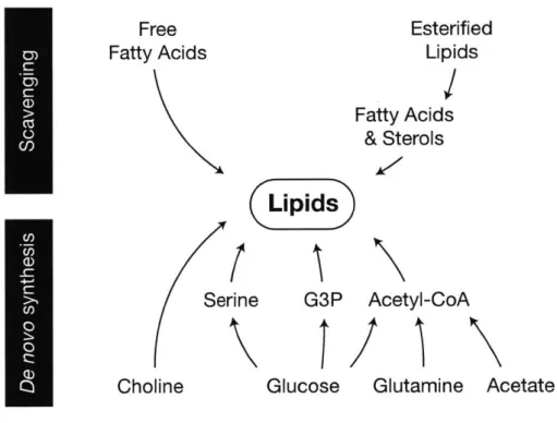
Lipids are a diverse class of non-polar molecules, and their synthesis imposes a complex biosynthetic demand for proliferating cells (Baenke et al., 2013). Lipids comprise 50% of membrane mass, where they enable the separation of organelle contents and the intracellular and extracellular environments, and they are also critical components of multiple signalling pathways. Synthesizing
membranes of the correct composition is critical for cell function (Simons and Sampaio, 2011; Thibault et al., 2012). In mammalian cells, membrane lipids primarily comprise cholesterol, phospholipids, and sphingolipids. Fatty acids are core components of these latter two classes and are also present in cells as triglycerides. Each lipid species can be acquired exogenously or synthesized de novo, and nutrient supply and growth conditions influence how cells meet this biosynthetic demand (Figure 4).
A large fraction of cellular lipids are derived from the extracellular environment (Hosios et al., 2016; Yao et al., 2016). Early work with proliferating lymphocytes demonstrated that exogenously
supplied free fatty acids are incorporated into the lipid pool in these cells (Ardawi and Newsholme, 1984). Circulating free fatty acid levels increase when esterified fatty acids stored in adipose tissue are
hydrolyzed (Duncan et al., 2007). Breast and ovarian cancer cells stimulate lipolysis in co-cultured adipocytes and can consume the released fatty acids (Balaban et al., 2017; Nieman et al., 2011). In other contexts, fatty acid transport promotes cancer cell proliferation, and recent work has shown that
Free
Fatty Acids
Sei
Choline
Esterified
Lipids
Fatty Acids
& Sterols
(Lipids)
rine
G3P
Acetyl-CoA
Glucose
Glutamine
Acetate
Figure 4: De novo and scavenging pathways contributing to the cellular lipid pool. >1
hypoxic cells and cells expressing activated Ras mutants utilize lysophospholipids present in serum as a source of unsaturated fatty acids. Lipids are carried through plasma bound to albumin or in lipoproteins, and cells can use free fatty acids and esterified lipids from these complexes as well. Lymphocytes and fibroblasts express lipoprotein lipase, allowing them to consume and hydrolyze triglycerides as a source of fatty acids for biosynthesis (Calder et al., 1994; Oram et al., 1980), and some cancers express other lipases to utilize exogenous esterified lipids (Zaidi et al., 2013).
Although a large portion of cellular lipids originate from exogenous sources (Hosios et al., 2016; Yao et al., 2016), lipids synthesized de novo are derived mainly from glucose carbon (Kamphorst et al., 2013). Active lipogenesis was first observed in tumor tissue slices by Medes et al. (1953), who noted that hepatomas incorporated more glucose into lipids than adjacent liver tissue. Pyruvate generated by
glycolysis is decarboxylated by pyruvate dehydrogenase (PDH) in the mitochondria to produce acetyl-CoA, the basic component of lipids. Citrate synthase combines this acetyl-CoA with oxaloacetate to produce citrate, which can be exported from the mitochondria and cleaved by ATP-citrate lyase (ACLY) to generate cytosolic acetyl-CoA. In the cytosol, acetyl-CoA is used for de novo synthesis of palmitate (by
fatty acid synthase, FASN) and cholesterol. Acetyl-CoA is also used for fatty acid elongation, and cellular fatty acids are diversified through elongation and desaturation reactions. Sterols can be esterified to fatty
acids, increasing their complexity as well. Many cancers upregulate enzymes required for de novo lipid synthesis, and there is current interest in targeting these enzymes, in particular FASN, therapeutically (reviewed in Svensson and Shaw, 2017).
Glucose is not always the primary source of lipogenic acetyl-CoA. When cells are subject to mitochondrial inhibition, either as a result of hypoxia or genetic alterations, glutamine can serve as a source of acetyl-CoA for de novo lipid synthesis (Metallo et al., 2012; Mullen et al., 2012; Wise et al., 2011). Reductive carboxylation of glutamine-derived a-ketoglutarate by isocitrate dehydrogenase (IDH) produces citrate, which can be cleaved by ACLY to generate acetyl-CoA. PDH flux is reduced in hypoxic cells, decreasing the availability of glucose-derived acetyl-CoA (Kim et al., 2006). The hypoxia-sensing
constitutive HIF activation in some cancers allows them to derive lipids from glutamine when oxygen is abundant (Gameiro et al., 2013; Metallo et al., 2012). Cells proliferating with ample oxygen can also derive acetyl-CoA from glutamine, but it is not the primary contributor to lipid synthesis in this context. Mammalian cells express three IDH isoforms, and reductive carboxylation can be carried out by IDHI and IDH2, which localize to the cytosol and mitochondria respectively. Some controversy exists as to which isoform primarily enables glutamine-derived lipogenesis; studies have argued for IDHI (Metallo et al., 2012), IDH2 (Wise et al., 2011), or neither being predominant (Filipp et al., 2012; Mullen et al., 2012). Of note, it remains unclear whether reductive carboxylation flux is enhanced when mitochondria are impaired or if its relative contribution to lipogenesis increases due to a paucity of glucose-derived acetyl-CoA, although some have argued that decreased citrate levels are a determining factor (Fendt et al., 2013; Gameiro et al., 2013).
Free acetate can also serve as an additional lipogenic substrate for proliferating cells. Acetate can contribute to lipogenic acetyl-CoA when oxygen is abundant, but its contribution is increased in low oxygen (Comerford et al., 2014; Kamphorst et al., 2014; Schug et al., 2015). Similar to glutamine, it is unknown if active upregulation causes this increase in relative contribution or if it is the result of the lower relative contribution of glucose. Acetyl-CoA synthetases (ACSS) are required to convert acetate to acetyl-CoA, and cytosolic ACSS2 is the primary enzyme that supplies acetate for lipid biosynthesis (Comerford et al., 2014). Acetate concentration determines how much this substrate is used for
lipogenesis (Kamphorst et al., 2014), and it is present in human plasma at 50-200 tM (Hosios and Vander Heiden, 2014; Schug et al., 2016), which is lower than concentrations often used experimentally. Free acetate in cells is predominantly generated by protein deacetylation, and a major role of ACSS is to salvage this acetate (Bulusu et al., 2017), raising the possibility that utilization of exogenous tracer acetate for biosynthesis is, in part, a readout of exchange between extracellular and intracellular acetate pools. Exchange explains labeling from tracer acetate when cells do not net consume acetate. Other nutrients, such as ketogenic amino acids, can provide acetyl-CoA for lipid biosynthesis, but this is not often seen in proliferating cells in vitro. Recent work, for example, demonstrated that 3T3-L1 cells begin to use branch
chain amino acid carbon for fatty acid synthesis following in vitro differentiation into adipocytes (Green et al., 2016). Ketone bodies have also been observed to contribute to lipid pools in some contexts, such as in brains of neonatal rodents (Patel and Owen, 1976).
Cells can acquire fatty acids from different sources; however additional molecules are required to synthesize the complex lipid species required by cells to proliferate. Fatty acids are esterified to glycerol-3-phosphate (G3P) to produce phospholipids and triglycerides, for membrane biogenesis and lipid storage respectively. G3P is generated by the phosphorylation of exogenous glycerol or by the reduction of the glycolytic intermediate dihydroxyacetone-phosphate, and proliferating cells may obtain glycerol from both sources (Harding et al., 1975). Phospholipids also contain polar head groups (choline, ethanolamine, serine, and inositol), which can also be scavenged or synthesized de novo. The sources of serine can vary (see above), and phosphatidylethanolamine (PE) can be synthesized from phosphatidylserine by
decarboxylation. Conversely, choline, the most common phospholipid head group, is typically essential for cultured cells (van Meer et al., 2008; Yamamoto and Niwa, 1993). In the body, it is commonly obtained from the diet or methylation of PE to phosphatidyicholine (PC), but this reaction is insufficient to sustain cell-autonomous proliferation in culture. PC is readily produced by the liver, and choline may therefore rarely be limiting for cells in tissues. Inositol can be synthesized from glucose, and proliferating cells exhibit variable dependence on an exogenous supply of this nutrient (Eagle et al., 1956a).
A final important class of lipids derived from fatty acids is sphingolipids. Sphingolipids are synthesized at a minimum from palmitate and serine but can gain additional complexity from the addition of a second fatty acid, phosphate, and/or polar head groups. These molecules have important functions in cell signalling (Merrill, 2002), and inability to synthesize these molecules can impair proliferation through downstream signalling pathways (Lee et al., 2011).
Lipids can be obtained from both exogenous sources and de novo synthesis. For many cells proliferating in culture, de novo lipid synthesis is sufficient to fully support proliferation, as these cells can grow in lipid-free conditions. Conversely, lipids are abundant in the plasma, and proliferating cells
lipid mass and how de novo synthesis supports proliferation is still an open question. Understanding the interplay of consumed and synthesized lipids will provide insight into how cells acquire the mass they need to proliferate.
Nucleic acids
Nucleic acids are another integral biosynthetic requirement of proliferating cells. DNA is necessarily synthesized during the cell division cycle, and RNA, the majority of which is ribosomal, is
important for synthesis of protein required to complete the cell growth cycle. Nucleotides are the precursors for nucleic acid synthesis but also serve as cofactors for many redox, energetic, and
biosynthetic reactions. As with amino acids and lipids, nucleotides can be derived from de novo synthesis or an exogenous supply (Figure 5). Many extant cancer therapies target de novo nucleotide synthesis
pathways, and their ability to impair the proliferation of both cancer and normal cells underscores the essentiality of de novo nucleotide synthesis (Martinez-Outschoom et al., 2017; Vander Heiden, 2011). All nucleotides are composed of a purine or pyrimidine base joined to ribose (or deoxyribose), which is derived from ribose-5-phosphate (R5P). Free ribose can be scavenged and phosphorylated by only a minority of cell types, and R5P is therefore synthesized from glucose by most cells (Agranoff and Brady,
1956; Eagle et al., 1958). R5P can be generated through either the oxidative or non-oxidative pentose phosphate pathways, and recent work has identified cues, such as increased p53 and mTORC 1 activity, that can enhance flux through these pathways (Bensaad et al., 2006; Duvel et al., 2010). The oxidative pathway is expected to be the primary source of R5P for biosynthesis in most proliferating cells (Lee et al., 1998; Reitzer et al., 1980).
Nucleotide base biosynthesis is closely linked to amino acid metabolism, requiring both amino acids themselves and one-carbon units generated by their catabolism (Figures 1,5). Purine bases
incorporate glycine, derive additional carbon atoms from carbon dioxide and the one-carbon pool, and obtain their remaining nitrogen atoms from aspartate and glutamine. Pyrimidine bases are synthesized from aspartate, with additional nitrogen atoms from glutamine and a carbon atom from carbon dioxide.
Nucleobases
(Nucleic
Acids)
its Glycine
t fRibose
Aspartate
Serine
Glucose
Glutamine
Figure 5: De novo and salvage pathways that generate nucleotides for nucleic acid synthesis. Q
1C
Un
Thymidine nucleotide synthesis requires another one-carbon unit. Consequently, the modes of acquiring amino acids described above have a direct impact on nucleotide biosynthesis as well, dictating whether purine and pyrimidine bases derive entirely from glucose or incorporate material from exogenous amino acids.
One-carbon units, individual carbon atoms covalently linked to a folate cofactor, are required for both purine and pyrimidine biosynthesis pathways. Proliferating cells primarily catabolize serine for one-carbon units to sustain nucleotides synthesis (Labuschagne et al., 2014; Mattaini et al., 2016). As with protein synthesis, this serine can be derived from glycolytic carbon or from an exogenous source
(Maddocks et al., 2013). Recent work has highlighted the importance of mitochondrial function in one-carbon metabolism, and Lewis et al. (2014) demonstrated that serine is catabolized in the mitochondria to yield one-carbon units for nucleotide synthesis. When mitochondria are inhibited, however, the cytosol becomes the more important source of one-carbon units (Bao et al., 2016). Many cells use serine catabolism as a source of glycine as well. Glycine catabolism may also serve as a source of one-carbon units in some contexts, but this is likely not used to sustain nucleotide biosynthesis in most proliferating cells (Fan et al., 2014; Jain et al., 2012). Jain et al. surveyed nutrient consumption across the NCI-60 panel of cell lines, and observed that glycine consumption was most strongly correlated with proliferation rate. Importantly, they observed only direct incorporation of glycine into purines with no contribution to the one-carbon pool. Subsequent work has argued that glycine consumption correlates best with DNA synthesis rate rather than the acquisition of cell mass (Dolfi et al., 2013), underscoring the importance of glycine metabolism to nucleotide synthesis.
Serine sits at the intersection of multiple biosynthetic pathways, and how proliferating cells acquire this amino acid informs how they generate many macromolecule precursors. In addition to serine and glycine, aspartate is also required for synthesis of both purines and pyrimidines. As discussed above, aspartate must be synthesized de novo, and limitation for this amino acid impairs both nucleic acid and protein biosynthesis. Aspartate synthesis critically depends upon sustained mitochondrial activity, and cells are unable to proliferate due to aspartate auxotrophy when mitochondria are inhibited (Birsoy et al.,