HAL Id: inserm-00828983
https://www.hal.inserm.fr/inserm-00828983
Submitted on 1 Jun 2013
HAL is a multi-disciplinary open access
archive for the deposit and dissemination of
sci-entific research documents, whether they are
pub-lished or not. The documents may come from
teaching and research institutions in France or
abroad, or from public or private research centers.
L’archive ouverte pluridisciplinaire HAL, est
destinée au dépôt et à la diffusion de documents
scientifiques de niveau recherche, publiés ou non,
émanant des établissements d’enseignement et de
recherche français ou étrangers, des laboratoires
publics ou privés.
The C-terminus region of β-arrestin1 modulates
VE-cadherin expression and endothelial cell
permeability.
Jagoda Hebda, Héloïse Leclair, Sandy Azzi, Célestin Roussel, Mark Scott,
Nicolas Bidère, Julie Gavard
To cite this version:
Jagoda Hebda, Héloïse Leclair, Sandy Azzi, Célestin Roussel, Mark Scott, et al.. The C-terminus
region of β-arrestin1 modulates VE-cadherin expression and endothelial cell permeability.. Cell
Com-munication and Signaling, BioMed Central, 2013, 11 (1), pp.37. �10.1186/1478-811X-11-37�.
�inserm-00828983�
S H O R T R E P O R T
Open Access
The C-terminus region of β-arrestin1 modulates
VE-cadherin expression and endothelial cell
permeability
Jagoda K Hebda
1,2,3, Héloïse M Leclair
1,2,3, Sandy Azzi
1,2,3, Célestin Roussel
1,2,3,4, Mark GH Scott
1,2,3,
Nicolas Bidère
5,6,7and Julie Gavard
1,2,3*Abstract
Background: The endothelial specific cell-cell adhesion molecule, VE-cadherin, modulates barrier function and
vascular homeostasis. In this context, we have previously characterized that VEGF (vascular endothelial growth
factor) leads to VE-cadherin phosphorylation, β-arrestin2 recruitment and VE-cadherin internalization in mouse
endothelial cells. However, exactly how this VE-cadherin/β-arrestin complex contributes to VEGF-mediated
permeability in human endothelial cells remains unclear. In this study, we investigated in-depth the VE-cadherin/
β-arrestin interactions in human endothelial cells exposed to VEGF.
Findings: First, we demonstrated that VEGF induces VE-cadherin internalization in a clathrin-dependent manner in
human umbilical vein endothelial cells (HUVEC). In addition to the classical components of endocytic vesicles,
β-arrestin1 was recruited and bound to phosphorylated VE-cadherin. Molecular mapping of this interaction
uncovered that the C-terminus tail of β-arrestin1, that comprises amino acids 375 to 418, was sufficient to directly
interact with the phosphorylated form of VE-cadherin. Interestingly, the expression of the C-terminus tail of
β-arrestin1 induced loss of surface exposed-VE-cadherin, promoted monolayer disorganization and enhanced
permeability. Finally, this effect relied on decreased VE-cadherin expression at the transcriptional level, through
inhibition of its promoter activity.
Conclusions: Altogether, our results demonstrate that β-arrestin1 might play multiple functions collectively
contributing to endothelial barrier properties. Indeed, in addition to a direct implication in VE-cadherin endocytosis,
β-arrestin1 could also control VE-cadherin transcription and expression. Ultimately, understanding the molecular
mechanisms involved in VE-cadherin function might provide therapeutic tools for many human diseases where the
vascular barrier is compromised.
Keywords: VE-cadherin promoter, Endocytosis, β-arrestin, VEGF, Permeability
Findings
The endothelial cells that form blood vessels ensure
selec-tive exchanges between plasma and irrigated tissues.
Mac-romolecules and cells larger than 3 nm use the paracellular
pathway, chiefly orchestrated through cell-cell junctions [1].
Endothelial junction closing and opening maintain vascular
integrity and homeostasis, and coordinate vascular
perme-ability. Adjacent endothelial cells are connected through
adherens and tight junctions [2]. VE-cadherin (vascular
endothelial cadherin) is the adherens junction protein
ex-clusively expressed in vessels [3]. Besides its key role during
vascular network formation, VE-cadherin guarantees
bar-rier integrity and modulates endothelial plasticity in
adult-hood [4-7]. Many angiogenic molecules can destabilize the
organization of intercellular junctions, causing endothelial
barrier opening [8-13]. We previously described that
vascu-lar endothelial growth factor (VEGF)-induced permeability
operates through a signaling pathway involving the
sequen-tial activation of Src/Vav2/Rac/PAK [9]. This molecular
cascade culminates in VE-cadherin phosphorylation on the
* Correspondence:julie.gavard@inserm.fr
1Cnrs, UMR810, Institut Cochin, 22 rue Mechain, Rm. 306, Paris 75014, France 2Inserm, U1016, 22 rue Mechain, Paris 75014, France
Full list of author information is available at the end of the article
© 2013 Hebda et al.; licensee BioMed Central Ltd. This is an Open Access article distributed under the terms of the Creative Commons Attribution License (http://creativecommons.org/licenses/by/2.0), which permits unrestricted use, distribution, and reproduction in any medium, provided the original work is properly cited.
highly conserved serine 665 (S665) [9]. This S665
phos-phorylation marshals VE-cadherin internalization and
sub-sequent interaction with β-arrestin2, a molecule known for
its role in endocytosis of activated membrane receptors [9].
VE-cadherin internalization appears to be a key mechanism
that controls the overall organization of endothelial
mono-layers in vitro and vascular organization in vivo [9,14-19].
However, how the VE-cadherin/β-arrestin complex is
inter-nalized into clathrin-coated vesicles to ultimately cause
weakening of adherens junctions, loss of barrier integrity
and increased vascular permeability is not yet documented.
Here we extend our previous findings [9,19] by
demons-trating that, in addition to β-arrestin2, phosphorylated
VE-cadherin recruits β-arrestin1. We further mapped a
re-gion in the β-arrestin1 C-terminus tail comprising the
resi-dues 375 to 418 essential for this interaction. In addition,
this domain contributes to the loss of barrier integrity
through reduction of VE-cadherin promoter activity.
VEGF stimulation leads to VE-cadherin
phospho-rylation, β-arrestin2 recruitment and VE-cadherin
intern-alization in murine cells [9]. To determine whether
human endothelial cells also co-opt this mechanism, we
first performed VE-cadherin internalization assays in
hu-man umbilical vein endothelial cells (HUVEC) exposed to
VEGF (Additional file 1). As early as 15 min, we observed
that internalized VE-cadherin vesicles seemed to follow
microtubule routes within the cell, in regions where
actin filaments were also aligned (Figure 1A). The
three-dimensional representation also indicated that
VE-cadherin-containing vesicles accumulated in the
peri-nuclear zone, where they could have been transported
along microtubules (Figure 1B). VEGF also promoted
VE-cadherin internalization in confluent monolayers,
al-though to a lesser extent and with a different pattern
(Additional file 2: Figure S1A-B). To further characterize
the nature of endocytosed VE-cadherin structures, confocal
analysis of internalized VE-cadherin and organelle markers
was performed (Figure 1C, Additional file 2: Figure S1C-D).
In VEGF-stimulated cells, internalized VE-cadherin
co-localized with clathrin-coated vesicles. Accordingly,
inter-nalized VE-cadherin co-labeled with adaptin α from the
AP2 complex, which is involved in the formation of
clathrin-coated pits, and with rab5, a typical marker for
early endosomes (Figure 1C). By contrast, no overlap was
detected with cholera toxin, which illuminates raft
mem-brane microdomains, caveolin, an essential structural
pro-tein for caveolae, or rab11 from the recycling endosomes
(Additional file 2: Figure S1C-D). To further investigate the
role of the clathrin/AP2-dependent endocytic machinery in
this process, endogenous levels of adaptin α were silenced
Figure 1 VE-cadherin is internalized in clathrin coated-vesicles. (A) HUVEC were cultivated on collagen-coated slides, serum deprived, subjected to VE-cadherin internalization assay as described in [18] and stimulated with VEGF (50 ng/ml, 15 min). Cells were fixed and stained for VE-cadherin (iVEC, green), microtubule (red) and actin (purple). Cells were then analyzed by confocal microscopy. Scale bar: 10 μm. (B) VE-cadherin staining was performed as described above, analyzed by confocal microscopy and by 3D reconstitution using IMARIS software. Nuclei are presented in blue (DAPI) and iVEC in green. Scale bar: 10 μm. (C) Confocal analysis of iVEC (green), together with clathrin heavy chain (HC), adaptin α or rab5 (red) in HUVEC stimulated with VEGF (50 ng/ml, 15 min). Scale bar: 10 μm. (D-F) HUVEC received adaptin α-targeting (AP2si) and non-silencing (nsi) duplexes. Total cell lysates were analyzed by western-blot three days later for adaptin α and Tubulin protein expression levels. Confocal analysis of iVEC (green) was performed in the absence (Ctrl) or presence of VEGF (50 ng/ml, 15 min). Graph represents the mean ± s.e.m. of the percentage of cells with iVEC staining; n >300; T-test: ** p < 0.01. Scale bar: 10 μm. All panels are representative of at least 3 independent experiments.
Hebda et al. Cell Communication and Signaling 2013, 11:37 Page 2 of 7 http://www.biosignaling.com/content/11/1/37
by RNA interference (Figure 1D). In these settings,
VEGF-elicited VE-cadherin internalization was highly reduced
(Figure 1E-F). Our data clearly establish that short-term
VEGF stimulation induces VE-cadherin endocytosis in
clathrin-coated vesicles in human endothelial cells.
Because β-arrestin1 and β-arrestin2 share redundant
functions, we next asked whether β-arrestin1 was similarly
involved in VE-cadherin internalization [9,19]. In response
to VEGF, β-arrestin1-CFP drastically evolved from a
membrane/cytosol diffused localization to a vesicular
pat-tern, which co-localized with VE-cadherin-containing
vesi-cles (Figure 2A, Additional file 2: Figure S1D).
Co-immunoprecipitation experiments further showed that
VEGF treatment increased a modest but significant
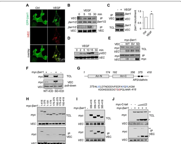
VE-Figure 2 β-arrestin1 C-terminus tail binds to S665D VE-cadherin mutant. (A) HUVEC were transfected with CFP-myc-tagged β-arrestin1 (CFP-βarr1, green). After serum deprivation, cells were subjected to VE-cadherin internalization assay, stimulated with VEGF (50 ng/ml, 15 min) and fixed for staining (iVEC, red). Cells were analyzed by confocal microscopy. Scale bar: 10 μm. (B-D) After overnight serum starvation, three day-old HUVEC were stimulated with VEGF (50 ng/ml) for the indicated times. (B-C) The anti-VE-cadherin (VEC) and anti-β-arrestin1/2 (βarr1/2) immunoprecipitated (IP) fractions were analyzed by western-blot. Densitometry analysis was done with Image J software. T-test: * p < 0.05. (D) Total cell lysates (TCL) were analyzed by western-blot for S665 phosphorylation of VE-cadherin (pVEC) and total VEC. (E) HEK-293T cells were co-transfected with CFP-myc-tagged β-arrestin1 (myc-βarr1) together with control plasmid (−), wild-type (WT), non-phosphorylable (SV) or phosphomimetic (SD) VE-cadherin mutants. TCL and IP fractions were analyzed by western-blot as indicated. (F) HEK-293T cells were
transfected with CFP-myc-β-arrestin1 (myc-βarr1, +) or CFP-myc control plasmid (−). Cellular extract was incubated with recombinant histidine-fused VE-cadherin intracellular domain (ICD), either WT or SD. TCL and pull-down fractions were analyzed by western-blot as indicated. (G) Schematic representation of human β-arrestin1, with two arrestin (arr) domains. The C-terminus tail (375–418) comprises an AP2 binding site (LIE and RXR in blue) and two phosphorylation sites (in red). (H-J) HEK-293T cells were co-transfected with VEC SD mutant together with control plasmid (−), full-length and deleted forms of myc-βarr1. IP fractions and TCL were analyzed by western-blot. (J) HEK-293T cells were co-transfected with VEC SD mutant together with a constant amount of full-length myc-βarr1 (**) and increasing concentrations of CFP-myc-tagged β-arrestin1 C-terminus tail comprising amino acids 475–418 (myc-C-tail, *). TCL and IP fractions were analyzed by western-blot. All panels are representative of at least 3 independent experiments.
cadherin binding to β-arrestin1 (Figure 2B-C). This was
accompanied
by
enhanced
VE-cadherin
phospho-rylation on residue S665 (Figure 2D). We then explored
whether VE-cadherin serine phosphorylation impacts
on β-arrestin1 recruitment by developing an ectopic
expression system in human embryonic kidney cells
(HEK-293T). We found that the interaction between
β-arrestin1 and a non-phosphorylable mutant (SV) of
VE-cadherin was much more limited than with the
wild-type (WT) form of VE-cadherin (Figure 2E). By contrast,
β-arrestin1 massively bound to a phosphomimetic mutant
(SD) of VE-cadherin. Combined, this data strongly
sug-gests that VE-cadherin serine phosphorylation enhances
β-arrestin1 recruitment. We next tested the ability of the
recombinant WT or SD versions of the intracellular
do-main of VE-cadherin to pull-down cellular β-arrestin1.
Again, SD VE-cadherin strongly bound to β-arrestin1 when
compared to WT VE-cadherin (Figure 2F). To further map
this association, co-immunoprecipitations were performed
in HEK-293T cells co-transfected with SD VE-cadherin and
deletion mutants of β-arrestin1 (Figure 2G). While SD
VE-cadherin barely bound to β-arrestin1 mutants lacking
its C-terminus tail (375–418 amino acids), it efficiently
interacted with all deletion mutants that retained this
re-gion (Figure 2H). Hence, we defined a 43 amino
acid-containing domain required for VE-cadherin/β-arrestin1
interaction. Consistent with this finding, the C-terminus
tail of β-arrestin1 was sufficient to interact with SD
VE-cadherin (Figure 2I), and efficiently outcompeted
full-length β-arrestin1 binding (Figure 2J).
We further investigated the effect of β-arrestin1
C-terminus tail enforced expression in HUVEC. To this end,
we engineered HUVEC cells stably expressing β-arrestin1
C-terminus tail (C-tail), along with empty plasmid (mock),
full-length (FL) and deleted C-tail (ΔC-tail). The stable
expression of CFP-myc-tagged β-arrestin1 constructs
was validated by flow cytometry and confocal analysis
(Additional file 3: Figure S2A-B). Of note, C-tail
prefer-entially accumulated in the nucleus (Additional file 3:
Figure S2B). Flow cytometric analysis further unveiled
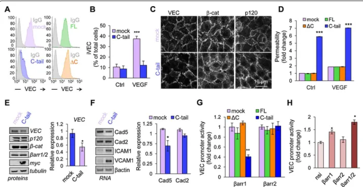
Figure 3 β-arrestin1 C-terminus tail induces loss of barrier integrity and VE-cadherin down-regulation. (A) VE-cadherin surface expression was tested by flow cytometry in HUVEC that stably expressed mock (pink) or CFP-myc-tagged β-arrestin1 full-length (1–418, green), ΔC (1–374, orange) and C-tail (375–418, blue). Isotype control (Ig) is shown in grey. (B) Mock and C-tail HUVEC were subjected to VE-cadherin internalization assay (iVEC) and analyzed by confocal microscopy. Graph represents the mean + s.e.m. of the percentage of cells with iVEC staining; n >300; T-test: *** p < 0.001. (C) Three day-old mock and C-tail HUVEC were stained for VE-cadherin (VEC), β-catenin (β-cat) and p120-catenin (p120) and analyzed by confocal microscopy. Scale bar: 10 μm. (D) Relative permeability was measured in mock, FL, ΔC and C-tail HUVEC by the fluorescence of 40 kDa FITC-dextran passage. Graph shows the mean ± s.e.m. of 3 independent experiments. Two-way ANOVA test: *** p < 0.001. (E-F) Protein and RNA expression of indicated targets were tested using western-blot (E) and RT-PCR (F) in mock and C-tail HUVEC. Cad5: cadherin-5, VE-cadherin; Cad2: cadherin-2, N-cadherin. Densitometry analysis was done with Image J software. T-test: * p < 0.05. (G-H) HUVEC were transfected with luciferase reporter for VE-cadherin promoter activity and Renilla. (G) They also received a control CFP-myc plasmid (mock) and the CFP-myc-tagged FL, ΔC and C-tail β-arrestin1. Alternatively, they received pGFP (mock) and GFP-tagged β-arrestin2 comprising amino acids 1–410 (FL), 1–359 (ΔC) and 317–410 (C-tail). (H) Two days prior plasmid transfection, cells were treated with non-silencing duplexes (nsi) and siRNA targeting βarr1, βarr2, or both (βarr1/2). Graphs show the mean ± s.e.m. of 3 independent experiments. Two-way and one-way ANOVA test: ** p < 0.01; * p < 0.05. All panels are representative of at least 3 independent experiments.
Hebda et al. Cell Communication and Signaling 2013, 11:37 Page 4 of 7 http://www.biosignaling.com/content/11/1/37
that surface-exposed VE-cadherin was minimized in C-tail,
but not in FL and ΔC-expressing HUVEC (Figure 3A). In
agreement with this data, VE-cadherin internalization was
not observed in C-tail HUVEC, while VEGF triggered a
four-fold increase in VE-cadherin endocytosis in control
cells (Figure 3B). These results prompted us to analyze
adherens junction organization by confocal microscopy.
Corroborating our data, VE-cadherin, p120 and β-catenin
appeared less cohesively organized at the cell-cell
junc-tions in C-tail HUVEC, where gaps, zigzags and weaker
VE-cadherin staining were observed, resulting in an overall
alteration of endothelial monolayer architecture (Figure 3C).
Importantly, this impacted on endothelial permeability as
measured in vitro with a 40 kDa fluorescent tracer
(Figure 3D). Indeed, C-tail cells exhibited higher basal
endothelial permeability, which could not be further
in-creased by VEGF, when compared to mock, FL and ΔC
HUVEC (Figure 3D). To gain insights into the mechanisms
involved, we investigated the protein expression levels of
adherens junction molecules in C-tail HUVEC.
Interest-ingly, VE-cadherin protein expression was diminished in
C-tail cells, while β-catenin, p120-catenin and β-arrestin1/2
remained unchanged (Figure 3E). Following these
observa-tions, we examined mRNA levels of VE-cadherin (Cad5),
N-cadherin (Cad2) and other endothelial adhesive
mole-cules, such as ICAM1 and VCAM1 (Figure 3F). Our data
show that VE-cadherin mRNA was reduced by a quarter
in C-tail cells, arguing in favor of a transcriptional effect
of the β-arrestin1 C-terminus tail. The lack of change in
N-cadherin mRNA suggests that it cannot substitute
for VE-cadherin. Interestingly, whereas TNFα challenge
strongly increased ICAM1 and VCAM1 expression
con-comitantly with AP1 and NF-κB activation, neither of
these pathways, nor p38 phosphorylation were turned on
in C-tail expressing cells (Additional file 4: Figure S3A-D).
Finally, the effect of the β-arrestin1 C-terminus tail on the
activity of the VE-cadherin promoter [20] was tested in
HeLa and HUVEC cells. The β-arrestin1 C-terminus
tail expression resulted in a significant decrease in
VE-cadherin promoter activity (Figure 3G, Additional
file 5: Figure S4A) in both cell lines, although milder in
HUVEC. This is most likely due to differences between
endothelial and non-endothelial cells [20]. This prompted
us to test the possibility that the C-terminus tail of
β-arrestin1 binds to the VE-cadherin promoter. Indeed,
chromatin immunoprecipitation experiments showed that
β-arrestin1 C-tail interacted with the proximal region of
the VE-cadherin promoter (Additional file 3: Figure S2C).
Interestingly, the expression of the corresponding
con-structs in β-arrestin2 was unable to alter VE-cadherin
pro-moter activity (Figure 3G, Additional file 5: Figure S4A).
This is in agreement with studies showing a predominant
role for β-arrestin1 in transcriptional regulation,
com-pared to β-arrestin2, which is actively exported from the
nucleus [21,22]. Conversely, reducing β-arrestin1, but
not β-arrestin2, expression with siRNA resulted in
ele-vated VE-cadherin promoter activity (Figure 3H,
Add-itional file 5: Figure S4B-C). Hence, the effects of
endogenous β-arrestin1 siRNA on VE-cadherin promoter
cannot mirror its overexpression. Indeed, β-arrestin1 is
expressed in a closed conformation in which the
C-terminus tail is masked [23]. Upon activation,
con-formational changes allow transition to an open state,
unleashing the C-tail. This mechanism has been
pro-posed to regulate β-arrestin1 functions [23,24]. In
agree-ment, ectopic C-tail but not full-length β-arrestin1
overexpression curtailed VE-cadherin promoter activity.
Altogether, our data extend our previous findings [9,19]
by demonstrating that VEGF induces the recruitment of
β-arrestin1 in the course of VE-cadherin internalization
in clathrin-containing vesicles. Molecular mapping of
VE-cadherin/β-arrestin1 interaction supports the idea of
a specific and direct interaction between the C-terminus
tail of β-arrestin1 and the phospho-mimicking mutant of
VE-cadherin. Additionally, β-arrestin1 C-terminus tail
re-duces VE-cadherin expression through the inhibition of
its promoter activity, suggesting that this multifaceted
protein could control endothelial barrier properties by
several means. Although our study is mainly based on
β-arrestin1 C-terminus tail overexpression, our results
might be therapeutically relevant in the context of
path-ologies exhibiting sustained increase of vascular
perme-ability, especially where endothelial cells are exposed to
aberrant levels of VEGF, such as in cancers.
Additional files
Additional file 1: Methods description.
Additional file 2: Figure S1. Characterization of internalized VE-cadherin. (A-C) HUVEC were cultivated on collagen-coated slides, either sparse or confluent, serum deprived (Ctrl), subjected to VE-cadherin internalization assay and stimulated with VEGF (50 ng/ml, 15 min). Cells were fixed and stained for VE-cadherin (iVEC, gray) and analyzed by confocal microscopy. Scale bar: 10 μm. (B) Graph represents the mean ± s.e.m. of the percentage of cells with iVEC staining; n > 200; T-test:**p < 0.01. (C) Confocal analysis of
iVEC (green), together with caveolin, cholera toxin (CTX), or rab11 (red). Scale bar: 10 μm. (D) Pearson’s coefficients were calculated for each indicated staining with iVEC; n > 10. Panels are representive of 3 independent experiments.
Additional file 3: Figure S2. β-arrestin1 expression and localization. (A-B) HUVEC were transfected with the indicated CFP-fused β-arrestin1 constructs, and analyzed by flow cytometry (A) and confocal microscopy (B). Scale bar: 10 μm. Panels are representive of 3 independent experiments. (C) Chromatin immunoprecipitations (ChIP) were performed with anti-GFP and preimmune (lg pAb) antibodies on shear cross-linked chromatin preparation from C-tail HUVEC. Three sets of primers flanking regions -541/-390, -132/+18 and -785/-670 on cadherin-5 (Cad5, VE-cadherin) promoter were used. Additional controls included anti-PoIII IP and premmune (lg mAb) and GADPH primers. Panels are representative of 3 independent experiments.
Additional file 4: Figure S3. Status of inflammatory signaling pathways in β-arrestin1 C-tail expressing cells. (A) HEK-293 T transfected with luciferase reporter for the indicated promoter activity (NF-KB and AP1)
and Renilla, together with mock, full lenght (FL) and C-tail β-arrestin1 (C-tail). Alternatively, cells were treated with the TNFα (10 μg/ml, 6 h). Graphs shows the mean + s.e.m. of 3 independent experiments, normalized to Renilla. Two-way ANOVA test:***p < 0.001. (B) HeLa
transfected with C-tail β-arrestin1 (green) were stained (red) for p65 and cFos and analyzed by confocal. Alternatively, cells were treated with TNFα for 1 h. (C) RT-PCR for ICAM1 and VCAM1 was performed in starved HUVEC (-), treated with TNFα (+, 10 μg/ml,6 h) or expressing C-tail and FL β-arrestin1. GAPDH serves as an internal control. (D) HEK-293 T (-) transfected with FL and C-tail β-arrestin1 were analyzed by western-blot for phosphorylated (p) p38. Anisomycin-treated cells (60 μm, 15 min) were used as a positive control. Total ERK2 serves as a loading control. All panels are representive of 3 independent experiments.
Additional file 5: Figure S4. Impact of β-arrestin1 and β-arrestin2 on VE-cadherin promoter activity. (A-C) HeLa were transfected with luciferase reporter for VE-cadherin (VEC) promoter activity and Renilla, together with either, a control CFP-myc plasmid (mock) and CFP-myc-tagged β-arrestin1 (βarr1) comprising amino acids 317-410 (C-tail). (B-C) Alternatively, HeLa (B) and HUVEC (C) received non-silencing duplexes (nsi) and β-arrestin1 (βarr1), β-arrestin2 (βarr2), and β-arrestin1/2 (βarr1/2)-targeting siRNA. VEC promoter activity was measured in a luciferase-based assay (B), and siRNA efficiency was evaluated by western-blot (C), using anti-βarr1 and anti-βarr2 antibodies. Tubulin serves as a loading control. Graph shows the mean ± s.e.m. of 3 independent experiments. Two-way and one-way ANOVA tests:***p < 0.001;**p < 0.01;*p < 0.05.
Panels are representative of 3 independent experiments. Abbreviations
CFP:Cyan fluorescent protein; HUVEC: Human umbilical vein endothelial cell; ICAM: Intercellular adhesion molecule; VCAM: Vascular cell adhesion molecule; VE-cadherin: Vascular endothelial cadherin; VEGF: Vascular endothelial growth factor.
Competing interests
The authors declare no competing interests. Authors’ contributions
JKH, HML, SA, CR, NB and JG performed experiments; JKH, HML, SA, NB, and JG analyzed data; MGHS contributed essential reagents; JKH and JG wrote the manuscript. All authors read and approved the final manuscript. Acknowledgments
The authors are thankful to the members from JG laboratory (Institut Cochin, Paris, France), especially Dr. SS. Smith for help with imaging, L. Treps for helpful discussion, and Dr. J. Dwyer for comments on the manuscript. We are grateful to Dr. P. Huber (CEA, Grenoble, France) for the VE-cadherin promoter luciferase construct. This research was funded by Ligue nationale contre le cancer comite de Paris, Fondation ARC, Fondation pour la Recherche Medicale, ANR JCJC and by a Marie Curie International Reintegration Grant within The Seventh Framework Programme. JKH is supported by doctoral fellowship from Universite Paris Descartes and SA by a post-doctoral fellowship from Fondation ARC.
Author details
1Cnrs, UMR810, Institut Cochin, 22 rue Mechain, Rm. 306, Paris 75014, France. 2Inserm, U1016, 22 rue Mechain, Paris 75014, France.3Universite Paris
Descartes, Sorbonne Paris Cite, 6 rue de l’Ecole de Medecine, Paris 75006, France.4Mitologics, Hopital Robert Debre, 48 Boulevard Serurier, Paris 75019,
France.5INSERM UMR_S1014, Hopital Paul Brousse, Villejuif 94800, France. 6Universite Paris-Sud P11, Orsay 91400, France.7Equipe Labellisée Ligue
contre le Cancer, Villejuif 94800, France.
Received: 8 February 2013 Accepted: 16 May 2013 Published: 28 May 2013
References
1. Vestweber D, Winderlich M, Cagna G, Nottebaum AF: Cell adhesion dynamics at endothelial junctions: VE-cadherin as a major player. Trends Cell Biol 2009, 19(1):8–15.
2. Gavard J: Breaking the VE-cadherin bonds. FEBS Lett 2009, 583(1):1–6. 3. Dejana E: Endothelial cell-cell junctions: happy together. Nat Rev Mol Cell
Biol 2004, 5(4):261–270.
4. Carmeliet P, Lampugnani MG, Moons L, Breviario F, Compernolle V, Bono F, Balconi G, Spagnuolo R, Oostuyse B, Dewerchin M, et al: Targeted deficiency or cytosolic truncation of the VE-cadherin gene in mice impairs VEGF-mediated endothelial survival and angiogenesis. Cell 1999, 98(2):147–157. 5. Corada M, Mariotti M, Thurston G, Smith K, Kunkel R, Brockhaus M,
Lampugnani MG, Martin-Padura I, Stoppacciaro A, Ruco L, et al: Vascular endothelial-cadherin is an important determinant of microvascular integrity in vivo. Proc Natl Acad Sci USA 1999, 96(17):9815–9820. 6. Crosby CV, Fleming PA, Argraves WS, Corada M, Zanetta L, Dejana E, Drake
CJ: VE-cadherin is not required for the formation of nascent blood vessels but acts to prevent their disassembly. Blood 2005, 105(7):2771–2776.
7. Vittet D, Buchou T, Schweitzer A, Dejana E, Huber P: Targeted null-mutation in the vascular endothelial-cadherin gene impairs the organization of vascular-like structures in embryoid bodies. Proc Natl Acad Sci USA 1997, 94(12):6273–6278.
8. Esser S, Lampugnani MG, Corada M, Dejana E, Risau W: Vascular endothelial growth factor induces VE-cadherin tyrosine phosphorylation in endothelial cells. J Cell Sci 1998, 111(Pt 13):1853–1865.
9. Gavard J, Gutkind JS: VEGF controls endothelial-cell permeability by promoting the beta-arrestin-dependent endocytosis of VE-cadherin. Nat Cell Biol 2006, 8(11):1223–1234.
10. Gavard J, Hou X, Qu Y, Masedunskas A, Martin D, Weigert R, Li X, Gutkind JS: A role for a CXCR2/phosphatidylinositol 3-kinase gamma signaling axis in acute and chronic vascular permeability. Mol Cell Biol 2009, 29(9):2469–2480.
11. Weis S, Cui J, Barnes L, Cheresh D: Endothelial barrier disruption by VEGF-mediated Src activity potentiates tumor cell extravasation and metastasis. J Cell Biol 2004, 167(2):223–229.
12. Le Guelte A, Galan-Moya EM, Dwyer J, Treps L, Kettler G, Hebda JK, Dubois S, Auffray C, Chneiweiss H, Bidere N, et al: Semaphorin 3A elevates endothelial cell permeability through PP2A inactivation. J Cell Sci 2012, 125(Pt 17):4137–4146.
13. Alexander JS, Alexander BC, Eppihimer LA, Goodyear N, Haque R, Davis CP, Kalogeris TJ, Carden DL, Zhu YN, Kevil CG: Inflammatory mediators induce sequestration of VE-cadherin in cultured human endothelial cells. Inflammation 2000, 24(2):99–113.
14. Orsenigo F, Giampietro C, Ferrari A, Corada M, Galaup A, Sigismund S, Ristagno G, Maddaluno L, Young Koh G, Franco D, et al: Phosphorylation of VE-cadherin is modulated by haemodynamic forces and contributes to the regulation of vascular permeability in vivo. Nat Commun 2012, 3:1208. 15. Gaengel K, Niaudet C, Hagikura K, Siemsen BL, Muhl L, Hofmann JJ, Ebarasi
L, Nystrom S, Rymo S, Chen LL, et al: The sphingosine-1-phosphate receptor S1PR1 restricts sprouting angiogenesis by regulating the interplay between VE-cadherin and VEGFR2. Dev Cell 2012, 23(3):587–599. 16. Nanes BA, Chiasson-MacKenzie C, Lowery AM, Ishiyama N, Faundez V, Ikura
M, Vincent PA, Kowalczyk AP: p120-catenin binding masks an endocytic signal conserved in classical cadherins. J Cell Biol 2012, 199(2):365–380. 17. Chiasson CM, Wittich KB, Vincent PA, Faundez V, Kowalczyk AP:
p120-catenin inhibits VE-cadherin internalization through a Rho-independent mechanism. Mol Biol Cell 2009, 20(7):1970–1980.
18. Xiao K, Allison DF, Buckley KM, Kottke MD, Vincent PA, Faundez V, Kowalczyk AP: Cellular levels of p120 catenin function as a set point for cadherin expression levels in microvascular endothelial cells. J Cell Biol 2003, 163(3):535–545.
19. Gavard J, Patel V, Gutkind JS: Angiopoietin-1 prevents VEGF-induced endothelial permeability by sequestering Src through mDia. Dev Cell 2008, 14(1):25–36.
20. Prandini MH, Dreher I, Bouillot S, Benkerri S, Moll T, Huber P: The human VE-cadherin promoter is subjected to organ-specific regulation and is activated in tumour angiogenesis. Oncogene 2005, 24(18):2992–3001. 21. DeWire SM, Ahn S, Lefkowitz RJ, Shenoy SK: Beta-arrestins and cell
signaling. Annu Rev Physiol 2007, 69:483–510.
22. Scott MG, Le Rouzic E, Perianin A, Pierotti V, Enslen H, Benichou S, Marullo S, Benmerah A: Differential nucleocytoplasmic shuttling of beta-arrestins. Characterization of a leucine-rich nuclear export signal in beta-arrestin2. J Biol Chem 2002, 277(40):37693–37701.
Hebda et al. Cell Communication and Signaling 2013, 11:37 Page 6 of 7 http://www.biosignaling.com/content/11/1/37
23. Nobles KN, Guan Z, Xiao K, Oas TG, Lefkowitz RJ: The active conformation of beta-arrestin1: direct evidence for the phosphate sensor in the N-domain and conformational differences in the active states of beta-arrestins1 and −2. J Biol Chem 2007, 282(29):21370–21381. 24. Shukla AK, Manglik A, Kruse AC, Xiao K, Reis RI, Tseng WC, Staus DP, Hilger
D, Uysal S, Huang LY, et al: Structure of active beta-arrestin-1 bound to a G-protein-coupled receptor phosphopeptide. Nature 2013, 497(7447):137–141.
doi:10.1186/1478-811X-11-37
Cite this article as: Hebda et al.: The C-terminus region of β-arrestin1 modulates VE-cadherin expression and endothelial cell permeability. Cell Communication and Signaling 2013 11:37.
Submit your next manuscript to BioMed Central
and take full advantage of:
• Convenient online submission
• Thorough peer review
• No space constraints or color figure charges
• Immediate publication on acceptance
• Inclusion in PubMed, CAS, Scopus and Google Scholar
• Research which is freely available for redistribution
Submit your manuscript at www.biomedcentral.com/submit
![Figure 1 VE-cadherin is internalized in clathrin coated-vesicles. (A) HUVEC were cultivated on collagen-coated slides, serum deprived, subjected to VE-cadherin internalization assay as described in [18] and stimulated with VEGF (50 ng/ml, 15 min)](https://thumb-eu.123doks.com/thumbv2/123doknet/14650109.551276/3.892.87.809.597.952/cadherin-internalized-clathrin-cultivated-subjected-internalization-described-stimulated.webp)