HAL Id: hal-02540215
https://hal.umontpellier.fr/hal-02540215
Submitted on 10 Apr 2020
HAL is a multi-disciplinary open access
archive for the deposit and dissemination of
sci-entific research documents, whether they are
pub-lished or not. The documents may come from
teaching and research institutions in France or
abroad, or from public or private research centers.
L’archive ouverte pluridisciplinaire HAL, est
destinée au dépôt et à la diffusion de documents
scientifiques de niveau recherche, publiés ou non,
émanant des établissements d’enseignement et de
recherche français ou étrangers, des laboratoires
publics ou privés.
DNA polymorphism and epigenetic marks modulate the
affinity of a scaffold/matrix attachment region to the
nuclear matrix
Natalia Kisseljova, Petr Dmitriev, Alexey Katargin, Elena Kim, Daria
Ezerina, Diana Markozashvili, Daria Malysheva, Emmeline Planche, Richard
Lemmers, Silvere van der Maarel, et al.
To cite this version:
Natalia Kisseljova, Petr Dmitriev, Alexey Katargin, Elena Kim, Daria Ezerina, et al.. DNA
poly-morphism and epigenetic marks modulate the affinity of a scaffold/matrix attachment region to the
nuclear matrix. European Journal of Human Genetics, Nature Publishing Group, 2014, 22,
pp.1117-1123. �10.1038/ejhg.2013.306�. �hal-02540215�
ARTICLE
DNA polymorphism and epigenetic marks modulate
the affinity of a scaffold/matrix attachment region
to the nuclear matrix
Natalia P Kisseljova
1,2,7, Petr Dmitriev
2,3,4,7, Alexey Katargin
1, Elena Kim
2,3, Daria Ezerina
1,2,
Diana Markozashvili
2,3, Daria Malysheva
2,3, Emmeline Planche
2,3, Richard JLF Lemmers
5,
Silve`re M van der Maarel
5, Dalila Laoudj-Chenivesse
4, Marc Lipinski
2,3and Yegor S Vassetzky*
,2,3,6Mechanisms that regulate attachment of the scaffold/matrix attachment regions (S/MARs) to the nuclear matrix remain largely unknown. We have studied the effect of simple sequence length polymorphism (SSLP), DNA methylation and chromatin organization in an S/MAR implicated in facioscapulohumeral dystrophy (FSHD), a hereditary disease linked to a partial deletion of the D4Z4 repeat array on chromosome 4q. This FSHD-related nuclear matrix attachment region (FR-MAR) loses its efficiency in myoblasts from FSHD patients. Three criteria were found to be important for high-affinity interaction between the FR-MAR and the nuclear matrix: the presence of a specific SSLP haplotype in chromosomal DNA, the methylation of one specific CpG within the FR-MAR and the absence of histone H3 acetylated on lysine 9 in the relevant chromatin fragment.
European Journal of Human Genetics (2014) 22, 1117–1123; doi:10.1038/ejhg.2013.306; published online 22 January 2014 Keywords: nuclear matrix; SSLP; facioscapulohumeral dystrophy; epigenetics; cancer cells
INTRODUCTION
Scaffold/matrix attachment regions (S/MARs) are specialized genomic DNA sequences that exhibit high affinity to the nuclear matrix (NM) in vitro and in vivo. S/MARs often colocalize with or are situated in close proximity to specific sequences, including replication origins, insulators and enhancers (for a review see Vassetzky et al1 and Razin et al2). S/MARs anchor chromatin onto
the NM, thereby organizing the genomic DNA into topologically distinct loop domains that are important for the regulation of replication and transcription.2
Despite numerous genomic studies,3–7 no consensus S/MAR
sequence has been identified so far.8 S/MARs can be both A/T9or
G/C rich.10 Moreover, the strength and specificity of S/MAR
attachment to the NM varies during ontogenesis,11in cancer12and
various genetic diseases such as facioscapulohumeral dystrophy (FSHD).13,14 Several factors potentially affect the association of
DNA with the NM. These include the DNA sequence itself and its epigenetic state.15–18
We have recently characterized an S/MAR within the subtelomeric region of chromosome 4q.13,14This region (Figure 1a) has a complex
structure and includes the D4Z4 macrosatellite repeat array, which consists of 3.3 kb repeated units with a number of units varying between individuals.19 This S/MAR, designated as FR-MAR for
FSHD-related matrix attachment region,13is located centromerically
to the D4Z4 array and includes a simple sequence length
polymorphism (SSLP) (for a review see Dmitriev et al20). The 4q
subtelomeric region is subject to extensive epigenetic changes between normal and pathological cells. Thus, we have shown that in FSHD the FR-MAR is partially detached from the NM.13,14 The mechanisms
underlying the observed interaction between the FR-MAR and the NM remain largely unknown. Several factors could have a role. First, the protein composition of the NM could vary between normal and pathological tissues. For example, levels of the NM protein vimentin were found to differ between FSHD and normal myoblasts.21 Also, the nucleotide sequence in the FR-MAR region
could affect the affinity for the NM. Indeed, several SSLPs have been identified that are specifically associated with FSHD.22–24Finally, the
chromatin structure and DNA methylation level could also modify the FR-MAR binding to the NM. Other genetic and epigenetic abnormalities affecting the subtelomeric region of chromosome 4q have also been observed in various pathologies including the Rett and ICF syndromes,25as well as in several types of cancer.26–32
Here, we have tested whether mechanisms controlling the associa-tion of the FR-MAR to the NM could be similar in different cell types. We have used primary myoblasts from FSHD patients and cervical carcinoma cell lines where the D4Z4 repeat array can be either strongly methylated or demethylated.30 From our experiments, the
FR-MAR binds the NM depending upon a combination of SSLP and chromatin modifications including DNA CpG methylation, histone H3 modifications and MeCP2 binding.
1NN Blokhin Russian Cancer Research Center, RAMS, Moscow, Russia;2UMR 8126, Universite´ Paris Sud, CNRS, Institut de cance´rologie Gustave Roussy, Villejuif, France; 3LIA 1066, Laboratoire Franco-Russe de recherche en oncologie, Villejuif, France;4INSERM U1046, Montpellier, France;5Department of Human Genetics, Leiden University
Medical Center, Leiden, The Netherlands;6NK Koltsov Institute of Developmental Biology, RAS, Moscow, Russia.
*Correspondence: Dr YS Vassetzky, UMR 8126, Universite´ Paris Sud, CNRS, Institut de cance´rologie Gustave Roussy, 39, rue Camille-Desmoulins, 94805 Villejuif, France. Tel: +33(0)1 42 11 62 83; Fax: +33(0)1 42 11 54 94; E-mail: [email protected]
7These authors contributed equally to this work.
MATERIALS AND METHODS Cell lines
The human cervical carcinoma cells CaSki and C-33A (American Type Culture Collection, Manassas, VA, USA) were maintained in DMEM supplemented with 5–10% FCS. Primary human myoblasts were isolated and differentiated as described.33The primary human myoblasts are described in Table 1. Cells
were treated with DNA methyltransferase inhibitor 5-aza-20-deoxycytidine
(5-aza-dC; Sigma, St Louis, MO, USA) for 3 days. The medium containing 5mM 5-aza-dC was changed daily. Cells were treated with trichostatin A
(Sigma) at a concentration of 1mMfor 1 day before harvesting.
Analysis of SSLP
DNAs (total and associated with NM) were PCR amplified using oligonucleo-tide primers: SSLP-F, 50-GCTCTTGAGCTCGTCTTGGACA-30 and SSLP-R,
50-CTTCAGAGGCATTTGGCAGAAG-30. The PCR products were cloned into
pGEM-T Easy vector (Promega, Madison, WI, USA), 65–100 clones were used for automated sequencing (‘Genome’ Center, Moscow, Russia; Millegen, Toulouse, France), analyzed and the quantity of clones with or without eight-nucleotide (8nt) insert was calculated.
Nuclei and nuclear matrices
Nuclei were isolated from the cell lines essentially as described elsewhere.34
Nuclear matrices were prepared by treatment of the isolated nuclei with NaCl as follows: digestion buffer (100 mMNaCl, 25 mM KCl, 10 mMTris-HCl at pH 7.5, 0.25 mMspermidine) was added to 107nuclei to a final volume of 400ml.
DNase I was added to a final concentration of 100mg/ml, and the samples were digested for 2 h at 41C, followed by the addition of CuCl2 to a final
concentration of 1 mMfor 10 min at 41C. The nuclei were then extracted by
the addition of one volume of a buffer containing 4MNaCl, 20 mMEDTA and
40 mM Tris-HCl at pH 7.5. The resulting nuclear matrices were spun in a
microfuge at 2000 g for10 min at 41C and then washed three times with a buffer containing 2M NaCl, 10 mM EDTA and 20 mM Tris-HCl at pH 7.5.
Nuclear matrices were digested with proteinase K and DNA was extracted with phenol–chloroform. The medium molecular weight of the MAR fraction was B400 bp. Total DNA was prepared from the intact nuclei by proteinase K digestion, followed by phenol–chloroform extraction. Two or three indepen-dent experiments were carried out in each case and the data were either averaged or pooled.
Chromatin immunoprecipitation
Chromatin immunoprecipitation (ChIP) was performed on 5! 106cells fixed
with 1% formaldehyde using the ChIP it express kit with protein G magnetic beads (Active Motif, Carlsbad, CA, USA) according to the manufacturer’s protocol. Chromatin was sheared by sonication and precipitated with MeCP2 antibodies (chip grade, ab2828; Abcam, Cambridge, UK) or anti-histone H3 acetylated on lysine 9 (H3K9ac) antibodies (ab4441; Abcam) according to the manufacturer’s protocol. After the removal of formaldehyde crosslinks, MeCP2-precipitated chromatin were treated with proteinase K (0.5 mg/ml) and used in qPCR amplification via primers: G17-F, 50-GGAAC
GACCCTTCTCAGACAGTA-30 and G17-R, 50-GCCTAAAGTTGAAAAC
TAAAATCACACATGA-30 together with the TaqMan probe G17: 50
-FAM-CACCCTGCCAACTATT-30 and analyzed using ABIPrism 7900 real-time
PCR device (Applied Biosystems, Saint Aubin, France). Alternatively, MeCP2 antibody-immunoprecipitated DNA was used in bisulfite conversion reactions and sequencing.
Bisulfite-based cytosine methylation analysis
The bisulfite conversion reaction was carried out using an EZ DNA Methylation Kit (Zymo Research, Irvine, CA, USA) according to the manufacturer’s instructions under the following conditions: 15 cycles (30 s at 951C and 15 min at 50 1C). Total DNAs digested overnight with BamHI and HindIII restriction endonucleases or with HindIII only (Fermentas, Vilnius, Lithuania) to improve the bisulfite conversion, while the NM DNAs and immunoprecipitated DNAs were used without preliminary digestion. Then, the converted DNA samples were used for the PCR amplification by Maxima Hot StartTaq DNA polymerase according to the manufacturer’s instructions (Fermentas). Two sets of FR-MAR-specific primers were used. To analyze the methylation status of CpG1–CpG4, we used a set of primers corresponding to
the upper strand of bisulfite-converted DNA: F1*, 50-ATTTTTAGGTGAGATG
GTTTG-30and R1*, 50-CCTAAAATTAAAAACTAAAATCACAC-30. To analyze
simultaneously the methylation status of CpG2–CpG4and obtain the sequence
of SSLP, the second set of primers corresponding to the lower strand of bisulfite-converted DNA was used: F2*, 50-CAAACTTTAAACAATATATATAACTC-30and
R2*, 50-TTTTAGAGGTATTTGGTAGAAG-30; primer positions are shown in
Figure 1. The resulting PCR products were cloned into pGEM-T Easy vector (Promega) and used for automated sequencing (‘Genome’ Center; Millegen).
Statistical analysis
Fisher’s exact test (two-tailed) andw2-test were used to assess the statistical
significance of differences between methylation levels of FR-MAR and SSLP variation in different samples, respectively. All statistical procedures were performed using ‘SATISTICA 6’ software (StatSoft, Boston, MA, USA).
RESULTS
The subtelomeric region of human chromosome 4q, represented in Figure 1a, is the subject of particular attention because of its association with several pathologies, including FSHD, a highly frequent muscular dystrophy. FR-MAR is a HincII 368-bp-long DNA segment located proximally to the D4Z4 repeat array. Four CpG dinucleotides (numbered CpG1–CpG4) are prone to DNA
methylation within the FR-MAR. All four are contained within a 348-bp-long PCR fragment amplifiable using the F1* and R1* oligonucleotides (Figure 1a). On its telomeric side, the MAR exhibits an SSLP with four major haplotypes referred to as 161, 163, 166
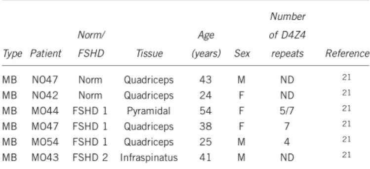
Table 1 Primary human myoblasts used in the study
Type Patient Norm/ FSHD Tissue Age (years) Sex Number of D4Z4 repeats Reference
MB NO47 Norm Quadriceps 43 M ND 21
MB NO42 Norm Quadriceps 24 F ND 21
MB MO44 FSHD 1 Pyramidal 54 F 5/7 21
MB MO47 FSHD 1 Quadriceps 38 F 7 21
MB MO54 FSHD 1 Quadriceps 25 M 4 21
MB MO43 FSHD 2 Infraspinatus 41 M ND 21
Abbreviations: MB, myoblasts; ND, not determined.
Figure 1 Organization of the 4q35 region and the FR-MAR. (a) Schematic representation of the subtelomeric region of chromosome 4 (4q35) showing FRG1 gene, D4Z4 repeat array, isolated D4Z4 repeat homolog (D4Z4*), FR-MAR region that comprises an SSLP and its partial sequence showing major restriction sites, including HincII (GTY^RAG) that delimits the 50 border of the FR-MAR, four CpG sites within FR-MAR (circles), PCR primers (arrowheads)
and a TaqMan probe. *Primers used for amplification of a bisulfite-converted DNA. Sites required for MeCP2 binding ([A/T]Z4) are framed. (b) Schematic representation of the FR-MAR polymorphic region and its partial sequence showing four major SSLPs: 161, 163, 166 and 168, which can be generally described by the formula (CA)n" (AA þ / ") "(CA)n"(G/T) "(CT)n"(8nt þ / "), HincII restriction site that delimits the 30 border of the FR-MAR, PCR
primers used to amplify non-converted (arrowheads) or bisulfite-converted DNA (*). Nucleotide numbering is according to AF117653.2 (GenBank), the first nucleotide of AF117653.2 corresponds to the nucleotide 115529279 of GRCh37.p10 (hg19) human genome reference assembly (NT_016354, GenBank).
Mechanism of attachment of S/MARs to the nuclear matrix NP Kisseljova et al 1118
and 168. An 8nt-long segment being present in haplotypes 166 and 168 but not in haplotypes 161 and 163 (Figure 1b), these are grouped as 8ntþ and 8nt ", respectively. Of note, the same haplotypes are
also found in the duplicated FR-MAR present within the q26 region of human chromosome 10.22–24Consequently, any cell contains four
copies and up to four haplotypes of the FR-MAR. All four copies of
FR-MAR are attached to the NM in normal primary human myoblasts, as was previously demonstrated by in situ fluorescent hybridization on nuclear halos.13
Preferential association of 8ntþ SSLP haplotypes with the NM We have tested whether the presence of the 8nt sequence impacted the efficiency of FR-MAR attachment to the NM. For this purpose, we have used a normal myoblast cell line, NO47, which contains three of the most frequent polymorphisms: 161 and 163, both 8nt", and 166, 8ntþ . In this cell line, the three haplotypes are present in a 2:1:1
ratio, respectively. Total and NM-associated DNA were isolated, the SSLP region of the FR-MAR was PCR amplified and cloned. The relative abundance of 8ntþ and 8nt " haplotypes was then compared between clones derived from total vs NM-attached DNA. As expected, approximately one-fourth of the clones isolated from total DNA (27 clones) had an 8ntþ status vs almost 75% being 8nt " (73 clones) (Figure 2a, left panel). Strikingly, the latter percentage fell to 45% of clones derived from NM-associated fraction (29 clones), with the percentage of 8ntþ clones increasing twofold to 55% (36 clones) (Figure 2a, Po10"3). These findings suggest that DNA with
Figure 2 Effect of SSLP and DNA methylation on the NM binding. (a) The efficiency of FR-MAR binding in vivo to the NM interaction depends on the presence of the 8nt sequence in the SSLP. DNA isolated from the NM fraction or total genomic DNA was PCR amplified using SSLP-F and SSLP-R primers, cloned into pGEM-T vector and sequenced; 100 and 65 clones were analyzed for total DNA and the NM, respectively. The percentage of clones with 8nt þ and 8nt " FR-MAR variants is indicated. The data represent a sum of two independent matrix isolation experiments. (b and c) Schematic representation of results of bisulfite sequencing of total genomic DNA isolated from normal human primary myoblasts and cervical carcinoma cell line. Sodium bisulfite treatment of DNA followed by PCR amplification (primers F1* and R1*; Figure 1a) converts all cytosines of the original DNA into thymines, but all 5-methylcytosines remain unchanged. After this procedure, PCR products were cloned and sequenced: each row of circles represents a cloned individual DNA molecule; closed circles – methylated CpG, open circles – unmethylated CpG and uncharacterized CpGs are represented as gaps. Numbers above the panels correspond to CpG1–4 within FR-MAR from 50 to 30 end. The percentage of methylation is shown under each sample. (b) Total genomic and
NM-associated DNA was isolated from normal human myoblasts NO47 and carcinoma cell line CaSki bisulfite-converted and PCR-amplified using primers F1* and R1*, cloned and sequenced. (c) There are no clear differences between methylation statuses of three FR-MAR variants. Total genomic DNA was isolated from FSHD myoblasts MO44 with three SSLPs, 161,163,166,24 and bisulfite-converted and PCR-amplified using primers F2* and R2*, cloned and
sequenced. The methylation status of CpG2–4is represented. (d) DNA isolated from CaSki chromatin immunoprecipitated with MeCP2-specific antibody (IP).
Mechanism of attachment of S/MARs to the nuclear matrix NP Kisseljova et al 1120
the 8ntþ SSLP haplotype had a much higher affinity to the NM than the 8nt" haplotypes (Figure 2a, right panel).
Methylated FR-MAR sequences are associated with NM
We then asked whether this differential binding of the FR-MAR sequence to the NM could be dependent on the methylation status in this DNA region. It is known that the methylation level of the D4Z4 array varies between normal cells, FSHD myoblasts and cervical carcinoma cells.19,29–32 However, the methylation status of the
FR-MAR sequence was not studied earlier. The total DNA was isolated from several muscular and cervical cell lines and treated as described in Materials and methods section. Seventy-five to 100% of the clones obtained from normal and FSHD myoblasts exhibited a methylated CpG4, with the other three CpGs being methylated to a
lower extent. A strong demethylation of four CpGs was revealed in the cervical carcinomas compared with normal cervix tissues (Po0.001; Supplementary Figure S1). We then isolated NM-asso-ciated DNA from two cell lines, NO47, a primary myoblast cell line, and CaSki, a cervical carcinoma cell line. The total DNA from these two cell lines contained a mixture of methylated and unmethylated CpG within the FR-MAR. In both cases, the clones obtained from the NM-associated fraction were more frequently methylated in each of the four CpGs than those produced from total DNA. Of the four CpGs, CpG4remained the most frequently methylated with 100% of the clones methylated in both cell lines (Figure 2b). Interestingly, when tested in vitro, an unmethylated FR-MAR was unable to bind NM (data not shown). Taken together, these results suggest that a high level of methylation could play a positive role in NM attachment. We then tested the hypothesis that 8ntþ haplotypes bind the NM more efficiently than 8nt" haplotypes because of a higher methyla-tion status of the DNA sequence. Three of the four SSLP haplotypes could be tested using the FSHD-derived myoblast cell line MO44, which contains the 161 (two alleles), 163 and 166 haplotypes. No statistically significant differences could be noticed between the DNA methylation patterns associated with each of the three haplotypes (P40.05, Fisher exact test; Figure 2c). In these experiments, the methylation status of CpG1could not be determined as we could not
obtain stable clones of PCR products that would include all four CpGs and the SSLP region. These experiments demonstrated that all FR-MAR alleles have similar methylation pattern independent of their haplotypes (8ntþ or 8nt ") or location (4q or 10q). From these data, it appears that the differential binding of the different FR-MAR haplotypes to the NM is not directly related to their DNA methylation level.
FR-MAR binds the NM and the MeCP2 protein depending upon its chromatin structure
Having found that DNA methylation affects FR-MAR attachment to the NM, we wondered whether other features of chromatin structure could further contribute to this association. We thus used ChIP to assess whether FR-MAR-containing chromatin was enriched in H3K9ac, an epigenetic mark specific of active chromatin. In normal myoblasts, no H3K9ac was identified within the FR-MAR, contrasting with FSHD cell lines (Figure 3a). The observed chromatin pattern was FR-MAR-specific as it did not spread toward the neighboring regions (Supplementary Figure S3).
A specific association of DNA with the NM also relies on matrix proteins such as the methyl-CpG-binding protein MeCP2,35 which
participates in the formation of chromatin loops.36,37 A methylated
CpG duplex flanked by [A/T]Z4 motif is required for high-affinity MeCP2 binding.38Such a motif is indeed present within the FR-MAR,
one was between CpG3 and CpG4 and the other after CpG4
(Figure 1a). We have looked for the presence of MeCP2 within the FR-MAR region in normal and FSHD myoblasts. High levels of MeCP2 were detected in normal myoblasts, whereas a lower level of MeCP2 was present in FSHD myoblasts (Figure 3a). Interestingly, the presence of MeCP2 appeared to be inversely related to that of H3K9ac not only in primary myoblasts but also in carcinoma cell lines, suggesting that it indicates a common mechanism of MeCP2/ chromatin binding operates in two types of cells (Supplementary Figure S2). We next analyzed the methylation status of MeCP2-associated FR-MAR in carcinoma cell line CaSki. The clones obtained from the DNA fraction immunoprecipitated by MeCP2-specific antibodies were more frequently CpG4 methylated than those produced from total DNA (Figures 2b and d). These data suggest a common mechanism of MeCP2-mediated chromatin attachment to NM in the two types of cells.
Chromatin-modifying drugs affect the FR-MAR association with the NM
To further explore these possible relationships between various features of the chromatin structure and FR-MAR affinity to the NM, we then treated CaSki cells with the methylation inhibitor 5-aza-dC, the histone deacetylation inhibitor trichostatin A (TSA) or a combination there of. Nuclear matrices were isolated from both control and drug-treated cells followed by qPCR analysis of the FR-MAR DNA. In control cells, the NM fraction contained approxi-mately 8% of the total input FR-MAR DNA. This percentage decreased twofold in 5-aza-dC-treated cells and approximately 25% in TSA-treated cells (Figure 3b). An additive effect was obtained when
0.05 0.1 0.15 0.2 0.25 H3K9ac 0.5 1 1.5 2 2.5 3 3.5 MeCP2 % input -beads 2 3 4 5 6 7 8 9 10 % in pu t DNA Nuclear matrix 0 Nor mal FSHD Normal FSHD 0 0 1 AzaC TSA - + - -- + + + ** * * * * ***
Figure 3 FR-MAR chromatin structure analysis and the effect of chromatin-modifying drugs on the affinity of the FR-MAR for the NM. (a) ChIP analysis of the FR-MAR region of normal and FSHD myoblasts. The chromatin was immunoprecipitated with antibodies against MeCP2, and H3K9ac, DNA was isolated from immunoprecipitates and quantified via TaqMan qPCR using G17-F/G17-R primers (Figure 1a). The results of the quantification are presented as an enrichment relative to input DNA (total chromatin) expressed in percentage after subtraction of unspecific chromatin absorption on magnetic beads; the experiments have been carried out in triplicate; *P-value o0.05. (b) Chromatin-modifying drugs affect the affinity of the FR-MAR for the NM. Cells of the cervix carcinoma cell line CaSki were treated either with 5mMDNA methyltransferase inhibitor AzaC, with 1mM
TSA or a combination of both; nuclear matrices were isolated and the amount of the FR-MAR in the input DNA and the NM fraction was quantified using qPCR. The data are presented as an enrichment relative to input DNA expressed in percentage; the average of three independent experiments is shown; *P-value o0.05; **P-value o0.01; ***P-valueo0.001.
the two drugs were combined (Figure 3b). Taken together, these results are consistent with the notion that the FR-MAR attachment to the NM depends on both the methylation of the DNA and the acetylation levels in the chromatin-contained histones.
DISCUSSION
An important aspect of the regulation of gene expression depends upon the organization of chromosomal DNA into chromatin loops (for a review see Razin et al2). This relies upon S/MAR regions that
have a major role in defining the limits of various chromatin loops. However, the molecular mechanism underlying this loop organization remains poorly characterized. Previously, we have demonstrated that the organization of chromatin loops was altered in myoblasts from patients with FSHD.13 FSHD is also associated with a partial
loss of DNA methylation and heterochromatin-specific histone modifications in D4Z4 repeats.19,39,40 In this chromosomal region,
altered levels of DNA methylation have also been associated with other genetic diseases such as the Rett’s syndrome and the ICF syndrome,25as well as in several types of cancer.28–30
Here, we have first studied the NM association of a 368-bp-long DNA segment containing four different SSLPs. Taken together, these SSLPs account for the majority of the genotypes identified within the FR-MAR. We found that the presence of an 8ntþ haplotypes within the polymorphic region was associated with a higher affinity for the NM. These 8ntþ haplotypes are generally not associated with the FSHD phenotype.22–24 Next, we have analyzed the level of DNA
methylation at four CpGs in the FR-MAR region in relation with NM attachment. One of the four CpGs, CpG4, was found to be 100%
methylated in the NM fraction. In total DNA, CpG4was also by far
the most frequently methylated CpG, although with variable frequencies depending on the cells analyzed. When the cells were treated with the DNA methylation inhibitor 5-aza-dC, the affinity of the FR-MAR to the NM was reduced more than twofold, indicating that DNA methylation probably favors an efficient interaction
between FR-MAR and the NM. Owing to the fact that a copy of FR-MAR is present within the q26 region of human chromosome 10,22–24data obtained here are valid for both 4q35 and 10q26
FR-MARs.
Chromosome looping is known to be mediated, at least in part, by MeCP2,36,37 a methyl-cytosine-binding protein that associates with nuclear matrices.41MeCP2 binding sites were indeed identified within
the FR-MAR. Using ChIP, we have observed that the level of MeCP2 binding to the FR-MAR was directly related with NM binding, consistent with a role of MeCP2 in FR-MAR attachment to NM. Additional elements could still have a role in NM attachment. Indeed, in the FR-MAR, the levels of histone H3 acetylated at lysine 9 were found to be inversely related with NM binding. Furthermore, when cells were treated with the histone deacetylase inhibitor TSA, the binding of the FR-MAR to the NM was significantly reduced, a direct experimental evidence of the impact of a chromatin-modifying agent on NM attachment. This effect was additive with that of 5-aza-dC, consistent with a combined role of DNA methylation and chromatin structure in NM attachment. Of note, 5-aza-dC and TSA are both known to provoke a decompaction of chromatin, which thus appears to inhibit the interaction between the FR-MAR and the NM.
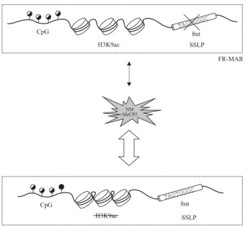
In summary, three criteria seem to be important for high-affinity interaction between the FR-MAR and the NM: (i) the presence of an 8ntþ haplotype in chromosomal DNA; (ii) the methylation of one specific CpG, namely CpG4, within the FR-MAR; and (iii) the
absence of H3K9ac in the relevant chromatin fragment (Figure 4). The presence of MeCP2 molecules provides another favoring factor. Whether other proteins have an additional role in this process will require further studies.
CONFLICT OF INTEREST
The authors declare no conflict of interest.
ACKNOWLEDGEMENTS
This research was supported by grants from the Association Franc¸aise contre les Myopathies (AFM) to YSV and DL, by Grants No. 10-04-00489 from RFBR (Russia) to NK, AK, DE, by a grant from Protek groups of companies to AK and by the exchange Grant No. 23981 from the CNRS to NK and ML. PD was supported by the Association ‘Amis FSH’ and the University Montpellier I. EK was supported by the ‘Stichtung FSHD’ fund (The Netherlands).
1 Vassetzky YS, Hair A, Razin SV: Rearrangement of chromatin domains in cancer and development. J Cell Biochem 2000; S35: 54–60.
2 Razin SV, Iarovaia OV, Sjakste N et al: Chromatin domains and regulation of transcription. J Mol Biol 2007; 369: 597–607.
3 Brun C, Dang Q, Miassod R: Studies of an 800-kilobase DNA stretch of the Drosophila X chromosome: comapping of a subclass of scaffold-attached regions with sequences able to replicate autonomously in Saccharomyces cerevisiae. Mol Cell Biol 1990; 10: 5455–5463.
4 Iarovaia O, Hancock R, Lagarkova M, Miassod R, Razin SV: Mapping of genomic DNA loop organization in a 500-kilobase region of the Drosophila X chromosome by the topoisomerase II-mediated DNA loop excision protocol. Mol Cell Biol 1996; 16: 302–308.
5 Ioudinkova E, Petrov A, Razin SV, Vassetzky YS: Mapping long-range chromatin organization within the chicken alpha-globin gene domain using oligonucleotide DNA arrays. Genomics 2005; 85: 143–151.
6 Linnemann AK, Platts AE, Krawetz SA: Differential nuclear scaffold/matrix attachment marks expressed genes. Hum Mol Genet 2009; 18: 645–654.
7 Drennan KJ, Linnemann AK, Platts AE, Heng HH, Armant DR, Krawetz SA: Nuclear matrix association: switching to the invasive cytotrophoblast. Placenta 2010; 31: 365–372.
8 Platts AE, Quayle AK, Krawetz SA: In silico prediction and observations of nuclear matrix attachment. Cell Mol Biol Lett 2006; 11: 191–213.
9 Mirkovitch J, Mirault ME, Laemmli UK: Organization of the higher-order chromatin loop: specific DNA attachment sites on nuclear scaffold. Cell 1984; 39: 223–232.
Figure 4 Three factors contribute, either independently or together, to the FR-MAR binding to the NM: DNA methylation, chromatin structure and the SSLP. Methylation of CpG4, low level of the H3K9ac and the presence of
the 8nt þ SSLP within the FR-MAR region are associated with a more efficient interaction of FR-MAR with the NM.
Mechanism of attachment of S/MARs to the nuclear matrix NP Kisseljova et al 1122
10 Vassetzky YS, Bogdanova AN, Razin SV: Analysis of the chicken DNA fragments that contain structural sites of attachment to the nuclear matrix: DNA–matrix interactions and replication. J Cell Biochem 2000; 79: 1–14.
11 Vassetzky Y, Hair A, Mechali M: Rearrangement of chromatin domains during development in Xenopus. Genes Dev 2000; 14: 1541–1552.
12 Oberhammer F, Wilson JW, Dive C et al: Apoptotic death in epithelial cells: cleavage of DNA to 300 and/or 50 kb fragments prior to or in the absence of internucleosomal fragmentation. EMBO J 1993; 12: 3679–3684.
13 Petrov A, Pirozhkova I, Laoudj D, Carnac G, Lipinski M, Vassetzky YS: Chromatin loop domain organization within the 4q35 locus in facioscapulohumeral dystrophy patients versus normal human myoblasts. Proc Natl Acad Sci USA 2006; 103: 6982–6987. 14 Petrov AV, Allinne J, Pirozhkova IV, Laoudj D, Lipinski M, Vassetzky YS: A nuclear matrix attachment site in the 4q35 locus has an enhancer-blocking activity in vivo: implications for the facio-scapulo-humeral dystrophy. Genome Res 2008; 18: 39–45. 15 Bode J, Maass K: Chromatin domain surrounding the human interferon-beta gene as
defined by scaffold-attached regions. Biochemistry 1988; 27: 4706–4711. 16 Forrester WC, Fernandez LA, Grosschedl R: Nuclear matrix attachment regions
antagonize methylation-dependent repression of long-range enhancer–promoter inter-actions. Genes Dev 1999; 13: 3003–3014.
17 Fernandez LA, Winkler M, Grosschedl R: Matrix attachment region-dependent function of the immunoglobulin mu enhancer involves histone acetylation at a distance without changes in enhancer occupancy. Mol Cell Biol 2001; 21: 196–208.
18 Martens JH, Verlaan M, Kalkhoven E, Dorsman JC, Zantema A: Scaffold/matrix attachment region elements interact with a p300-scaffold attachment factor A complex and are bound by acetylated nucleosomes. Mol Cell Biol 2002; 22: 2598–2606. 19 deGreef JC, Frants RR, van der Maarel SM: Epigenetic mechanisms of
facioscapulo-humeral muscular dystrophy. Mutat Res 2008; 647: 94–102.
20 Dmitriev P, Lipinski M, Vassetzky YS: Pearls in the junk: dissecting the molecular pathogenesis of facioscapulohumeral muscular dystrophy. Neuromusc Disord 2009; 19: 17–20.
21 Dmitriev P, Barat A, Cochet E et al: FSHD myoblasts fail to downregulate intermediate filament protein vimentin during myogenic differentiation. Biopolym Cell 2011; 27: 359–363.
22 Lemmers RJ, van der Vliet PJ, Klooster R et al: A unifying genetic model for facioscapulohumeral muscular dystrophy. Science 2010; 329: 1650–1653. 23 Lemmers RJ, Wohlgemuth M, van der Gaag KJ et al: Specific sequence variations
within the 4q35 region are associated with facioscapulohumeral muscular dystrophy. Am J Hum Genet 2007; 81: 884–894.
24 Lemmers RJ, van der Vliet PJ, van der Gaag KJ et al: Worldwide population analysis of the 4q and 10q subtelomeres identifies only four discrete interchromosomal sequence transfers in human evolution. Am J Hum Genet 2010; 86: 364–377.
25 Forlani G, Giarda E, Ala U et al: The MeCP2/YY1 interaction regulates ANT1 expression at 4q35: novel hints for Rett syndrome pathogenesis. Hum Mol Genet 2010; 19: 3114–3123.
26 Kondo T, Bobek MP, Kuick R et al: Whole-genome methylation scan in ICF syndrome: hypomethylation of non-satellite DNA repeats D4Z4 and NBL2. Hum Mol Genet 2000; 9: 597–604.
27 de Greef JC, Lemmers RJ, van Engelen BG et al: Common epigenetic changes of D4Z4 in contraction-dependent and contraction-independent FSHD. Hum Mutat 2009; 30: 1449–1459.
28 Cadieux B, Ching TT, VandenBerg SR, Costello JF: Genome-wide hypomethylation in human glioblastomas associated with specific copy number alteration, methylene-tetrahydrofolate reductase allele status, and increased proliferation. Cancer Res 2006; 66: 8469–8476.
29 Tsumagari K, Qi L, Jackson K et al: Epigenetics of a tandem DNA repeat: chromatin DNaseI sensitivity and opposite methylation changes in cancers. Nucleic Acids Res 2008; 36: 2196–2207.
30 Katargin AN, Pavlova LS, Kisseljov FL, Kisseljova NP: Hypermethylation of genomic 3.3-kb repeats is frequent event in HPV-positive cervical cancer. BMC Med Genom 2009; 2: 30.
31 Choi SH, Worswick S, Byun HM et al: Changes in DNA methylation of tandem DNA repeats are different from interspersed repeats in cancer. Int J Cancer 2009; 125: 723–729.
32 Tilman G, Loriot A, Van Beneden A et al: Subtelomeric DNA hypomethylation is not required for telomeric sister chromatid exchanges in ALT cells. Oncogene 2009; 28: 1682–1693.
33 Barro M, Carnac G, Flavier S, Mercier J, Vassetzky YS, Laoudj-Chenivesse D: Primary myoblasts derived from the facioscapulohumeral dystrophy patients are hypersensitive to oxidative stress and show defects upon terminal differentiation. J Cell Mol Med 2010; 14: 275–289.
34 Gasser SM, Vassetzky YS: Analysis of nuclear scaffold attachment regions. In: Gould H (ed) Chromatin: A Pratical Approach. Oxford, UK: Oxford University Press, 1998; pp 111–124.
35 Stratling WH, Yu F: Origin and roles of nuclear matrix proteins. Specific functions of the MAR-binding protein MeCP2/ARBP. Crit Rev Eukaryot Gene Expr 1999; 9: 311–318.
36 Horike S, Cai S, Miyano M, Cheng JF, Kohwi-Shigematsu T: Loss of silent-chromatin looping and impaired imprinting of DLX5 in Rett syndrome. Nat Genet 2005; 37: 31–40.
37 Eivazova ER, Gavrilov A, Pirozhkova I et al: Interaction in vivo between the two matrix attachment regions flanking a single chromatin loop. J Mol Biol 2009; 386: 929–937.
38 Klose RJ, Sarraf SA, Schmiedeberg L, McDermott SM, Stancheva I, Bird AP: DNA binding selectivity of MeCP2 due to a requirement for A/T sequences adjacent to methyl-CpG. Mol Cell 2005; 19: 667–678.
39 Zeng W, de Greef JC, Chen YY et al: Specific loss of histone H3 lysine 9 trimethylation and HP1gamma/cohesin binding at D4Z4 repeats is associated with facioscapulo-humeral dystrophy (FSHD). PLoS Genet 2009; 5: e1000559.
40 van Overveld PG, Lemmers RJ, Sandkuijl LA et al: Hypomethylation of D4Z4 in 4q-linked and non-4q-linked facioscapulohumeral muscular dystrophy. Nat Genet 2003; 35: 315–317.
41 Weitzel JM, Buhrmester H, Stratling WH: Chicken MAR-binding protein ARBP is homologous to rat methyl-CpG-binding protein MeCP2. Mol Cell Biol 1997; 17: 5656–5666.
Supplementary Information accompanies this paper on European Journal of Human Genetics website (http://www.nature.com/ejhg)