HAL Id: hal-01465453
https://hal.archives-ouvertes.fr/hal-01465453
Submitted on 12 Feb 2017HAL is a multi-disciplinary open access
archive for the deposit and dissemination of sci-entific research documents, whether they are pub-lished or not. The documents may come from teaching and research institutions in France or abroad, or from public or private research centers.
L’archive ouverte pluridisciplinaire HAL, est destinée au dépôt et à la diffusion de documents scientifiques de niveau recherche, publiés ou non, émanant des établissements d’enseignement et de recherche français ou étrangers, des laboratoires publics ou privés.
Red Wine and Pomegranate Extracts Suppress Cured
Meat Promotion of Colonic Mucin-Depleted Foci in
Carcinogen-Induced Rats
Nadia Bastide, Nathalie Naud, Gilles Nassy, Jean-Luc Vendeuvre, Sylviane
Taché, Françoise Guéraud, Ditte Hobbs, Gunter Kuhnle, Denis E. Corpet,
Fabrice H.F. Pierre
To cite this version:
Nadia Bastide, Nathalie Naud, Gilles Nassy, Jean-Luc Vendeuvre, Sylviane Taché, et al.. Red Wine and Pomegranate Extracts Suppress Cured Meat Promotion of Colonic Mucin-Depleted Foci in Carcinogen-Induced Rats. Nutrition and Cancer, Taylor & Francis (Routledge), 2017, 69 (2), pp.289 - 298. �10.1080/01635581.2017.1263745�. �hal-01465453�
NUTRITION AND CANCER
2017, VOL. 69, NO. 2, 289–298
http://dx.doi.org/10.1080/01635581.2017.1263745
Red Wine and Pomegranate Extracts Suppress Cured Meat Promotion of
Colonic Mucin-Depleted Foci in Carcinogen-Induced Rats
Nadia M. Bastide a, Nathalie Naud a, Gilles Nassy b, Jean-Luc Vendeuvre b, Sylviane Taché a, Françoise Guéraud a, Ditte A. Hobbs c, Gunter G. Kuhnle c, Denis E. Corpet a, and Fabrice H. F. Pierre a
A INRA UMR1331, TOXALIM (Research Center in Food Toxicology), Universite de Toulouse, ENVT, INP, Toulouse, France; b IFIP-Institut du Porc, Paris, France;
c Department of Food and Nutritional Sciences, University of Reading, Whiteknights, UK ABSTRACT
Processed meat intake is carcinogenic to humans. We have shown that intake of a workshop-made cured meat with erythorbate promotes colon carcinogenesis in rats. We speculated that
polyphenols could inhibit this effect by limitation of endogenous lipid peroxidation and nitrosation. Polyphenol-rich plant extracts were added to the workshop-made cured meat and given for 14 days to rats and 100 days to azoxymethane-induced rats to evaluate the inhibition of preneoplastic lesions. Colons of 100-d study were scored for precancerous lesions (mucin-depleted foci, MDF), and biochemical end points of peroxidation and nitrosation were measured in urinary and fecal samples. In comparison with cured meat-fed rats, dried red wine, pomegranate extract, a-tocopherol added at one dose to cured meat and withdrawal of erythorbate significantly decreased the number of MDF per colon (but white grape and rosemary extracts did not). This protection was associated with the full suppression of fecal excretion of nitrosyl iron, suggesting that this nitroso compound might be a promoter of carcinogenesis. At optimized concentrations, the incorporation of these plant extracts in cured meat might reduce the risk of colorectal cancer associated with processed meat consumption.
Introduction
Colorectal cancer (CRC) is the third most common type of cancer worldwide and the second cause of cancer death in affluent countries (1). Epidemiological
studies show that processed meat intake is linked to the risk of CRC (2). The World Cancer Research Fund panel considers this risk as convincing and recommends avoiding processed meat consumption (3,4).
The World Health Organization classified consumption of processed meat as “carcinogenic to humans”
(IARC Group 1) based on sufficient evidence for colorectal cancer (5). Making safer meat products could be an alternative to banning cured meat (6,7). In
carcinogen-initiated rats, given a low-calcium diet freeze-driedcooked ham and moist hot-dog increase significantly the number of mucin-depleted foci (MDF) (8,9). The intake of an experimental cured pork meat, similar to an air-exposed cooked shoulder-ham (DCNO for dark cooked meat with nitrite, oxidized, described below), also promotes carcinogenesis in rats (10). In human volunteers, cured meat intake increases endogenous nitrosation and fat peroxidation and fecal waterinduced oxidative DNA damage (7,11).
We have speculated that heme-iron could explain in part the promoting effect of processed meat (12,13), and added experimental support to this hypothesis: Dietary hemin (free heme stabilized by a chloride ion) promotes azoxymethane-induced aberrant crypt foci (ACF) in the colon of rats (14). Hemin, but not hemoglobin, mimics
the effect of ham on biomarkers associated with carcinogenesis (8).
Heme-iron catalyzes the formation of apparent total nitroso compounds (ATNC) (15) and of lipid peroxidation end products, e.g., 4-hydroxynonenal and other
alkenals (16). 4-hydroxynonenal is cytotoxic and genotoxic to the intestinal epithelial cells (17,18). Potentially carcinogenic ATNC are formed in the gastrointestinal tract by N-nitrosation of peptide-derived amines or amides. Nitrosylated heme-iron present in processed red meat also represents a significant part of measured ATNC (13,19,20). ATNC and alkenals could explain tumor promotion by dietary heme and by cured meat (6,21,22).
Our starting hypothesis here was that lipid peroxidation end products would promote carcinogenesis (23), and that polyphenols would decrease heme-induced luminal peroxidation (24) and hence carcinogenesis. There is ample evidence that polyphenols and plant extract can block heme-induced fat peroxidation: For instance, quercetin, red wine, and a-tocopherol suppress myoglobin-induced peroxidation in a fat/water emulsion that mimics the gastric environment and blocks the accumulation of conjugated dienes (25,26). In human volunteers, red wine polyphenols strongly decrease postprandial plasma malondialdehyde after a red meat meal, probably by suppressing heme-induced
peroxidation in the stomach (27–29). In rats, a mix of rutin and butylated hydroxyanisole inhibits hemin-induced lipid peroxidation in the gut and suppresses carcinogenesis promotion (14). In addition, polyphenols
such as punicalagin and ellagic acid from pomegranate can chelate iron through catechol groups (30,31), while propyl gallate, tannic acid, thymol, vanillin, and ascorbate and a-tocopherol can inhibit nitrosation and ATNC formation (32,33).The present study was designed to test the hypothesis that polyphenols can prevent the promotion of colon tumorigenesis by processed meat, by suppressing lipid peroxidation in the gut. In a short-term screening study, several agents were added to DCNO cured meat during the manufacturing process. Such diets were given to rats for 14 days. Early lipid peroxidation end points were measured in feces and urine. Most promising agents selected during these screening studies were added to DCNO and tested for chemoprevention in a 100-day carcinogenesis study in rats. Tumorigenesis end points were azoxymethane-induced preneoplastic lesions (ACF and MDF) in rats. The results showed that dried red wine, pomegranate extract, and a-tocopherol prevented
meatassociated formation of fecal nitrosyl iron and promotion of preneoplastic lesions.
Materials and Methods
Animal Study Design
Two sequential studies were performed on male Fischer 344 rats purchased at 4–5 weeks of age from Charles River (St Germain l’Arbresle, France): A 14-day study investigated the effect of plant extracts added to an experimental cured meat on early fecal and urinary biomarkers in rats. A 100-day study measured the antipromoting effect of four plant extracts added to the same cured meat, on preneoplastic lesions in carcinogen initiated rats. Animal care was in accordance with the guidelines of the European Council on animals used in experimental studies. Study was done in an accredited animal colony (French A 31504) by approved staff (e.g., P.I. Corpet: Certificat d’autorisation d’expérimenter sur animaux vertébrés vivants #31-121).
Short-term Study Design (14 Days-long)
Forty-three rats were housed individually in metabolic cages. They were kept at 22_C and 12 h–12 h lightdark
cycle. After 3 days of acclimatization to the animal colony and to a standard AIN76 diet, rats were randomly allocated to eight groups. There were five rats in each experimental group given DCNO cured meat with plant extracts (described below), and eight rats in the control group fed DCNO. Rats were fed the experimental diets described below during 14 days and allowed free access to tap water. Body weight was monitored every week. Food and water intakes were measured at day 13. Feces and urine were collected at days 11 and 12 and frozen at ¡20_C. Animals were terminated by CO2 asphyxiation
on day 14. Fecal water samples (preparation
described below) were analyzed for heme, cytotoxicity, and thiobarbituric acid reactive substances
(TBARS). Urine samples were analyzed for 1,4-dihydroxynonane mercapturic acid (DHN-MA).
Carcinogenesis Study (100 Day-long): Animals and Design
Eighty-six rats were housed individually in stainless steel, wire-bottomed cage (same animal colony as above). After 7 days of acclimatization, each rat received a single i.p. injection of azoxymethane (20 mg/kg i.p.; Sigma Chemical) in NaCl (9 g/L). Seven days later, they were randomly allocated to seven groups (N D 10 rats per group, except control group, N D 26) and fed the experimental DCNObased diets described below. Body weights were monitored every week for four weeks, then every two
weeks. Food and water intakes were measured at days 20 and 80. Feces were collected daily between days 18 and 21, and 80 and 91 and frozen at ¡20_C. Between
days 74 and 76, each rat was put in a metabolic cage, and urine was collected and frozen at ¡20_C. Rats were killed by CO2 asphyxiation in a random order
at day 96–98. Colons were removed and fixed in 10% buffered formalin (Sigma Chemical) between two sheets of filter paper with a blinding code. ACF and MDF were scored. Fecal water samples were analyzed for heme, TBARS, cytotoxicity, and ATNC. Urine samples were analyzed for DHN-MA.
Animal Diets
The type of meat and the additives that were given to groups of rats during the short-term and carcinogenesis studies are listed in the first column of Tables 1 and 2, respectively.
Pork meat was cured in a specialized workshop by IFIP-Institut du Porc (14-day study) and in a ham factory by Fleury Michon (Pouzauges-France) (100-day
study). Meat was given as such (moist piece) to the rats because freeze-drying boosts peroxidation of fat in meat (34). The experimental cured meat, which was similar to air-exposed picnic ham and called DCNO, was chosen because it promotes carcinogenesis in rats
(10). DCNO was made from Musculus vastus
intermedius, cured with 2.19 g salt with 0.6% sodium
nitrite (131 ppm NaNO2), and 1.4 g sodium erythorbate
(an ascorbate isomer) per 100 g meat. DCNO was then heated at 70_C for 3 h in vacuum-sealed plastic bags
in a water bath. The final product contained 12 mg heme-iron/kg, 71 mg sodium nitrite/kg, and 500 mg ascorbate/kg. One group of rats was given an erythorbate- free DCNO. The processed meat was divided into
1.3-cm thick slices of 300 g, that were stored separately at ¡20_C in air-tight plastic bags with low-oxygen
permeability (14 day study) or under CO2/N2 50/50
atmosphere to avoid further fat oxidation (100 day study). Before being given to rats, each slice was exposed to air for five days in a dark refrigerator (4_C), then cut into ten 30-g portions that were given to rats at 5:00 p.m. for 14 or 100 days. A low-calcium powdered diet
(35) was given in a separated feeder, 7.6 g/d/rat, so that each rat would eat roughly half meat/half powder (dry matter). This modified AIN76 diet was prepared by UPAE (INRA, Jouy, France) as follows (g/100 g):
sucrose, 59.5; corn starch, 15.0; cellulose, 12.5; AIN76 mineral mix without calcium, 8.7; AIN76 vitamin mix, 2.5; methionine, 0.75; calcium phosphate, 0.52; choline bitartrate, 0.5. Safflower oil (5 g) was mixed with 100- g powder to provide polyunsaturated fatty acids (MP Biomedicals, Illkirch, France).
Six polyphenols-rich plant extracts were added to DCNO during the curing process, at a concentration recommended by the supplier: white grape extract (NutriPhy_ white grape 100, 72% of total polyphenols, CHR Hansen, Horsholm, Denmark; 0.055% w/w in DCNO), carnosic acid (StabilEnhance_ OSR5 extracted
from rosemary leaves, 10% carnosic acid, Naturex, Avignon, France; 1% w/w in DCNO), and a water soluble rosemary extract, containing 7% of rosmarinic acid (Stabilenhance_ WSR6, Naturex; 0.66% w/w in DCNO),
red wine concentrate, 10% of total polyphenols
(Avvinr9005_, Diana Naturals, Antrain, France; 2% w/w in DCNO), pomegranate extract, 12% ellagic acid (Naturex, Ultimate Botanical Benefits; 0.6% w/w in DCNO), green tea extract, 98% of total polyphenols (Naturex; 0.08% w/w in DCNO). Polyphenol data were given by the suppliers, and the composition of extracts was not determined more precisely in this pilot study. Another group was given DCNO supplemented with a-tocopherol (Covitol_, Nutrition & Health, Cognis, BASF; 0.045%): this fat-soluble antioxidant agent suppresses MDF in carcinogen-induced rats and was used as a positive control for protection (7). A last group of rat given DCNO without sodium erythorbate was added to the carcinogenesis study.
Meat Composition
Processed meat was analyzed by Lareal (Vannes, France, laboratory specialized in physicochemical and
microbiological analyzes) for total iron, total pigments, nitrosylated pigments (36). Hexanal, a marker of
secondary products of lipid peroxidation, was analyzed by Lareal by gas chromatography of the headspace of the sample dispersed in phosphate buffer at 37_C, with
solid-phase micro-extraction fiber. Trolox equivalent antioxidative capacity (TEAC-1), malondialdehyde (MDA by HPLC), and TBARS (after acidic extraction) were measured by ADIV (Clermont-Ferrand, France). The oxygen radical absorbance capacity (ORAC) was measured by Naturex (Avignon, France). Two measures per processed meat batch were done.
Fecal and Urinary Measures
Analysis of Heme, Thiobarbituric Acid Reactive Substances in, and Cytotoxicity of Fecal Water, and 1,4-Dihydroxynonane Mercapturic Acid in Urine
Fecal pellets were collected under each cage for 24 h, at day 11 of the short-term study and days 88–91 of the carcinogenesis study. TBARS value was used as a global measure of lipid peroxidation end products. Fecal water was prepared, and heme and TBARS were measured in fecal water exactly as previously described (7) except that
1 mL of distilled water was added to 0.42 g of crushed fresh feces, but not to 0.3 g of dried feces.
1,4-Dihydroxynonane mercapturic acid
(DHN-MA) is themain urinary metabolite of
4-hydroxynonenal, which is a major toxic end product of endogenous fat peroxidation (16). The 24-h urine was collected under each metabolic cage, at day 11 of the short-term study and days 74–76 of the carcinogenesis study. DHN-MA assay was done (n D 5–8 for the 14-day study and n D 10–26 for the 100-day study) as previously described (7). To determine cytotoxicity of fecal water (n D 6), the
3(4,5-dimethylthiazol-2-yl)-2,5-diphenyltetrazolium bromide (MTT) assay was used on a cancerous mouse colonic epithelial cell line, CMT93 (European Collection of Animal Cultures), as previously described (7).
ATNC Analysis
ATNC were analyzed using a modification of the method previously used (37), using a CLD88 Exhalyzer
(Ecomedics, Duernten, Switzerland). Sulfamic acid solution (500 ml, 5%) was added to 100 ml of fecal water to remove nitrite, and samples were injected into a purged vessel kept at 60_C and filled with a standard tri-iodide reagent (38mg I2 was added to a solution of 108 mg KI in
1 ml water; to this mixture, 13.5 ml of glacial acetic acid was added) to determine total ATNC. To determine mercury(II) stable compounds, 100 ml of 10-mM aqueous HgCl2 was added prior to analysis; to determine
mercury(II) and ferricyanide stable compounds, 100 ml each of 10 mMaqueousHgCl2 and 10-mM
aqueous K3Fe(CN)6 solution were added prior to analysis.
Nitrosothiols were determined as the difference between total ATNC and mercury(II) stable ATNC; nitrosyl iron was determined as a difference between mercury(II) stable ATNC and mercury(II) and K3Fe(CN)6 stable
compounds. Data are concentrations (in mM), measured in triplicate in 100 mL of each sample.
ACF and MDF Assays
ACF and MDF were scored by a single observer blinded for the origin of the colon, exactly as described previously
(7). Number of lesions and number of crypts per lesions (i.e., size of ACF and MDF) were numbered.
Statistical Methods
Results were analyzed using Systat 10 software for Windows, and all data were reported as mean § SD (except Fig. 1B). Values were considered firstly usingone-way analysis of variance. If a significant difference was found between all groups (P < 0.05), comparison of each experimental group with the control group was made using Dunnett’s test. For ORAC analysis, data show results of twomeasures per processed meat batch, but Student t-test statistics could be done because the within-pair correlation was high; however, P values should be taken cautiously (38).
Results
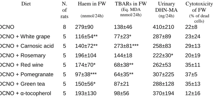
Fourteen-Day Study: Effect of Plant Extracts on Peroxidation Biomarkers
Fecal and Urinary Fat Peroxidation Biomarkers
Dietary DCNO cured meat increases the number of carcinogen-induced precancerous lesions, and urinary and fecal water peroxidation biomarkers in rats (10,14). These early peroxidation biomarkers were thus measured here because they correlate with heme-induced promotion of colon carcinogenesis (8,39). Extracts of pomegranate, red wine, white grape, green tea, rosemary,
and carnosic acid and a-tocopherol were added to DCNO before being fed to rats for 14 days. As shown in
Table 1, fecal water from rats given DCNO added with pomegranate, red wine, or white grape extracts contained half TBARS than control rats given DCNO. In contrast, fecal water from rats given DCNO plus carnosic acid contained surprisingly twice more TBARS than controls. All tested extracts led to some reduction in urinary DHN-MA, but only rosemary extract significantly decreased the excretion of this 4-hydroxynonenal metabolite. All the tested plant extracts reduced fecal heme excretion in DCNO-fed rats, except for
a-tocopherol and rosemary extract. Finally, addition of plant extracts in DCNO did not affect the cytotoxicity
associated with DCNO consumption.
Choice of Polyphenol Additives for the Carcinogenesis Study
Pomegranate, red wine, and white grape extracts that decreased TBARS in fecal water of DCNO-fed rats (Table 1) were chosen to be tested in the carcinogenesis study because our starting hypothesis was that
polyphenols would exert their protective action by inhibiting lipid peroxidation (25,40).We also chose to test carnosic acid, a common additive to brine in Europe, because it surprisingly increased TBARS in fecal water. In addition, we tested a-tocopherol as a protection control because it suppresses cured meat promotion in rats (7). Finally, a special DCNO meat, cured without erythorbate, was given to a group of rat to test the effect of this common additive.
Carcinogenesis Study: Effect of Plant Extracts
General Observation
All rats survived and were healthy, except rats given carnosic acid that had diarrhea. Moist meat and powdered diet were given to the rats in separated feeders: the relative intake of meat and of powder that was 48:52 (dry weight) on day 18 of the study slowly changed to 39:61 on day 82. The final body weight of rats was 343 ± 19 g without significant difference between groups except rats fed cured meat plus carnosic acid (321 § 16 g, P < 0.05). Rats in this group ate and drank less than the rats in others groups: their average food intake per day was 12±1 g comparedwith13±1 g in other groups (P < 0.05).Water intake was reduced in rat fed carnosic acid and increased
in rat fed a-tocopherol or white grape extract, compared with the other groups (full data not shown, P<0.0001).
Quantification of ACF and MDF
A DCNO-based diet increases the number of MDF and ACF in the colon of carcinogen-injected rats, in comparison with a no-meat control diet (7,10). The DCNO diet was thus chosen as a promoting control to test potentially protective plant extracts. At the doses tested, all plant extracts decreased the number of MDF per colon in comparison with DCNO diet, but only a-tocopherol, pomegranate, and red wine extracts led to a significant protection (Fig. 1B). Neither the number of ACF nor the MDF and ACF multiplicity was different between groups (Table 3). Surprisingly, the removal of erythorbate from DCNO curing brine led to a significant reduction in the number of colonic MDF. Mean number of large ACF or of large MDF, with 4 or more crypts per foci, was similar in all dietary groups. In an attempt to explain the
observed protection, diets, fecal water, and urine were analyzed for lipoperoxides and nitroso compounds.
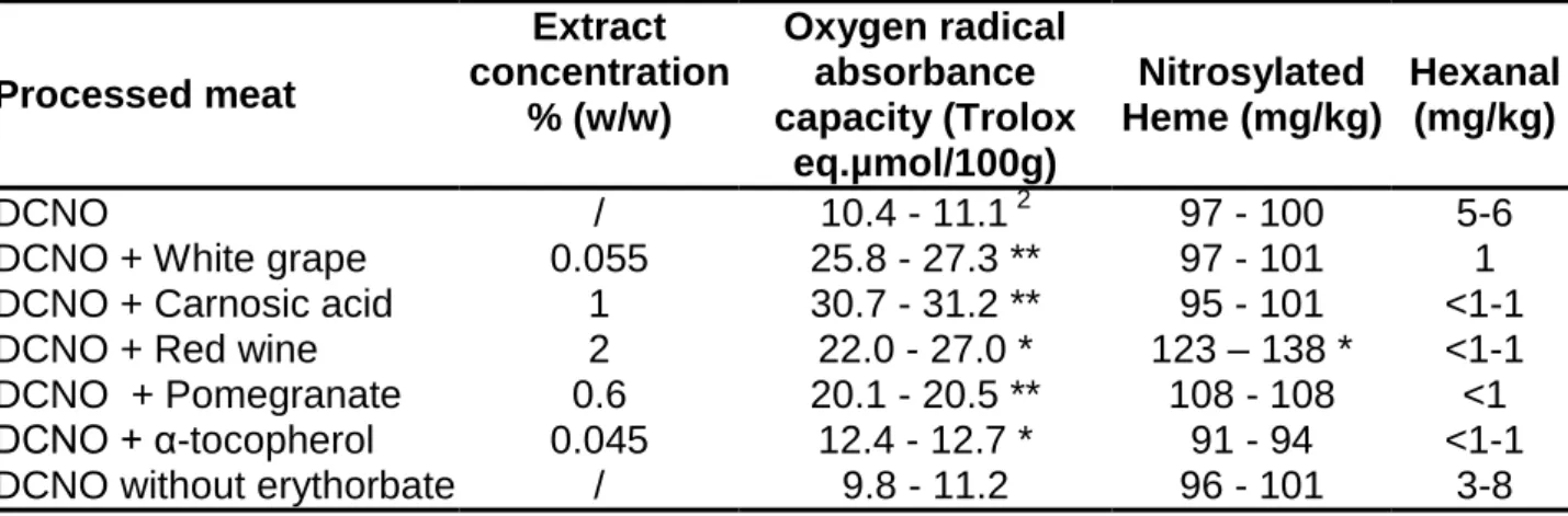
Meat Analyses
Trolox, MDA, TBARS, ORAC, hexanal, and nitrosylated heme were analyzed, but only ORAC, hexanal, and nitrosylated heme values were reported here, because they are not significantly affected by the modification of process of meats. ORAC is a measure of antioxidant power. As expected, all tested plant extracts increased cured meat ORAC value 2–3 times and suppressed hexanal production, a measure of meat peroxidation (Table 4). Heme and NO from nitrite can form nitrosyl heme (19) that might be the promoting factor in cured meat (8). The tested plant extracts did not change much nitrosylated heme concentration in meat, except red wine that increased it (Table 4).
Fecal and Urinary Fat Peroxidation Biomarkers
Fecal water from rats fed DCNO added with pomegranate, red wine, or white grape extracts, or a-tocopherol
contained 1.5–2 times less TBARS, and about 1.5 times less heme than fecal water from rats fed DCNO alone (Table 2). Pomegranate extract, carnosic acid, and a-tocopherol also decreased urinary DHN-MA, a metabolite of 4-hydroxy-nonenal. Carnosic acid that significantly increased fecal TBARS in the first study (Table 1) tended to increase it in this second study (not significant).
Addition of plant extracts had not modified the cytotoxicity of fecal water in DCNO group except for carnosic acid that induces a significant increase in fecal water cytotoxicity (Table 2). Surprisingly, the absence of erythorbate in DCNO significantly decreased fecal TBARS and heme compared with
erythorbate-supplemented DCNO, without modification of cytotoxic activity of fecal water (Table 2).
Fecal Nitroso Compounds
ATNC concentration in fecal samples was reduced by the addition of a plant extract to the curing brine of DCNO
(Fig. 1A). The reduction was more than a three-fold (except white grape). Significance could not be formally established since only one value was obtained per group because feces from all rats in one dietary group had been pooled, so results were interpreted with caution.
“ATNC” are a complex mixture of nitrite-derived products, and the ATNC composition was not identical in the feces from different groups. Fecal ATNC from rats fed DCNO cured meat plus carnosic acid or white grape extract were made of 100% nitrosyl iron (Fig. 1A). In contrast, fecal ATNC from rats fed DCNO with
pomegranate or red wine extracts were 100% nitrosothiols (data not shown). Tocopherol fully suppressed nitrosation, but the removal of erythorbate from DCNO curing brine led to a fifty percent increase in fecal ATNC, no nitrosyl iron being detected (Fig. 1A).
Discussion
This study shows that polyphenol-rich plant extracts can inhibit the promotion of colonic mucin-depleted foci by cured meat that had been demonstrated repeatedly in this model (7–10): dried red wine and pomegranate extract suppressed cured meat-induced colon
tumorigenesis promotion as well as a-tocopherol, while white grape extract and carnosic acid extracted from rosemary did not. Promotion was evidenced on a
surrogate end point biomarker, mucin-depleted foci. MDF, formed by dysplastic crypts devoid of mucin, have been identified in the colon of humans at high risk for colon cancer (41). Like tumors, MDF harbor mutations in genes affecting colon carcinogenesis (Apc and K-ras) and show Wnt signaling activation (42), a dramatic reduction of MUC2 expression (43), and a strong activation of the inflammatory process (44), all features suggesting that MDF are precancerous. Several rodent studies suggest that MDF are better predictors of colorectal cancer than ACF are (45), and respond more consistently than ACF to promotion by red and processed meat and by dietary heme (9,20,39); this is why we focused on MDF data. The promotion of colon carcinogenesis by fresh, moist cured meat (DCNO) in rats has been associated with increased fecal nitroso-compound (ATNC) concentrations and increased fecal biomarkers of fat peroxidation (TBARS) (10). Hence, we chose to use DNCO to identify prevention strategies aiming at
normalizing fecal biomarkers. However, the prediction of cancer-promoting properties in food by simple chemical analysis would be a great step toward cancer prevention. Unfortunately, no correlation was seen between cured meat composition and the number of MDF: neither hexanal, ORAC, nitrosylated heme, nor any other meat component was associated with MDF promotion. This supports the hypothesis that CRC promotion by processed meat is not directly due to a factor present in food. Meat-induced endogenous factors would thus promote MDF, e.g., aldehydes or N-nitroso compounds (6). Our starting hypothesis was that lipid peroxidation end products would promote carcinogenesis, and that polyphenols would
decrease luminal peroxidation. Polyphenols can scavenge oxygen radicals, preventing the damage toward
macromolecules and peroxidation of fatty acids, and they can bind iron, thus reducing catalytic properties of heme
(46). The measurement of ORAC is commonly used to study the radical-scavenging ability of polyphenols. Here, the tested plant extracts doubled or tripled the ORAC of meat (Table 4). In addition, fecal excretion of heme-iron was reduced in rats given polyphenol-supplemented meat (Table 2). However, neither the antioxidant effect nor the reduced fecal heme-iron was linked with MDF reduction: for instance, carnosic acid tripled ORAC value in meat but did not reduce MDF number in rats
(Fig. 1B).
Our previous studies strongly suggest that fecal aldehydes collectively measured by the TBARS assay would participate to carcinogenesis promotion in meat-fed rats
(8,39). Present data support this hypothesis because all plant extracts that decreased MDF number significantly reduced fecal TBARS concentration. In this way, previous in vitro data of our team allowed to propose that a
premalignant cell selection by heme-induced aldehydes explains the heme-induced promotion of MDF (20). Thus, limitation of peroxydation and aldehydes formation by antioxydant could explain the protective effect by limitation of the selection of preneoplastic cells. In addition, carnosic acid that increased TBARS did not reduce MDF number (Tables 1 and 4, Fig. 1B). In contrast, white grape extract reduced fecal TBARS but had
no effect on MDF number. This discrepant group may suggest that TBARS are not the only parameter involved in CRC promotion.
Although data on ATNC were obtained on fecal pools, our results support the hypothesis of Cross et al on the role of endogenous ATNC in the promotion of CRC by processed meat. The presence of nitrosyl iron in feces, but not the other types of ATNC, was associated with the promotion of CRC by cured meat (Fig. 1). Several human studies strongly suggest that the formation of ATNC can explain the positive links between
processed meat intake and CRC (10,11,47). Here, a-tocopherol fully inhibited the formation of fecal ATNC and suppressed MDF promotion in rats. Similarly, the reduction of MDF by red wine and pomegranate extracts was associated with reduced fecal ATNC and lack of nitrosyl iron in feces (Fig. 1). Nitrosyl iron was indeed the only fecal marker that was consistently associated with MDF promotion. However, no dose-response relationship was seen, since white grape and carnosic acid that boosted nitrosyl iron formation did not increase the MDF number over the control number (Fig. 1). We nevertheless suggest that nitrosyl iron might be used as a short-term biomarker to screen additives added to cured meat to reduce cancer risk.
To test the hypothesis that nitrosation can explain promotion, an artificial model of cured meat was made with no erythorbate. Currently, all commercial processed
meats contain erythorbate. Indeed, this additive is usually added to brine during the curing process to increase nitrosylation and to block nitrosation (19). As expected, fecal ATNC value was higher in rats given erythorbate-free DCNO than in control rats given DCNO with erythorbate (C55%, Fig. 1A). Surprisingly, this ATNC increase was associated with a decrease in the number of MDF per colon, which shows that all ATNC types do not promote tumorigenesis. However, no nitrosyl iron was detected in rats given erythorbate-free DCNO (Fig. 1A), as already observed by Hotter, Zhou and Mirvish (Abstract B-111, Frontiers in Cancer prevention, Am. Ass. Cancer Res., Boston, Oct. 2011). We thus suggest a central role of luminal nitrosyl iron in the promotion of colorectal tumorigenesis by cured meat, associated with a minor role of luminal aldehydes. This contradicts Hogg’s hypothesis that the sequestration of the “nitrosating potential” of diet as nitrosyl iron is a
protective mechanism (48). In contrast, it supports Kuhnle’s hypothesis that nitrosyl heme may cause the formation of DNA adduct O6-carboxymethyl guanine in colonic cells (49), which are found in stools of volunteers given red meat (37) with a stimulating effect of heme-iron on adduct production during in vitro fermentation of meat
(50). The evidence presented here is weak, however, since no statistics could be done on ATNC data, and because no dose-effect relation was seen between nitrosyl heme excretion and MDF numbers (Fig. 1).
Major weaknesses of this study are the pooling of fecal samples before ATNC analysis and the lack of a no-meat arm. Hence, no statistics could be done on ATNC data: significance of the three-fold reduction in total ATNC by plant extracts, and of full suppression of nitrosyl iron by three of the extracts is unknown. In addition, the present study was not designed to confirm MDF promotion by cured meat (DCNO). This promoting effect had repeatedly been shown in the same model (7–10), but could not be tested again in the present study.
The finding that it is possible to counteract the cancer-promoting effect of processed meat by adding selected plant extracts into the meat should have consequences on public health and on dietary recommendations.
The World Cancer Research Fund’s advice to
avoid processed meat may be updated with the advice that any cured meat meal should also include a polyphenol- or tocopherol-rich plant food. Despite recommendations, individuals, particularly those in low socioeconomic groups, consume large amounts of processed meat. These people are at a higher risk of CRC, early disability, and death. We suggest that the meat industry should use specific protective plantbased additives during the curing process, as this
could reduce cancer risk in all consumers. Making safer meat products might be a better approach than banning meat (6,7).
Conclusions
This study shows that the incorporation of polyphenol rich plant extracts (pomegranate or red wine) or of a-tocopherol inhibited the promoting effect of cured meat on preneoplastic lesions in carcinogen-induced rats. If these results were confirmed in volunteers’ study, these agents might be added to meat during the curing process to make functional processed meat. This study represents an informative starting point; however, future research should address dose dependence and potential efficacy of modified meats that might induce effects ranging from protection, lack of protection to possible cancer-promoting effect at other doses. The use of the protective agents would reduce colorectal cancer risk compared with processed meat. This study also shows that fecal excretion of a specific class of nitroso compounds, nitrosyl iron was associated with tumorigenesis promotion by cured meat.
Acknowledgments
This work was supported by French Institut National de la Recherche Agronomique.
Working costs of the fourteen-day DCNO study were paid by Institut Fran¸cais du Porc (IFIP). We thank Florence Blas-YEstrada for animal care. This article is written in memoriam of J. L. Vendeuvre.
Declaration of Interest
G. Nassy and J. L. Vendeuvre were employed by the Institut Fran¸cais du Porc (IFIP).
N. Bastide, N. Naud, S. Tach_e, F. Gu_eraud, D. Hobbs, G. Kuhnle, D. E. Corpet, F. H. F. Pierre: No conflicts of interest.
Author Contributions
N. Bastide and N. Naud contributed equally to this work; F. H. F. Pierre, D. E. Corpet, G. Nassy, and J. L. Vendeuvre designed research; Bastide, Naud, S. Tach_e, F. Gu_eraud, D. A. Hobbs, and G. Kuhnle conducted research; Bastide, Corpet, and Pierre analyzed data and wrote the paper; Corpet and Pierre had primary responsibility for final content. All authors have read and approved the final manuscript.
References
1. Siegel R, Naishadham D, and Jemal A: Cancer statistics, 2012. CA Cancer J Clin 62, 10–29, 2012.
2. Chan DS, Lau R, Aune D, Vieira R, Greenwood DC, et al.: Red and processed meat and colorectal cancer incidence: Meta-analysis of prospective studies. PLoS One 6, e20456, 2011.
3. WCRF WCRF: Food, nutrition, physical activity, and the prevention of cancer: A global perspective. WCRF and American Institute for Cancer Research, Washington DC, 1–537, 2007.
4. WCRF WRCF: WCRF/AICR Systematic Literature Review Continuous Update Project Report: The Associations
between Food, Nutrition and Physical Activity and the Risk of Colorectal Cancer. WCRF and American Institute for Cancer Research, Washington DC, 1–855, 2010. 5. Bouvard V, Loomis D, Guyton KZ, Grosse Y, Ghissassi FE, et al.: Carcinogenicity of consumption of red and processed meat. Lancet Oncol 16, 1599–1600, 2015.
vegetarians, or can we make meat safer? Meat Sci 89, 310– 6, 2011.
7. Pierre FH, Martin OC, Santarelli RL, Tache S, Naud N, et al.: Calcium and alpha-tocopherol suppress cured-meat promotion of chemically induced colon carcinogenesis in rats and reduce associated biomarkers in human volunteers. Am J Clin Nutr 98, 1255–1262, 2013.
8. Pierre FH, Santarelli RL, Allam O, Tache S, Naud N, et al.: Freeze-dried ham promotes azoxymethane-induced
mucin-depleted foci and aberrant crypt foci in rat colon. Nutr Cancer 62, 567–573, 2010.
9. Santarelli RL, Naud N, Tache S, Gueraud F, Vendeuvre JL, et al.: Calcium inhibits promotion by hot dog of 1,2-
dimethylhydrazine-induced mucin-depleted foci in rat colon. Int J Cancer 133, 2533–2541, 2013.
10. Santarelli RL, Vendeuvre JL, Naud N, Tache S, Gueraud F, et al.: Meat processing and colon carcinogenesis: Cooked, nitrite-treated, and oxidized high-heme cured meat promotes mucin-depleted foci in rats. Cancer Prev Res (Phila) 3, 852–864, 2010.
11. Joosen AM, Kuhnle GG, Aspinall SM, Barrow TM, Lecommandeur E, et al.: Effect of processed and red meat on endogenous nitrosation and DNA damage. Carcinogenesis 30, 1402–1407, 2009.
12. Parnaud G, Peiffer G, Tache S, and Corpet DE: Effect of meat (beef, chicken, and bacon) on rat colon carcinogenesis. Nutr Cancer 32, 165–173, 1998.
13. Bastide NM, Pierre FH, and Corpet DE: Heme iron from meat and risk of colorectal cancer: A meta-analysis and a review of the mechanisms involved. Cancer Prev Res (Phila) 4, 177–184, 2011.
14. Pierre F, Tache S, Petit CR, Van der Meer R, and Corpet DE: Meat and cancer: Haemoglobin and haemin in a low calcium diet promote colorectal carcinogenesis at the aberrant crypt stage in rats. Carcinogenesis 24, 1683–1690,
2003.
15. Cross AJ, Pollock JRA, and Bingham SA: Heme, not protein or inorganic iron, is responsible for endogenous intestinal n-nitrosation arising from red meat. Cancer Res 63, 2358–2360, 2003.
16. Pierre F, Peiro G, Tache S, Cross AJ, Bingham SA, et al.: New marker of colon cancer risk associated with heme intake: 1,4-dihydroxynonane mercapturic acid. Cancer Epidemiol Biomarkers Prev 15, 2274–2279, 2006. 17. Pierre F, Tache S, Gueraud F, Rerole AL, Jourdan ML, et al.: Apc mutation induces resistance of colonic cells to lipoperoxide-triggered apoptosis induced by faecal water from haem-fed rats. Carcinogenesis 28, 321–327, 2007. 18. Baradat M, Jouanin I, Dalleau S, Tache S, Gieules M, et al.: 4-Hydroxy-2(E)-nonenal metabolism differs in Apc(C/C) cells and in Apc(Min/C) cells: It may explain colon cancer promotion by heme iron. Chem Res Toxicol 24, 1984– 1993, 2011.
19. Santarelli RL, Pierre F, and Corpet DE: Processed meat and colorectal cancer: A review of epidemiologic and experimental evidence. Nutr Cancer 60, 131–144, 2008. 20. Bastide NM, Chenni F, Audebert M, Santarelli RL, Tache S, et al.: A central role for heme iron in colon carcinogenesis associated with red meat intake. Cancer Res 75, 870– 879, 2015.
21. Demeyer D, Mertens B, De Smet S, and Ulens M: Mechanisms linking colorectal cancer to the consumption of (processed) red meat: A review. Crit Rev Food Sci Nutr doi: 10.1080/10408398.2013.873886, 2015.
22. Hammerling U, Laurila JB, Grafstrom R, and Ilback NG: Consumption of red/processed meat and colorectal carcinoma:
Possible mechanisms underlying the significant association. Crit Rev Food Sci Nutr doi: 10.1080/ 10408398.2014.972498, 2015.
23. Sawa T, Akaike T, Kida K, Fukushima Y, Takagi K, et al.: Lipid peroxyl radicals from oxidized oils and
heme-iron: Implication of a high-fat diet in colon carcinogenesis. Cancer Epidemiol Biomarkers Prev 7,
1007–1012, 1998.
24. Sawa T, Nakao M, Akaike T, Ono K, and Maeda H: Alkylperoxyl radical-scavenging activity of various flavonoids and other phenolic compounds: Implication for the antitumor- promoter effect of vegetables. J Agric Food Chem
47, 397–402, 1999.
25. Vulcain E, Goupy P, Caris-Veyrat C, and Dangles O: Inhibition of the metmyoglobin-induced peroxidation of linoleic acid by dietary antioxidants: Action in the aqueous vs.
lipid phase. Free Radic Res 39, 547–563, 2005.
26. Lorrain B, Dangles O, Genot C, and Dufour C: Chemical modeling of heme-induced lipid oxidation in gastric conditions and inhibition by dietary polyphenols. J Agric Food
Chem 58, 676–683, 2010.
27. Gorelik S, Ligumsky M, Kohen R, and Kanner J: The stomach as a “bioreactor”: When red meat meets red wine. J Agric Food Chem 56, 5002–5007, 2008.
28. Gorelik S, Ligumsky M, Kohen R, and Kanner J: A novel function of red wine polyphenols in humans: Prevention of absorption of cytotoxic lipid peroxidation products. FASEB J 22, 41–46, 2008.
29. Goupy P, Bautista-Ortin AB, Fulcrand H, and Dangles O: Antioxidant activity of wine pigments derived from anthocyanins: Hydrogen transfer reactions to the
DPPH radical and inhibition of the heme-induced peroxidation of linoleic acid. J Agric Food Chem 57, 5762–
5770, 2009.
30. Kulkarni AP, Mahal HS, Kapoor S, and Aradhya SM: In vitro studies on the binding, antioxidant, and cytotoxic actions of punicalagin. J Agric Food Chem 55, 1491–1500, 2007.
31. Sestili P, Martinelli C, Ricci D, Fraternale D, Bucchini A, et al.: Cytoprotective effect of preparations from various parts of Punica granatum L. fruits in oxidatively injured mammalian cells in comparison with their antioxidant capacity in cell free systems. Pharmacol Res 56, 18–26, 2007.
32. Douglass ML, Kabacoff BL, Anderson GA, and Cheng MC: The chemistry of nitrosamine formation, inhibition and destruction. J Soc Cosmet Chem 29, 581–606, 1978. 33. Mirvish SS: Effects of vitamins C and E on N-nitroso compound formation, carcinogenesis, and cancer. Cancer 58, 1842–1850, 1986.
34. Gasc N, Tache S, Rathahao E, Bertrand-Michel J, Roques V, et al.: 4-hydroxynonenal in foodstuffs: Heme concentration, fatty acid composition and freeze-drying are determining factors. Redox Rep 12, 40–44, 2007.
35. Pierre F, Santarelli R, Tache S, Gueraud F, and Corpet DE: Beef meat promotion of dimethylhydrazine-induced colorectal carcinogenesis biomarkers is suppressed by dietary
calcium. Br J Nutr 99, 1000–1006, 2008.
36. Hornsey HC: The colour of cooked cured pork. I.Estimation og the nitric oxide-haem pigments. J Sci Food Agric 7, 534–540, 1956.
37. Kuhnle GG, Story GW, Reda T, Mani AR, Moore KP, et al.: Diet-induced endogenous formation of nitroso compounds in the GI tract. Free Radic Biol Med 43, 1040–
1047, 2007.
small sample sizes. Pract Assess Res Eval 18, 1–12, 2013. 39. Pierre F, Freeman A, Tache S, Van der Meer R, and Corpet DE: Beef meat and blood sausage promote the formation of azoxymethane-induced mucin-depleted foci and aberrant crypt foci in rat colons. J Nutr 134, 2711–2716, 2004. 40. Gorelik S, Lapidot T, Shaham I, Granit R, Ligumsky M, et al.: Lipid peroxidation and coupled vitamin oxidation in simulated and human gastric fluid inhibited by dietary polyphenols: Health implications. J Agric Food Chem 53, 3397–3402, 2005.
41. Femia AP, Giannini A, Fazi M, Tarquini E, Salvadori M, et al.: Identification of mucin depleted foci in the human colon. Cancer Prev Res (Phila) 1, 562–567, 2008.
42. Femia AP, Dolara P, Giannini A, Salvadori M, Biggeri A, et al.: Frequent mutation of Apc gene in rat colon tumors and mucin-depleted foci, preneoplastic lesions in experimental colon carcinogenesis. Cancer Res 67, 445–449, 2007. 43. Femia AP, Tarquini E, Salvadori M, Ferri S, Giannini A, et al.: K-ras mutations and mucin profile in preneoplastic lesions and colon tumors induced in rats by
1,2-dimethylhydrazine.
Int J Cancer 122, 117–123, 2008.
44. Femia AP, Dolara P, Luceri C, Salvadori M, and Caderni G: Mucin-depleted foci show strong activation of inflammatory markers in 1,2-dimethylhydrazine-induced carcinogenesis and are promoted by the inflammatory agent
sodium dextran sulfate. Int J Cancer 125, 541–547, 2009.
45. Caderni G, Femia AP, Giannini A, Favuzza A, Luceri C, et al.: Identification of mucin-depleted foci in the unsectioned colon of azoxymethane-treated rats: Correlation with carcinogenesis. Cancer Res 63, 2388–2392,
2003.
46. Perron NR and Brumaghim JL: A review of the antioxidant mechanisms of polyphenol compounds related to
iron binding. Cell Biochem Biophys 53, 75–100, 2009. 47. Davis ME, Lisowyj MP, Zhou L, Wisecarver JL, Gulizia JM, et al.: Induction of colonic aberrant crypts in mice by feeding apparent N-nitroso compounds derived from hot dogs. Nutr Cancer 64, 342–349, 2012.
48. Hogg N: Red meat and colon cancer: Heme proteins and nitrite in the gut. A commentary on “diet-induced endogenous formation of nitroso compounds in the GI tract.”
Free Radic Biol Med 43, 1037–1039, 2007. 49. Fahrer J and Kaina B: O6-methylguanine-DNA methyltransferase
in the defense against N-nitroso compounds
and colorectal cancer. Carcinogenesis 34, 2435–2442, 2013.
50. Vanden Bussche J, Hemeryck LY, Van Hecke T, Kuhnle GG, Pasmans F, et al.: O(6)-carboxymethylguanine DNA adduct formation and lipid peroxidation upon in vitro gastrointestinal
digestion of haem-rich meat. Mol Nutr Food Res 58, 1883–1896, 2014.
Figure 1: Faecal excretion of nitroso compounds and promotion of preneoplastic lesions in
the colon of rats.
A: Mucin Depleted Foci (MDF) in the colon of azoxymethane-initiated rats given cured meat added
with plant extracts for 100 days. Values are mean ± SEM (same data in Table 3). * Significantly
different from DCNO by Dunnett’s t test.
B: Apparent Total N-nitroso Compounds (ATNC) and nitrosyl iron (FeNO) values were obtained on
pooled faecal samples from all rats in one group: error bars show analytical SD. ND: not detected.
Table 1. Faecal and urinary biomarkers in rats given cured meat with plant extracts during
the fourteen-day study
Diet
N.
of
rats
Haem in FW
(nnmol/24h)TBARs in FW
(Eq. MDA nnmol/24h)Urinary
DHN-MA
(ng/24h)Cytotoxicity
of FW
(% of dead cells)DCNO
8
279±90
138±46
410±210
22±8
DCNO + White grape
5
116±54**
77±23*
287±89
23±24
DCNO + Carnosic acid
5
140±72**
273±81***
258±83
29±13
DCNO + Rosemary
5
196±104
144±18
222±30*
20±19
DCNO + Red wine
5
174±70*
68±38**
262±53
35±11
DCNO + Pomegranate
5
97±38***
64±35**
307±225
37±5
DCNO + Green tea
5
150±56*
87±21
288±128
35±13
DCNO + α-tocopherol
5
193±130
98±56
370±194
12±16
FW: faecal water. TBARS: thiobarbituric acid reactive substances; MDA: malondialdehyde;
DHN-MA: dihydroxynonene mercapturic acid; DCNO: dark meat, cooked, cured with sodium nitrite,
oxidized by air.
Table 2. Effect of cured meat diets on fecal and urinary biomarkers in carcinogen-initiated
rats 80 days after an azoxymethane injection
FW
:
fae
cal
wat
er.
TB
AR
S:
thio
bar
bituric acid reactive substances; MDA: malondialdehyde; DHN-MA: dihydroxynonene mercapturic
acid; DCNO: dark meat, cooked, cured with sodium nitrite, oxidized by air.
Significantly different from DCNO by the Dunnett’s t test: * P < 0.05; ** P < 0.01; *** P < 0.001.
Diet
N. of
rats
Haem in FW
(nnmol/24h)TBARs in
FW
(Eq. MDA nnmol/24h)Urinary
DHN-MA
(ng/24h)Cytotoxicity
of FW
(% of dead cells)DCNO
26
238±84
153±16
243±99
52±20
DCNO + White grape
10
145±40***
99±19***
235±80
57±32
DCNO + Carnosic acid 10
135±61***
164±39
122±34***
100±1***
DCNO + Rosemary
10
172±51**
76±11***
191±60
59±16
DCNO + Red wine
10
162±55**
85±11***
172±64**
66±17
DCNO + Pomegranate 10
135±41***
120±19**
124±44***
46±20
Table 3 Preneoplastic lesions (ACF and MDF) in the colon of rats fed cured meat added with plant
extracts for 98 d, 105 d after an azoxymethane injection
1Diet
No. of
rats
MDF/Colon Crypt/MDF
ACF/colon
Crypt/ACF
Mean SD
Mean
SD
Mean
SD
Mean
SD
DCNO
26 16.0 5.8
2.6
0.8
106
22
3.4
0.2
DCNO + White grape
10 14.2 7.3
2.3
0.2
105
14
3.4
0.2
DCNO + Carnosic acid
10 12.5 7.4
2.6
0.7
95
17
3.5
0.2
DCNO + Red wine
10 10.0 3.5**
2.6
0.5
108
19
3.4
0.3
DCNO + Pomegranate
10 10.0 6.8**
2.3
0.4
108
28
3.6
0.2
DCNO + α-tocopherol
10 10.3 3.6**
2.4
0.4
99
21
3.4
0.3
DCNO without erythorbate
10 11.1 3.1*
2.6
0.5
103
24
3.5
0.3
1
Table 4 Processed meat analysis: Antioxidant activity, nitrosyl heme and hexanal concentrations
after air exposure for five days at 4°C in cured meat added with plant extracts
1Processed meat
Extract
concentration
% (w/w)
Oxygen radical
absorbance
capacity (Trolox
eq.µmol/100g)
Nitrosylated
Heme (mg/kg)
Hexanal
(mg/kg)
DCNO
/
10.4 - 11.1
297 - 100
5-6
DCNO + White grape
0.055
25.8 - 27.3 **
97 - 101
1
DCNO + Carnosic acid
1
30.7 - 31.2 **
95 - 101
<1-1
DCNO + Red wine
2
22.0 - 27.0 *
123 – 138 *
<1-1
DCNO + Pomegranate
0.6
20.1 - 20.5 **
108 - 108
<1
DCNO + α-tocopherol
0.045
12.4 - 12.7 *
91 - 94
<1-1
DCNO without erythorbate
/
9.8 - 11.2
96 - 101
3-8
1
Plant extracts were added during the curing process to DCNO (dark meat, cured with sodium
nitrite, cooked and oxidized).
2