HAL Id: tel-01561961
https://tel.archives-ouvertes.fr/tel-01561961
Submitted on 13 Jul 2017
HAL is a multi-disciplinary open access archive for the deposit and dissemination of sci-entific research documents, whether they are pub-lished or not. The documents may come from teaching and research institutions in France or
L’archive ouverte pluridisciplinaire HAL, est destinée au dépôt et à la diffusion de documents scientifiques de niveau recherche, publiés ou non, émanant des établissements d’enseignement et de recherche français ou étrangers, des laboratoires
Setting up a method to detect ligand-macromolecule
interaction through 19F NMR
Raphaël Recht
To cite this version:
Raphaël Recht. Setting up a method to detect ligand-macromolecule interaction through 19F NMR. Biomolecules [q-bio.BM]. Université de Strasbourg, 2016. English. �NNT : 2016STRAJ055�. �tel-01561961�
UNIVERSITÉ DE STRASBOURG
École Doctorale des Sciences de la Vie et de la Santé
IGBMC – CNRS UMR 7104 – Inserm U 964
THÈSE
présentée par :Raphaël RECHT
soutenue le : 23 septembre 2016
pour obtenir le grade de :
Docteur de l’université de Strasbourg
Discipline/ Spécialité
: Biophysique
Mise au point de méthodes de détection
d’interaction ligand-macromolécule par RMN du
19
F
Setting up a method to detect ligand-macromolecule
interaction through
19F NMR
THÈSE dirigée par :
M. KIEFFER Bruno Directeur de recherche, Université de Strasbourg M. YUSUPOV Marat Directeur de recherche, Université de Strasbourg RAPPORTEURS :
M. JAHNKE Wolfgang Senior Research Investigator, Novartis Institues for Biomedical Research
M. MARTINS José Professor, Ghent University
THANKS A thesis is everything but a straight line. In my case, I would define it as driving a car that cannot steer on its own so the trajectory is corrected with each crash (illustrated in figure 1). Fig 1. Schematic of the path I took during my PhD. So I will thank all the people that assisted me in taking the right direction. I must start by thanking my jury members: Dr. Wolfgang Jahnke, Pr. José Martins, Dr. Esther Kellenberger, Dr. Martial Piotto, for accepting to review my work, I feel truly honored by this. Then I’ll thank my PhD supervisors, Pr. Marat Yusupov and Pr. Bruno Kieffer. Marat, thanks for your kindness and your wise advices. Bruno, congratulations on your patience with me. You could have killed me a hundred times and be justified to do so, due to my lack of scientific rigor, yet I am still breathing. So you are either the worst killer ever or the best pedagogue ever. I tend to think that it is the later…
I thank all of my colleagues, that helped me, at a moment or another: Claude Ling (who set up with me record times to install/uninstall cryoprobes), Lionel Chiron (absolute expert in Python), Yves Nominé (unstoppable life-form, helped me especially in maths & stats), Isabelle Lebars (who always had the right advice), Marc-Andre Delsuc (the reference in signal
processing), Emmanuel Oppong (works in the wet lab, but I still like him), Juan Ramirez (for all the help on the PDZ) … And I’m forgetting many many colleagues…
In regards to the other PhD students that I had the chance to work with, I thank Sandra Meyer (who never stops, I was always impressed), Julia Asencio-Hernandez (too shy to admit that she is a talented chemist, and now NMR spectroscopist), Justine Vieville (that lent me the BBFO probe many times!). In the same topic, the NMR Chemistry team (Lionel Allouche, Bruno Vincent…), thanks you for having tolerated my numerous BBFO kidnappings.
Outside of the NMR magnetic field, I spent the last 8 years studying alongside Jérôme Eberhardt so I reward him quickly by a mention, this way, when I’ll be a world-famous scientist, he will be able to prove that he knew me. There are also Blum Matthias and Chollet Pierre-Etienne that I know from my Master Thesis and now work at the IGBMC, in company of Valerya Malysheva, Anna Belorusova and Mickael Dumas. I have to thank all the members of the Student and Post-Doc association of the IGBMC, where I was proud to help a little bit. I started at the same time as David Bruchlen and Karima Tazibt and I’ll see them at their own thesis defense. I was happy to collaborate with Oyindamola Oladosu, a very fresh PhD student. It is the same for Iskander Khusainov, who was a great partner in attack during our football sessions. Valerio Taverniti and Simone Pellegrino were always nice to me, as were Michel Ney, Perrine Strasser, Justine Mailiot, etc. Anyway, I forget again many peoples and I apologize for it. The key message is that the IGBMC is a big institute, that is an incredible luck for a young student, as many friendships can start and from a scientific viewpoint, it is easy to learn a little bit of everyone’s work. I will miss this environment. I thanks all members of my family, especially my wife (for keeping my daughter busy when I needed to write) and my daughter (for keeping my wife busy when I needed to write). All of this wouldn’t be worth it without them. *
RESUME DE LA THESE Mise au point de méthodes de détection d’interaction ligand-macromolécule par RMN du 19F Recht Raphaël Superviseurs : Kieffer Bruno et Yusupov Marat
Les interactions biologiques sont régies par des mécanismes complexes, qui mêlent différentes échelles, de temps comme de taille. C’est le cas du ribosome, un complexe nucléoprotéique responsable de la traduction de l’ARNm en protéines, et ce faisant, une cible thérapeutique primordiale. Or la taille du ribosome procaryote 70S (2.4 MDa) rend difficile l’applications des techniques classiques de criblage de ligands. Au cours de ma thèse, j’ai exploré la possibilité d’utiliser la RMN du fluor pour caractériser les interactions entre des ligands et le ribosome procaryote. Cette approche a été motivée par l’apport de nouvelles méthodes de détection pouvant coupler la versatilité de la RMN (Résonance Magnétique Nucléaire) avec les propriétés de l’atome de fluor. L’atome 19F se prête parfaitement à la RMN, avec son rapport gyromagnétique proche du proton et son abondance isotopique naturelle de 100%. De plus, le fluor est bio-orthogonal au Vivant. Enfin, les caractéristiques physico-chimiques du fluor sont bien exploitées dans la pharmacopée (un quart des antibiotiques en possèdent un groupement).
Ces travaux ont été conduits sous la double direction de Bruno Kieffer, pour l’expertise spectroscopique, et Marat Yusupov, pour la production des échantillons de ribosome. La méthode spectroscopique a été mise au point à partir du modèle théorique FAXS (Fluorine chemical shift Anisotropy and eXchange for screening) (Dalvit et al, 2003). Cette méthode repose sur l’observation de la relaxation transversale d’un atome de fluor présent sur un ligand. Cette relaxation est très différente, selon que le ligand soit libre ou lié à sa cible. Les conditions d’étude ont demandé une phase de mise au point significative du fait des particularités du ribosome. Ainsi, le projet a nécessité le développement de molécules témoins négatifs de l’interaction, adapté aux caractéristiques particulières du ribosome. Il a également fallu optimiser les quantités de ligand et de ribosome à mettre en solution. La principale qualité de l’approche envisagée au sein de ce projet est de cribler les
ligands directement sur le ribosome entier. Cela permet de se rapprocher au plus près des conditions in-vivo.
Les molécules testées initialement ont été le Linezolid (famille des oxazolidinones), le Florfenicol (phénicolés) ainsi que la Flurithromycine (macrolides). Le Linezolid est un antibiotique disponible commercialement sous le nom « Zyvox », de Pfizer. Il se lie dans le peptidyl-transferase center (PTC) du ribosome comme le montre la figure 1. Fig 1. Structures chimiques du linezolid et du florfenicol, avec leurs atomes de fluor mis en évidence. Un schéma de la structure du ribosome bactérien est dessiné en-dessous avec un encart reproduisant le site de liaison du linezolid, au cœur du site de transfert catalytique de la liaison peptidique.
La liaison du linezolid au ribosome a pu être observée dans différentes conditions (température, concentration). La compétition pour son site de liaison a pu être testée avec des ligands connus se liant également dans le PTC, la Lincomycine et le Chloramphénicol. Ces expériences de compétition entre la molécule testée et un ligand connu, dit « espion », ont
d’expériences reprenant des paramètres identiques a pu être conduit sur le Florfenicol, une molécule dont la formule chimique diffère très peu du Chloramphenicol. Les résultats obtenus lors de titrations avec le ribosome ont montré des similarités globales avec le Linezolid mais également des différences fines. Ces distinctions permettent de mettre en évidence les affinités relatives de chacun des deux antibiotiques. La méthode créée dans le cadre de ce projet a également été appliquée sur des composés en phase de développement. Ce fut le cas pour une molécule membre de la famille des 4HPP, une nouvelle classe d’inhibiteurs de la synthèse procaryotique (Ribble et al., 2010). Le site de la molécule étudiée n’est cependant pas encore connu. Les expériences menées suivant la méthode ont permis de caractériser par RMN la structure chimique de la molécule de 4HPP puis de confirmer sa liaison au ribosome.
De manière similaire des peptides antibiotiques fluorés, synthétisés par la compagnie française Nosopharm, ont pu être criblés via notre méthode. Nous avons pu démontrer que le site de liaison de ces peptides se trouvait bien sur le ribosome. En effet, l’addition répétée de doses croissantes de ribosome conduit à une altération distincte des pics de fluor des peptides en solution comme le montre la figure 2. De plus, différentes molécules témoins présentes également dans la solution n’ont elles montrées aucune réaction. Enfin, l’expérience de criblage a été menée sur le ribosome de Thermus thermophilus mais également sur celui d’un eucaryote, Escherichia coli, avec des résultats identiques.
Les différentes expériences menées au cours de cette étude ont révélé la difficulté à établir le caractère spécifique de l’interaction. En effet, la mise en évidence de l’interaction compétitive est brouillée par la présence d’un grand nombre d’interactions non spécifiques se produisant à différentes échelles d’affinité. Ce phénomène est accentué par la différence de taille entre les interactants (quelques centaines de Dalton pour un antibiotique contre des millions de Dalton pour le ribosome). Afin de finaliser la méthode mise au point durant la thèse, la fin de 3e année ainsi que la 4e ont été consacrées à réduire ce problème de spécificité. Plusieurs paramètres ont pu être testés dont la concentration de ligand ou de ribosome, et la température.
Fig 2. Superposition de spectres RMN issus de la titration de deux peptides (N374 de Nosopharm et un peptide
contrôle) par des quantités croissantes de ribosome 70S de Thermus thermophilus.
Au cours de ma thèse, j’ai également étudié d’autres problématiques biologiques. Notamment l’interaction entre un domaine riche en prolines situé sur le DBD (DNA Binding Domain) de RARγ (Retinoic Acid Receptor) et le domaine SH3 d’un co-régulateur de la Vinexineβ. Cette interaction est modulée par la phosphorylation d’une sérine située au milieu du domaine riche en proline. Ainsi, des peptides comportant des fluoroprolines construits à partir du DBD de RAR-γ ont été étudiés par RMN du proton mais également du fluor. La figure 3 illustre l’interaction entre un de ces peptides fluorés et le domaine SH3. L’utilisation du noyau fluor permet d’étudier les conséquences de la phosphorylation sur le comportement de la région polyproline et d’expliquer les affinités observées. De plus, les paramètres cinétiques de l’interaction peuvent également être obtenus à partir de l’étude des spectres fluor. Ces développements ont pu être directement utilisés dans le criblage des peptides de Nosopharm, ceux-ci possédant également une fluoroproline.
Fig 3. Représentation de la structure du domaine SH33 de la vinexinβ et d’un peptide de séquence PPP19FPPAP19FPPPPRVYK. Les atomes de fluor sont représentés en vert et ils sont orientés non pas vers le domaine mais en direction du solvant afin de ne pas perturber l’interaction. En conclusion, la méthode de criblage de ligands sur le ribosome modèle de Thermus bâtie autour de la RMN du fluor a donné des résultats probants sur des ligands antibiotiques connus, ainsi que sur de nouveaux candidats. Différents types d’interactions ont pu mettre en évidence les domaines d’application de la méthode mais aussi ses limites, ouvrant des voies de développement. Ces pistes de recherche sont maintenant complétées par des tests de techniques NMR différentes (Water-Logsy, DEST). Une publication détaillant la méthode ainsi que les résultats obtenus est en cours d’écriture. Au cours du doctorat, deux papiers scientifiques ont pu être publiés, l’un résultant de travaux effectués sur le papillomavirus humain lors du stage de Master 2 et un autre se concentrant sur la méthode optimale pour effectuer un dosage ligand-protéine par RMN.
LISTE DES PUBLICATIONS
Köhler C, Recht R, Quinternet M, De lamotte F, Delsuc MA, Kieffer B. Accurate protein-peptide titration experiments by nuclear magnetic resonance using low-volume samples. Methods Mol Biol. 2015;1286:279-96.
Ramírez J, Recht R, Charbonnier S, et al. Disorder-to-order transition of MAGI-1 PDZ1 C-terminal extension upon peptide binding: thermodynamic and dynamic insights. Biochemistry. 2015;54(6):1327-37.
TABLE OF CONTENTS INTRODUCTION 11 1. Structural and functional overview of the ribosome 11 1.1 Historical discovery 11 1.2 Functional representation 12 1.3 Structural model 15 2. A therapeutic crossroad 18 2.1 Ribosomal antibiotics 18 2.2 Bacterial ways of resistance 21 2.3 Eukaryotic organism 23 3. Screening on the ribosome 26 3.1 NMR in Drug discovery 26 3.2 Screening through NMR 27 3.3 Our research goal 30 CHAPTER 1: FLUORINE IN NMR 36 1.1 The fluorine atom 38 1.1.1 Physicochemical properties of fluorine 38 1.1.2 Specific aspects of fluorine chemistry 39 1.1.3 Presence of fluorine in pharmaceuticals 43 1.2 Fluorine NMR 48 1.2.1 19F NMR properties 48 1.2.1.1 Fluorine chemical shifts 49 1.2.1.2 Fluorine couplings 52 1.2.2 NMR relaxation of 19F 56 1.2.2.1 Relaxation mechanisms 56 1.2.2.2 CSA effect on the fluorine relaxation 59 1.2.3 Fluorine NMR and molecular interaction 62 1.2.3.1 Thermodynamic and kinetic models of molecular interaction62 1.2.3.2 Solving the equilibrium equations 63 1.2.3.3 Kinetic treatment of the equilibrium 65 1.2.3.4 Impact of molecular interaction on NMR parameters 67 1.3 19F NMR Practical considerations 71 1.3.1 Fluorine probes 71 1.3.2 NMR tubes 73 1.3.3 Samples concentration evaluation 74 1.3.4 Spin-Echo sequence optimal echo delay 75 1.3.5 Uniform excitation of the wide 19F chemical shift range 76 CHAPTER 2: NMR characterization of the ligands used in the screening process 85 2.1 Fluorinated antibiotics 85 2.1.1 Linezolid 85 2.1.2 Florfenicol 91 2.1.3 Compound 323230 from the 4HPP family 95
2.2 Control molecules 103 2.2.1 3-Fluorophenol 103 2.2.2 2,2,2-Trifluoroethanol 104 2.2.3 Lincomycin 104 2.2.4 Kanamycin 105 2.3 Ribosomes and buffers 107 CHAPTER 3: Fluorine NMR study of ligand binding to 70S Thermus Ribosome 112 3.1. Introduction 112 3.2. Material and Methods 114 3.3. Results 116 3.3.1. Ligand binding to the ribosome 116 3.3.2. Effect of temperature and ligand concentration 119 3.3.3. Competition experiments 120 3.4. Conclusion 123 CHAPTER 4: NMR structural characterization and ribosome screening test of a new class of antimicrobial peptides 127 4.1 Introduction 127 4.2 Material and Methods 130 4.3 Results 130 4.3.1. Structural characterization of the peptides 130 4.3.1.1 Conformational analysis 134 4.3.1.2 K5-P6 cis-trans conformation 136 4.3.2 Interaction of Nosopharm peptides with ribosome 138 4.3.2.1 Control experiments 139 4.3.2.2 Titration with ribosome 141 4.4 Conclusion 147 CHAPTER 5: NMR study of the interaction between a proline-rich ligand from RARγ and a SH3 domain of the human Vinexinβ 149 5.1 Introduction 149
5.1.1 Regulation of transcriptional activity of the Retinoic Acid Receptor by phosphorylation 149 5.1.2 Structural properties of polyproline peptides 150 5.1.3 Using Flp to study the RARγ PRD and vinexinβ SH33 interaction 153 5.2 Results 153 5.2.1 Fluorinated NTD peptides 153 5.2.2 Binding impact on the SH33 domain of Vinexinβ 157 5.2.3 The binding event through ligand-observed 19F NMR 161 5.3 Conclusion 163 CHAPTER 6: 15N NMR relaxation study of the dynamic of a protein domain upon ligand binding 166
Introduction 166
INTRODUCTION
1. Structural and functional overview of the ribosome 1.1 Historical discovery
In a few simple words, ribosomes are sub-microscopic -but dense- molecules found in all living cells and they are responsible for the protein production. Ribosomes are nowadays known to be the cellular protein factory and still are the focus of many biological studies. As a testimony to the intense scientific unrest that aimed at deciphering the core mechanism of the protein synthesis, 17 years passed between the first observation of the ribosomes (in 1941) and their final naming (in 1958) (reviewed by (Tissières-1974) and (Bąkowska-Żywicka-2009)). In that timespan, they had different names, adopted from the scientific pioneers of that era: Claude’s particles, Palade’s particles… The discovery of ribosomes was preceded by two related observations made in the thirties: RNA was found mainly located in the cytoplasm by Brachet as soon as 1933 (Brachet-1933) and Caspersson established a relationship between the amount of RNA and tissue growth in 1938 (Caspersson – 1938). The work on cell fractionation by Albert Claude led to its observation in 1941 of particles (Claude-1941), that he named “small granules” then “microsomes” (from Greek σῶμα,
sỗma, meaning body). Claude was limited in its discovery by its instrumentation, the optic
microscope, unable to resolve the cell’s interior detail. Ultracentrifugal analysis of microsome fractions (from mice spleen in 1952) by Petermann and Hamilton showed discrete peaks, ranging from 40S to 76S (Petermann-1952). Those fractions after electrophoresis showed high content of RNA as well as aminoacids, hinting at a connection between the microsomes and the protein biosynthesis. The use of an electronic microscope allowed in 1955 George Palade (a student of Claude) to show that the microsomes are fragments of endoplasmic reticulum and that on the surface of those, dense granules are present, in all the tissues examined (Palade-1955). Palade and Claude, in association with de Duve, were recompensed by the 1974 Nobel Prize in Physiology or Medicine for their work. The name “ribosome” was proposed and adopted in the 1958 meeting of the Biophysical Society by Richard Brooke Roberts, pointing out the ribonucleic acid part. At this time, scientists purified ribosomes from many organisms and started to explore their physico-chemical properties. In 1956, Schachmann and Chao isolated stable eukaryotic ribosomes
dissociate into two portions of 60S and 40S (Chao-1956). Two years later, Alfred Tissières and James Watson characterized the 70S particle of prokaryotes (with Escherichia coli), as well as the dissociation in 50S and 30S sub-units (Tissières-1958). The Svedberg coefficient units of the particles stuck as their names. 1.2 Functional representation
Hans Noll, one of the scientists involved in the early stages of the ribosome discovery, remembers a stroll he took with Jim Watson in which Noll challenged his friend to explain how a single ribosome could translate a messenger RNA many times its diameter (Noll-2008). Watson showed Noll that the messenger strand could be read by moving like a tape through a fixed read-out site. While rudimentary, Watson’s model is right in its core, an analogy can be made between the ribosome operating mode and a machine, that is a linotype or a factory. The ribosome, formed by the association of two sub-units, will ensure three functions in one cycle: the translation of a genetic information obtained from the nucleus through messenger RNA (mRNA), the collect of amino-acids from the cytoplasm through their transfer RNA (tRNA), and finally the linking between those residues to form a nascent polypeptide and export it to the cytoplasm. The ribosome can join up peptidic bonds at a rate of 10-20 peptide bonds/sec (Kjeldgaard – 1974) with a ~10-4 error rate (Loftfield-1972) but while the energy required for formation of the peptide bond comes from the ester bond of tRNA charged with its cognate amino acid, the ribosome also requires the GTPase activities of two G-protein elongation factors, EF-Tu and EF-G (Vanzi-2003). The three functions of the ribosome can be described as three stages and three operational sites involved in the protein production line (BSCB-2016): the “Initiation”, “Elongation” and “Termination” using the sites “A”, “P” and “E” in reading order of the mRNA strand. Table 1 resumes the positions and their roles. During the initiation stage, a small ribosomal sub-unit, typically alongside a formylmethionine-tRNA bound to an EF-Tu with GTP, will bind to the AUG codon of the 5’ end of a mRNA strand. The ternary complex is completed by a large sub-unit. The start codon is recognized by the ribosome through specific sequences in the mRNA: the Shine-Dalgarno sequence in the prokaryotes and the Kozak box for the eukaryotes.
Binding site mRNA strand entry site Biological term Main processes A site 1st
Aminoacyl-tRNA strand of tRNA. Checking and decoding and Admission of codon of mRNA & ‘charged’ start of ‘handing over’ one amino acid
molecule.
P site 2nd Peptidyl-tRNA Peptide synthesis, consolidation, elongation and transfer of peptide chain to site A
E site 3rd Exit Preparation of ‘uncharged’ tRNA for exit to the cytoplasm Tab 1. Binding sites, positions and functions in a ribosome During the peptidyl-transfer, the catalysis of the peptide bond will involve the C2 hydroxyl of RNA’s P-site adenosine. The RNA catalytic activity earned ribosome the term “ribozyme” and in an early RNA world, RNA provided both genetic information and catalytic function, later embellished by additional proteins (Cech-2000). The ribosome brings about a 107-fold enhancement in the rate of the peptidyl-transfer reaction compared with the second-order reaction between model substrates in solution, by lowering the entropy of activation. By contrast, enzymes that employ general acid–base or covalent catalysis act by lowering the activation enthalpy of the catalyzed reaction (Rodnina-2007). Other steps in the protein synthesis are caused by changes in protein conformations, like the translocation, the shift of the mRNA/tRNA position by one codon, accompanied by a ratchet-like motion of the ribosomal subunits. The elongation factor EF-G in complex with GTP bind to the ribosome to facilitate the tRNAs translocation to the P and E sites. The release of the EF-G after GTP hydrolysis leaves the ribosome in the post-translocational state. It is now ready to accept a new aminoacyl-tRNA. In case of a match, EF-Tu with the hydrolyzed GTP and the E-site tRNA leave the ribosome. The ribosome can now enter the recycling phase, after which a 70S initiation complex is formed again. Figure 1 help illustrate the functional cycle of the ribosome (Yamamoto and al.-2013), though a supplementary elongation factor is presented, EF4-GTP, that can catalyze a reversal of the translocation, in order to mobilize stalled ribosomes.
Fig 1. Functional phases of the prokaryotic ribosome during translation (Yamamoto and al.-2013).
To conclude this part as it was started and to make the transition towards the next part, Noll was present when Francis Crick said, during a sailing session, “If you don’t understand function, study structure” that got around as Crick’s second dogma (Noll-2008).
1.3 Structural model
The ribosomal system is one of the hottest focal areas in molecular cell biology, due to its functional role and progresses in the resolution of its structure, despite its large size. Both prokaryotic and eukaryotic ribosomes are made of two sub-units but they differ in their structures. The prokaryotic ribosome is smaller than its eukaryotic counterpart, at around 20 nm of diameter versus 25-30 nm. The size is correlated with the mass, with around 2 MDa in bacteria and 3.2 MDa in eukaryotic organisms. To designate the ribosome and its subunits, their sedimentation coefficient is usually preferred: 70S for the prokaryotic, divided in a 50S large subunit and a 30 small subunit; 80S for the eukaryotic, divided in 60S and 40S (Svedberg units, a measure of the rate of sedimentation in centrifugation, do not add up). Those subunits are composed of a balance of proteins and ribosomal RNAs (rRNA), in a ratio of 35%/65% in the prokaryotic world while the rRNA-to-protein ratio is close to 1 for the eukaryotic world. For the prokaryotes, their small subunit has a 16S RNA (consisting of 1540 nucleotides) bound to 21 proteins. The large subunit is composed of a 5S RNA subunit (120 nucleotides), a 23S RNA subunit (2900 nucleotides) and 31 proteins (Alberts-2002). For the eukaryotes, their 40S subunit has an 18S RNA (1900 nucleotides) and 33 proteins. The large subunit is composed of a 5S RNA (120 nucleotides), 28S RNA (4700 nucleotides), a 5.8S RNA (160 nucleotides) subunits and 46 proteins (Alberts-2002). Those structural properties are resumed in table 2.
Prokaryotic Eukaryotic
Ribosome
Diameter ~20 nm ~25-30 nm
Molecular Mass ~2 MDa ~3.2 MDa
Sedimentation 70S 80S
Large subunit
Sedimentation 50S 60S
rRNAs 23S (2839 nucleotides) 5S (122 nucleotides) 28S (3354 nucleotides) 5S (154 nucleotides) 5.8S (120 nucleotides)
Proteins 33 47
Small subunit
Sedimentation 30S 40S
rRNAs 16S (1504 nucleotides) 18S (1753 nucleotides)
Proteins 20 32
Tab 2. Main structural properties of the prokaryotic and eukaryotic ribosomes
The ribosome undergoes several conformational changes and binds to different factors that catalyze specific reactions. Atomic details are crucial to understanding its function. Although the general organization of the ribosome was known since the 1970s, 3D structures of it came thirty years later. The initial structures of ribosomes were obtained through electron microscopy, then cryo-electron microscopy, on eukaryotic ribosomes from the yeast (Verschoor-1998). The resolution on the first structures was still low, in the 30 Å range, but progress in crystallography quickly allowed to reach higher resolutions in the order of a few Å. There was a scientific race to publish the first ribosomal structure at atomic resolution. In the year 2000, the journals Science, Cell and Nature published the ribosomal structures at atomic resolution from respectively the 50S subunit of the archaeon Haloarcula marismortui (Ban-2000), the bacterium Deinococcus radiodurans (Schluenzen-2000), and the 30S subunit from Thermus thermophilus (Wimberly-2000). Nine years later, the Nobel Prize in Chemistry would reward Venkatraman Ramakrishnan, Thomas A. Steitz and Ada E. Yonath for their work in the structure and function studies of the ribosome. Shortly after those publications, the entire Thermus thermophilus 70S structure was published in Science by Marat Yusupov team (Yusupov-2001). A decade later, due to difficulties in obtaining crystals of sufficient quality, the team led by Yusupov finally published the first atomic structure of the whole 80S of a eukaryotic organism, Saccharomyces cerevisiae (Ben-Shem-2011). Their atomic model (PDB ids: 3U5B to 3U5I) revealed the architecture of eukaryote-specific elements and their interaction with the universally conserved core, and described eukaryote-specific bridges between the two ribosomal subunits. The precise structural data between eukaryotic and
prokaryotic ribosomes allow to compare them and pinpoint their differences, as illustrated in figure 2 (adapted from (Ben-Shem-2011)). Fig 2. Characteristics of the 80S ribosome surface, the two subunits being viewed from their solvent-side. The eukaryote-specific elements are in red while conserved core elements are in gray. Landmarks include head, body (Bd), and platform (Pt) of 40S as well as central protuberance (CP), P-stalk and L1-stalk on the 60S.
The solvent-exposed sides of the 40S and 60S subunits contain the majority of the eukaryote-specific RNA and protein elements. The subunit interface, as well as important functional regions such as the peptidyl transferase center (in the large subunit) and the decoding site (in the small subunit) are mostly conserved. Eukaryote-specific protein segments engage in a multitude of protein-protein interactions. Long distance interactions are mediated by eukaryote-specific helical extensions of ribosomal proteins, and several eukaryotic ribosomal proteins can form inter-protein beta-sheets (Ben-Shem-2011). This enhanced interactivity of the eukaryotic ribosome can be linked to a more complex regulation of the biosynthesis happening in eukaryotic life forms (Topisirovic-2011). The targeting of newly-synthetized proteins and the translocation machinery are also much more complex in eukaryotes (Bohnsack-2010). Finally, the increased cancer susceptibility that
2. A therapeutic crossroad
2.1 Ribosomal antibiotics
The differences between the bacterial and eukaryotic ribosomes are exploited by pharmaceutical chemists to create antibiotics that can destroy a bacterial infection without harming the cells of the infected person. Due to the differences in their structures, the bacterial 70S ribosomes are vulnerable to these antibiotics while the eukaryotic 80S ribosomes are not. Even though mitochondria possess ribosomes similar to the bacterial ones, mitochondria are not affected by these antibiotics because they are surrounded by a double membrane that does not easily admit these antibiotics into the organelle (O’Brien-1971). Most antibiotics are microbial secondary metabolites mainly produced by actinomycetes. Under unfavorable growth production, actinomycetes see their metabolic activity slowed down and can release antibiotics to protect themselves. Indeed, by blocking the growth of better adapted competitors, stressed actinomycetes can improve their survival chances (Cundliffe-2010). In term of semantics, “antibiotics” are only natural products, “chemotherapeutics” describes semi or full-synthetic molecules and “antimicrobial” is the term with the widest definition (Nikolay et al., 2016). There are many different antibiotics and also many different targets: a hundred of cellular goals were identified from high-throughput screening campaigns by GlaxoSmithKline (Payne et al., 2006). The target can either be the replication, the transcription, the translation or the cell wall synthesis (with for example, in respective order, nalidixic acid, rifamycin, chloramphenicol or penicillin). Every step of the ribosomal cycle can be targeted by an antibiotic but the functional complexity doesn’t really translate in a wild variety of binding sites. The ribosomal sub-units have specific area where many inhibitors are binding, concentrated in functional key sites. Given the fundamental importance of the rRNA, it is not surprising that most inhibitors target the rRNA-rich surfaces on the 30S and 50S subunits (Poehlsgaard and Douthwaite, 2005). Figure 3 illustrates the possibilities of inhibition of the ribosomal cycle through different antibiotics while figure 4 locates those molecules binding sites on the ribosomal structure (both adapted from (Wilson-2013)).
Fig 3. Antibiotic inhibition of the ribosomal cycle. The protein synthesis steps were described in the first part of this chapter. Each process can be blocked through one or more molecules. The antibiotics names are avilamycin (Avn), blasticidin S (Bls), capreomycin (Cap), chloramphenicol (Cam), clindamycin (Cln), edeine (Ede), erythromycin (Ery), evernimicin (Evn), fusidic acid (Fus), hygromycin B (HygB), linezolid (Lnz), kasugamycin (Ksg), neomycin (Neo), pactamycin (Pct), paromomycin (Par), pleuromutilins (Plu), puromycin (Pmn), sparsomycin (Spr), spectinomycin (Spt), streptogramin A (SA), streptogramin B (SB), streptomycin (Stp), telithromycin (Tel), tetracyclines (Tet), tigecycline (Tig), thermorubin (Thb), thiostrepton (Ths), and viomycin (Vio).
Fig 4. Overview and enlargement of antibiotic binding sites on the 70S ribosome. Both subunits are presented at their binding interface. The enlargements focus on the mRNA binding channel of the 30S subunit and the peptidyl-transferase center of the 50S. Supplementary antibiotic names in this figure concern the lincomycin (Lin), macrolides (Mac) and the tuberactinomycins (Tub). Antibiotic binding of the small subunit may hinder its principle function of deciphering the genetic information encoded in the mRNA. In the A site area of the decoding center, the group of the aminoglycosides (kasugamycin (Ksg), neomycin (Neo), streptomycin (Stp)) meddles with the aa-tRNAs selection fidelity and before that, the assembly of the subunit (Mehta-2002). The tetracyclines group acts by disrupting aa-tRNAs binding stability but the
can interfere with its main functions in controlling GTP hydrolysis, the formation of peptide bonds, and channeling the peptide through the subunit tunnel. Regarding the peptidyl-transferase center (PTC), diverse reasons can explain its prevalence as an antibiotic target. It is located at the bottom of a large structural cavity and many anfractuosities allow the binding of diverse small molecules. The PTC is the host of the two principal chemical reactions of protein synthesis: peptide bond formation and peptide release, needing an important structural adaptability. High fitness costs are correlated with the binding of small molecules in the peptidyl-transferase center (Polacek-2005). Some antibiotics, like the fusidic acid, shown in figure 3 but not figure 4, can also act not by binding directly to the ribosome but on associated proteins, like on the second elongation factor G (EF-G).
2.2 Bacterial ways of resistance
Bacteria originated over 3.8 billion years ago and based on the genetic divergence of antibiotic biosynthetic gene clusters, antibiotics are at least hundreds of millions of years old. The resistance mechanisms have been evolving for the same large amount of time and thus, predates the industrial use of antibiotics (Wright and Poinar-2012). The antibiotic properties of the Penicillium mold are used by mankind since antiquity but with Fleming ‘s demonstration and purification of penicillin in 1929, a period of drug innovation and wide implementation in human and animal health started. Antibiotics quickly became mankind’s most effective therapeutic, but only three quarters of a century later, a looming antibiotic-resistance crisis might make diseases that were once easily treatable deadly again. The timeline in figure 5 shortly resumes the history of drug discovery, with the prevalent drug discovery models for each era (from (Brown and Wright - 2016)). The roots of the pandemic are multiple and complex. One could cite the misuse and overuse of antibiotics (especially in veterinary applications) that allowed resistant germs to spread through HGT (horizontal gene transfer) but mainly, it is due to a lack of basic knowledge on the biochemical and biophysical mechanisms of resistance (Davies and Davies - 2010). There are two main ways for bacteria to withstand the action of an antibiotic: stop the antibiotic from reaching its target at a high enough concentration or modify/bypass the target that the antibiotic inhibits. Over time, bacteria have evolved many different resistance strategies to accomplish
Fig 5. Development of the drug-discovery, divided by era models. The discovery of penicillin and later sulfonamides started the golden era. In subsequent years, termed the medicinal-chemistry era, these scaffolds were modified chemically, but without finding new classes of antibiotics. In the future, a focus on innovative methods must help to create narrow-spectrum agents. Fig 6. Resistance strategies in bacteria (Courtesy of Dr. E. Gullberg – ReAct group)
To stop the antibiotic from reaching its target, the bacteria may pump the antibiotic out or decrease the permeability of the membrane. Mutations in bacterial DNA can make the bacteria produces more efflux pumps. The antibiotic can be modified or simply degradated, as it this the case with the β-lactamase that destroys the active component (the β-lactam ring) of penicillins. The antiobiotic target can be modified in its structure or composition, or a variant can be produced as it is the case with Vancomycin-resistant bacteria that produce a different cell wall. In MRSA (methicillin-resistant Staphylococcus aureus), a new penicillin-binding protein is produced, with a low affinity to β-lactams. In order to pump drugs outside the bacteria, ABC transporters (for ATP-binding cassette) are often used. This is one of the most common bacterial resistance mechanisms. Resistance to many ribosomal drugs was found to be obtained through the ABC-F family, recent studies have shown that the ABC-F proteins can displace drugs from the binding site of the ribosome (Wilson-2016). Mutation rates for bacterial products are in the range of 10-6 to 10-8, meaning that one bacterium out of one or a hundred million can be resistant to a specific drug and later transferring that resistance. With 1013 bacteria in the human body, in a rough 1:1 ratio with human cells (Sender, fuchs and Milo - 2016), it is therefore obvious that new antibacterial drugs are needed.
2.3 Eukaryotic organisms
The selectivity of antibiotics on prokaryotic inhibition is based on differences between the prokaryotic (i.e, bacterial) and eukaryotic (i.e., protozoan parasite, parasitical worms, animal, human…) ribosomes. In some cases, the eukaryotic and prokaryotic worlds overlap a little. For example, malaria and leishmaniasis are diseases caused by the parasites (plasmodium and leishmania) that possess a eukaryotic 80S ribosome but also an organelle, an apicoplast, that uses its own 70S-like ribosome. Apicoplasts are similar in some points with mitochondria and chloroplasts, found in plants. Doxycycline, an antibiotic first used in 1967, is also employed as a defensive measure against malaria. Crucial apicoplasts proteins will see their synthesis inhibited through the action of doxycycline on the 70S ribosome (Dahl-2008). The paromycin, known since the 1950s to bind the 30S subunit, is now thought to interact with both the 80S (in the cytosol) and the 70S ribosome in leishmania parasites, but with an enhanced affinity for the 70S version (Fernandez et al.-2011). Mitochondria,
reasons can explain this: mitochondria possess a double membrane that protect from some antibiotics, mitochondrial rRNAs differ from bacterial ones, and finally, in the drug-discovery process, if antibiotics had been found to be harmful to human cells through mitochondria inhibition, they would not be used. However, they are reports of side-effects in widely used antibiotics (Kalghatgi et al., 2013). Inhibitors of eukaryotic translation may also hold therapeutic potential for application as antifungal (against Candida or Aspergillus) but also antitumor agents. Indeed, the deregulation of global translation has emerged as an important component of cancer chemo-responsiveness. Some antitumor agents can act as modulators of the eukaryotic ribosome, neglecting the effect of premature STOP codons found in genetic diseases, in order to restore the synthesis of the full and viable protein. This action aims at “tripping” the ribosome, rather than “stopping” it. Other inhibitors of eukaryotic protein synthesis have entered the cancer drug development pipeline since the antitumor activities of elongation inhibitors such as mycalamide B (MycB) or cycloheximide (CHX), that binds to the 60S E-site, have been found in 'in vivo' and 'in vitro' settings (Dang et al. – 2010). Some advanced to Phase I and Phase II clinical trials, but those are plagued by high failure rates due to toxicity problems (Pelletier-2007). The atomic resolution of the human ribosome (Ben-Shem - 2011) enables now the use of structure-based methods for the design of novel therapeutics, and allows the structural differences to the bacterial ribosome to be exploited, improving the selectivity of drugs and therefore reducing adverse effects. More than 16 high-resolution structures of 80S ribosomes from Saccharomyces
cerevisiae in complexes with 12 eukaryote-specific and 4 broad-spectrum inhibitors were
recently obtained by the Yusupov team (Garreau de Loubresse et al.-2014). Figures 7 and 8, in a symmetrical manner to figures 3 and 4, show the steps of protein synthesis in eukaryotes impaired by small molecule inhibitors as well as their binding sites.
Fig 7. Inhibitors action during protein synthesis in eukaryotes. Schematic showing the steps of protein synthesis in eukaryotes impaired by 16 small-molecule inhibitors (Garreau de Loubresse et al.-2014). Fig 8. Binding sites of inhibitors on the yeast ribosome. The 80S ribosome is split to show the 60S and 40S subunit interface. Eukaryotic specific inhibitors are marked with an asterisk. (From (Garreau de Loubresse et al.-2014)).
3. Screening on the ribosome
3.1 NMR in Drug discovery
The technological and scientific advancements seen in the late twentieth century (high throughput screening, genomic sequencing, combinatorial chemistry on compound libraries) created new approaches for drug design and discovery to find new drug leads (Hajduk-2007). Nuclear magnetic resonance (NMR) offers several screening possibilities and can be combined with X-ray crystallography and computational modelling (Pellecchia-2008). NMR took then new roles in the drug discovery process, that can be defined as the identification and optimization of drug candidates. Drug discovery is a long (around a decade) and costly (estimated in hundreds of millions of US dollars) process that can be segmented in different stages: the program selection (the disease to remedy), the target identification, the assay development, the lead generation, and the clinical trials. The lead generation step identifies “hits” with binding activity against the target. The hit compounds are used as a starting point for optimization in order to improve potency, selectivity, and pharmacokinetic parameters. NMR is mostly used, through different strategies, in the early stages, from the hit discovery to the lead optimization, as illustrated in figure 9 (from (Pellecchia-2008)). The hits identification is usually done by high throughput screening (HTS) or fragment based drug design (FBDD), a new approach that emerged as an alternative (Jhoti-2013). HTS involves screening a large chemical library for compounds that bind to the target with a high affinity. Although a large number of compounds can be screened, it is limited by the size and characteristics of the chemical libraries used. FBDD is based on a screening of a smaller number of compounds but trying to find binders that can be expanded to generate a high affinity lead. NMR is well adapted to the screening of compounds with weak to strong binding affinities (with Kd ranging respectively from the mM to the µM) and it can be done by two approaches: receptor-based screening or ligand-based screening.
Fig 9. NMR roles in the drug discovery process. NMR can be employed in primary screening, in the validation of the binding site or later in the process, in structural analysis of the complex structure. (ADME-tox stands for Absorption, Distribution, Metabolism, Excretion and TOXicity)(from (Pellecchia-2008)). 3.2 Screening through NMR NMR is a popular tool in fragment-based techniques. The methods employed can be divided into two groups whether it is the resonances of the target or the ligand that are monitored to detect the binding event. In target or receptor-based screening, one of the oldest and most widely used method to detect binding is the use of chemical shifts perturbations (CSPs). The target chemical shifts are monitored upon the interaction with the ligand and the changes observed give information on the binding parameters. The structure-activity relationship (SAR) by NMR uses the chemical shift mapping for the identification of a high affinity binding ligand on the basis of two ligand fragments that bind to two neighboring
the identification of ligands with a sufficient binding affinity (Kd under the mM range) from a library of compounds by using 2D 1H-15N HSQC, then the optimization of ligands by chemical modification. The operation is repeated for a neighboring site and finished by the linking of the two ligands. Receptor-based techniques usually require relatively high concentration of protein, that may need isotopic labelling. This can lead to technical difficulties and/or high associated costs. The target size is also an important factor since the spectral overlap increases with the molecular weight and the resonances get broader due to reduced tumbling. There have been NMR developments to tackle those issues, namely the increased magnetic fields, cryoprobes, optimized acquisition protocols (Renaud-2009). Thus, NMR experiments on the ribosome have been realized, but due to the molecular weight of the ribosome, sharp resonances are confined to specific regions of the macromolecule (Waudby – 2013). Techniques based on the ligand observation offer complementary advantages. In ligand-based screenings, the interaction is monitored through the modifications of the ligand signals upon addition of the target, not only its chemical shift difference. Cocktails of ligands can be screened at the same time, if the resonances can be properly attributed. Different techniques have been developed: the saturation-transfer difference (STD), methods based either on the relaxation or the NOE, diffusion-edited screenings… The STD method is often found in NMR screenings. It consists in the selective saturation of a spectral area that belongs only to the receptor (Mayer and Meyer – 1999). If a ligand binds, the saturation will propagate from the receptor protons to other protons of receptor (via spin diffusion) and then the saturation is transferred to binding compounds (by a cross relaxation mechanism) at the ligand-receptor interface. A control experiment must be done by saturating a region of the spectrum that does not contain any signal. The resulting difference spectrum yields only the resonances that have experienced saturation, namely those of the receptor and those of the compound that binds to the receptor. But the receptor is typically present at very small concentrations so its resonances will not be visible, leaving thus only the bound ligand signal. The WaterLOGSY experiment is a variant of the STD where saturation transfer involves bound water molecules instead of the protein so it is the water resonance that will be saturated (Dalvit-2000). Two-dimensional screening experiment exist also among ligand-observed techniques, such as the trNOE (Chen and Shapiro – 1998). Transferred NOE (Nuclear Overhauser Effect) can be used to screen for small molecules that
receptor is necessary. Even under conditions where there is only a small percentage of bound ligand in solution, the “memory” of NOEs present within the bound state conformation of the ligand are carried over to the ligand free in solution under conditions of fast exchange and nOes will appear if binding occurs. From the ligand viewpoint, 1H-NMR T2 (CPMG) measurements have been realized on MLSB antibiotics (macrolides, lincosamides and streptogramin B) but the binding-induced line broadening circumscribed the ligand-target molar ratio to 600 at minima (Verdier – 2000).
Finally, NMR screenings experiments can be conducted by exploiting the properties of the fluorine isotope 19F, which are reviewed in the first chapter of this thesis. 19F-based screening methods are relatively recent (Dalvit-2003) and offer interesting advantages compared to other screening techniques, as listed in table 3 (adapted from (Dalvit-2009)). Properties and requirements NMR techniques Fluorescence Anisotropy SPR Ligand detection Protein detection STD and WaterLOGSY HR 2 FR 2 SAR by NMR Response
dependence [EL] [EL]/[LT] [EL]/[LT] [EL]/[ET] [EL]/[ET] [EL]/[ET]
Intrinsic
sensitivity Low Low Low Low High High
Dynamic range Large Large large Very Medium-large Small Small
Protein size
sensitivity increase with size with size increase with size increase decrease with size increase with size unpredictable
Fragment
concentration µM to mM Low µM Low µM µM to mM µM to mM µM to mM
Protein
concentration Low µM Low µM low µM nM to µM nM nM
Volumes 30-500 µL 5-50 µl 50-100 µl Tab 3. Biophysical techniques frequently applied to fragment screening (adapted from (Dalvit-2009)). The dynamic range describes the possibility of detecting an interaction between different concentration scales of target and ligand. If the dynamic range of a technique is small, the interaction can be detected only around ligand concentration value similar to the Kd.
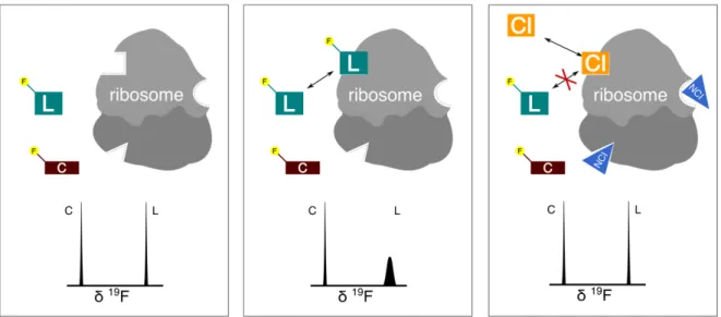
3.3 Our research goal The goal targeted through this thesis project is to develop a method capable of screening ligands on the ribosome using 19F NMR. The screening of chemical libraries is performed on the target (in this case, the whole ribosome) in the presence of a weak- to medium-affinity ligand referred in this role as the "spy" molecule. It is also necessary to experiment in the presence of a control molecule that does not interact with the target. Screening is carried out simply by monitoring the relative intensities of the spy and control in the presence of the target and the various candidate molecules successively tested. If the spy is bound to the target (it is supposed to be at the beginning of the experiment) its NMR signal disappears, only to reappear if the spy is specifically displaced of its binding site by another ligand. This method, originally performed with the proton, can also be used while detecting the resonance of the fluorine atom. Indeed, the use of the signals of only our two fluorinated molecules allows simple and rapid detection. In addition, the properties of fluorine in NMR guarantee a very high contrast between the free and bound states. This very sensible approach is called FAXS (Fluorine chemical shift Anisotropy and eXchange for Screening) and was introduced by the works of Claudio Dalvit (Dalvit - 2003). This method has since then been successfully used by Marc-André Delsuc, now in Bruno Kieffer lab, on a project on the Glucokinase (Assemat et al., 2015).
The development of this method of detection by NMR screening of new ligands on the ribosome will first start on the bacterial ribosome. Indeed, model organisms such as
Thermus thermophilus exists in the bacterial world in extreme environments, from which a
very stable ribosome can be extracted, ideal for testing different experimental conditions. Once experimental parameters will be well-defined (temperature, concentration, signal to noise ratio, etc.) the FAXS experiments will be able to begin. It will be relevant to test different fluorinated antibiotics as "spies" and various antibiotics competitors for each site. It will also be necessary to define a control molecule. Purification of ribosomes is a long and difficult process, but it is much more complicated for eukaryotic than prokaryotic organisms, even with a eukaryotic organism as simple as the yeast Saccharomyces cerevisiae. Thus, the validation of the experimental model with bacterial ribosomes is required before testing on
eukaryotic ribosomes. It is the same for the passage of the yeast ribosome to the human ribosome.
This thesis manuscript will then start by reviewing the properties of fluorine and present how they can be exploited in NMR. The characterization through NMR of the ligands used in our screening campaigns are detailed in the second chapter. The third chapter will present the screening results obtained on the ribosome, that will soon be submitted as a scientific publication. In the fourth chapter is related a collaboration done with a biotech company, Nosopharm, as our screening method was put to the test.
During my thesis, I had the opportunity to work on other projects and two of them are presented in the fifth and sixth chapter of this thesis. One is the observation of a binding event between peptides containing fluorinated polyprolines and a SH3 domain while another project, that I jumped on during my Master thesis, revolves around the dynamic properties of a protein domain and led to a publication.
References Assemat, O., Antoine, M., Fourquez, J., Wierzbicki, M., Charton, Y., Hennig, P., Perron-Sierra, F., Ferry, G., Boutin, J. and Delsuc, M. (2015). 19F nuclear magnetic resonance screening of glucokinase activators. Analytical Biochemistry, 477, pp.62-68. Bruce Alberts, et al. (2002). The Molecular Biology of the Cell, fourth edition. Garland Science pg. 342
Bąkowska-Żywicka K, Tyczewska A. (2009). The structure of the ribosome – short history. Biotechnologia; 1:14 – 23 Ban N, Nissen P, Hansen J, Moore P, Steitz T. (2000). "The complete atomic structure of the large ribosomal subunit at 2.4 Å resolution". Science. 289 (5481): 905–20. Ben-Shem A, Garreau de Loubresse N, Melnikov S, Jenner L, Yusupova G, Yusupov M (2011). "The structure of the eukaryotic ribosome at 3.0 Å resolution". Science. 334 (6062): 1524– 1529. Bohnsack, Markus T.; Schleiff, Enrico. "The evolution of protein targeting and translocation systems". Biochimica et Biophysica Acta (BBA) - Molecular Cell Research. 1803 (10): 1115– 1130.
Brachet, J. (1933). Recherches sur la synthase de l'acide thymonucleique pendant Ie developpement de l'oeuf d'oursin. Arch. Biol. 44:519.
“Ribosome.” British Society of Cell Biology. British Society for Cell Biology, n.d. Web. http://bscb.org/learning-resources/softcell-e-learning/ribosome/
Caspersson, T. and J. Schultz. (1938). Nucleic acid metabolism of the chromosomes in relation to gene reproduction. Nature 142:294. Cech T. (2000). "Structural biology. The ribosome is a ribozyme". Science. 289 (5481): 878–9. Claude, A. (1941). Particulate components of cytoplasm. Cold Spring Harbor Symp. Quant. Biol. 9:263. Chao FC S. H. (1956). Arch. Biochem. Biophys., 61, 220-230. Chen A. and M. J. Shapiro. (1998). NOE pumping: a novel NMR technique for identification of compounds with binding affinity to macromolecules. Journal of the American Chemical Society, 120(39):10258–10259.
Cundliffe, E.; Demain, A.L. (2010). Avoidance of suicide in antibiotic-producing microbes. J. Ind. Microbiol. Biotechnol. 37, 643–672.
Dahl EL and Rosenthal PJ. (2008). “Apicomplast translation, transcription and genome replication: targets for antimalarial antibiotics.” TRENDS in Parasitology 24: 279-284. Dalvit, C. (2009). NMR methods in fragment screening: theory and a comparison with other biophysical techniques. Drug Discovery Today, 14(21-22), pp.1051-1057. Dalvit C., P. E. Fagerness, D. T. Hadden, R. W. Sarver, and B. J. Stockman. (2003). Fluorine-NMR experiments for high-throughput screening: theoretical aspects, practical considerations, and range of applicability. Journal of the American Chemical Society, 125(25):7696–7703,.
Dalvit, C., P. Pevarello, M. Tato, M. Veronesi, A. Vulpetti and M. Sundstrom. (2000). "Identification of compounds with binding affinity to proteins via magnetization transfer from bulk water." Journal of Biomolecular Nmr 18(1): 65-68.
Dang, Y., Schneider-Poetsch, T., Eyler, D., Jewett, J., Bhat, S., Rawal, V., Green, R. and Liu, J. (2011). Inhibition of eukaryotic translation elongation by the antitumor natural product Mycalamide B. RNA, 17(8), pp.1578-1588.
Davies, J. and Davies, D. (2010). Origins and Evolution of Antibiotic Resistance. Microbiology and Molecular Biology Reviews, 74(3), pp.417-433.
Hajduk, P. J. and Greer J. (2007). A decade of fragment-based drug design: strategic advances and lessons learned. Nature reviews Drug discovery, 6(3):211–219.
Jhoti H., G. Williams, D. C. Rees, and C. W. Murray. (2013) The ‘rule of three’ for fragment-based drug discovery: where are we now? Nature Reviews Drug Discovery, 12(8):644–644.
Fernandez MM et al. (2011) “Differential effects of paromomycin on ribosomes of Leishmania mexicana and mammalian cells.” Antimicrobial Agents and Chemotherapies 55: 86-92.
Garreau de Loubresse, N., Prokhorova, I., Holtkamp, W., Rodnina, M., Yusupova, G. and Yusupov, M. (2014). Structural basis for the inhibition of the eukaryotic ribosome. Nature, 513(7519), pp.517-522.
Kalghatgi, S., Spina, C., Costello, J., Liesa, M., Morones-Ramirez, J., Slomovic, S., Molina, A., Shirihai, O. and Collins, J. (2013). Bactericidal Antibiotics Induce Mitochondrial Dysfunction and Oxidative Damage in Mammalian Cells. Science Translational Medicine, 5(192), pp.192ra85-192ra85.
Kjeldgaard, N.O. and Gausing, K. (1974). Regulation of biosynthesis of ribosomes. In Ribosomes (eds. M. Nomura et al.), pp. 369–392. Cold Spring Harbor Laboratory, Cold Spring Harbor, NY.
Noll, H. (2008). The discovery of polyribosomes. Bioessays, 30(11-12), pp.1220-1234. Loftfield RB, Vanderjagt D. (1972). The frequency of errors in protein biosynthesis. Biochem J.; 128(5):1353-6. Mayer, M. and B. Meyer (1999). "Characterization of ligand binding by saturation transfer difference NMR spectroscopy." Angewandte Chemie-International Edition 38(12): 1784-1788.
Mehta, R.; Champney, W.S. (2002). 30S ribosomal subunit assembly is a target for inhibition by aminoglycosides in Escherichia coli. Antimicrob. Agents Chemother. 46, 1546–1549.
O'Brien, T.W. (1971). "The General Occurrence of 55S Ribosomes in Mammalian Liver Mitochondria". J. Biol. Chem. 245: 3409.
Palade, G. E. (1955). A small particulate component of the cytoplasm. J. Biophys. Biochem. Cytol. 1:59.
Pelletier, J.; Peltz, S.W. (2007). "Therapeutic Opportunities in Translation". Cold Spring Harbor Monograph Archive. 48: 855–895.
Petermann, M. L. and M. G. Hamilton. (1952). An ultracentrifugal analysis of the macromolecular particles of normal and leukemic mouse spleen. Cancer Res. 12:373
Poehlsgaard, J. and Douthwaite, S. (2005). The bacterial ribosome as a target for antibiotics. Nature Reviews Microbiology, 3(11), pp.870-881.
Polacek, N.; Mankin, A.S. (2005). The ribosomal peptidyl transferase center: Structure, function, evolution, inhibition. Crit. Rev. Biochem. Mol. Biol., 40, 285–311.
J.-P. Renaud and M.-A. Delsuc (2009). Biophysical techniques for ligand screening and drug design. Current opinion in pharmacology, 9(5):622–628.
Rodnina MV, Beringer M, Wintermeyer W (2007). "How ribosomes make peptide bonds". Trends Biochem. Sci. 32 (1): 20–6.
Schluenzen F, Tocilj A, Zarivach R, Harms J, Gluehmann M, Janell D, Bashan A, Bartels H, Agmon I, Franceschi F, Yonath A (2000). "Structure of functionally activated small ribosomal subunit at 3.3 Åresolution". Cell. 102 (5): 615–23. Sender, R., Fuchs, S. and Milo, R. (2016). Revised estimates for the number of human and bacteria cells in the body. bioRxiv, [online] p.036103. Shuker, S. B., P. J. Hajduk, R. P. Meadows and S. W. Fesik (1996). "Discovering high-affinity ligands for proteins: SAR by NMR." Science 274(5292): 1531-4.
Stumpf, CR., Ruggero, D. (2011). "The cancerous translation apparatus". Curr Opin Genet Dev. 21 (4): 474–83. doi:10.1016/j.gde.2011.03.007.
Tissieres A., Watson J. D., (1958), Nature, 182, 778-780.
Tissières, A. (1974). Ribosome Research: Historical Background. Cold Spring Harbor Monograph Archive, 04(0). Topisirovic, I. Sonenberg, N. (Apr 2011). "Translational control by the eukaryotic ribosome". Cell. 145 (3): 333–4. Vanzi, F. (2003). Protein synthesis by single ribosomes. RNA, 9(10), pp.1174-1179. Verdier, L., Gharbi-Benarous, J., Bertho, G., Evrard-Todeschi, N., Mauvais, P., & Girault, J.-P. (2000). Dissociation–equilibrium constant and bound conformation for weak antibiotic binding interaction with different bacterial ribosomes. Journal of the Chemical Society, Perkin Transactions 2, (12), 2363–2371. Verschoor, A; Warner, JR; Srivastava, S; Grassucci, RA; Frank, J. (1998). "Three-dimensional structure of the yeast ribosome". Nucleic Acids Res. 26 (2): 655–61 Waudby CA, Launay H, Cabrita LD, Christodoulou J. (2013). Protein folding on the ribosome studied using NMR spectroscopy. Prog Nucl Magn Reson Spectrosc.;74:57-75.
Wilson, D. (2013). Ribosome-targeting antibiotics and mechanisms of bacterial resistance. Nature Reviews Microbiology, 12(1), pp.35-48.
Wilson, D. (2016). The ABC of Ribosome-Related Antibiotic Resistance. mBio, 7(3), pp.e00598-16.
Wimberly BT, Brodersen DE, Clemons WM Jr, Morgan-Warren RJ, Carter AP, Vonrhein C, Hartsch T, Ramakrishnan V . (2000). "Structure of the 30S ribosomal subunit". Nature. 407 (6802): 327–39.
Wright, G. and Poinar, H. (2012). Antibiotic resistance is ancient: implications for drug discovery. Trends in Microbiology, 20(4), pp.157-159.
Yamamoto, H., Qin, Y., Achenbach, J., Li, C., Kijek, J., Spahn, C. and Nierhaus, K. (2013). EF-G and EF4: translocation and back-translocation on the bacterial ribosome. Nature Reviews Microbiology, 12(2), pp.89-100. Yusupov MM, Yusupova GZ, Baucom A, et al. (May 2001). "Crystal structure of the ribosome at 5.5 A resolution". Science. 292 (5518): 883–96.
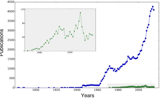
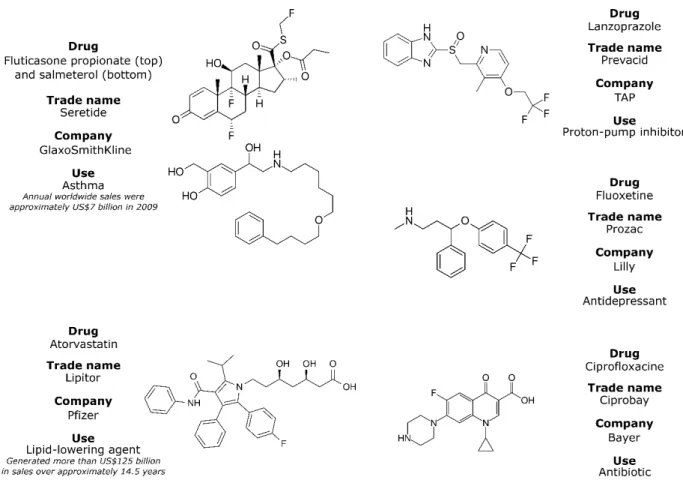
CHAPTER 1: Fluorine in NMR Introduction Fluorine (symbol F and atomic number 9) is a very peculiar chemical element. Most of the time, fluorine is only known for its dental care applications. But its specific chemical properties lead to its widespread introduction in various molecules used in everyday life. Ranging from refrigerants (Freon) to cookware (Teflon), not mentioning toothpaste, fluorochemical sales are worth 15 billion $ per year (Freedonia Industry study – 2012). The pharmaceutical industry has currently a fifth of its drugs that contain at least one fluorine (Arnston – 2015). The natural 19F isotope is well suited for NMR spectroscopy, although the corresponding number of scientific publications did not raise over recent years at the same pace as fluorine applications (figure 1.1). Fig 1.1 – Number of scientific papers published in the PubMed database per years about fluorine applications (blue curve, key word “Fluorine”) and 19F NMR (in green, key words “19F NMR”). The insert magnifies the publication number related to NMR. Figure 1.1 shows the modern history of fluorine, a little bit before and after the twentieth century and highlights the major steps of fluorine applications. World War 2 and the Manhattan Project triggered the development of industrial production of fluorine. Uranium
corrosive and reactive that all valves and seals from the separation plants were coated with Teflon, a polymer made of tetrafluoroethylene. (Takashi – 2009). Public academic work on fluorine raised interest when safe fluorinating agents were available for ordinary laboratories (Müller – 2007). The sixties were favorable to the development of many applications of fluorine chemistry. Teflon was used in cooking pans, but also in fabric, with the Gore-Tex technology, in bulletproof vest and in spaceships shields. Fluorine played also a role in cooling, with the chlorofluorocarbons initially, later replaced by hydrochlorofluorocarbons (HCFCs) and hydrofluorocarbons (HFCs) for ecological reasons. Nowadays, many commercial applications rely on fluorine compounds. Half of the annual mined fluorite (predicted 2016–18 production figures of 3.5 to 5.9 million tons) goes toward the steelmaking industry. The other half is transformed in organic fluorides, as previously mentioned, or into artificial cryolite (used for aluminium refining). All mentioned applications use the fluorine-19 isotope, but the fluorine-18 isotope has also applications. It is used in the radiopharmaceutical industry, and is primarily synthesized into fluoro-2-deoxy-D-glucose (FDG) to be used in positron emission tomography (PET scans). Its interest is due to both its short half-life and the emission of positrons (Jacobson – 2015). 19F NMR represents only a very small portion of studies reported on fluorine. Initially restricted to the chemical analysis of organofluorine compounds, 19F NMR is currently expanding in biology. 19F NMR finds applications in metabolic studies, in protein-ligand binding assays (supported by the works of Dalvit around 2000) and even in structural analysis of protein and macromolecules.
This chapter aims at presenting how the combination of fluorine and NMR emerges as an efficient technique in biology. We will review the fluorine atom properties and how those lead to its wide use in pharmaceutical compounds. A second part will present how NMR can exploit the peculiar properties of fluorine, for ligand screening studies. Some practical aspects of 19F NMR will be finally presented in the third part of the chapter.