HAL Id: hal-02346891
https://hal.archives-ouvertes.fr/hal-02346891
Submitted on 12 Nov 2019
HAL is a multi-disciplinary open access archive for the deposit and dissemination of sci-entific research documents, whether they are pub-lished or not. The documents may come from teaching and research institutions in France or abroad, or from public or private research centers.
L’archive ouverte pluridisciplinaire HAL, est destinée au dépôt et à la diffusion de documents scientifiques de niveau recherche, publiés ou non, émanant des établissements d’enseignement et de recherche français ou étrangers, des laboratoires publics ou privés.
Variability in the virulence of specific Mycobacterium
tuberculosis clinical isolates alters the capacity of human
dendritic cells to signal for T cells
Ana Gabriela Ramos-Martinez, Monica Alejandra Valtierra-Alvarado,
Mariana Haydee Garcia-Hernandez, Rogelio Hernandez-Pando, Julio Enrique
Castañeda-Delgado, Céline Cougoule, Bruno Rivas-Santiago, Olivier
Neyrolles, Jose Antonio Enciso-Moreno, Geanncarlo Lugo-Villarino, et al.
To cite this version:
Ana Gabriela Ramos-Martinez, Monica Alejandra Valtierra-Alvarado, Mariana Haydee Garcia-Hernandez, Rogelio Hernandez-Pando, Julio Enrique Castañeda-Delgado, et al.. Variability in the virulence of specific Mycobacterium tuberculosis clinical isolates alters the capacity of human dendritic cells to signal for T cells. Memórias do Instituto Oswaldo Cruz, Instituto Oswaldo Cruz, Ministério da Saúde, 2019, 114, �10.1590/0074-02760190102�. �hal-02346891�
online | memorias.ioc.fiocruz.br
Variability in the virulence of specific Mycobacterium tuberculosis
clinical isolates alters the capacity of human dendritic cells
to signal for T cells
Ana Gabriela Ramos-Martinez1,2, Monica Alejandra Valtierra-Alvarado1,2,
Mariana Haydee Garcia-Hernandez1, Rogelio Hernandez-Pando3,
Julio EnriqueCastañeda-Delgado4, Céline Cougoule5, Bruno Rivas-Santiago1, Olivier Neyrolles5,
Jose Antonio Enciso-Moreno1, Geanncarlo Lugo-Villarino5, Carmen Judith Serrano1/+
1Instituto Mexicano del Seguro Social, Unidad de Investigación Biomédica Zacatecas, Zacatecas, México
2Universidad Autónoma de San Luis Potosí, Escuela de Medicina, Departamento de Inmunología, San Luis Potosí, México 3Instituto Nacional de Ciencias Médicas y de la Nutrición Salvador Zubirán, Departamento de Patología, Sección de Patología
Experimental, Ciudad de México, México
4Catédras Consejo Nacional de Ciencia y Tecnología, en Instituto Mexicano del Seguro Social, Unidad de Investigación Biomédica
Zacatecas, Zacatecas, México
5Université de Toulouse, Institut de Pharmacologie et de Biologie Structurale, Centre National de la Recherche Scientifique,
Université Paul Sabatier, Toulouse, France
BACKGROUND Once in the pulmonary alveoli, Mycobacterium tuberculosis (Mtb) enters into contact with alveolar macrophages and dendritic cells (DCs). DCs represent the link between the innate and adaptive immune system owing to their capacity to be both a sentinel and an orchestrator of the antigen-specific immune responses against Mtb. The effect that the virulence of Mtb has on the interaction between the bacilli and human DCs has not been fully explored.
OBJECTIVE To evaluate the effect of Mtb virulence on human monocyte-derived DCs.
METHODS We exposed human monocyte-derived DCs to Mtb clinical strains (isolated from an epidemiological Mtb diversity study in Mexico) bearing different degrees of virulence and evaluated the capacity of DCs to internalise the bacilli, control intracellular growth, engage cell death pathways, express markers for activation and antigen presentation, and expand to stimulate autologous CD4+ T cells proliferation.
FINDINGS In the case of the hypervirulent Mtb strain (Phenotype 1, strain 9005186, lineage 3), we report that DCs internalise and neutralise intracellular growth of the bacilli, undergo low rates of apoptosis, and contribute poorly to T-cell expansion, as compared to the H37Rv reference strain. In the case of the hypovirulent Mtb strain (Phenotype 4, strain 9985449, lineage 4), although DCs internalise and preclude proliferation of the bacilli, the DCs also display a high level of apoptosis, massive levels of apoptosis that prevent them from maintaining autologous CD4+ T cells in a co-culture system, as compared to H37Rv.
MAIN CONCLUSIONS Our findings suggest that variability in virulence among Mtb clinical strains affects the capacity of DCs to respond to pathogenic challenge and mount an immune response against it, highlighting important parallels to studies previously done in mouse models.
Key words: tuberculosis - dendritic cells - virulence - apoptosis - antigen presentation
Tuberculosis (TB) remains a challenging infectious disease and was rated by the World Health Organization in 2017 as one of the top ten causes of death worldwide. In 2019, TB was responsible for more deaths than HIV
doi: 10.1590/0074-02760190102
Financial support: Fondo de Investigación en Salud from Instituto Mexicano del Seguro Social, México (grant nº FIS/IMSS/PROT/MD15/1489), Agence Nationale de la Recherche, France (grant n ANR-15-CE15-0012, MMI-TB). AGRM and MAVA were supported by Consejo Nacional de Ciencia y Tecnología, México (CONACyT) during their PhD studies (fellowship numbers 417995 and 584982). For an internship at the IPBS in Toulouse, France, AGRM received sponsoring from CONACyT (Beca Mixta) and from the host laboratories of Isabelle Maridonneau-Parini and Olivier Neyrolles. B Rivas-Santiago is a scholar of Fundación IMSS, México.
AGR-M and MAV-A contributed equally to this work; GL-V and CJS are co-senior authors.
+ Corresponding author: carmenyuyu2000@yahoo.com.mx https://orcid.org/0000-0002-7047-9120
Received 22 March 2019 Accepted 08 July 2019
and malaria, accounting for 1.6 million deaths
world-wide.(1) Therefore, there is a need to better understand
the interaction between Mycobacterium tuberculosis (Mtb) and host cells in order to design better preventive and therapeutic strategies.
TB is mainly transmitted among humans when Mtb
is inhaled via the aerosol route.(2) Once in the pulmonary
alveoli, Mtb enters into contact with alveolar macro-phages and dendritic cells (DCs), which are the
domi-nant cell targets for this pathogen.(3) In particular, DCs
represent the link between the innate and adaptive im-mune systems due to their capacity to act as both a “sen-tinel” and an “orchestrator” of the antigen-specific im-mune responses against the bacilli. As a “sentinel”, DCs patrol peripheral sites and are capable of recognising and internalising microorganisms that invade the mucosal
barrier.(4) As an “orchestrator”, DCs efficiently acquire
and process Mtb-specific antigens at the site of infec-tion, and then traffic them to the lymph nodes, where
T-Ana Gabriela Ramos-Martinez et al. 2|9
cell priming occurs. There is a keen interest in the field to investigate in detail how Mtb interacts with DCs, and how this interaction can be modulated for the efficient elimination of Mtb infection.
Upon encountering and internalising a pathogen, DCs must activate an intracellular microbicidal
environ-ment to restrict pathogen growth and spread.(5) In vitro
studies in monocyte-derived dendritic cells (MDDCs) suggest low levels of bacterial replication within these
cells.(6) While Mtb is known to affect cell death of
in-fected DCs,(7,8) the pathways related to this process have
not been extensively studied in DCs within the context of TB. Upon neutralising the intracellular pathogen, DCs undergo a maturation process that allows them to process antigens derived from the pathogen, and subse-quently relay this information to naïve T cells in the form of antigen presentation. To counteract this process, Mtb impairs DC maturation, reduces the capacity of DCs to secrete IL-12, and inhibits the ability of DCs to stimulate
T-cell proliferation.(9,10,11) However, further studies are
required to understand the degree of the effect of Mtb virulence on the capacity of the bacteria to inhibit DCs. This is precisely the focus of this study.
The relationship between the virulence of Mtb strains and their transmissibility in humans, as well as its consequences on the quality of the host immune re-sponse, has been studied, among others, by
Marquina-Castillo et al.(12). They conducted a 10-year
population-based prospective study of pulmonary TB in Southern Mexico by performing an in-depth analysis of the epi-demiological and clinical data of household contacts of TB patients. The authors selected a panel of isolates representing clinical and epidemiological diversity in the population of Mtb strains found in Mexico and tested them in two mouse models. Based on strain viru-lence, immune response (defined by cytokine expres-sion), and transmissibility, four phenotypes were identi-fied. The strains representing the two extremes, from hypervirulence to hypovirulence, were Phenotype 1 (strain 9005186, lineage 3, family EAI) and Phenotype 4 (strain 9985449, lineage 4, family Haarlem), respec-tively. On the one hand, Phenotype 1 (Ph1) was highly transmissible; it grew rapidly in the lung and caused high mortality rates in mice along with significantly more pneumonic areas, as compared to the H37Rv ref-erence strain. In addition, Ph1 induced a poor protective immune response in the host. On the other hand, Pheno-type 4 (Ph4) exhibited a long survival index, low bacil-lary load, and little pneumonia. Moreover, Ph4 induced
a delayed acquired protective immune response.(13) The
human leukocyte response against these Mtb clinical isolate strains is still poorly understood.
Here, we focused our investigation on the interac-tion of Mtb clinical isolates Ph1 and Ph4 with human DCs. Altogether, our study showed that Mtb virulence has an important effect on the interaction with human DCs and the capacity of those DCs to then stimulate to T lymphocytes expansion, suggesting that these clinical isolate strains are optimal candidates for the prospective identification of Mtb genes associated with virulence and human immunogenicity.
MATERIALS AND METHODS
Human blood peripheral mononuclear cells, MD-DCs and T lymphocytes - Mononuclear cells were
iso-lated from buffy coats provided by the State Blood Transfusion Center of Zacatecas, SSA (Guadalupe, Zac., Mexico) by density gradient using Ficoll-Paque PLUS (GE Healthcare, UK). The donors were healthy individu-als complying with the following criteria: 18-55 years old, minimum weight of 50 kg, fasting conditions for at least 8 h, and with no history of Hepatitis type B or C, HIV/AIDS, syphilis, organ transplants, epilepsy, tu-berculosis, cardiovascular disease, or cancer. Exclusion criteria included recreational drug use, mental disease, women who were pregnant or lactating, tattoos or skin perforations (12 months previous to the donation), his-tory of diabetes, surgery, mononucleosis, toxoplasmosis or meningitis (in the last six months), having received vaccines (in the last 28 days), alcohol or narcotics use (in the last 12 h). Different sets of individuals were used for the distinct parameters evaluated in the present protocol. The number of individuals evaluated in each assay is an-notated in the corresponding figure legend. Whole blood from individuals positive for tuberculin was used only for T-cell proliferation assays.
In order to generate MDDCs, mononuclear cells (5
× 106) were seeded in a 75 cm2 flask using RPMI-1640
medium supplemented with 10% foetal calf serum (FCS) (Gibco Life Technologies, USA), 1% pyruvate (Sigma Al-drich, USA), 0.1% 2-beta-mercaptoethanol (Gibco Life Technologies, USA), recombinant IL-4 (15 ng/mL) (Pe-protech Inc., USA), and granulocyte macrophage colony stimulating factor (GM-CSF, 200 ng/mL) (GRAMAL, Probiomed Lab, Mexico), and cultured at 37ºC in a
hu-midified atmosphere at 5% CO2. At days 3 and 5, the cells
were additionally supplemented with IL-4 and GM-CSF. At day 7, MDDCs were collected by centrifugation and used for functional analyses. Alternatively, mononuclear cells isolated from healthy volunteers with positive tuber-culin tests (range of 15 to 25 mm) were used to generate
MDDCs and to purify CD4+ T cells by a magnetic bead
approach using the CD4+ negative selection kit (Miltenyi,
USA) according to manufacturer’s instructions.
Mtb strains, culture and storage - All
manipula-tions with the Mtb strains were performed in a dedi-cated BSL-3 laboratory. The Mtb strain H37Rv was used in order to establish an experimental reference for the comparative analyses done with the clinical isolate strains Ph1 (highly virulent, with no induction of pro-tective immune response in mice) and Ph4 (less viru-lent, provoking a protective adaptive immune response).
(12) The strain Ph1, for spoligotyping, belongs to the
fam-ily EAI and it is susceptible to all the first-line antibiot-ics used for TB treatment, while strain Ph4 belongs to the Haarlem family and is streptomycin-monoresistant. All strains were grown in Middlebrook 7H9 medium (BD-Diagnostic Systems, USA) supplemented with 10% Middlebrook Oleic Albumin Dextrose Catalase Growth Supplement (OADC) (BD-Diagnostic Systems, USA). Strains were grown to reach exponential phase, and culture concentrations were measured by
measur-ing optical density at 600 nm. Bacterial aliquots were stored at -80ºC until their use. Bacillary viability was tested using the colony forming unit (CFU) assay, grow-ing serial dilutions in 7H10 agar plates (BD-Diagnostic Systems, USA) supplemented with 10% OADC (BD-Diagnostic Systems, USA) for 14 and 21 days.
Internalisation of Mtb and infection of MDDCs -
Bac-terial aliquots (H37Rv, Ph1, and Ph4) were thawed at room temperature, and the bacterial aggregate declumping was achieved by vortexing with borosilicate beads for 5 min and centrifuging at 2040 × g for 5 min. MDDC infection was performed at a multiplicity of infection (MOI) of 5 (5 bacteria to 1 cell). At 2 h post-infection (hpi), MDDCs were washed with RPMI-1640 medium to remove non-internalised bacteria. At 24 hpi, MDDCs were recollected by centrifugation and fixed with 4% paraformaldehyde (PFA) for 30 min at room temperature. In order to evalu-ate internalisation of Mtb strains, the fixed MDDCs were centrifuged in pretreated slides (Biocare Medical, Con-cord CA, USA) using a Cytocentrifuge (Wescor Cytopro 7620, USA), and Ziehl-Neelsen staining was performed to identify the bacilli associated/within cells. The percent-age of MDDCs with at least one associated bacterium was calculated after counting at least 100 MDDCs/sample in high power fields using a Carl Zeiss inverted Axiovert M-200 microscope (Zeiss, Germany).
Mtb intracellular growth in human MDDCs - The
bacterial strains were cultured at 37ºC in Middlebrook 7H9 medium supplemented with 10% OADC and 0.05% Tween-80 (Sigma-Aldrich, USA). During exponential growth, the bacteria were centrifuged (2000 × g) for 15 min and resuspended in 1× phosphate buffered saline (PBS). Clumps were dissociated by passages through a 26-G needle, and then resuspended in RPMI-1640 me-dium containing 10% FBS. The mycobacterial concen-tration was determined by measuring optical density at 600 nm (OD600). To test the intracellular growth capac-ity of these strains, MDDCs were collected at day 7 of culture and seeded in 24-well plates at a density of 5 ×
105 cells per well. These cells were then infected with
each Mtb strain individually at a multiplicity of infection (MOI) of 0.2 bacteria per cell in RPMI-1640 medium with 10% FBS for 4 h. Cells were then washed twice with 1× PBS before addition of RPMI-1640/10% FBS. At the indicated time points, the cells were lysed in a 0.1% Triton (Sigma-Aldrich, USA) lysing solution. Serial di-lutions of the resulting bacterial suspension were plated on Middlebrook 7H11 solid agar supplemented with 10% OADC (BD-Diagnostic Systems, USA) and incubated for 14-21 days at 37ºC for CFU scoring.
Cell-death assessment of MDDCs infected with Mtb strains - To evaluate apoptosis and necrosis, we
anal-ysed MDDCs at day 7 of differentiation using the FITC Annexin V Apoptosis Detection Kit II (BD Biosciences, USA), according to the manufacturer’s instructions. Briefly, MDDCs were infected with each Mtb strain in-dividually at a MOI of 5 in RPMI-1640 medium with 10% FBS for 4 h. MDDCs were then washed twice with 1× PBS before addition of RPMI-1640/10% FBS. After 24 h, MDDCs were washed twice with cold PBS and
resuspended in 1× binding buffer, stained with 5 µL of Annexin V and 5 µL of Propidium Iodide, and gently vortexed and incubated for 15 min at room temperature, protected from light. MDDCs were then and washed us-ing PBS and resuspended in 4% PFA for 30 min at room temperature for fixation. Finally, the viability status of all MDDC populations was acquired using the FACS Canto II cytometer (BD Biosciences, USA). For data analysis, Flow Jo VX (Tristar, USA) was used.
T-cell proliferation assay - The ability of
Mtb-in-fected MDDCs to stimulate CD4+ T cells was assessed
using an autologous co-culture system, as previously
Fig. 1: virulence of Mycobacterium tuberculosis (Mtb) strains does not affect recognition/internalisation by human monocyte-derived dendritic cells (MDDCs). Human monocytes were differentiated into MDDCs until day 7. (A) Percentage of Mtb bacilli internalised by MD-DCs after infection with the Mtb experimental H37Rv strain (white) or clinical isolate strains Ph1 (black) and Ph4 (grey). Results were analysed using Kruskal-Wallis tests. (B) Categorisation of infected MDDCs according to the number of bacilli (1-5, white; 6-10, black; > 10, grey) internalised per cell, illustrated as percentage of infected MDDCs. Each bar represents mean ± SEM of four independent donors. Results were analysed using two-way analysis of variance (ANOVA).
Ana Gabriela Ramos-Martinez et al. 4|9
described.(14) Briefly, autologous CD4+ T cells (the
re-sponders) were freshly isolated and purified from healthy donors positive for the tuberculin test on the
day that the co-culture was initiated. A total of 1.5 × 10⁴
5-(and 6)-Carboxyfluorescein diacetate succinimidyl
ester (CFSE) stained CD4+ T cells (responder cells) were
added to each well in 200 μL complete media on round-ed-bottom, 96-well plates (BD, Pharmingen, USA). Au-tologous MDDCs (the stimulators) were differentiated until day 6 and pulsed for 24 h with LPS (100 ng/mL) (Sigma, Germany). Later, MDDCs were infected with each Mtb strain individually at a MOI of 0.2 in RPMI-1640 medium with 10% FBS for 4 h, washed twice with 1× PBS, harvested, and added to the responder cells at a ratio (stimulator:responder) of 1:2.5, 1:5, 1:10, or 1:20. As a control for the response to Mtb antigens, a group of non-infected MDDCs were grown until day 6 and were then pulsed for 24 h with a peptide corresponding to the partial sequence of the Mtb early secretory antigenic target (ESAT-6) protein (20 μg/mL) for 24 h. Of note, the partial ESAT-6 protein a peptide with the sequence H2N-LNNALQNLARTISEAG-COOH was synthesised at Fundación Instituto de Inmunología de Colombia,
Bogotá Colombia. After six days of culture in 5% CO2
at 37ºC, the cells were harvested, stained, and gated for CD3 and CD4 positivity, and then analysed for dilution of the CFSE intensity by flow cytometry.
Statistical analyses - The corresponding statistical
analysis is annotated in each figure legend. Data was compared using two-way ANOVA analysis with Bonfer-roni’s post-hoc test for internalisation index, Wilcoxon analysis for CFU, while comparison inside the groups was performed with the Kruskal-Wallis test and Dunn’s post-test assuming non-normally distributed data for all the other analysed variables. Statistical analysis was per-formed with GraphPad Prism software v.5.0 (San Diego, CA, USA). P ≤ 0.05 was considered as the level of statis-tical significance.
Ethical considerations - The present protocol was
re-viewed and authorised by the National Research Commit-tee of Instituto Mexicano del Seguro Social (The Mexi-can Institute of Social Security, IMSS), that includes a subcommittee for ethical approval (agreement number R-2014-785-042). Informed consent was obtained from all individual participants included in the study.
RESULTS
Virulence of Mtb strains does not affect recognition/ internalisation by human MDDCs - In order to assess
whether the virulence of the Mtb clinical isolates Ph1 and Ph4 affect their capacity to be recognised/inter-nalised by human MDDCs, we infected MDDCs with each bacterial strain, and the bacillary load was de-termined by Ziehl-Neelsen staining. Our results show only a tendency towards a higher recognition/internali-sation of low virulence strains compared to the refer-ence H37Rv strain (Fig. 1A). This was confirmed by an analysis of the number of bacilli bound/internalised per MDDC, which yielded no difference among any clinical isolate compared to H37Rv (Fig. 1B). Therefore, these
results suggest that variations in virulence do not sig-nificantly affect the capacity of Mtb clinical strains to be recognised or internalised by MDDCs.
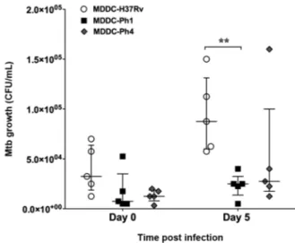
MDDCs control the intracellular growth of the Mtb clinical strains independent of virulence - In order to
investigate whether the virulence of Mtb strains modu-lates their capacity to proliferate in human MDDCs, we performed CFU assays. After 4 hpi (day 0), we con-firmed there was no significant difference in the bacte-rial charge in MDDCs infected with the different Mtb strains (Fig. 2). More specifically, we observed that MD-DCs were able to control the intracellular growth of both Ph1 and Ph4 Mtb clinical strains after 120 hpi (day 5). However, at day 5, we did observe a higher inhibition of growth of the Ph1 strain compared to the reference strain (Fig. 2). Altogether, the results suggest that these Mtb clinical isolate strains fail to colonise human MDDCs, and that the difference in virulence among these strains does not play a role in this process.
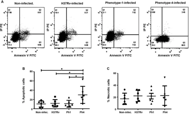
Mtb clinical strain bearing low virulence leads to high levels of apoptosis in MDDCs - We examined
whether the difference in virulence among the Mtb clini-cal isolate strains affects the mode of cell death in infect-ed MDDCs. Flow cytometry revealinfect-ed that at 24 hpi, the strain with lower virulence (Ph4) induced more apopto-sis in MDDCs compared to the other Mtb strains and to the uninfected cells (Fig. 3A-C). As shown in Fig. 3C, the virulence among the Mtb strains does not signifi-cantly affect the induction of necrosis in MDDCs. Over-all, these results show that the hypovirulent Mtb clinical
Fig. 2: human monocyte-derived dendritic cells (MDDCs) control the intracellular growth of the Mycobacterium tuberculosis (Mtb) clini-cal strains independent of virulence. MDDCs were infected with the Mtb experimental strain H37Rv (black circles) the Ph1 clinical isolate strain (white squares), or the Ph4 clinical isolate strain (black dia-monds). The intracellular growth of the bacteria was determined at 4 h (day 0) and 120 h (day 5) post-infection, as measured by colony forming unit (CFU) assays. Vertical scatter plots show the number of CFU per mL with each symbol representing a single donor run in triplicate, n = 5 different donors. Data were analysed with two-way analysis of variance (ANOVA) for repeated measures and the Bonfer-roni post-hoc test was applied. Bars represent median and interquar-tile range for five different donors. **p ˂ 0.01.
isolate strain (Ph4) differs considerably in its capacity to induce apoptosis, but not necrosis, in infected MDDCs when compared to the hypervirulent strain (Ph1) and the experimental H37Rv reference strain.
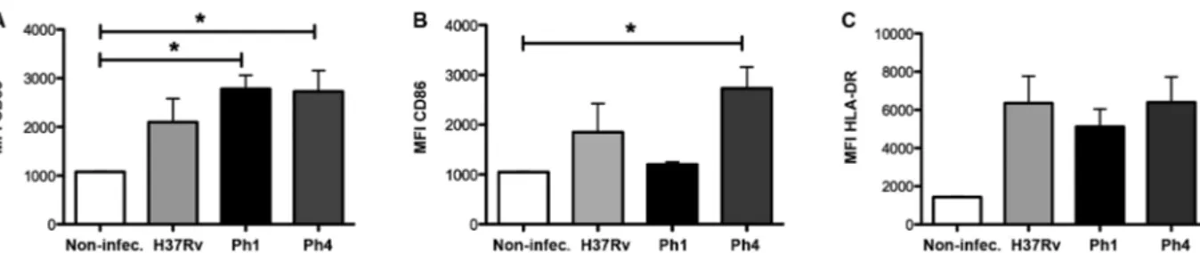
Virulence of Mtb strains does not affect the activa-tion of MDDCs - In order to assess whether the virulence
of the Mtb clinical isolate strains affects the activation of human DCs, we evaluated the expression of cell sur-face markers during infection by flow cytometry. As ex-pected, MDDCs infected with any Mtb strain displayed significantly higher levels (albeit only a tendency for H37Rv) of the activation marker CD83, as compared to uninfected cells. Yet, there were no differences observed between the Mtb clinical isolate strains or compared to the reference H37Rv strain (Fig. 4A). In addition, we as-sessed the expression levels of receptors that are impor-tant for antigen presentation, such as CD86 and HLA-DR. Although MDDCs infected with the clinical isolate bear-ing low virulence (Ph4) displayed higher levels of CD86 compared to uninfected cells, we noticed there were no significant differences detected among all the infected groups (Fig. 4B). The same was true for the expression levels of HLA-DR among all the infected groups, al-though there was a tendency for higher expression com-pared to uninfected cells (Fig. 4C). Altogether, these re-sults suggest that the difference in virulence among the Mtb strains does not affect the activation of MDDCs.
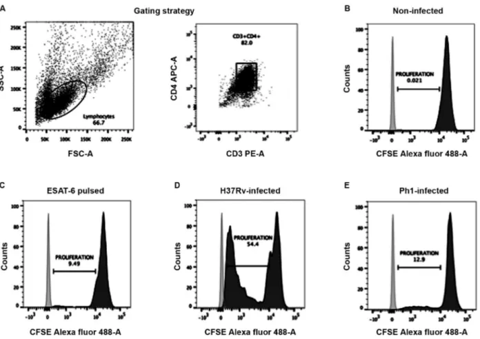
Virulence of Mtb strains affects the capacity of MDDCs to activate CD4+ T cells - In order to evaluate
whether the virulence of the Mtb clinical isolate strains modulates the capacity of MDDCs to activate T lym-phocytes, we set up an autologous co-culture system to evaluate T-cell proliferation based on the dilution of the CFSE dye, as measured by flow cytometry. As expect-ed, T cells that were co-cultured with uninfected MD-DCs did not proliferate, as measured by the undiluted CFSE dye (Fig. 5A-B). By contrast, we observed T-cell proliferation with MDDCs pulsed with the ESAT-6 pep-tide, which served as positive control, at any tested ratio (MDDC:T cell), confirming the presence of an antimy-cobacterial immune response in healthy donors positive for the tuberculin test (Fig. 5C). Likewise, we noticed that MDDCs infected with the H37Rv reference strain were also capable of inducing robust T-cell proliferation (Fig. 5D). However, comparison of MDDCs infected with the different Mtb clinical isolates resulted in strik-ing differences. On the one hand, MDDCs infected with the clinical isolate bearing low virulence (Ph4) were not able to prevent cell death of autologous T cells, as the co-cultures did not remain viable for the entire six day co-culture period (data not shown). On the other hand, MDDCs infected with the clinical isolate bearing high virulence (Ph1) were capable of inducing T-cell prolif-eration, but less efficiently than MDDCs infected with
Fig. 3: the Mycobacterium tuberculosis (Mtb) clinical strain bearing low virulence induces high levels of apoptosis in monocyte-derived dendritic cells (MDDCs). MDDCs were infected for 24 h with the Mtb experimental strain H37Rv, clinical isolate Ph1, clinical isolate Ph4, or uninfected control. The MDDCs were then stained using the FITC Annexin V Apoptosis Detection Kit II. (A) Representative dot plot for the gating strategy to assess MDDC cell death in non-infected, H37Rv-infected, Ph1-infected, and Ph4-infected MDDCs. The plots are illustrative of cell death measurements as analysed by flow cytometry. The amount of MDDC cell death due to (B) apoptosis (propidium iodide-/Annexin V+ cells) or (C)
necrosis (propidium iodide+ cells) was quantified. Analysis for comparison among groups was performed in parts B and C using one-way analysis
Ana Gabriela Ramos-Martinez et al. 6|9
the H37Rv reference strain (Fig. 5A,D). These results were consistent for the five independent tuberculin-positive donors tested in the antigen presentation assays, even when there were not statistic differences between the rates of proliferation induced by Ph1 compared to H37Rv. Collectively, these results suggest that the viru-lence difference among the Mtb clinical strains is an im-portant factor that can affect antigen presentation.
DISCUSSION
Most of the information about Mtb strain varia-tion and immunopathology derives from
experimen-tal animal models.(13) Yet, this information cannot be
extrapolated directly to human TB infection, and thus highlights the need to study Mtb strain variation using human leukocytes. In the present study, we investigated whether Mtb clinical isolates obtained from a prospec-tive population-based study of pulmonary TB patients in Southern Mexico, whose virulence varied dramatically
from high (Ph1) to low (Ph4) in a mouse model,(12)
dif-fer in their interaction with human DCs in comparison to the standard experimental strain H37Rv. We believe this study makes the following contributions in advanc-ing our understandadvanc-ing of how these Mtb clinical isolates differ in their interaction with human DCs.
First, we showed that the Ph1 hypervirulent strain diminishes the capacity of human DCs to activate
au-tologous CD4+ T cells. Compared to DCs infected with
H37Rv, Ph1-infected DCs displayed a poor capacity to induce proliferation of autologous T cells in our in
vi-tro co-cultures. This is not due to a deficiency in DCs
to recognise or internalize the Ph1 strain in comparison to H37Rv. Additionally, judging from the induction of antigen presenting molecules (e.g. HLA-DR, CD86) and cell death pathways (e.g. apoptosis, necrosis), we can rule out a problem with the ability of the Ph1 strain to activate DCs. Rather, we believe that the diminished capacity of Ph1-infected DCs to activate autologous T cells may be due to their inability to colonise these APCs. Based on our CFU assays, we determined that Ph1 failed to achieve a significant level of intracellu-lar growth in human DCs. Therefore, we can infer that the Ph1-infected DCs may have less antigenic material available to present and activate naïve T cells. This is in contrast to the results obtained in the study by
Mar-quina-Castillo et al.(12) conducted in a mouse model. In
their study, the Ph1 strain grew rapidly in the lung and peaked at 21 dpi, exhibiting a bacterial burden (via CFU readings) twice as high in comparison to those mice
in-fected with the H37Rv strain.(12) We infer this phenotype
is likely due to the capacity of the Ph1 strain to colonise and grow within other leukocytes besides DCs, because, compared to H37Rv, the Ph1 strain displays similar in
vitro phenotypes in terms of cord formation, growth
curves, and response to hydrogen peroxide exposure.(12)
However, the authors did not examine the type of mu-rine leukocytes serving as reservoirs for the bacteria. As previously reported, DCs exhibit a non-permissible
phe-notype against Mtb intracellular growth,(6) and this
char-acteristic is not influenced by the degree of virulence as we are showing in this study. Therefore, our results suggest that the Ph1 strain may have a better capacity to colonise macrophages in contrast to DCs.
Nevertheless, our observation that Ph1-infected DCs do not optimally activate autologous T cells is in line with the main finding in the study conducted by Marquina-Castillo et al.,(12) which concluded that the Ph1 strain does not induce a protective immune response in a mouse model, such as the one promoted by the H37Rv strain. Indeed, infection of mice with the Ph1 strain was charac-terised by a delayed IFNγ expression, which is indicative
of poor activation of Th1 cells.(12) Infection of mice with
other hypervirulent Mtb strains, such as those belonging to the W-Beijing lineage, also results in a poor protec-tive Th1-driven immune response distinguished by the low and temporal expression of IFNγ, TNFα, and iNOS. (15,16) Suboptimal antigen presentation has been shown to contribute to the virulence of Mtb in an in vivo mouse
model.(17) Moreover, DCs generated from monocytes after
Type I IFN exposure (IFN-DCs) highly resemble natu-rally occurring DCs induced in vivo, for example, in a
chronic infection context.(18) In active TB patients,
IFN-DCs showed a diminished capacity to induce Ag-specific
T-cell responses against Mtb.(19) Collectively, our results
argue that the interaction of the Ph1 hypervirulent strain with human DCs differs from that of H37Rv, resulting in a lower capacity for these antigen-presenting cells to opti-mally activate an adaptive immune response.
The second key contribution of our study is the dem-onstration that the Ph4 hypovirulent strain induces apop-tosis in human MDDCs, preventing them from support-ing an autologous co-culture system with T lymphocytes.
Fig. 4: virulence of Mycobacterium tuberculosis (Mtb) strains does not affect the activation of monocyte-derived dendritic cells (MDDCs). MD-DCs were infected with H37Rv (light grey), clinical isolate Ph1 (black), clinical isolate Ph4 (dark grey), or left uninfected as a control (white). After 24 h, MDDCs were harvested and stained for activation markers, as assessed by flow cytometry. Bar graphs show the fluorescent intensity (MFI) (mean ± SEM) for cell-surface expression of (A) CD83, (B) CD86, and (C) HLA-DR. Intragroup comparisons were performed with a Kruskal-Wallis test followed by Dunn’s multiple comparison post-test. For each group, n = 4 different donors. *p ˂ 0.05.
Similar to the cells infected with H37Rv, MDDCs were able to recognise, internalise, and control the intracellular growth of the Ph4 strain. However, MDDCs infected with this hypovirulent strain underwent rapid and massive levels of apoptosis, as compared to the other Mtb strains. These results suggest that less virulent Mtb strains can induce apoptosis in DCs, as well as macrophages, and in this manner, become a source of antigen for other anti-gen-presenting cells to pick up and promote an efficient
adaptive immune response, as postulated previously.(7,20)
Interestingly, while the Ph4 strain did not differ in its ca-pacity to activate MDDCs in terms of the up-regulation of cell surface receptors involved in antigen presentation (albeit always displaying the highest tendency among the tested strains), MDDCs infected with this strain were not able to support the established autologous co-culture
sys-tem with CD4+ T cells. In the context of TB, infected
mi-gratory DCs are poor antigen-presenting cells,(21) which
suggests that infected cells have a mechanism to transfer antigen to uninfected cells so that uninfected cells could
prime CD4+ T cells. In vivo studies demonstrated that
mi-gratory DCs must collaborate with one or more resident
Fig. 5: virulence of Mycobacterium tuberculosis (Mtb) strains affects the capacity of monocyte-derived dendritic cells (MDDCs) to activate CD4+ T cells. MDDCs were activated with LPS and then infected with the different Mtb strains (or left uninfected as a control). MDDCs were
added at different ratios to autologous CD4+ T cells that were previously labelled with CFSE dye. After culturing for six days, cells were
har-vested and stained for flow cytometry analysis. (A) Gating strategy to select cells by size and granularity, and double positivity for the receptors CD3 (PE-A) and CD4 (APC-A) to specifically determine the T-cell population. Representative histograms showing the MFI for the CFSE dye in the T-cell populations co-cultured with (B) uninfected MDDCs (negative control), (C) MDDCs pulsed with the peptide from ESAT-6 (control for response to Mtb antigens), (D) MDDCs infected with the H37Rv strain, and (E) MDDCs infected with the Ph1 strain. The cells were plated at a ratio of 1 MDDC to 10 CD4+ T lymphocytes.
DCs to successfully prime CD4+ T cells.(22,23) Cultured
DCs and macrophages release multiple Mtb protein anti-gens into the extracellular medium that can be taken up,
processed, and presented to CD4+ T cells by uninfected
DCs.(23) This form of antigen transfer occurs without
transfer of the pathogen itself, providing a mechanism for host cells to bypass the inhibitory effects of Mtb on antigen presentation and allow for effective priming of
antigen-specific CD4+ T cells. We inferred that MDDCs
infected with the Ph4 strain become the source of my-cobacterial antigens for uninfected bystander antigen-presenting cells, which may consequently activate an ef-ficient T-cell response. Moreover, we believe that these results are in line with the characterisation performed in the mouse model by Marquina-Castillo and colleagues.
(12) Indeed, the authors determined that infection of mice
with the Ph4 strain culminated in an increased surviv-al rate of the mice in comparison to infection with the H37Rv strain. This was also correlated with a better pro-tective acquired immune response characterised by high and sustained (albeit delayed) IFNγ expression, and early
Ana Gabriela Ramos-Martinez et al. 8|9
It remains to be determined what signalling path-ways promote the rapid and high levels of apoptosis triggered by the Ph4 strain in MDDCs. In the context of murine: bone marrow-derived-dendritic cells (BMDC) for example it was demonstrated that Mtb induces
cas-pase-1/11-independent apoptosis but not necrosis.(7) In
the context of macrophages, avirulent Mtb strains in-duce strong expression of prostaglandin-endoperoxide synthase 2 (PTGS2), which consequently leads to high
rates of apoptosis-promoting prostaglandin E2 (PGE2)
production.(20) Lipoxin A4 (LXA4) is known to inhibit
PTGS2 and PGE2, leading to an increase in necrosis
stead of apoptosis, which is common in macrophages
in-fected by virulent Mtb strains.(5) Altogether, our results
argue that the interaction of the Ph4 hypovirulent strain with human DCs greatly differs from that of the H37Rv strain, resulting in rapid and massive rates of apoptosis that may facilitate the acquisition of antigenic material, thus leading hypothetically towards an efficient activa-tion of the adaptive immune response.
In conclusion, this study represents the first prospec-tive assessment of the human leukocyte response to the Mtb strains endemic in Southern Mexico that vary in virulence and transmission as previously assessed in mouse models. In general, our findings in human DCs suggest that the variability in virulence among these Mtb strains affects the capacity of leukocytes to respond to pathogenic challenge and mount an immune response against it, highlighting important parallels from stud-ies performed in mouse models. The association of the observed effects in this study and specific virulence factors previously described for Mtb warrant further in-vestigation. We support the notion that our Mtb clinical isolate strains make good candidates for further investi-gation using genome sequencing, transcriptome hybridi-sation, and comparative proteomics, probably leading to the eventual identification of Mtb genes associated with virulence and the interference of human DC biological
functions, as previously proposed.(13)
ACKNOWLEDGEMENTS
To Dr Hudrisier for his help and sharing of reagents and equipment for the antigen-presentation assays.
AUTHORS’ CONTRIBUTION
AGRM, MAVA and MHGM carried out the experiments and participated in the analysis of data, in the discussion of the global results, and wrote the paper; RHP, CC, ON, JEM and BRS participated in the discussion of the global results; JECD participated in the analysis and discussion of data, and in writing the paper; GLV and CJS designed the study, ob-tained the funds to conduct the project, analysed the global results, and wrote the paper. All of the authors read and ap-proved the manuscript. The authors declare that they have no conflicts of interest.
REFERENCES
1. WHO - World Health Organization. Global Tuberculosis Report. Geneva: World Health Organization; 2018.
2. Wolf AJ, Desvignes L, Linas B, Banaiee N, Tamura T, Takatsu K, et al. Initiation of the adaptive immune response to
Mycobacteri-um tuberculosis depends on antigen production in the local lymph node, not the lungs. J Exp Med. 2008; 205(1): 105-15.
3. Mayer-Barber KD, Barber DL. Innate and adaptive cellular im-mune responses to Mycobacterium tuberculosis infection. 2015; 5(12): 1-20.
4. Iwasaki A. Mucosal dendritic cells. Annu Rev Immunol. 2007; 25(1): 381-418.
5. Lugo-Villarino G, Neyrolles O. Manipulation of the mononuclear phagocyte system by Mycobacterium tuberculosis. Cold Spring Harb Perspect Med. 2014; 4(11): a018549.
6. Tailleux L, Neyrolles O, Honoré-Bouakline S, Perret E, Sanchez F, Abastado J-P, et al. Constrained intracellular survival of My-cobacterium tuberculosis in human dendritic cells. J Immunol. 2003; 170(4): 1939-48.
7. Abdalla H, Srinivasan L, Shah S, Mayer-Barber KD, Sher A, Sutter-wala FS, et al. Mycobacterium tuberculosis infection of dendritic cells leads to partially caspase-1/11-independent IL-1β and IL-18 secretion but not to pyroptosis. PLoS One. 2012; 7(7): e40722. 8. Ryan RCM, O’Sullivan MP, Keane J. Mycobacterium
tuberculo-sis infection induces non-apoptotic cell death of human dendritic cells. BMC Microbiol. 2011; 11: 237.
9. Hanekom WA, Mendillo M, Manca C, Haslett PAJ, Siddiqui MR, Barry C, et al. Mycobacterium tuberculosis inhibits maturation of human monocyte-derived dendritic cells in vitro. J Infect Dis. 2003; 188(2): 257-66.
10. Dulphy N, Herrmann JL, Nigou J, Réa D, Boissel N, Puzo G, et al. Intermediate maturation of Mycobacterium tuberculosis LAM-ac-tivated human dendritic cells. Cell Microbiol. 2007; 9(6): 1412-25. 11. Balboa L, Romero MM, Yokobori N, Schierloh P, Geffner L,
Ba-sile JI, et al. Mycobacterium tuberculosis impairs dendritic cell response by altering CD1b, DC-SIGN and MR profile. Immunol Cell Biol. 2010; 88(7): 716-26.
12. Marquina-Castillo B, Garcia-Garcia L, Ponce-de-Leon A, Jime-nez-Corona ME, Bobadilla-Del Valle M, Cano-Arellano B, et al. Virulence, immunopathology and transmissibility of selected strains of Mycobacterium tuberculosis in a murine model. Immu-nology. 2009; 128(1): 123-33.
13. Hernandez-Pando R, Marquina-Castillo B, Barrios-Payan J, Ma-ta-Espinosa D. Use of mouse models to study the variability in virulence associated with specific genotypic lineages of Myco-bacterium tuberculosis. Infect Genet Evol. 2012; 12(4): 725-31. 14. Balboa L, Kviatcovsky D, Schierloh P, García M, de la Barrera S,
Sasiain MC, et al. Monocyte-derived dendritic cells early exposed to Mycobacterium tuberculosis induce an enhanced T helper 17 response and transfer mycobacterial antigens. Int J Med Microbi-ol. 2016; 306(7): 541-53.
15. Lopez B, Aguilar D, Orozco H, Burger M, Espitia C, Ritacco V, et al. A marked difference in pathogenesis and immune response induced by different Mycobacterium tuberculosis genotypes. Clin Exp Immunol. 2003; 133(1): 30-7.
16. Manca C, Tsenova L, Bergtold A, Freeman S, Tovey M, Musser JM, et al. Virulence of a Mycobacterium tuberculosis clinical iso-late in mice is determined by failure to induce Th1 type immuni-ty and is associated with induction of IFN-alpha /beta. Proc Natl Acad Sci USA. 2001; 98(10): 5752-7.
17. Grace PS, Ernst JD. Suboptimal antigen presentation contributes to virulence of Mycobacterium tuberculosis in vivo. J Immunol. 2016; 196(1): 357-64.
18. Vermi W, Fisogni S, Salogni L, Schärer L, Kutzner H, Sozzani S, et al. Spontaneous regression of highly immunogenic molluscum
contagiosum virus (MCV)-induced skin lesions is associated with plasmacytoid dendritic cells and IFN-DC infiltration. J Invest Dermatol. 2011; 131(2): 426-34.
19. Parlato S, Chiacchio T, Salerno D, Petrone L, Castiello L, Romag-noli G, et al. Impaired IFN-α-mediated signal in dendritic cells differentiates active from latent tuberculosis. PLoS One. 2018; 13(1): e0189477.
20. Behar SM, Divangahi M, Remold HG. Evasion of innate immuni-ty by Mycobacterium tuberculosis: is death an exit strategy? Nat Rev Microbiol. 2010; 8(9): 668-74.
21. Baena A, Porcelli SA. Evasion and subversion of antigen pres-entation by Mycobacterium tuberculosis. Tissue Antigens. 2009; 74(3): 189-204.
22. Samstein M, Schreiber HA, Leiner IM, Sušac B, Glickman MS, Pamer EG. Essential yet limited role for CCR2+ inflammatory monocytes during Mycobacterium tuberculosis-specific T cell priming. Elife. 2013; 2: e01086.
23. Srivastava S, Ernst JD. Cell-to-cell transfer of M. tuberculosis antigens optimizes CD4 T cell priming. Cell Host Microbe. 2014; 15(6): 741-52.