HAL Id: hal-02322811
https://hal.archives-ouvertes.fr/hal-02322811
Submitted on 2 Jun 2020
HAL is a multi-disciplinary open access
archive for the deposit and dissemination of sci-entific research documents, whether they are pub-lished or not. The documents may come from teaching and research institutions in France or abroad, or from public or private research centers.
L’archive ouverte pluridisciplinaire HAL, est destinée au dépôt et à la diffusion de documents scientifiques de niveau recherche, publiés ou non, émanant des établissements d’enseignement et de recherche français ou étrangers, des laboratoires publics ou privés.
Golgi trafficking defects in postnatal microcephaly: The
evidence for “Golgipathies”
Sandrine Passemard, Franck Perez, Emilie Colin-Lemesre, Sowmyalakshmi
Rasika, Pierre Gressens, Vincent El Ghouzzi
To cite this version:
Sandrine Passemard, Franck Perez, Emilie Colin-Lemesre, Sowmyalakshmi Rasika, Pierre Gressens, et al.. Golgi trafficking defects in postnatal microcephaly: The evidence for “Golgipathies”. Progress in Neurobiology, Elsevier, 2017, 153 (3), pp.46-63. �10.1016/j.pneurobio.2017.03.007�. �hal-02322811�
Passemard et al, Golgi in Postnatal Microcephaly Final Version-22Feb2017 1
2 3 4
Golgi trafficking defects in postnatal microcephaly: the
5
evidence for "Golgipathies"
6 7 8
Sandrine Passemard
1,2, Franck Perez
3, Emilie Colin-Lemesre
1,3,
Sowmyalakshmi
9Rasika
1,
Pierre Gressens
1,4, Vincent El Ghouzzi
1* 1011 12
1. PROTECT, INSERM, Université Paris Diderot, Sorbonne Paris Cité, Paris, France 13
2. AP HP, Hôpital Robert Debré, Service de Génétique Clinique, Paris, France 14
3. Institut Curie, PSL Research University, CNRS, UMR144, Paris, France 15
4. Centre for the Developing Brain, Division of Imaging Sciences and Biomedical 16
Engineering, King’s College London, King’s Health Partners, St. Thomas’ Hospital, 17
London, United Kingdom 18 19 20 21 22 23 *Corresponding author: 24 Vincent El Ghouzzi 25
Address: Inserm U1141, Hôpital Robert-Debré, 48 Boulevard Sérurier, F-75019, 26 Paris, France. 27 E-mail: [email protected] 28 Phone: +331 40031973, Fax: +331 40031995 29 30
Key words: Golgi apparatus; RAB GTPase; neuronal trafficking; postnatal microcephaly;
31
intellectual disability; Golgipathies 32
33
Number of words in the abstract: 225
34
Number of words in the body of the manuscript: 9,641
35 36
Abstract
1 2
The Golgi apparatus plays a central role in cell homeostasis, not only in processing and 3
maturing newly synthesized proteins and lipids but also in orchestrating their sorting, packing, 4
routing and recycling on the way to their final destination. These multiple secretory pathways 5
require a complex ballet of vesicular and tubular carriers that continuously bud off from donor 6
membranes and fuse to acceptor membranes. Membrane trafficking is particularly prominent in 7
axons, where cargo molecules have a long way to travel before they reach the synapse, and in 8
oligodendrocytes, which require an immense increase in membrane surface in order to sheathe 9
axons in myelin. Interestingly, in recent years, genes encoding Golgi-associated proteins with a 10
role in membrane trafficking have been found to be defective in an increasing number of 11
inherited disorders whose clinical manifestations include postnatal-onset microcephaly (POM), 12
white matter defects and intellectual disability. Several of these genes encode RAB GTPases, 13
effectors or regulating proteins, linking POM and intellectual disability to RAB-14
dependent Golgi trafficking pathways and suggesting that their regulation is critical to postnatal 15
brain maturation and function. Here, we review the key roles of the Golgi apparatus in post-16
mitotic neurons and the oligodendrocytes that myelinate them, and provide an overview of 17
these Golgi-associated POM-causing genes, their function in Golgi organization and trafficking 18
and the likely mechanisms that may link dysfunctions in RAB-dependent regulatory pathways 19
with POM. 20
Contents
1
1. Introduction... 4 2
3
2. TheGolgi apparatus in post-mitotic neurons and oligodendrocytes... 6 4
2.1 Role of the Golgi apparatus in neuronal polarity... 6 5
2.1.1 Distribution of microtubules in neuronsand their relationship to the Golgi 6
apparatus...8 7
2.1.2 Specificity of Golgi-derived transport carriers...9 8
2.1.3 Golgi outposts in dendrites...10 9
2.1.4 Golgi components in axons...11 10
2.2 Role of the Golgi apparatus in myelination... 11 11
2.3 Role of the Golgi apparatus in autophagy...14 12
2.4 Role of Golgi-associated RAB proteins in the brain... 16 13
14
3. Syndromes with postnatal onset microcephaly (POM) and causative genes... 20 15
3.1 Postnatal onset microcephaly ... 20 16
3.2 Golgi-associated proteins implicated in POM... 22 17
3.2.1 Cohen syndrome and COH1/VPS13B... 22 18
3.2.2 PCC2 syndrome and VPS53...23 19
3.2.3 Warburg-Micro syndrome and RAB3GAP1/2, RAB18 and TBC1D20... 24 20
3.2.4 Autosomal recessive mental retardation 13 (MRT13) and TRAPPC9...25 21
3.2.5 A neuromuscular syndrome with microcephaly and GOLGA2/GM130...26 22
3.2.6 Dyggve-Melchior-Clausen syndrome and DYMECLIN... 27 23
3.2.7 Congenital disorders of glycosylation and COG complex...28 24
25
4. Possible mechanisms underlying POM... 30 26 27 5. Conclusion...33 28 29 6. Acknowledgements... 33 30 31 7. References...34 32 33
1. Introduction
1
Microcephaly affects about 2% of the population worldwide and represents the most 2
frequent neurological sign encountered in developmental brain disorders. It is characterized by 3
a small brain size, indirectly diagnosed by an occipito-frontal or head circumference (OFC) 4
smaller than the age- and gender-adjusted mean by more than 2 standard deviations (SD) at 5
birth and/or 3 SD as measured at 6 months of age or later; it is frequently associated with 6
intellectual disability of variable severity. Among the many kinds of microcephaly, genetic 7
forms have yielded essential information as to how the human brain develops during 8
embryonic/fetal and postnatal periods. While primary microcephaly is defined by a congenital 9
failure of brain growth detectable before birth, secondary (or postnatal-onset) microcephaly 10
(POM) is characterized by normal brain size at birth and the subsequent deceleration of brain 11
growth, and in particular the white matter, during infancy and/or childhood. In the past 15 12
years, considerable efforts have led to the identification of genes and pathways whose 13
deficiency causes hereditary primary microcephaly, also known as MCPH. The vast majority of 14
MCPH genes (17 identified to date, see updated review by (Alcantara and O'Driscoll, 2014)) 15
play a role in the regulation of cell division and/or centrosome function in neural progenitors, 16
and a large number of functional studies now converge on common mechanisms that affect the 17
mode and/or extent of cortical progenitor division and their subsequent survival and 18
differentiation during the development of the neocortex. In contrast, POM or acquired 19
microcephaly, which often appears to be only one of many clinical signs in complex and 20
divergent syndromes, is not always considered a disorder on its own. As such, it is 21
underdiagnosed and underinvestigated, and the cellular mechanisms leading to it are poorly 22
understood. 23
The timing of POM suggests that these cellular mechanisms presumably involve 24
processes and pathways that occur later during development than neuronal progenitor division, 25
the major process implicated in primary microcephaly. Indeed, several principally postnatal 26
mechanisms that could lead to POM, such as defective gliogenesis or myelination, the 1
impairment of neuronal maturation or synaptic pruning, the arrest of normal development or 2
degenerative processes, have received much attention from the scientific community in recent 3
years. One candidate process worth noting is membrane trafficking and secretion through the 4
Golgi apparatus. Indeed, several recent studies have implicated Golgi-associated proteins in 5
genetic disorders that include POM among their characteristics, suggesting that the regulation 6
of Golgi trafficking and secretory functions are critical to postnatal brain maturation. 7
Intriguingly, a number of these POM-causing genes encode either RAB proteins - members of 8
the RAS superfamily of small GTPases which play a central role in membrane trafficking 9
including Golgi organization, vesicle formation, transport and fusion – or RAB-associated or 10
RAB-tethering factors whose fast and reversible recruitment facilitates such trafficking. 11
Interestingly, all these POM-causing genes are associated with the defective development of 12
white matter, which consists principally of the axons of neurons and the myelinating 13
oligodendrocytes that ensheathe them, highlighting the link between the heavy membrane 14
trafficking and secretory activity of these two interdependent cellular components and postnatal 15
brain development. 16
In this review, we describe the key roles played by the Golgi apparatus in post-mitotic 17
neurons and oligodendrocytes, describe recently identified POM-causing genes associated with 18
the Golgi apparatus, and discuss the intriguing fact that many of these appear to encode RAB 19
proteins or their molecular partners. In light of their role in Golgi organization and trafficking 20
and the mechanistic links between RAB proteins, white matter defects and the development of 21
POM, we propose a new term for these disorders based on their similar pathophysiology: 22
"Golgipathies"/"Golgipathic microcephalies". 23
2. The Golgi apparatus in post-mitotic neurons and oligodendrocytes
1
The Golgi apparatus is a multifunctional organelle essential to ensure differentiated 2
cellular functions as well as to maintain cell homeostasis. In mammalian cells, about one-third 3
of newly synthesized proteins are destined to be secreted following the conventional secretory 4
pathway. The Golgi apparatus is primarily involved in the processing of secretory proteins and 5
lipids as they transit through it, effecting posttranslational modifications such as glycosylation, 6
sulfation and proteolytic cleavage. The Golgi apparatus also acts in the sorting, packing, 7
routing and recycling of these cargo molecules for their final destination. Depending on the cell 8
type and stage of development, Golgi-dependent trafficking routes and secretory cargos have 9
become diversified to fulfill specific secretory functions (Boncompain and Perez, 2013a). This 10
is especially true of two cell types that are heavily affected in POM: post-mitotic projection 11
neurons and the oligodendrocytes that enwrap their axons in myelin, with several studies 12
showing that the Golgi apparatus plays a key role in the dynamic trafficking specific to the 13
axonal and dendritic compartments of these neurons, as well as the extensive plasma membrane 14
extensions of oligodendrocytes required for myelin formation. Besides its involvement in 15
protein and lipid trafficking/processing in these two cell types, the Golgi apparatus is also 16
involved in the determination and maintenance of neuronal polarity, as well as in autophagy, 17
another process essential both for brain development and homeostasis of mature neural cells. 18
19
2.1 Role of the Golgi apparatus in neuronal polarity
20 21
In mammalian cells, the Golgi apparatus is a ribbon-shaped organelle made up of 22
flattened cisternae organized into polarized stacks, flanked on either side by fenestrated tubular 23
reticular membranes called the cis-Golgi network (CGN) and the trans-Golgi network (TGN) 24
(Nakamura et al., 2012; Papanikou and Glick, 2014). In most cells, the Golgi apparatus is 25
positioned near or around the centrosome, with which it is dynamically associated through the 26
action of cytoplasmic dynein motor proteins and Golgi anchor proteins (Yadav and Linstedt, 27
2011). In developing neurons, centrosomes, the Golgi apparatus and endosomes cluster 28
together at one pole of the cell body before neurites form, and play a key role in axon 1
specification (Caceres, 2007). Although the existence of a direct correlation between 2
Golgi/centrosome positioning and the area where the future axon will form has remained 3
controversial (de Anda et al., 2005; Distel et al., 2010; Horton et al., 2005; Lowenstein et al., 4
1994; Zmuda and Rivas, 1998), this asymmetric pericentrosomal confinement of the Golgi 5
apparatus likely leads to a local concentration of neuronal growth potential both in terms of 6
cytoskeletal infrastructure and of newly synthesized proteins, two components essential for the 7
elongation of the axon. Axonal outgrowth also requires a huge expansion of the plasma 8
membrane surface (Horton and Ehlers, 2003), which is achieved by the progressive integration 9
of Golgi-derived vesicles. Such vesicles have been shown to accumulate and polarize before 10
axonogenesis in cultured hippocampal neurons (Bradke and Dotti, 1997). In line with this 11
mechanism, brefeldin A treatment, which disassembles the Golgi apparatus, results in the 12
selective inhibition of axonal growth (Jareb and Banker, 1997). Similarly, the genetic 13
invalidation of certain Golgi-related proteins leads to altered neuronal polarity and death and/or 14
dysfunction. For instance, in mice in which the expression of the golgin GM130 is invalidated 15
by shRNA treatment or genetically knocked out, the polarity of the Golgi apparatus is altered, 16
leading to altered dendritic polarization in granule cells of the hippocampus (Huang et al., 17
2014), as well as altered ER-to-Golgi transport, inducing the atrophy and death of Purkinje 18
cells of the cerebellum, and consequently, ataxia (Liu et al., 2017). The loss of expression of 19
two other golgins, Golgin-160 and GMAP210, which disrupt pericentrosomal Golgi positioning 20
without affecting either the microtubule network or general secretion, also strongly affects cell 21
polarity in vitro (Yadav et al., 2009). However, the effect of GMAP120 deletion on Golgi 22
structure or function might depend on the cellular subtype being examined (Smits et al., 2010). 23
24
In addition to its involvement in neuronal development, the Golgi apparatus is required 25
for the maintenance of axodendritic polarity throughout the lifespan of mature post-mitotic 26
neurons. These highly specialized cells possess specific architectural features that make the 1
secretory pathway central to their structural maintenance, dynamics and function. In particular, 2
their strongly polarized axons and dendrites are characterized by morphologically and 3
functionally distinct components and pathways. This necessitates the asymmetric transport of 4
membranes and the continuous targeting of distinct repertoires of cargo proteins and lipids to 5
these distinct subcellular compartments. Mature neurons also often develop extensive dendritic 6
branching accompanied by a huge increase in membrane surface area (Ye et al., 2006). In 7
addition, the long axons possessed by some neurons pose a perennial challenge to the 8
movement of proteins, lipids, vesicles and organelles between cell bodies and synaptic sites. 9
Although the mechanisms through which this differential targeting is specifically achieved and 10
regulated are complex and only partially understood, a number of key findings show that the 11
Golgi apparatus lies at the core of processes that elicit distinct secretory features in the axons 12
and dendrites of post-mitotic neurons, thereby maintaining neuronal polarity. 13
14
2.1.1 Distribution of microtubules in neurons and their relationship to the Golgi
15
apparatus
16
Microtubules, which themselves are polarized and serve as rails for active vesicular 17
cargo transport driven by molecular motors, are asymmetrically distributed in axons and 18
dendrites. While axons usually display long, uniformly oriented microtubules with their minus 19
ends towards the soma and the plus ends facing outwards, proximal dendrites contain shorter 20
microtubules oriented in both directions (Baas, 1999) (Figure 1A). In dendrites, the minus-end-21
out microtubules are generally more stable (Yau et al., 2016), which contributes to generating 22
directionality. This implies a difference in the organization of molecular motors involved in 23
trafficking in the two compartments. For example, dynein, which moves along microtubules 24
towards their minus end, drives retrograde transport in axons but bidirectional transport in 25
dendrites, while kinesin motors seem to predominantly drive anterograde transport in axons 26
(Kapitein et al., 2010). Interestingly, the Golgi apparatus not only sorts and provides the 1
various cargos to be conveyed to specific destinations but also acts as a microtubule-organizing 2
center (MTOC), independently of the centrosome (Chabin-Brion et al., 2001; Zhu and 3
Kaverina, 2013), and itself generates a distinct population of microtubules called Golgi-derived 4
microtubules. During the development of rodent hippocampal neurons, the centrosome actually 5
loses its function as an MTOC, and it is microtubules of non-centrosomal origin that enable 6
axon extension and serve as rails for directional post-Golgi trafficking (Stiess et al., 2010). 7
Similarly, microtubule organization is independent of the centrosome in developing and mature 8
Drosophila neurons (Nguyen et al., 2011), and the Golgi apparatus has been proposed as a 9
possible source of dendritic microtubules (Ori-McKenney et al., 2012), a process promoted by 10
the golgin GM130 (Zhou et al., 2014). Interestingly, directional trafficking defects have been 11
observed in human RPE1 cells lacking Golgi-derived microtubules, suggesting that the latter 12
are essential for post-Golgi transport (Miller et al., 2009; Vinogradova et al., 2012). Thus, 13
while further evidence is still required to confirm this possibility, the Golgi apparatus might 14
also be directly involved in the maintenance of neuronal polarity in postmitotic neurons 15
through its role as an MTOC. 16
17
2.1.2 Specificity of Golgi-derived carriers 18
The differential distribution of cargo proteins and lipids between dendrites and axons is 19
largely due to specific and reciprocal interactions between cargos, their carriers and molecular 20
motors. This occurs through the docking of motor proteins onto their specific cargos either 21
directly or via adaptor molecules, including scaffolding proteins, receptors and Rab GTPases 22
that regulate neuronal transport(Franker and Hoogenraad, 2013; Maeder et al., 2014; Schlager 23
and Hoogenraad, 2009). Interestingly, the identity of the various carriers is in large part 24
conferred by the specific cargos they carry. The sorting of axonal and dendritic cargo proteins 25
and lipids occurs in the TGN, where they are physically segregated into specific clusters that 26
define specific dynamic TGN subdomains, ultimately leading to vesicle budding. This physical 1
segregation of cargos appears to rely on both the intrinsic affinity of different cargos for 2
specific lipid microenvironments provided by the TGN (Brugger et al., 2000; Klemm et al., 3
2009; Orci et al., 1987; Paladino et al., 2004; Schuck and Simons, 2004); reviewed in (Anitei 4
and Hoflack, 2011; De Matteis and Luini, 2008; Guo et al., 2014; Lingwood and Simons, 2010; 5
Surma et al., 2012), and the presence of sorting signals on cargo molecules that target them to 6
TGN-specific adaptors such as small ADP ribosylation factors, Rab and Rho GTPases, and 7
Golgi-localized tethering factors (De Matteis and Luini, 2008). In other words, the selective 8
targeting of cargos that contribute to the axodendritic polarity of neurons starts as soon as the 9
cargos reach the TGN (Guo et al., 2014). 10
11
2.1.3. Golgi outposts in dendrites 12
In addition to the somatic Golgi apparatus, the Golgi complex forms smaller satellite 13
structures called Golgi outposts that are found in about 20% of the dendrites of mature neurons 14
(Gardiol et al., 1999; Horton et al., 2005; Pierce et al., 2001) (Figure 1A). Several studies have 15
provided evidence that these Golgi outposts lack continuity with the somatic Golgi apparatus 16
and are functionally independent. Golgi outposts ensure the post-translational modifications, 17
trafficking and sorting of locally synthesized proteins (Horton et al., 2005; Jeyifous et al., 18
2009; Torre and Steward, 1996; Ye et al., 2007), as well as local microtubule nucleation (Ori-19
McKenney et al., 2012), thereby playing a major role both in shaping dendritic arbor 20
morphology and in serving as platforms for the local delivery of postsynaptic molecules such as 21
synaptic receptors. In line with this role, and consistent with the recent demonstration that 22
Golgi outposts destined for the major dendrite are generated by a sequential process that 23
involves the polarized deployment and fission of tubules derived from the somatic Golgi 24
(Quassollo et al., 2015), markers of cis, medial and trans Golgi compartments have all been 25
detected in dendrites (Horton et al., 2005; Pierce et al., 2001). Reinforcing the role of the Golgi 26
apparatus in the functional specialization of dendrites, a recent study by Mikhaylova provides 1
evidence for a Golgi-related satellite microsecretory system in dendrites that is even more 2
widespread than Golgi outposts and would permit the autonomous local control of membrane 3
protein synthesis and processing within dendrites (Mikhaylova et al., 2016). 4
5
2.1.4. Golgi components in axons 6
Besides the well-described transport mechanisms that direct cargos to axons through 7
molecular motors and microtubules, and ensure their activity, function and plasticity (Hirokawa 8
and Takemura, 2005), growing evidence suggests that a number of axonal proteins are locally 9
synthesized from mRNAs and ribosomes present in axons and presynaptic elements (Sotelo-10
Silveira et al., 2006; Yoo et al., 2010). The existence of such decentralized protein synthesis 11
could allow axons to meet local demands in a fast and energy-efficient manner, as is the case 12
with dendrites (Donnelly et al., 2010; Holt and Bullock, 2009; Jung et al., 2012). However, 13
whether this process also involves the presence of Golgi outpost-like structures in axons is a 14
matter of debate. The presence of early secretory components, including markers of the ER, the 15
ER-Golgi intermediate compartment (ERGIC), Golgi apparatus and TGN, has been observed 16
by some authors in the distal axoplasm of rat peripheral axons (Gonzalez et al., 2016; Merianda 17
et al., 2009), raising the possibility that these components self-organize into small functional
18
organelles in situ. Although rough ER and Golgi stacks have not so far been observed in axons 19
at the ultrastructural level (reviewed in (Ramirez and Couve, 2011)), the occurrence of local 20
protein synthesis suggests that protein processing and secretory needs could also be met locally, 21
rendering axons at least partially independent of the somatic early secretory pathway and 22
facilitating, for example, fast membrane receptor recycling in response to local conditions. 23
24 25 26
2.2 Role of the Golgi apparatus in myelination
Most neurons are characterized by a myelin sheath that enwraps their axons and is 1
responsible for the whitish appearance of the white matter of the brain. In the central nervous 2
system (CNS), the myelin sheath is a multilamellar structure consisting of the plasma 3
membrane extensions of oligodendrocytes, with a single mature oligodendrocyte ensheathing 4
several axons. These lipid-rich processes are extremely long and packed in tight spirals around 5
axons, forming a dense sheath to protect and insulate them and thus ensure the high-speed 6
propagation of electrical impulses. A stereological study carried out in a 20 year-old man has 7
revealed a total myelinated fiber length of 170,000 kilometers (Marner et al., 2003); the 8
dimensions of the oligodendrocytic processes required to ensheathe these fibers must therefore 9
be many times greater. The biogenesis and maintenance of this vast quantity of myelin implies 10
an intensive and sustained supply of membrane proteins and lipids. In oligodendrocytes, as in 11
neurons, this is achieved both through the local synthesis of myelin components close to the 12
site of their assembly, and through intensive vesicle trafficking mechanisms involving the 13
traditional ER-Golgi-TGN pathway (Kramer et al., 2001). For example, myelin basic protein 14
(MBP), which represents approximately 30 percent of myelin proteins and plays a major role in 15
myelin compaction (Privat et al., 1979) and composition by regulating its protein to lipid ratio 16
(Aggarwal et al., 2011), is synthesized on the spot by the local translation of MBP mRNAs 17
(Colman et al., 1982), following their packing in a translationally repressed state (Bauer et al., 18
2012; Kosturko et al., 2006) into large ribonucleoprotein complexes called RNA transport 19
granules (Muller et al., 2013), and their transport along microtubules into the myelin 20
compartment (Ainger et al., 1993; Carson et al., 1997). In contrast, myelin-specific lipids and 21
other major myelin proteins, such as the proteolipid protein PLP, are synthesized in the soma of 22
mature oligodendrocytes and pass through the Golgi where they are processed and self-23
assemble with cholesterol and sphingolipids to form a type of preformed myelin modules called 24
lipid-enriched liquid ordered membrane microdomains or lipid "rafts", which are transported 25
through the secretory pathway (Gielen et al., 2006; Simons et al., 2000). However, the 26
technical complications inherent in observing nanoscale molecular organizations such as lipid 1
microdomains in a reliable manner, i.e. without altering the object of the observation, has no 2
doubt contributed to their being viewed by some authors as hypothetical rather than real 3
structures for the present (see (Guo et al., 2014)). Reciprocal communication between axons 4
and oligodendrocytes is also required for the generation of the myelin sheath. In 5
oligodendrocytes, such lipid microdomains, in addition to being components of myelin, behave 6
as dynamic signaling modules in recruiting specific signaling proteins that integrate axon-7
derived soluble or membrane-bound signals to regulate myelination spatiotemporally (White 8
and Kramer-Albers, 2014). The nodes of Ranvier, non-myelinated axon segments that are 9
regularly placed along myelinated fibers, constitute privileged zones where molecular 10
interchanges take place across the axonal membrane. In addition to specific cell adhesion 11
molecules and cytoskeletal scaffold molecules that maintain the proper function and 12
architecture of nodes (Susuki and Rasband, 2008), these nodes are also the sites of release of 13
several axon-derived signaling molecules that have been shown to regulate the proliferation, 14
differentiation and survival of oligodendrocytes, and control the onset and timing of myelin 15
membrane growth (Simons and Trajkovic, 2006). For example, both the stability and the site-16
specific translation of MBP mRNA are promoted by the recruitment of the tyrosine kinase Fyn 17
by oligodendrocytic lipid microdomains (White and Kramer-Albers, 2014), and its activation 18
occurs in response to the binding of the axonal cell adhesion molecule L1 (White et al., 2008). 19
Interestingly, the myelin membrane protein TPO1, which has also been proposed to activate 20
Fyn, is highly enriched both in the Golgi and in the Fyn-positive sheets of myelinating 21
oligodendrocytes (Fukazawa et al., 2006; Jain and Ganesh, 2016). Thus, the fine regulation of 22
myelin formation and maintenance appear to depend on trafficking through the Golgi-23
dependent secretory pathway and microtubule network and signaling pathways in both 24
oligodendrocytes and the neurons, and on their functional interactions at specific sites. 25
2.3 Role of the Golgi apparatus in autophagy
1
Autophagy or "self-eating" is an evolutionarily conserved catabolic process by which 2
cytosolic contents are delivered to acidic lysosomes for degradation. It serves various purposes: 3
the maintenance of cellular homeostasis by eliminating waste or toxic products and recycling 4
cellular components and nutrients, especially during conditions of starvation, for protection 5
against certain pathogens, as well as the facilitation of cellular remodeling. In contrast to the 6
ubiquitin-proteasome system (which achieves the regulated degradation of individual 7
ubiquitinated proteins), autophagy leads to the bulk degradation of whole organelles and large 8
amounts of proteins. Of the three main types of autophagy – microautophagy, chaperone-9
mediated autophagy and macroautophagy – the last is the best studied, and is characterized by a 10
newly formed "isolation membrane" or "phagophore" that grows to envelop the components to 11
be degraded in a double-walled structure called the autophagosome, which subsequently fuses 12
with the lysosome (for a broad review, see (Feng et al., 2014; Mizushima and Komatsu, 2011)). 13
For this reason, the term "autophagy" is often used to refer specifically to macroautophagy. 14
In the central nervous system with its specialized long-lived cells characterized by extensive 15
membrane processes, in addition to its traditional role in maintaining cellular homeostasis (Hu 16
et al., 2015; Tooze and Schiavo, 2008), autophagy plays several other roles: the modulation of
17
synaptic plasticity (Hernandez et al., 2012), the maintenance of the pool of neural stem cells 18
required for postnatal neurogenesis (Wang et al., 2013), and finally, the normal development of 19
the CNS, including neural progenitor proliferation, neuronal maturation, connectivity and 20
myelination (Ban et al., 2013; Hara et al., 2006; Jang et al., 2015; Kadir et al., 2016; Kim et 21
al., 2016; Komatsu et al., 2006; Liang et al., 2010; Rangaraju et al., 2010; Schwarz et al.,
22
2012; Smith et al., 2013; Song et al., 2008). As could be expected, defects in autophagy-related 23
genes or dysfunctions of autophagy are reflected in a number of human neurological disorders 24
(for review, see (Bockaert and Marin, 2015; Ebrahimi-Fakhari et al., 2016; Yamamoto and 25
Yue, 2014)). 26
Despite the fact that neurons were among the first cell types in which autophagosomes were 1
observed (Dixon, 1967; Holtzman and Novikoff, 1965), most of the research into the 2
mechanisms of autophagy has focused on other cell types and/or non-mammalian species. 3
However, keeping in mind the highly conserved nature of this process, there is evidence from 4
neuronal and non-neuronal models to show that, at the structural level, the nucleation and the 5
elongation of the phagophore or isolation membrane might occur directly from the Golgi 6
apparatus, although, depending on the cell type involved, the ER or the ERGIC have been 7
proposed as alternative sources (Ge et al., 2015; Lamb et al., 2013). In alternative forms of 8
autophagy (Atg5/Atg7-independent autophagy (Nishida et al., 2009) or the recently discovered 9
Golgi membrane-associated degradation (Yamaguchi et al., 2016)), autophagosomes have been 10
shown to bud directly from Golgi membranes. Using 3D electron tomography of cryopreserved 11
brain tissue, Fernandez-Fernandez et al. have further described distinct engulfing Golgi 12
structures as a potential site for the degradation of cytoplasmic contents in neurons (Fernandez-13
Fernandez et al., 2017). At the functional level also, there are numerous links between Golgi-14
related proteins and autophagic processes. Beclin1 is involved in endosome-to-Golgi recycling 15
but also plays a crucial early role in autophagosome formation (reviewed in (He and Levine, 16
2010)). The membrane-bound protein Atg9, normally involved in TGN-to-endosome transport, 17
is found in vesicles that contribute to autophagosome formation (Longatti et al., 2012), and the 18
regulation of its trafficking plays a crucial role in the induction of autophagy pathways (Young 19
et al., 2006; Zhou et al., 2017). The clathrin adaptor proteins AP1/2, involved in the clathrin
20
coating of secretory vesicles and known to interact with Atg9, are also necessary for 21
autophagosome formation at specific TGN domains (Guo et al., 2012). UVRAG (UV radiation 22
resistance-associated gene), which normally mediates Golgi-to-ER retrograde transport through 23
the tethering of COPI-coated vesicles, is dissociated from the ER and used for the generation of 24
autophagosomes during autophagy (He et al., 2013). As discussed further below, several Golgi-25
associated RAB GTPases and their partners, involved in various stages of trafficking, also play 26
key roles in the formation of the autophagosome (Geng et al., 2010; Itoh et al., 2008; Longatti 1
et al., 2012; Oda et al., 2016; Wen et al., 2017) (see also Table I). In addition, SNAREs 2
(soluble N-ethylmaleimide-sensitive fusion protein attachment proteins), small membrane-3
bound protein labels that help target vesicles to the Golgi apparatus, are also involved in the 4
fusion of autophagosomes (reviewed in (Reggiori and Ungermann, 2017)). It appears thus that 5
the membrane trafficking role of the Golgi apparatus and its role in autophagy are two sides of 6
the same coin, with the molecular machinery involved in one function being requisitioned to 7
serve the other according to cellular needs. 8
9 10
2.4 Role of Golgi-associated RAB proteins in the brain
11
RAB proteins are small GTPases that regulate the docking of cargo vesicles to their 12
target compartments through specific interactions with tether, motor, and coat proteins at 13
almost every step of membrane trafficking, and in both anterograde (secretory) and retrograde 14
(endocytic and recycling) pathways. RAB proteins are considered to be molecular switches, 15
cycling between an active form (bound to GTP) and an inactive form (bound to GDP). The 16
switching between the two forms is regulated by guanine nucleotide exchange factors (GEFs), 17
which promote the active GTP-bound state, and by GTPase-activating proteins (GAPs), which 18
inactivate RABs by promoting hydrolysis of GTP to GDP (Barr and Lambright, 2010). Among 19
the ~60 RAB GTPases identified so far in mammalian cells, 20 have been localized to the 20
Golgi complex (Golgi-associated RABs) and 12 appear to be enriched in TGN membranes or to 21
act between the TGN and recycling endosomes (Table I). 22
Golgi-associated RABs play critical roles in two tightly linked processes that jointly 23
contribute to Golgi homeostasis - Golgi structural organization and membrane trafficking - as 24
the maintenance of ribbon organization is essential for cargo proteins to be correctly modified 25
and efficiently sorted (Liu and Storrie, 2012). An increasing number of studies show that each 26
Golgi-associated RAB fulfils more than one function and can recruit a large number of 27
effectors in several different locations of the Golgi apparatus. Interestingly, many RABs, 1
including several that are associated with the Golgi apparatus, appear to play a role in the 2
morphogenesis or function of post-mitotic neurons, for example by promoting neurite 3
elongation and/or enhancing dendritic growth and branching in neuronal cultures (Villarroel-4
Campos et al., 2014). The involvement of some of these Golgi-associated RABs in the 5
autophagic pathway could also be important for the maturation and maintenance of post-mitotic 6
neurons and glia, as mentioned in the previous section (see also Table I). The flip side of this 7
observation is that defects in some of these RABs or their effectors could be expected to lead to 8
the abnormal morphogenesis or function of post-mitotic neurons, as seen for instance in 9
disorders characterized by POM. This is precisely the case with RAB6, RAB1, RAB18, 10
RAB33 and RAB39, whichwe will examine further below. 11
12
RAB6 is one of the most abundant and best-characterized Golgi-associated RABs 13
(Goud, 2012). The RAB6 subfamily consists of 4 different isoforms, RAB6A, RAB6A', 14
RAB6B and RAB6C. RAB6A and A', two isoforms encoded by the same gene, localize to the 15
medial and trans-Golgi cisternae, cytoplasmic vesicles and TGN, and can recruit at least 15
16
different effectors through which they regulate Golgi vesicle biogenesis (Miserey-Lenkei et al., 17
2010), vesicle tethering at the Golgi (Short et al., 2002), intra-Golgi transport and retrograde 18
transport from late endosomes via the Golgi to the ER (Heffernan and Simpson, 2014). Recent 19
studies, however, suggest that the main function of RAB6 is to ensure the generation of post-20
Golgi carriers and their exocytosis (Grigoriev et al., 2007; Grigoriev et al., 2011). 21
Nevertheless, in the context of microcephaly, the role of RAB6 in regulating retrograde 22
transport and its functional interactions with other molecules involved in this process, RAB33B 23
and the COG complex (Starr et al., 2010; Sun et al., 2007), are particularly intriguing (see next 24
section). A second gene encodes the brain-specific isoform RAB6B, which is localized to 25
structures similar to RAB6A/A', but is preferentially expressed in neuronal cells (Opdam et al., 26
2000), where it also mediates retrograde membrane transport in neurites (Opdam et al., 2000; 1
Wanschers et al., 2007); however, whether this transport also involves the Golgi-to-ER 2
compartment has not been confirmed. Interestingly, RAB6A/A' and B are thought to play a key 3
role in the regulation of neurite outgrowth during the early phase of neuronal differentiation, 4
through the recruitment Bicaudal-D-related protein 1 and dynamic interactions with the kinesin 5
motor Kif1C and the dynein/dynactin retrograde motor complex (Schlager et al., 2010). 6
RAB6C is encoded by a primate-specific intronless gene and is expressed in a limited number 7
of human tissues (including brain). In contrast to other RAB6 proteins, RAB6C associates with 8
the centrosome and is involved in cell cycle progression (Young et al., 2010). 9
RAB1 is known to regulate anterograde membrane trafficking mediated by vesicles 10
coated with the coatomer COPII between the ER and the Golgi, where its two isoforms RAB1A 11
and RAB1B are predominantly expressed (Plutner et al., 1991; Saraste et al., 1995), but it is 12
also present in lipid microdomains and in autophagosomes (Wang et al., 2010; Zoppino et al., 13
2010). As for RAB6, RAB1 can recruit many different effectors such as the golgins p115, 14
GM130, GIANTIN, GRASP65 and GOLGIN-84, which act as tethers to help COPII-coated 15
vesicles dock to cis-Golgi membranes (Alvarez et al., 2001; Diao et al., 2003; Moyer et al., 16
2001; Satoh et al., 2003; Weide et al., 2001). Interestingly, Drosophila neurons lacking a 17
functional dar6 gene (the Drosophila RAB1 homolog) show reduced dendritic arborization (Ye 18
et al., 2007). Conversely, over-expression of RAB1 rescues defective vesicular trafficking in
19
models of Parkinson disease with a-Synuclein-induced disruption of ER-to-Golgi transport 20
(Cooper et al., 2006). This suggests that RAB1 is critical to both neuronal differentiation and 21
homeostasis. 22
RAB18, although less well studied, appears to have multiple roles as well, depending on 23
cell type and differentiation stage, and a combination of effectors (Vazquez-Martinez and 24
Malagon, 2011). In certain non-neuronal cells, e.g. adipocytes and hepatic stellate cells, RAB18 25
is associated with lipid droplets and functions in cell activation and lipid metabolism (Martin et 26
al., 2005; O'Mahony et al., 2015; Ozeki et al., 2005). In neuroendocrine cells, RAB18 cycles
1
between the cytosol and the surface of a discrete population of secretory granules to reduce 2
their transport, and thereby negatively modulates the secretory activity of the cells (Vazquez-3
Martinez et al., 2007). In most cells, RAB18 is also present in the cis-Golgi and ER 4
compartments (Dejgaard et al., 2008) and is required to maintain the morphology of the 5
perinuclear ER (Gerondopoulos et al., 2014). There is good evidence that RAB18 can bind the 6
ER-resident Dsl1 protein complex (Gillingham et al., 2014), which tethers and fuses vesicles 7
returning from the Golgi. This suggests that RAB18 may participate in the tethering of COPI-8
coated vesicles to the ER (Gillingham et al., 2014; Schroter et al., 2016). Interestingly, RAB18 9
is expressed in the developing mouse brain from E14.5, and its expression markedly increases 10
around birth (Wu et al., 2016). The depletion of RAB18 impairs the radial migration of neurons 11
to the cortical plate in vivo and alters cortical neuron morphogenesis in vitro (Wu et al., 2016), 12
providing evidence that RAB18 is critical to neuronal positioning and maturation. 13
RAB33, another RAB of particular interest for brain maturation, exists as two closely 14
related and conserved proteins encoded by distinct genes, RAB33A and RAB33B. Both are 15
Golgi-associated proteins but RAB33A is found only in the brain, lymphocytes and 16
melanocytes (Cheng et al., 2006; Lee et al., 2006; Zheng et al., 1997), whereas RAB33B is 17
ubiquitous (Zheng et al., 1998). In the mouse brain, RAB33A is particularly highly expressed 18
throughout all cell layers of the cortex and hippocampus (Cheng et al., 2006). In neurons, the 19
protein preferentially accumulates in growing axons and is found both in Golgi membranes and 20
in synaptophysin-positive vesicles that are transported along the growing axons (Nakazawa et 21
al., 2012). RAB33A downregulation inhibits the anterograde axonal transport of these vesicles
22
while its overexpression results in their excessive accumulation and the formation of 23
supernumerary axons (Nakazawa et al., 2012), suggesting that RAB33A mediates 24
axonogenesis and anterograde axonal transport of post-Golgi vesicles. Although RAB33B 25
shares strong sequence homology with RAB33A (especially in the effector domain, which is 26
perfectly conserved), functional studies have assigned a role for RAB33B in the regulation of 1
the retrograde transport of vesicles between the Golgi and the ER (Starr et al., 2010). 2
Interestingly, RAB33B and RAB6A cooperate in regulating Golgi-to-ER trafficking and are 3
thought to act through a common RAB cascade in which the active form RAB33B recruits the 4
GEFs necessary to activate RAB6A (Pusapati et al., 2012). In addition, RAB33B plays a role in
5
autophagy by modulating autophagosome formation through an interaction with Atg16L (Ao et
6
al., 2014). 7
Like RAB33B, RAB39B is a neuron-specific protein that is localized to the Golgi 8
apparatus (Giannandrea et al., 2010). Interestingly, both its downregulation and overexpression 9
in mouse primary hippocampal neurons significantly affect neuronal branching, the density of 10
presynaptic boutons and subsequent synapse formation (Giannandrea et al., 2010; 11
Vanmarsenille et al., 2014; Wilson et al., 2014). This suggests that the tightly tuned expression 12
of RAB39B is required for proper neuronal maturation and further illustrates the direct link 13
between Golgi-associated RABs and the specification and maintenance of post-mitotic neurons. 14
15
3. Syndromes with postnatal onset microcephaly (POM) and causative genes
16
3.1 Postnatal-onset microcephaly
17
Postnatal-onset microcephaly (POM) reflects a failure of the brain to achieve its normal 18
growth after birth, implicating mechanisms occurring during infancy or childhood and involved 19
in its maturation rather than those involved in its formation. At birth, the human brain is only at 20
around 60% of its adult size. The processes critical to ensure the establishment of a functional 21
neuronal network largely take place postnatally, throughout childhood, adolescence and even 22
into adulthood (Figure 2): while most neurons are produced and migrate during corticogenesis 23
(i.e. during the first two trimesters), synaptogenesis, which starts at mid-gestation, massively 24
increases during the first two years of life and continues throughout childhood. Synaptic 25
pruning, the process by which extra synapses are selectively eliminated, starts during the third 26
trimester, increases in childhood and lasts until adulthood. Similarly, the myelination process, 1
by which oligodendrocytes enwrap axons to generate a myelin sheath, starts during the third 2
trimester of gestation and peaks around two to three years of age, but persists throughout 3
childhood and adolescence and continues into adulthood (Back et al., 2002; Bercury and 4
Macklin, 2015). In line with this prolonged role of glial cells, gliogenesis, though very active 5
around 32-40 weeks of gestation, largely continues after birth, especially during the first two 6
years of life (Stiles and Jernigan, 2010). POM, which likely results from the impairment of one 7
or several of these maturation processes, thus consistently becomes apparent during the first 8
two years of age (Figure 2). In most cases, POM is associated with cognitive impairments of 9
variable severity and outcome, collectively referred as to intellectual disability. Regardless of 10
the pathophysiological mechanism involved, as for primary microcephaly, POM has multiple 11
etiologies that may be genetic or environmental. A good classification has been proposed in the 12
review by Ashwal and colleagues (Ashwal et al., 2009), and distinguishes, among the genetic 13
causes of POM, inborn errors of metabolism from the syndromic forms of POM. 14
The most famous syndrome consistently associated with POM is undoubtedly Rett 15
syndrome (RTT). Among the many neurological and behavioral features that characterize the 16
complex clinical spectrum of this neurodevelopmental disorder, typical criteria include a 17
normal period of development followed by a deceleration of head growth in the first two years 18
of life, associated with cognitive deterioration and seizures (Liyanage and Rastegar, 2014; 19
Pohodich and Zoghbi, 2015). Neuropathological examinations reveal reduced cortical thickness 20
associated with smaller and more closely packed neuronal cell bodies, but no active 21
neurodegeneration (Bauman et al., 1995). Local myelin abnormalities and abnormal 22
membrane-bound inclusions in oligodendrocytes have also been reported in several RTT cases, 23
suggesting an involvement of white matter defects in the microcephaly associated with RTT 24
patients (Lekman et al., 1991; Papadimitriou et al., 1988). MeCP2, the major RTT gene, 25
encodes a methyl-CpG binding protein that binds methylated DNA. Initially thought to act as a 26
transcriptional repressor to modulate the transcription of target neuronal genes (Ausio et al., 1
2014), MeCP2 has turned out to be a multifunctional protein with many interactors and several 2
roles in the CNS. It is expressed in microglia, astrocytes and oligodendrocytes in addition to 3
neurons (Cronk et al., 2016), and is located in cellular compartments other than the nucleus, 4
such as the cytosol (Miyake and Nagai, 2007), the post-synaptic compartments of neurons 5
(Aber et al., 2003) and even the centrosome (Bergo et al., 2015). In line with this, a role for 6
MeCP2 in microtubule stability and vesicular transport has been suggested recently (Delepine 7
et al., 2013; Roux et al., 2012).
8 9
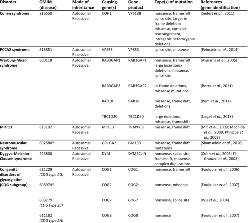
3.2 Golgi-associated proteins implicated in POM
10
Vesicular routing within the cell is highly dependent on microtubules, and the Golgi 11
apparatus is central to this process as it not only drives translational modifications of freshly 12
synthesized proteins and lipids but also orchestrates the complex process that allows them to be 13
packed into specific transport vesicles and routed to their final destinations (Boncompain and 14
Perez, 2013b). As detailed in the introduction, this is even more relevant in the case of neurons 15
and oligodendrocytes. In line with the involvement of the secretory pathway in brain 16
maturation, an increasing number of genes that have been recently associated with syndromic 17
or isolated POM appear to encode Golgi proteins involved in the regulation of the Golgi-18
mediated traffic machinery, including vesicle targeting and membrane recycling (Table II). 19
20
3.2.1 Cohen syndrome and COH1/VPS13B 21
Cohen syndrome (COH, MIM 216550) is an autosomal recessive disorder characterized 22
by motor delays, retinal dystrophy appearing by mid-childhood, progressive severe myopia, 23
hypotonia, joint hypermobility and progressive POM associated with intellectual disability 24
(Wang et al., 1993). Brain MRI reveals a relatively large corpus callosum in some patients, 25
associated with markedly smaller sagittal diameters of the brain stem (Kivitie-Kallio et al., 26
1998). While the neurological signs are most prominent, additional features such as short 1
stature, small hands and feet or childhood-onset obesity have also been reported in some 2
patients but are not constant (Falk et al., 2004). COH1, the only gene associated with Cohen 3
syndrome so far, encodes VPS13B, a large peripheral membrane protein that displays regions 4
homologous to yeast vacuolar protein sorting-associated protein 13 (Vps13p), and is active in 5
the Golgi (Seifert et al., 2011). VPS13B has recently been found to colocalize and interact 6
physically with the active form of RAB6 (Seifert et al., 2015). Depletion experiments using 7
RNAi against RAB6 show that it is required for VPS13B recruitment to Golgi membranes. 8
Conversely, the downregulation of VPS13B or a blockade of its recruitment to the Golgi 9
apparatus results in the fragmentation of Golgi ribbons and a simultaneous inhibition of neurite 10
outgrowth in hippocampal neurons (Seifert et al., 2011; Seifert et al., 2015). Thus, the gene 11
responsible for Cohen syndrome likely encodes an effector protein of RAB6 with a specific 12
role in the dynamics and function of the Golgi apparatus in particular during neuronal 13
maturation (Figure 1B). 14
15
3.2.2 PCCA2 syndrome and VPS53 16
Progressive Cerebello-Cerebral Atrophy type 2, also named Ponto-Cerebellar 17
Hypoplasia type 2E (PCCA2/PCH2E, MIM 615851) is an autosomal recessive 18
neurodegenerative disorder characterized by normal development during the first three to five 19
months of life, followed by motor delays, progressive POM, progressive spasticity leading to 20
contracture and epileptic seizures prior to two years of age (Ben-Zeev et al., 2003). Patients 21
have a normal head circumference at birth and undergo progressive growth deceleration, 22
resulting in microcephaly during the first year of life. Brain MRI reveals a gradual decrease in 23
cerebral white matter associated with delayed myelination and thinning of the corpus callosum 24
(Ben-Zeev et al., 2003). The responsible gene, mapped and identified in 2014, encodes VPS53, 25
a vacuolar protein-sorting protein that participates in the transport and recycling of endosome-26
derived transport vesicles (Feinstein et al., 2014). VPS53 is part of two large multisubunit 1
complexes named Golgi-associated retrograde protein (GARP) and Endosome-associated 2
recycling protein (EARP). Both GARP and EARP ensure the proper tethering between 3
endosomes and their acceptor compartment. GARP is a peripheral complex associated with the 4
TGN and is involved in tethering retrograde transport carriers from endosomes to the TGN 5
(Bonifacino and Hierro, 2011). EARP, characterized more recently, is localized to recycling 6
endosomes and promotes their fast recycling back to the plasma membrane (Schindler et al., 7
2015). Both complexes cooperate with SNAREs for subsequent membrane fusion. RAB 8
proteins play an essential role during these tethering-fusion steps as they recruit the required 9
tethering factors. In line with this role, GARP has been found to interact with RAB6A at the 10
TGN (Liewen et al., 2005) and EARP associates with RAB4-containing vesicles (Schindler et 11
al., 2015). Thus, the gene responsible for PCCA2 syndrome encodes a subunit of tethering
12
complex proteins that specifically interact with RAB GTPases during endosomal transport in 13
between the TGN and the plasma membrane (Figure 1B). 14
15
3.2.3 Warburg-Micro syndrome and RAB3GAP1/2, RAB18 and TBC1D20 16
Warburg-Micro syndrome (WARBM1, MIM 600118) is an autosomal recessive 17
disorder characterized by neurodevelopmental defects, severe visual impairment and 18
hypogonadism (Warburg et al., 1993). Neurodevelopmental features generally include POM 19
with profound intellectual disability and progressive limb spasticity associated with progressive 20
peripheral axonal neuropathy (Bem et al., 2011). Brain MRI shows predominantly frontal 21
polymicrogyria bilaterally, and hypoplasia of the corpus callosum and cerebellar vermis 22
(Handley et al., 2013; Liegel et al., 2013). Loss-of-function mutations in four distinct genes, 23
RAB3GAP1, RAB3GAP2, RAB18 and TBC1D20, have been implicated in WARBM1 in 24
recent years (Aligianis et al., 2005; Borck et al., 2011; Liegel et al., 2013). RAB18 has been 25
linked to several distinct membrane-bound organelles such as endosomes (Lutcke et al., 1994), 26
peroxisomes (Gronemeyer et al., 2013), secretory granules (Vazquez-Martinez et al., 2007) and 1
the ER, and to lipid droplet formation (Martin et al., 2005; Ozeki et al., 2005), depending upon 2
circumstances and cell types. More recent studies have confirmed its localization in the ER and 3
the cis-Golgi compartment (Dejgaard et al., 2008). The RAB3GAP complex, initially identified 4
as a GTPase activating protein (GAP) specific to the RAB3 subfamily of small G proteins 5
(Fukui et al., 1997; Nagano et al., 1998), is also a GEF (guanine nucleotide exchange factor) of 6
RAB18 (Gerondopoulos et al., 2014). TBC1D20, an ER-localized GAP that promotes the 7
hydrolysis of RAB1 GTP (Haas et al., 2007; Sklan et al., 2007), is thought to act on RAB18 as 8
well (Handley et al., 2015). Thus, RAB3GAP1, RAB3GAP2 and TBC1D20 all play a role in 9
the regulation of the RAB18 activity, directly linking WARBM1 to RAB18 deficiency or 10
dysregulation. RAB3GAP and TBC1D20 also regulate the ER localization of RAB18, an 11
essential step to support the function of RAB18 in the control of ER structural integrity and 12
retrograde membrane recycling from the Golgi apparatus to the ER (Gerondopoulos et al., 13
2014; Handley et al., 2015). Thus, the genes involved in WARBM1 all pinpoint a specific 14
RAB-dependent pathway directly associated with ER-Golgi trafficking (Figure 1B). 15
16
3.2.4 Autosomal recessive mental retardation 13 (MRT13) and TRAPPC9 17
Loss-of-function mutations in the TRAPPC9 gene were originally identified by 18
autozygosity mapping in four families with a nonsyndromic autosomal recessive intellectual 19
disability (MRT13, MIM 613192) (Mir et al., 2009; Mochida et al., 2009; Philippe et al., 20
2009). Since then, 3 additional cases have been reported. Although initially referred to as 21
nonsyndromic, the phenotype that is starting to emerge appears to be quite distinctive, 22
including moderate to severe POM, a peculiar facial appearance, obesity and hypotonia. 23
Reported brain anomalies consistently include a reduced volume of the cerebral white matter 24
with a hypersignal on FLAIR sequences, and a marked thinning of the corpus callosum (Abou 25
Jamra et al., 2011; Kakar et al., 2012; Marangi et al., 2013). TRAPPC9 is one of the subunits 26
of the Trafficking Protein Particle (TRAPP) complex, which mediates the tethering of COPII-1
coated ER-derived vesicles to allow their fusion with cis-Golgi membranes (Barrowman et al., 2
2010). The TRAPP complex acts through the recruitment and activation of the GTPase RAB1, 3
which in turn recruits specific cis-Golgi effectors such as p115 and GM130, allowing the 4
tethering of the vesicles to Golgi membranes (Barnekow et al., 2009). During this anterograde 5
ER-to-Golgi transport, the TRAPP complex is dynamically associated with the microtubules 6
through a physical interaction with p150Glued, a subunit of dynactin. A recent study has
7
proposed that TRAPPC9 in particular mediates the interaction between p150Glued and COPII-8
coated vesicles until they reach their target membrane (Zong et al., 2012), evoking an 9
additional paradigm in which RAB-associated ER-Golgi trafficking linked to POM and white 10
matter defects (Figure 1B). 11
12
3.2.5 A neuromuscular syndrome with microcephaly and GOLGA2/GM130 13
A homozygous frame-shift deletion in the GOLGA2 gene that results in a loss of gene 14
function has recently been identified in an individual with a neuromuscular phenotype 15
characterized by developmental delays, seizures, progressive microcephaly starting at 4 months 16
of age, hypotonia and muscular dystrophy (Shamseldin et al., 2016). Here also, brain MRI has 17
revealed delayed myelination and a thinning of the corpus callosum, but with no other specific 18
loss of cerebral volume. GOLGA2 encodes the Golgi matrix protein GM130, which is a 19
peripheral membrane protein located on the cis-side of the Golgi apparatus and involved in 20
both the assembly/maintenance of Golgi structure and the regulation of the secretory pathway 21
(Nakamura, 2010). As mentioned above in the case of TRAPPC9, GM130 participates in 22
membrane-tethering events at the Golgi complex through dynamic interactions with RAB1 and 23
other tethering proteins such as p115, to ensure efficient anterograde cargo delivery to the cis-24
Golgi compartment. Moreover, GM130 binds to other RAB proteins involved in membrane 25
traffic regulation at the ER/Golgi interface, such as RAB2 and RAB33B (Short et al., 2001; 26
Valsdottir et al., 2001). Thus, GOLGA2/GM130 deficiency appears to be yet another situation 1
highlighting the link between Golgi-associated RABs, POM and white matter defects (Figure
2
1B). 3
4
3.2.6 Dyggve-Melchior-Clausen syndrome and DYMECLIN 5
Dyggve-Melchior-Clausen syndrome (DMC, MIM #223800) is an autosomal recessive 6
skeletal dysplasia associated with POM and intellectual disability, and caused by loss-of-7
function mutations in the DYM gene encoding DYMECLIN, a Golgi protein involved in 8
intracellular trafficking (Dimitrov et al., 2009; Osipovich et al., 2008; Paupe et al., 2004). 9
Brain MRI in DMC patients with a truncating mutation in DYM reveals a marked thinning of 10
the corpus callosum and brain stem (Dupuis et al., 2015). In line with this finding, recent data 11
from our group show a significant reduction in white matter volume associated with defects in 12
the way the myelin sheath is wrapped, and a reduced thickness of myelinated axons in Dym-/- 13
mutant mice (Dupuis et al., 2015). Interestingly, Dym-deficient neurons display a fragmented 14
Golgi apparatus and impaired ER-to-Golgi trafficking (Dupuis et al., 2015). However, an 15
impairment of the retrograde transport of vesicles from the Golgi to the ER has also been 16
suggested in Dym-/- mouse embryonic fibroblasts (Osipovich et al., 2008). Although 17
DYMECLIN function is still elusive at the molecular level, several lines of evidence suggest 18
that it has a tethering role during vesicle trafficking between the ER and the Golgi: (i) 19
DYMECLIN localizes to both the cytosol and the periphery of cis-Golgi membranes, and 20
permanently shuttles between these two compartments (Dimitrov et al., 2009), (ii) 21
DYMECLIN colocalizes and directly interacts with GIANTIN (Dimitrov et al., 2009; 22
Osipovich et al., 2008), a giant Golgi-resident protein of the golgin family that forms 23
complexes with RAB1 or RAB6 to tether Golgi membranes with membrane structures derived 24
from the ER (anterograde pathway) or returning to the ER (retrograde pathway), respectively 25
(Goud and Gleeson, 2010; Koreishi et al., 2013; Rosing et al., 2007) (Figure 1B). 26
Interestingly, Smith McCort dysplasia, a clinical variant of DMC syndrome with identical 1
skeletal defects but normal intelligence and no microcephaly, has been found to result either 2
from specific missense mutations in DYM that could result in some residual activity of the 3
protein (SMC1, MIM #607326) (Cohn et al., 2003; Dimitrov et al., 2009) or from loss-of-4
function mutations in the small GTPase RAB33B (SMC2, MIM #615222) (Alshammari et al., 5
2012; Dupuis et al., 2013). Given that the main functional domains of RAB33A and RAB33B 6
proteins are perfectly conserved (Zheng et al., 1998), it is tempting to speculate that the 7
cerebral phenotype in SMC is rescued by the partial activity of DYMECLIN (SMC1) or a 8
complementation of RAB33B deficiency by RAB33A in the brain (SMC2). Interestingly, both 9
RAB33A and RAB33B are present in the Golgi complex (Cheng et al., 2006; Zheng et al., 10
1998) and are involved in the regulation of vesicular transport: while RAB33B functions in 11
concert with RAB6 to coordinate bidirectional intra-Golgi and retrograde Golgi-to-ER transport 12
(Starr et al., 2010), RAB33A has been shown to mediate the anterograde transport of post-13
Golgi vesicles in growing hippocampal axons (Nakazawa et al., 2012). Although the precise 14
link between DYM, RAB33A/B and RAB6 is yet to be understood, these factors likely function 15
in the regulation of common Golgi-driven secretory pathways. 16
17
3.2.7 Congenital disorders of glycosylation and the COG complex 18
Congenital Disorders of Glycosylation (CDG) represent a huge and still growing family 19
of multisystemic autosomal recessive pathologies involving dysfunctions in the processing of 20
N- and O-linked glycans, with most of the genes identified so far encoding glycosylation 21
enzymes (Freeze and Ng, 2011). However, one subgroup of these diseases involves the 22
Conserved Oligomeric Golgi (COG) complex, a hetero-octameric protein complex, which, as 23
its name suggests, is localized to the cis and medial Golgi as well as surrounding vesicles 24
(Climer et al., 2015). The COG complex is thought to act as a tethering factor, in particular 25
during intra-Golgi and retrograde Golgi-to-ER trafficking, where it mediates the recycling of 26