R E S E A R C H A R T I C L E
Open Access
Localization and expression of EDS5H a
homologue of the SA transporter EDS5
Nonglak Parinthawong
1,2, Stéphanie Cottier
1, Antony Buchala
1, Christiane Nawrath
1,3and Jean-Pierre Métraux
1*Abstract
Background: An important signal transduction pathway in plant defence depends on the accumulation of salicylic acid (SA). SA is produced in chloroplasts and the multidrug and toxin extrusion transporter ENHANCED DISEASE SUSCEPTIBILITY5 (EDS5; At4g39030) is necessary for the accumulation of SA after pathogen and abiotic stress. EDS5 is localized at the chloroplast and functions in transporting SA from the chloroplast to the cytoplasm. EDS5 has a homologue called EDS5H (EDS5 HOMOLOGUE; At2g21340) but its relationship to EDS5 has not been described and its function is not known.
Results: EDS5H exhibits about 72 % similarity and 59 % identity to EDS5. In contrast to EDS5 that is induced after pathogen inoculation, EDS5H was constitutively expressed in all green tissues, independently of pathogen infection. Both transporters are located at the envelope of the chloroplast, the compartment of SA biosynthesis. EDS5H is not involved with the accumulation of SA after inoculation with a pathogen or exposure to UV stress. A phylogenetic analysis supports the hypothesis that EDS5H may be an H+/organic acid antiporter like EDS5.
Conclusions: The data based on genetic and molecular studies indicate that EDS5H despite its homology to EDS5 does not contribute to pathogen-induced SA accumulation like EDS5. EDS5H most likely transports related substances such as for example phenolic acids, but unlikely SA.
Introduction
The signal transduction for induced resistance to many pathogens including viruses, bacteria, fungi and oomy-cetes involves the phenolic compound salicylic acid (SA) [1–3]. The importance of SA for the activation of de-fences has been repeatedly demonstrated with a number of mutants or transgenic plants impaired in the accu-mulation of SA. In particular, the Arabidopsis thaliana eds5/sid1 (enhanced disease susceptibility5/salicyclic acid-deficient 2) and sid2 mutants accumulate only 10 % of the SA produced in wild-type plants after induc-tion and exhibit increased susceptibility to Pseudo-monas syringae and Hyaloperonospora parasitica and fail to express the pathogenesis-related gene PR1 [4]. The eds5/sid1 mutation was found in a gene encoding a member of the MATE (MULTIDRUG AND TOXIN EX-TRUSION) transporter family [1], while the sid2 muta-tion was found in the ISOCHORISMATE SYNTHASE1 gene (ICS1) [5].
The identification of ICS1 in A. thaliana, definitely demonstrated the importance of the isochorismate path-way for SA biosynthesis, similar to the pathpath-way de-scribed in some Pseudomonas species [5]. The ICS1 gene product was confirmed to possess ICS activity and to be targeted to the plastidic compartment [6]. Synthe-sis of SA following exposure to ozone in ArabidopSynthe-sis was also proposed to proceed through the activity of ICS enzymes [7]. The involvement of isochorismate in the synthesis of SA was confirmed in transgenic tobacco plants overexpressing an ICS of Catharanthus roseus [8] as well as in tomato [9] and Nicotiana benthamina [10]. The second ICS gene present in the Arabidopsis genome named ICS2 [5] encodes a protein that is also localized in the chloroplast and has ICS activity [11]. ICS2 partici-pates in the synthesis of SA in partial redundancy with ICS1 since ics1/ics2 double mutants produce only 36 % of the SA amount found in the ics1 single mutant. The conversion from isochorismate to SA has not yet been described in plants. In Pseudomonas aeruginosa it is cat-alyzed by a bifunctional enzyme displaying isochorismate pyruvate-lyase and chorismate mutase activities [12]. * Correspondence:[email protected]
1Department of Biology, University of Fribourg, 1700 Fribourg, Switzerland
Full list of author information is available at the end of the article
© 2015 Parinthawong et al.; licensee BioMed Central. This is an Open Access article distributed under the terms of the Creative Commons Attribution License (http://creativecommons.org/licenses/by/4.0), which permits unrestricted use, distribution, and reproduction in any medium, provided the original work is properly credited. The Creative Commons Public Domain Dedication waiver (http://creativecommons.org/publicdomain/zero/1.0/) applies to the data made available in this article, unless otherwise stated.
In fact, MATE-transporters are present in almost all prokaryotes and eukaryotes and are thus one of the most conserved families in nature [15, 16]. Plants have the lar-gest gene family of MATE-transporters with 58 genes in Arabidopsis, while prokaryotes have approximately 10 genes and mammals only 2. Plant MATE–transporters have not only been found at the plasma membrane, but also at the vacuolar membrane acting in the sequestra-tion of toxic metabolites. For example, the MATE-transporter TT12 acts as flavonoid/H+ antiporter at the vacuole and is active in proanthocyanidin accumulation in the seed coat of Arabidopsis [17, 18]. In tobacco, the alkal-oid nicotine is sequestered into the vacuole in exchange with protons by the action of NtMATE1 and NtMATE2 in the roots and by the MATE-transporter NtJAT1 in the shoots [19, 20]. These MATE-transporters may also trans-port other alkaloids, such as anabasine, hyoscyamine, sco-polamine or berberine, but no flavonoids. Here we report a detailed characterization of EDS5H, a close homologue of the SA transporter EDS5.
Results
Identification and characterization of EDS5H
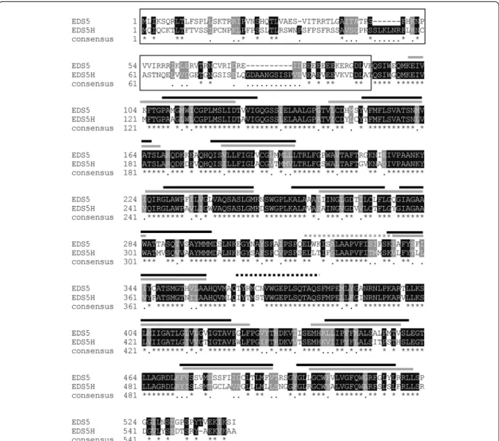
The analysis of the Arabidopsis genome revealed a homologue of EDS5 that is encoded by the gene At2g21340 and was therefore named EDS5H. The EDS5H gene was characterized by amplification of a 1680 bp cDNA by reverse transcriptase-mediated polymerase chain reaction (RT-PCR) and subsequently sequenced. The ana-lysis of the EDH5 sequence confirmed that EDS5H has an open reading frame (ORF) of 1680 bp encoding for a pro-tein of 559 amino acids. The genomic region of EDS5H consists of 14 exons and 13 introns based on the anno-tated Arabidopsis genome. The alignment between the predicted protein sequences of EDS5 and EDS5H showed an overall 72 % similarity and 59 % identity. However, the 100 aa at the N-terminus showed less conservation (20 % identity) (Fig. 1).
Expression of EDS5H
The expression of EDS5H was studied in leaves of plants
inoculated with Pseudomonas syringae pv tomato
avrRpt2. As indicated in Fig. 2, the expression shows no change in response to infection. The activity of the pro-moter of EDS5H was also studied using propro-moter-GUS fusions. Promoter fragments of 500, 1000 or 2000 bp
CaMV 35S promoter upstream of a triple cMyc epitope tag. In tissue sections of transgenic plants carrying the CaMV35S::EDS5H::3xcMyc construct, the cMyc-epitope tag was labelled with a primary anti-myc antibody that was identified by a goat anti-mouse IgG conjugated with Alexa Fluor 488 (green). RCCR (red chlorophyll catalase reductase) was used as an example for a protein targeted to the chloroplast [21]. Tissues were labelled with a pri-mary antibody against RCCR and secondary antibody goat anti-rabbit IgG conjugated with Alexa Fluor 568 (red). Fig. 4 shows an intense and fine green labelling lo-calized around the chloroplast, while the red label was in the middle of the chloroplast. The co-localization of the EDS5H with RCCR shows that EDS5H is located to the chloroplast. cMyc-tag could not be detected in plant cells when the natural EDS5H promoter was used, al-though the transcription of the EDS5H::cMyc could be determined by RT-PCR (data not shown). This suggests that the EDS5H promoter, which is constitutive, was too weak to drive the whole cassette of the EDS5H::3xcMyc to its target organelle or the amount was below detec-tion limits. Taken together, these studies show that EDS5H is localized at the chloroplast as was shown pre-viously for EDS5 [11].
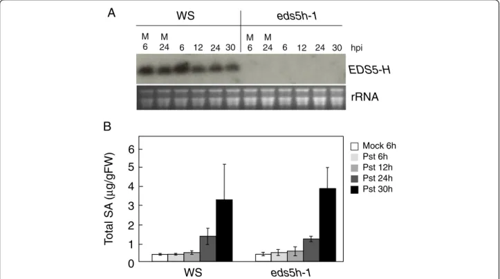
Analysis of plants with downregulated EDS5H expression Since both EDS5 and EDS5H are expressed in the same tissues and in the same organelle it is possible that EDS5H may also function in SA accumulation since it has been observed that eds5-3 mutants still contain ca 10 % of the SA level found in wild-type plants [2]. This level of SA could potentially be due to the functional EDS5H. A mutant carrying a T-DNA insertion in the EDS5Hgene, called eds5h-1, was isolated after screening the population of T-DNA insertion pools generated by the Arabidopsis knock-out facility service at the University of Wisconsin-Madison. Two mutant lines were obtained, eds5h-1carrying the T-DNA insertion at 229 bp (exon 1) and eds5h-10 carrying the insertion at 1360 bp (exon 10). Both were transcriptional null alleles, and the eds5h-1 mu-tant was used for further studies. The expression of EDS5H in Ws wild-type plants was strong in non-inoculated leaves (Fig. 5a). The expression of EDS5 was not different between wild type and the eds5h-1 mutant (data not shown). No difference was detectable in the ac-cumulation of SA after inoculation with P. syringae pv.
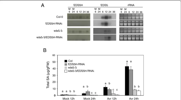
tomato DC3000 in Ws and eds5h-1, most likely due to EDS5 function (Fig. 5b). Since the induction kinetics of SA accumulation in Col-0 and Ws showed strong differences, the generation of a double mutant between eds5h-1 (in the Ws background) and eds5-3 (in the Col-0 background) was not useful (data not shown). Therefore, we used RNA interference (RNAi) to downregulate the transcription of EDS5Hin the Col-0 background. For the construction of the RNAi construct, the 350 bp 5’-non-conserved region of EDS5H was cloned into the plasmid pHANNIBAL [22] in sense and antisense orientations. The RNAi construct was transformed into both Col-0 and eds5-3. Independent lines from eds5h (EDS5H-RNAi) and from the double
mutant eds5-3/eds5h (eds5-3/EDS5H-RNAi) were selected for further investigation because they did not accumulate EDS5H mRNA (Fig. 6a). These lines showed no changes in phenotype compared to wild-type plants. Transgenic plants in which no EDS5H transcription was detected after infection with P. syringae pv. tomato DC3000 with or without the avirulence gene avrRpt2 were analysed for their SA content in comparison to Col-0 plants. A slight decrease in SA accumulation (ca 20 %) was observed in EDS5H-RNAi plants 12 h after infection with P. syringae pv. tomato DC3000 avrRpt2 (Avr) compared to Col-0 but this difference disappeared 24 h after infection. Similarly, no changes were observed when EDS5H was knocked Fig. 1 Alignment of the predicted protein sequences of EDS5H and EDS5. The predicted membrane-spanning domains are indicated above the alignment with a grey bar for EDS5H and black bars for EDS5. Identical amino acids are indicated with an asterisk, and conserved amino acids are indicated with a single dot. The N-terminus region has a low degree of homology and is indicated by a black frame
down by RNAi in the eds5-3 mutant (Fig. 6b). Similar re-sults have been found for the SA accumulation after UV-light exposure (data not shown). These results support that EDS5H is not responsible for the presence of the re-sidual SA in the eds5-3 mutant.
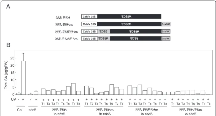
Analysis of transgenic eds5-3 plants overexpressing EDS5H and a EDS5::EDS5H fusion protein
In order to determine if overexpression of EDS5H can res-cue the SA-deficient phenotype of eds5-3, EDS5H was expressed in eds5-3 under the control of the 35S promoter (Fig. 7a). The lines overexpressing EDS5H did not display any phenotypic change compared to wild types. Trans-genic plants overexpressing EDS5H were exposed to UV-light to induce the SA-biosynthesis pathway and their SA content was measured 12 h after induction. Some of the independent eds5-3 lines overexpressing EDS5H accumu-lated up to 3 times more SA than the eds5-3 controls but still 5 times less than the wild-type plants (Fig. 7b). The rescue of the eds5-3 mutant phenotype by the CaMV35-S::EDS5Hconstruct was therefore only limited.
EDS5 and EDS5H show a very high percentage of homology at the C-terminal region containing the trans-membrane spanning domains. In contrast, a small do-main of approximately 60 aa that lies between the signal peptide for plastid targeting and the beginning of the transmembrane spanning domains shows little hom-ology. In EDS5, this domain contains a hepta-peptide potentially forming a coiled-coil domain (http://embnet.-vital-it.ch/software/COILS_form.html), while not in EDS5H. In order to test if the N-terminal domain con-fers specificity to the function of EDS5, eds5-3 plants were transformed with a CaMV35S::EDS5-EDS5H fusion that contained the plastid localization signal from EDS5 as well as the N-terminus of the mature protein. The
accumulation of SA was monitored in transgenic eds5-3 plants overexpressing the EDS5::EDS5H fusion protein after their exposure to UV-light followed by a 12-h incu-bation time. The expression of the EDS5::EDS5H fusion protein did not result in a better rescue of the eds5-3 phenotype than the original EDS5H (Fig. 7b), but still in-creased the SA amount of the eds5 mutant suggesting that the resulting fusion protein still had transport activ-ity. The fusion of the entire N-terminal domain of Fig. 2 Expression analysis of EDS5H. A. thaliana plants (Col-0 and Ws)
were inoculated with P. syringae pv. tomato avrRpt2 (Pst) and harvested for RNA extraction at 12, 24, 36, and 48 h after inoculation. Mock control was carried out by treatment with 10 mM MgCl2. The RNA blot
was probed with a gene-specific probe for EDS5H. Ethidium bromide staining of the RNA gel (rRNA) was used to show equal loading. The experiment was repeated 3 times with similar results
Fig. 3 Localization of GUS activity in transgenic Arabidopsis plants expressing the pEDS5H::GUS fusion construct. Seedlings, siliques or inflorescence parts of plants transformed with EDS5H::GUS constructs were stained with the substrate for GUS. The activity of a promoter of 2000 bp length is shown in a seedling (a), flowers (b), and a silique (c). The experiment was repeated 3 times with similar results
EDS5H to the EDS5 transporter region did not rescue SA accumulation (Fig. 7b). These results show that EDS5H does not function better in SA accumulation when the EDS5 N-terminus is present.
Potential transport function of EDS5 and EDS5H
Phylogenetic studies of the MATE-family of plants re-vealed that EDS5 and EDS5H are two transporters that are in the same subfamily of MATE-transporters as the Arabidopsis citrate transporters described recently [23,
24]. Therefore, EDS5 and EDS5H have been aligned in comparison to plant MATE transporters that have been shown to transport flavonoids (AtTT12) [18], alkaloids (NtMATE) [20] or other toxic molecules, such as poly-vinylpyrrolidone and pyrrolidinone (AtAFL5) [25], as well as citrate transporters from monocots (barley [26], sorghum [27] and rice [28]) and Arabidopsis (AtFRD3 [29] and AtMATE [24]). This alignment shows that EDS5 and EDS5H form their own subgroup clustering together with the citrate transporters and not with the Fig. 4 Subcellular localization of EDS5H-3myc in transgenic Arabidopsis. Mesophyll cells of transgenic plants carrying the CaMV35S::EDS5H::3xmyc construct. The two panes show the cMyc-epitope labelled with Alexa Fluor 488 excited at the wavelength of 488 nm and detected using the emission filter 522 DF32 (green) and Red chlorophyll catalase reductase labelled with Alexa Fluor 568 excited at a wavelength of 568 nm and detected using the emission filters 605 DF32 and 585 EFLP (red). Bar = 5μm. A representative picture out of 11 transformed lines is presented
eds5h-1
T
o
tal SA
(
µ
g/gFW)
0
2
4
3
1
WS
Mock 6h Pst 6h Pst 12h Pst 24h Pst 30h5
6
WS
M 6 M 24 6 12 24 30rRNA
EDS5-H
eds5h-1
M 6 M 24 6 12 24 30B
A
hpiFig. 5 Characterization of the eds5h-1 mutant. a) Expression analysis of the EDS5H. 10 mM MgCl2(M) or P. syringae pv. tomato DC3000 were
infiltrated into Ws and eds5h-1 leaves. Samples were taken at 6, 12, 24, and 30 h post-inoculation (hpi). Northern blots were hybridized with a gene-specific probe for EDS5H. Ethidium bromide staining of the RNA gel (rRNA) was used as control for the loading. The experiment was repeated 3 times with similar results. b) Accumulation of SA in plants infiltrated with 10 mM MgCl2(M) or P. syringae pv. tomato DC3000
flavonoid/alkaloids/toxin transporters (Fig. 8). Since EDS5 was shown to transport SA, EDS5H might trans-port related phenolic compounds, but unlikely SA. Discussion
A detailed characterization of the related MATE-transporter EDS5H was undertaken in order to obtain more information on its possible function relative to EDS5. The present studies confirm earlier results that EDS5 is only weakly expressed under non-inducing con-ditions and strongly after pathogen infection as reported for SA accumulation (Fig. 6a) [1]. The homologue EDS5H is constitutively expressed in all green tissues, independently of pathogen infection (Fig. 3). The fact that the eds5-3 mutant phenotype is clearly detectable under conditions where EDS5H is strongly expressed in-dicates that EDS5 and EDS5H may only have partially redundant functions.
The determination of the sub-cellular localization of EDS5H by fluorescence microscopy reveals that it is lo-calized at the plastid where most of the SA is synthe-sized [13, 30]. This is similar to EDS5 that was shown to localize within the chloroplast envelope and to function
as a multidrug and toxin extrusion-like transporter in the export of SA from the chloroplast to the cytoplasm where it controls the innate immune response [13].
In order to clarify the functional relationship between EDS5 and EDS5H, T-DNA insertion mutants and RNAi lines where the EDS5H transcript was knocked out have been isolated or produced in the Col-0 or in the eds5-3 mutant background. The analysis of all these lines clearly show that EDS5H is not involved in SA accumulation after pathogen or UV-light induction (Fig. 6). Two ex-planations may be proposed for why the eds5-3/eds5H double mutants do not have less SA than eds5-3. Firstly, EDS5H could have a very low SA transport activity and therefore not reduce the SA content in the plastid. Sec-ondly, the SA content that remains in the eds5-3 mutant is actually not produced in the plastid. Similarly, the sin-gle mutant ics1 or the double mutant ics1/ics2 contain a similarly low amount of SA indicating the existence of an alternative pathway for the production of SA [11]. Also, eds5-3 lines overexpressing EDS5H contain only slightly more SA independent of whether EDS5H was equipped with the N-terminus of EDS5 or not. These studies suggest that EDS5H has no, or at best a very
0 60
B
T o tal SA ( µ g/gFW)Mock 12h Mock 24h Avr 12h
Col EDS5H-RNAi eds5-3 eds5-3/EDS5H-RNAi 10 0 20 30 40 50 Avr 24h a a b b a b c c c c a b b b a a
Fig. 6 Characterization of the EDS5H-RNAi mutant and eds5-3/EDS5H-RNAi double mutant. a) Expression analysis of EDS5H and EDS5 in different genotypes. Plants were inoculated with 10 mM MgCl2(M) or P. syringae pv. tomato DC3000. Samples were taken at 6, 12, 24, and 36 hpi.
Northern blots were hybridized with gene-specific probes for EDS5H and EDS5. Ethidium bromide staining of the RNA gel (rRNA) was used as loading control. Note that some EDS5 expression can be detected as eds5-3 is not a transcriptional null mutant. The experiment was repeated 3 times with similar results. b) Accumulation of SA in different genotypes. Plants were inoculated with 10 mM MgCl2(Mock) or P. syringae pv.
tomato DC3000 carrying avrRpt2 (Avr). Samples were taken 12 and 24 hpi. Different letters above each bar represent statistically significant differences. The mean comparison of total SA was analyzed using Duncan’s multiple-range test (DMRT) with p-value ≤ 0.05. For each time point n = 4 (±SD), the experiment was repeated 3 times
weak transport activity for SA and is not involved in resist-ance to P. syringae pv. tomato or Botrytis cinerea (unpub-lished data). Since EDS5H is closely related to EDS5 (Fig. 8) it is most likely involved in transporting related substances, for example other phenolic acids. The bio-chemical characterization of the transport activities of EDS5H will be exciting objectives for future work.
Conclusions
Despite its homology to EDS5, EDS5H does not contrib-ute to pathogen-induced SA accumulation like EDS5. The phylogenetic relatedness of EDS5H with EDS5 sup-ports a function of EDS5H in the transport of phenolic substances.
Materials and methods
Growth conditions, bacterial inoculations and plant transformation
Arabidopsis accessions Columbia-0 (Col-0) and Wassilewskija (Ws) or transgenic plants were grown under either sterile or non-sterile conditions. Non-sterile plants were grown on a pasteurized soil mix of commercial potting soil:perlite (3:1), in a growth chamber at 22 ± 2 °C under a 12-h photoperiod. Seed dormancy was broken by stratification at 4 °C for 3 days. For sterile cultures, seeds were surface steril-ized by treating for 15 min in 2.5 % (v/v) commercial bleach containing 0.05 % Triton X-100 with continu-ous agitation and rinsed four times with sterile distilled water. Seeds were placed on solid medium consisting of ½ MS (Murashige and Skoog basal medium, Sigma) with 1 % sucrose, 0.1 % vitamins (Murashige and Skoog, Sigma). P. syringae pv tomato with or without avrRpt2 was cultured at 28 °C and 220 rpm in Luria-Bertani medium containing 30 μg/ml rifampicin and 35S-E5H in eds5 35S-E5Hm in eds5 35S-E5/E5Hm in eds5 35S-E5H/E5m in eds5
B
A
3xMYC CaMV 35S EDS5H CaMV 35S EDS5H 3xMYC CaMV 35S EDS5H 3xMYC CaMV 35S EDS5 EDS5 EDS5H 35S-E5H 35S-E5Hm 35S-E5/E5Hm 35S-E5H/E5m UV T otal SA ( µ g/gFW) Col 0 5 10 15 20 25 - + - + + + + + + + + + + + + + + + + + + + + + + + + + + + + + + + + + eds5 T1 T2 T3 T4 T5 T6 T7 T8 T1 T2 T3 T4 T5 T6 T7 T8 T1 T2 T3 T4 T5 T6 T7 T8 T1 T2 T3 T4 T5 T6 T7 T8Fig. 7 Characterization of transgenic eds5-3 plants carrying constructs for the overexpression of EDS5H or EDS5H variants. a) Schematic representation of the constructs used. b) Accumulation of total SA in different genotypes overexpressing EDS5H and its variants. Plants were exposed to UV-C light for 20 min and samples were taken 12 h later. For determinations with Col0 and eds5, each time point n = 3 (±SD), SA determinations on all transformants were carried out once in the T1 generation
Fig. 8 Phylogenetic studies of the MATE-family of plants. EDS5 and EDS5H have been aligned in comparison to plant MATE shown to transport flavonoids (AtTT12) [18], alkaloids (NtMATE) [20] or molecules such as polyvinylpyrrolidone and pyrrolidinone (AtAFL5) [25], as well as citrate transporters from monocots (barley [26], sorghum [27] and rice [28]) and Arabidopsis (AtFRD3 [29] and AtMATE [24]). The dendrogram was established with the ClustalW program
plants were selected on ½ MS plates, containing 30μg/ml hygromycin.
Cloning of EDS5H and sequence analysis
cDNA of EDS5H (At2g21340) was amplified from Col-0 plants using the Access RT-PCR system from Promega. The 5’ end of the cDNA was determined by RT-PCR using primers located 75 (75UpStream), and 150 (150UpStream) bp upstream of the predicted ATG on the annotated genomic sequence (for these and all other primers indicated below, see sequences in Additional file 1: Figure S1). The 3’ end of the cDNA was determined by RT-PCR using primers located 50 (50DownStream), 100 (100DownStream), 200 (200DownStream), 250 (250Down-Stream), and 300 (300DownStream) bp downstream of the stop codon of the annotated genomic sequence. RT-PCR products were cloned into pGEM-T Easy Vector (Promega) for sequencing.
Isolation of RNA and DNA, transcript analysis
For RNA gel blot analysis, RNA was isolated as previ-ously described [4]. For qPCR, RNA was isolated using the Qiagen RNA easy kit including the recommended DNAase treatment. Genomic DNA was isolated using a protocol modified from [32]. Briefly, a leaf was extracted with buffer (0.2 M Tris–HCl pH 9.0, 0.4 M LiCl, 25 mM EDTA, 1 % SDS). The extracted tissue was centrifuged and the DNA was precipitated with isopropanol. The airdried pellet was resuspended in TE (10 mM Tris pH 8.0, 1 mM EDTA).
For RNA gel blot analysis, total RNA (10μg) was sepa-rated in formaldehyde-agarose (1 %) gels, transferred to a Nylon membrane (Hybond-N, Amersham Biosciences, UK) and crosslinked by UV-light. Hybridization was car-ried out in hybridization buffer (0.5 M NaHPO4pH 7.2, 7 % SDS, 1 mM EDTA, 1 % BSA) at 65 °C. The membrane was washed twice with 2X SSC containing 0.1 % SDS and twice with 0.2X SSC containing 0.1 % SDS at 65 °C, before exposing to X-Omat or Bio Max film (Kodak).
Isolation of mutants carrying a T-DNA in the EDS5H region
Plants carrying a T-DNA fragment in the region of exon 1 and exon 10, called eds5h-1 and eds5H-10, respect-ively, were screened by PCR www.biotech.wisc.edu/ NewServicesAndResearch/Arabidopsis. eds5H-1 plants
fragment by using GSP4 and GSP3 as forward and re-verse primers, respectively.
Construction of the plasmid used for RNAi
A fragment of about 350 bp of the non-conserved region of EDS5H was amplified by RT-PCR and cloned into plasmid pHANNIBAL [22] in sense and anti-sense orientation. The sense strand was designed to include XhoI and KpnI as cloning sites (forward primer: E5H-sens-For; reverse primer: E5H-sens-Rev) and the anti-sense strand was designed to include BamHI and ClaI as cloning sites (forward primer: E5H-anti-For; reverse primer: E5H-anti-Rev). The construct was cloned into the binary vector pART27 using the NotI sites [33]. Construction of promoter:GUS and cMyc-tagged constructs Three fragments of the promoter region of EDS5H were amplified using the High Fidelity Kit (Roche) with gene-specific primers designed to introduce EcoRI at the 5’ ends and NcoI sites at the 3’ ends of the fragments. The forward primers used for the amplification of EDS5H
promoter fragments were: Pro500F, Pro1000F and
Pro2000F resulting in fragments of 546 bp, 1057 and 1984 bp, respectively. The reverse primer for the EDS5H promoter fragments was ProE5H-R. The PCR fragments were cloned into the plasmid pCAMBIA 1303 (www.CAM-BIA.org) from which the fragments of the promoter CaMV 35S and LacZ alpha had been removed.
The ORF of EDS5H was amplified by High-fidelity RT-PCR using the Omniscript RT-PCR system (Qiagen) and the forward primer Nco-E5H introducing a NcoI site as well the reverse primer E5H-Mycs introducing a triple cMyc-tag and the restriction sites SmaI and NdeI. The PCR fragment was cloned into pGEM-T Easy Vector (Promega) in the NcoI and NdeI sites. The clone was se-quenced and the NcoI-SmaI fragment that included EDS5H::3xcMyc-tagg was then cloned into pCAMBIA 1304 (www.CAMBIA.org) from which the GUS and GFP genes had been removed.
Construction of plasmid for overexpressing of EDS5H The cDNA of EDS5H was amplified by RT-PCR using the forward primer E5H-For and the reverse primer E5H-Rev and was sub-cloned into pGEM-T Easy Vector (Promega). The cDNA was digested with EcoRI, blunted,
and cloned into pART7 [33]. Clones were selected based on the correct orientation of the cDNA between the CaMV35S promoter and OCS terminator. The cassette of CaMV35S::EDS5H::OCS was digested with NotI and cloned into the binary vector pART27 [33].
Constructions of plasmids for protein domain swapping The constructs for the peptide-swapping experiments were designed to include the triple cMyc-epitope tag (3xcMyc-tag). Therefore, a fragment of 3xcMyc-tag was amplified using the synthesized oligonucleotide E5H-Mycs as template. The PCR fragment included a NotI site at 5’-end and a SmaI and NdeI sites at 3’-end and 4 alanine residues upstream of cMyc-tag (forward primer:
For-Ala-Myc; reverse primer: Rev-Myc-PCS). The
3xcMyc-tag was cloned into pGEM-T Easy vector at cor-responding sites.
EDS5and EDS5H genes were swapped at the NdeI site of EDS5 (281 bp downstream of ATG). The NdeI site was introduced into EDS5H by changing of a nucleotide T to A (336 bp downstream of ATG) that caused no change in amino acid residues. Therefore, the N-terminus of each protein was designed to include NcoI and NdeI as cloning sites. The fragments were amplified by RT-PCR using forward primer Nco-EDS5, reverse pri-mer EDS5-Nde for EDS5, and forward pripri-mer Nco-E5H, reverse primer E5H-Nde for EDS5H. The C-terminus of each protein was designed to include NdeI and NotI as cloning sites. The fragments were amplified using for-ward primer Nde-eds5, reverse primer EDS5-Rev for EDS5, and forward primer Nde-e5h, reverse primer E5H-Swap-Rev for EDS5H. The swapped domains of ei-ther EDS5::EDS5H or EDS5H::EDS5 were constructed in pBluescript (Stratagene).
The constructs were placed upstream of 3xcMyc-tag with NcoI and NotI sites. The cassettes of the swapped peptide domains and 3xcMyc-tag were then cloned into pCAMBIA 1304 (www.CAMBIA.org). The GFP and GUS reporter genes of the binary vector were replaced with ei-ther EDS5::EDS5H::3xcMyc-tag or EDS5H::EDS5::3xcMyc-tag cassettes at NcoI and PmlI sites.
Immunolabelling and fluorescence microscopy
For immunolabelling, plant tissue was prepared according to the protocol described in [34]. The PEG-embedded tis-sue was sectioned into 4–5 μm-thick slices and placed on poly-L-lysine-covered slides. The slides were incubated in PBS for 10 min in order to remove the PEG from the sec-tion. Each slide was blocked for free aldehyde by incuba-tion with 0.1 M NH4Cl (in PBS) for 5 min, and washed for 5 min with PBS. The unspecific binding sites were blocked by incubation with 5 % BSA (in PBS) for 30 min. Slides were then incubated overnight with primary antibody so-lution (mouse anti-cMyc diluted in 5 % BSA/PBS in ratio
1:500) at 4 °C, washed three times with 0.1 % BSA (in PBS) for 10 min each, and once with 1 % BSA (in PBS) for 10 min. The secondary antibody used was goat mouse-IgG conjugated with Alexa Fluor 488 or goat anti-rabbit-IgG conjugated with Alexa Fluor 568, diluted 1:500 in 5 % BSA (in PBS). Slides were placed in a humid cham-ber, incubated with secondary antibody for 2 h at 37 °C. After washing with PBS (4x10 min, samples were kept at 4 °C in the dark. Fluorescent signals were recorded with a BioRad MCR 1024 Kripton-Argon confocal microscope using an excitation wavelength of 488 nm with an emis-sion filter 522 DF32, or an excitation wavelength of 568 nm with emission filters 605 DF32 and 585 EFLP. GUS-assays and determination of SA
GUS assays and the determination of free and conju-gated SA were performed as described previously [4, 35]. Additional file
Additional file 1: List of primers.
Competing interests
The authors declare that they have no competing interests.
Authors’ contributions
NP carried out the molecular genetic studies, participated in the sequence alignment. SC contributed to the localization studies. AB carried out the SA analyses. CN designed the study, performed the statistical analysis, drafted the manuscript. JPM drafted the final version of manuscript. All authors read and approved the final manuscript.
Acknowledgements
We would like to thank Linda Grainger and Martine Schorderet for excellent technical assistance. This work was supported by grants from the Swiss National Science Foundation to Jean-Pierre Métraux and Christiane Nawrath. Author details
1
Department of Biology, University of Fribourg, 1700 Fribourg, Switzerland.
2Faculty of Agricultural Technology, King Mongkut’s Institute of Technology
Ladkrabang, Chalongkrung Rd., Ladkrabang, 10520 Bangkok, Thailand.
3Department of Plant Molecular Biology, University of Lausanne, 1015
Lausanne, Switzerland.
Received: 17 January 2015 Accepted: 1 May 2015
References
1. Nawrath C, Metraux JP. Salicylic acid induction-deficient mutants of Arabi-dopsis express PR-2 and PR-5 and accumulate high levels of camalexin after pathogen inoculation. Plant Cell. 1999;11:1393–404.
2. Vlot AC, Dempsey DMA, Klessig DF. Salicylic acid, a multifaceted hormone to combat disease. Annu Rev Phytopathol. 2009;47:177–206.
3. Yan S, Dong X. Perception of the plant immune signal salicylic acid. Curr Opin Plant Biol. 2014;20:64–8.
4. Nawrath C, Heck S, Parinthawong N, Metraux JP. EDS5, an essential component of salicylic acid-dependent signaling for disease resistance in Arabidopsis, is a member of the MATE transporter family. Plant Cell. 2002;14:275–86.
5. Wildermuth MC, Dewdney J, Wu G, Ausubel FM. Isochorismate synthase is required to synthesize salicylic acid for plant defence. Nature. 2001;417:562–5. 6. Strawn MA, Marr SK, Inoue K, Inada N, Zubieta C, Wildermuth MC.
Pseudomonas syringae pv. tomato DC3000. Mol Plant Microbe Interact. 2007;20:955–65.
10. Catinot J, Buchala A, Abou-Mansour E, Métraux J-P. Salicylic acid production in response to biotic and abiotic stress depends on isochorismate in Nicotiana benthamiana. FEBS Lett. 2008;582:473–8.
11. Garcion C, Lohmann A, Lamodiere E, Catinot J, Buchala A, Doermann P, et al. Characterization and biological function of the ISOCHORISMATE SYNTHASE2 gene of Arabidopsis. Plant Physiol. 2008;147:1279–87. 12. Gaille C, Kast P, Haas D. Salicylate biosynthesis in Pseudomonas aeruginosa.
Purification and characterization of PCHB, a novel bifunctional enzyme displaying isochorismate pyruvate-lyase and chorismate mutase activities. J Biol Chem. 2002;277:21768–75.
13. Serrano M, Wang BJ, Aryal B, Garcion C, Abou-Mansour E, Heck S, et al. Export of salicylic acid from the chloroplast requires the Multidrug and Toxin Extrusion-Like transporter EDS5. Plant Physiol. 2013;162:1815–21. 14. Yamasaki K, Motomura Y, Yagi Y, Nomura H, Kikuchi S, Nakai M, et al.
Chloroplast envelope localization of EDS5, an essential factor for salicylic acid biosynthesis in Arabidopsis thaliana. Plant Signal Behav. 2013;8:e23603. 15. Kuroda T, Tsuchiya T. Multidrug efflux transporters in the MATE family.
Biochim Biophys Acta. 2009;1794:763–8.
16. Moriyama Y, Hiasa M, Matsumoto T, Omote H. Multidrug and toxic compound extrusion (MATE)-type proteins as anchor transporters for the excretion of metabolic waste products and xenobiotics. Xenobiotica. 2008;38:1107–18.
17. Debeaujon I, Peeters AJM, Leon-Kloosterziel KM, Koornneef M. The TRANSPARENT TESTA12 gene of Arabidopsis encodes a multidrug secondary transporter-like protein required for flavonoid sequestration in vacuoles of the seed coat endothelium. Plant Cell. 2001;13:853–71.
18. Marinova K, Pourcel L, Weder B, Schwarz M, Barron D, Routaboul J-M, et al. The Arabidopsis MATE transporter TT12 acts as a vacuolar flavonoid/H + −antiporter active in proanthocyanidin-accumulating cells of the seed coat. Plant Cell. 2007;19:2023–38.
19. Morita M, Shitan N, Sawada K, Van Montagu MCE, Inzé D, Rischer H, et al. Vacuolar transport of nicotine is mediated by a multidrug and toxic compound extrusion (MATE) transporter in Nicotiana tabacum. Proc Natl Acad Sci. 2009;106:2447–52.
20. Shoji T, Inai K, Yazaki Y, Sato Y, Takase H, Shitan N, et al. Multidrug and toxic compound extrusion-type transporters implicated in vacuolar sequestration of nicotine in tobacco roots. Plant Physiol. 2009;149:708–18.
21. Rodoni S, Vicentini F, Schellenberg M, Matile P, Hortensteiner S. Partial purification and characterization of red chlorophyll catabolite reductase, a stroma protein involved in chlorophyll breakdown. Plant Physiol. 1997;115:677–82.
22. Helliwell C, Waterhouse P. Constructs and methods for high-throughput gene silencing in plants. Methods. 2003;30:289–95.
23. Li LG, He ZY, Pandey GK, Tsuchiya T, Luan S. Functional cloning and characterization of a plant efflux carrier for multidrug and heavy metal detoxification. J Biol Chem. 2002;277:5360–8.
24. Liu J, Magalhaes J, Shaff J, Kochian L. Aluminum-activated citrate and malate transporters from the MATE and ALMT families function independently to confer Arabidopsis aluminum tolerance. Plant J. 2009;57:389–99. 25. Diener AC, Gaxiola RA, Fink GR. Arabidopsis ALF5, a multidrug efflux
transporter gene family member, confers resistance to toxins. Plant Cell. 2001;13:1625–37.
26. Furukawa J, Yamaji N, Wang H, Mitani N, Murata Y, Sato K, et al. An aluminum-activated citrate transporter in barley. Plant Cell Physiol. 2007;48:1081–91.
27. Magalhaes J, Liu J, Guimaraes C, Lana U, Alves V, Wang Y, et al. A gene in the multidrug and toxic compound extrusion (MATE) family confers aluminum tolerance in sorghum. Nat Genet. 2007;39:1156–61.
32. Sussman MR, Amasino RM, Young JC, Krysan PJ, Austin-Phillips S. The Arabidopsis knockout facility at the University of Wisconsin-Madison. Plant Physiol. 2000;124:1465–1467.
33. Gleave AP. A versatile binary vector system with a T-DNA organizational-structure conducive to efficient Integration of cloned DNA into the plant Genome. Plant Mol Biol. 1992;20:1203–7.
34. Vanlammeren AAM, Keijzer CJ, Willemse MTM, Kieft H. Structure and function of the microtubular cytoskeleton during pollen development in Gasteria verrucosa (Mill) H Duval. Planta. 1985;165:1–11.
35. Ribot C, Wang Y, Poirier Y. Expression analyses of three members of the AtPHO1 family reveal differential interactions between signaling pathways involved in phosphate deficiency and the responses to auxin, cytokinin, and abscisic acid. Planta. 2008;227:1025–36.
Submit your next manuscript to BioMed Central and take full advantage of:
• Convenient online submission • Thorough peer review
• No space constraints or color figure charges • Immediate publication on acceptance
• Inclusion in PubMed, CAS, Scopus and Google Scholar • Research which is freely available for redistribution
Submit your manuscript at www.biomedcentral.com/submit