HAL Id: hal-03012634
https://hal.archives-ouvertes.fr/hal-03012634
Submitted on 18 Nov 2020
HAL is a multi-disciplinary open access
archive for the deposit and dissemination of
sci-entific research documents, whether they are
pub-lished or not. The documents may come from
teaching and research institutions in France or
abroad, or from public or private research centers.
L’archive ouverte pluridisciplinaire HAL, est
destinée au dépôt et à la diffusion de documents
scientifiques de niveau recherche, publiés ou non,
émanant des établissements d’enseignement et de
recherche français ou étrangers, des laboratoires
publics ou privés.
expression of the Pseudomonas aeruginosa σvreI ECF
factor and its target genes
Laura M Faure, Maria A Llamas, Karlijn C. Bastiaansen, Sophie de
Bentzmann, Sarah Bigot
To cite this version:
Laura M Faure, Maria A Llamas, Karlijn C. Bastiaansen, Sophie de Bentzmann, Sarah Bigot.
Phos-phate starvation relayed by PhoB activates the expression of the Pseudomonas aeruginosa σvreI ECF
factor and its target genes. Microbiology, Microbiology Society, 2013, 159 (Pt_7), pp.1315-1327.
�10.1099/mic.0.067645-0�. �hal-03012634�
Phosphate starvation relayed by PhoB activates the
expression of the Pseudomonas aeruginosa s
vreI
ECF factor and its target genes
Laura M. Faure,
13 Marı´a A. Llamas,
23 Karlijn C. Bastiaansen,
2,3Sophie de Bentzmann
1and Sarah Bigot
1Correspondence Sophie de Bentzmann bentzman@imm.cnrs.fr Sarah Bigot sbigot@imm.cnrs.fr Received 11 March 2013 Accepted 2 May 2013
1UMR7255, CNRS – Aix Marseille University, 31 Chemin Joseph Aiguier, 13402 Marseille, France 2Department of Environmental Protection, Estacio´n Experimental del Zaidı´n-CSIC, 18008 Granada,
Spain
3Section of Molecular Microbiology, Department of Molecular Cell Biology, VU University,
1081 HV Amsterdam, The Netherlands
The cell-surface signalling (CSS) system represents an important regulatory mechanism by which Gram-negative bacteria respond to the environment. Gene regulation by CSS systems is particularly present and important in the opportunistic human pathogen Pseudomonas aeruginosa. In this bacterium, these mechanisms regulate mainly the uptake of iron, but also virulence functions. The latter is the case for the P. aeruginosa PUMA3 CSS system formed by the putative VreA receptor, the sVreIextracytoplasmic function sigma factor and the VreR
anti-sigma factor. A role for this system in P. aeruginosa virulence has been demonstrated previously. However, the conditions under which this system is expressed and activated have not been elucidated so far. In this work, we have identified and characterized the global regulatory cascade activating the expression of the PUMA3 system. We show that the PhoB transcriptional regulator, part of the PhoB-PhoR two-component signalling system, can sense a limitation of inorganic phosphate to turn on the expression of the vreA, vreI and vreR genes, which constitute an operon. Upon expression of these genes in this condition, sVreIfactor mediates transcription of most, but not all, of the previously identified sVreI-regulated genes. Indeed, we found new sVreI-targeted genes and we show that sVreI-regulon genes are all located immediately downstream to the vreAIR gene cluster.
INTRODUCTION
Pseudomonas aeruginosa is the third most frequent pathogen associated with nosocomial infections and the principal one for cystic fibrosis (CF) patients. Its pathogenicity depends on its unique capacity to adapt to diverse environments by switching from a planktonic (swarming motility) to sedentary lifestyle (biofilms forma-tion) (Bordi & de Bentzmann, 2011) and on its production of an arsenal of proteins implicated in virulence. Efficient adaptation to environmental changes comes from sophist-icated regulatory networks, some of which involve sigma factors that allow tight regulation of gene expression. Sigma factors are small proteins that associate with the RNA polymerase core enzyme and direct it to specific
promoter sequences, thus initiating gene transcription. All bacteria contain a constitutively expressed primary sigma factor (s70), responsible for the expression of housekeeping genes, and a number of alternative sigma factors that are activated by suitable stimuli and control differential gene expression. The largest group of alternative sigma factors includes the so-called ECF (extracytoplasmic function) sigma factors (Lonetto et al., 1994), which respond to signals from within the cell envelope or from outside the cell to activate genes whose products are involved in cell envelope functions, such as secretion, iron transport or stress responses (Bastiaansen et al., 2012). P. aeruginosa contains a large number of ECF sigma factors (Potvin et al., 2008), which are post-transcriptionally activated in response to an environmental signal in contrast to constitutively activated s70 factor. In Gram-negative bacteria, the most common and important mechanism for the control of ECF sigma factor activity is the so-called cell-surface signalling (CSS) regulatory system (Braun et al., 2006). This system is composed not only of the
3These authors contributed equally to this work.
Abbreviations: CF, cystic fibrosis; CSS, cell-surface signalling; ECF, extracytoplasmic function; Fur, ferric uptake regulator; Pi, inorganic phosphate; PIA, Pseudomonas isolation agar; Pst, phosphate-specific ABC transport; TU, transcriptional unit.
ECF sigma factor but also by an outer membrane receptor and an anti-sigma factor, located in the inner membrane, which binds the ECF sigma factor, preventing its binding to the RNA polymerase. Receptors belong to a subfamily of TonB-dependent receptors that contain an N-terminal extension of ~80 amino acids, which interacts in the periplasm with the inner membrane anti-sigma factor to determine the specificity of the transduction pathway (Koebnik, 2005; Schalk et al., 2004). The signalling cascade starts by binding of the inducing signal to its cognate TonB-dependent outer membrane receptor, which trans-duces the signal to the anti-sigma factor, leading to the release and activation of the sigma factor. Once activated, the ECF sigma factor binds the RNA polymerase core enzyme and directs it to the promoter of a small number of genes. Most P. aeruginosa surface signalling systems regulate iron uptake via siderophores, haemophores or iron-citrate (Llamas & Bitter, 2010) and their expression is usually controlled by iron via the Fur (ferric uptake regulator) repressor protein (Escolar et al., 1999). These systems need both iron limitation to be synthesized and the presence of the inducing signal (i.e. ferrisiderophore) to be activated. In P. aeruginosa, the CSS system, termed the PUMA3 system, which is composed of the sVreIfactor, the VreR anti-sigma factor and the receptor component VreA, is an unusual CSS system. It seems to be involved not in the regulation of iron uptake but in the control of P. aeruginosa virulence (Llamas et al., 2009). The most obvious specific characteristic of this system is the receptor component VreA, which is considerably smaller than other receptors because of the lack of the 22-strand b-barrel outer membrane domain of TonB-dependent receptors and is therefore not located in the outer membrane but in the periplasm (Llamas et al., 2009). However, VreA contains the typical N-terminal extension of surface signalling receptors. As a consequence, VreA seems to be involved in signalling but not in the transport of the signal (i.e. ferrisiderophore).
Microarray analysis of cells overproducing sVreI factor revealed 27 PUMA3 target genes, all positively regulated and most of them genetically associated with the PUMA3 gene cluster (Fig. 1 and Llamas et al., 2009). Among those
are genes encoding the type II Hxc secretion system and LapA (low-molecular mass alkaline phosphatase A), the main substrate of this secretion system. Interestingly, expression of these genes is positively regulated by inorganic phosphate (Pi) limitation (Ball et al., 2002) and a link between regulation by sVreI and phosphate could exist. Pi limitation, as iron starvation, is an environmental condition that turns on the expression of genes encoding nutrient transport systems, but also genes implicated in virulence (Guerinot, 1994; Lamarche et al., 2008). The level of Pi in the environment is sensed by the phosphate-specific ABC transport (Pst) system. Upon phosphate limitation, the Pst system mediates Pi transport and activates expression of the Pho regulon via the two-component PhoR-PhoB signal transduction system (Lamarche et al., 2008). This regulatory system is inactive in excess of Pi through the interaction of the Pst component PhoU with PhoR. Low phosphate concentra-tion relieves this interacconcentra-tion and promotes autophosphor-ylation of PhoR, the transmembrane histidine kinase sensor, which subsequently donates its phosphate group to its cognate response regulator PhoB. Phosphorylated PhoB controls the expression of a large set of genes by binding to a pho box, a 22 bp specific DNA sequence on the promoter region of the phosphate-limitation regulon genes (Blanco et al., 2002).
In the present study, we have examined the link between phosphate-limiting conditions and the induction of the PUMA3 system in P. aeruginosa. Using reporter fusion and quantitative reverse transcriptase-PCR (RT-PCR) assays, we show that the PUMA3 genes (vreA, vreI and vreR) form an operon, whose expression is activated in low phosphate condition through the PhoB transcrip-tional regulator protein. We also show that, in this condition, both the PhoB and the sVreI proteins regulate the expression of 25 genes, all located immediately downstream to the PUMA3 locus. Based on our results we propose a model in which, in response to Pi limitation, PhoB enhances expression of the PUMA3 genes resulting in the production of sVreI. Free sVreI binds to the RNA polymerase and then mediates transcription of the low Pi induced sVreI-regulated genes.
PUMA3 CSS system Hxc PA0690 (tpsA) PA0692 (tpsB ) PA0693 (exbB ) PA0695 (tonB ) PA0696 PA0694 (exbD )
PA0697 PA0698 PA0699 PA0700 PA0701 PA0702 phdA
7.9 16.2 17.8 5 3.2 17 25.9 3.2 2.6
secretion system and its substrates
vreA vrel vreR
Fig. 1. Genetic organization of genes located downstream of the PUMA3 locus. Black arrows represent genes regulated by sVreI(Llamas et al., 2009) with their name or their respective PA number according to the PA01 genome annotation (http://
www.pseudomonas.com and Winsor et al., 2011). Numbers inside the genes indicate the fold change obtained by microarray analysis (Llamas et al., 2009). Grey arrows represent the neighbouring genes.
METHODS
Bacterial strains culture conditions.Strains used in this study are listed in Table 1. Strains were grown at 37uC in Luria-Bertani (LB) medium. As required strains were cultured in low and high Pi medium. Low Pi medium [3 g l21 proteose peptone (Difco Laboratories), 100 mM HEPES, 20 mM NH4Cl, 20 mM KCl,
3.2 mM MgCl2] contained 0.4 % glucose and was adjusted to
pH 7.2. For high Pi medium, 10 mM of KH2PO4was added to the
low Pi medium. Escherichia coli strains were used to propagate plasmids. Plasmids were introduced into P. aeruginosa using the conjugative properties of pRK2013 and pRK600 plasmids (de Lorenzo
& Timmis, 1994; Figurski & Helinski, 1979) or by electroporation (Choi & Schweizer, 2006). P. aeruginosa transconjugants were screened on Pseudomonas isolation agar (PIA; Difco Laboratories) supplemented with appropriate antibiotics. The following antibiotic concentrations were used. For E. coli: ampicillin, streptomycin, tetracycline, 15 mg ml21; kanamycin, 25 mg ml21; gentamicin, 20 mg
ml21. For P. aeruginosa: carbenicillin, 300 mg ml21; streptomycin,
2 mg ml21; tetracycline, 200 mg ml21; gentamicin, 75 mg ml21.
Molecular cloning and plasmids.Plasmids and primers are listed in Tables 1 and 2, respectively. PCR amplification on genomic P. aeruginosa PA01 DNA was performed using Phusion Hot Start
Table 1. Bacterial strains and plasmids
ApR, KmR, SmR, TcR, GmR, resistance to ampicillin, kanamycin, streptomycin, tetracycline and gentamicin, respectively.
Strain/plasmid Relevant characteristics Source Strains
E. coli
Top10 F2mcrA D(mrr-hsdRMS-mcrBC) Q80lacZDM15 DlacX74 nupG recA1 araD139 D(ara-leu)7697 galE15 galK16 rpsL(StrR) endA1 l2
Invitrogen SM10 thi-1 thr leu tonA lacY supE recA : : RP4-2-Tc : : Mu, KmR de Bentzmann’s lab collection CC118lpir araD139 D(ara leu)7697 DlacX74 phoA20 galE galK thi rpsE rpoB
argE(Am) recA1, lpir lysogen
Herrero et al. (1990) P. aeruginosa
PAO1 Wild-type, prototroph Lab collection
pPAO690 : : lacZ PAO1 with the plasmid pPAO690 : : lacZ transcriptional fusion inserted at attB site
This study pphdA : : lacZ PAO1 with the plasmid pphdA : : lacZ transcriptional fusion inserted at attB site This study pPAO692 : : lacZ PAO1 with the plasmid pPAO692 : : lacZ transcriptional fusion inserted
at attB site
This study DvreI Chromosomal vreI deletion in PAO1 This study DphoB Chromosomal phoB deletion in PAO1 This study Plasmids
pRK2013 ori ColE1, tra+mob+, KmR Figurski & Helinski (1979)
pRK600 Helper plasmid, oriColE1 mobRK2 traRK2; CmR de Lorenzo & Timmis (1994) pCR2.1 TA cloning vector for PCR products, lacZa ColE1 f1 ori, ApRKmR Invitrogen
miniCTX-lacZ Tcr lacZ+; self-proficient integration vector with tet, V-FRT-attPMCS, ori, int and oriT; TcR
This study miniCTX-pPAO690 : : lacZ PAO690 promoter fragment cloned upstream of lacZ gene in miniCTX-lacZ;
TcR
This study miniCTX-pphdA : : lacZ phdA promoter fragment cloned upstream of lacZ gene in miniCTX-lacZ; TcR This study
miniCTX-pPAO692 : : lacZ PAO692 promoter fragment cloned upstream of lacZ gene in miniCTX-lacZ; TcR
This study pMP220 IncP broad-host-range lacZ fusion vector; TcR Canter Cremers et al. (1989) pMPR3 vreA promoter fragment cloned upstream of the lacZ gene in pMP220 This study pMPR3mutPho vreA promoter fragment with a mutated pho box cloned upstream of
the lacZ gene in pMP220
This study pKNG101 Suicide vector in P. aeruginosa; sacB, SmR Kaniga et al. (1991) pKNG101-DvreI vreI deletion construct This study pKNG101-DphoB phoB deletion construct This study pFLP2 Site-specific excision vector, ApR Hoang et al. (1998) pJN105 araC-PBADcassette cloned in pBBR1MCS-5; GmR Newman & Fuqua (1999)
pUC18-miniTn7T-LAC pUC18 vector; construction of strains with chromosomal PLACexpression
cassette; ApRGmR
Choi & Schweizer (2006) pUC18-miniTn7T-LAC-vreI pUC18-miniTn7T-LAC vector carrying in BamHI/SpeI the 1.3 kb PCR
fragment containing the vreI gene
This study pJN105-vreI vreI gene cloned into pJN105 This study
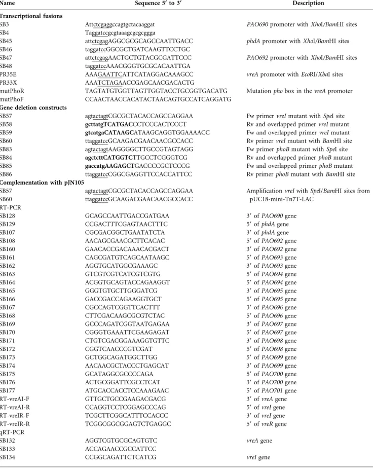
Table 2. Oligonucleotides used in this study
Fw, forward; Rv, reverse; restriction sites are underlined; complementary regions are in bold.
Name Sequence 5§ to 3§ Description
Transcriptional fusions
SB3 Attctcgaggccagtgctacaaggat PAO690 promoter with XhoI/BamHI sites SB4 Taggatccgcgtaaagcgcgcggga
SB45 attctcgagAGGCGCGCAGCCAATTGACC phdA promoter with XhoI/BamHI sites SB46 taggatccGGCGCTGATCAAGTTCCTGC
SB47 attctcgagAACTGCTGTACGCGATTCCC PAO692 promoter with XhoI/BamHI sites SB48 taggatccAAACGGGTGCGCACAATTGA
PR35E AAAGAATTCATTCATAGGACAAAGCC vreA promoter with EcoRI/XbaI sites PR33X AAATCTAGAACCGAGCAACGACACTG
mutPhoR TAGTATGTGGTTAGTTGGTACCTGCGGTGACATG Mutation pho box in the vreA promoter mutPhoF CCAACTAACCACATACTAACAGTGCCATCAGGATG
Gene deletion constructs
SB57 agtactagtCGCGCTACACCAGCCAGGAA Fw primer vreI mutant with SpeI site SB58 gcttatgTCATGACCCTCCCACTCCCT Rv and overlapped primer vreI mutant SB59 gtcatgaCATAAGCATAAGCAGGTGGAAAACC Fw and overlapped primer vreI mutant SB60 ttaggatccGCAAGACGAACAACGCCACC Rv primer vreI mutant with BamHI site SB83 agtactagtAAGGGGCTTGCCGTAGTAGG Fw primer phoB mutant with SpeI site SB84 agctcttCATGGTCTTGCCTCGGGTCG Rv and overlapped primer phoB mutant SB85 gaccatgAAGAGCTGACCCCGCTCCCG Fw and overlapped primer phoB mutant SB86 ttaggatccCGGCGAGGTTCCACCATTCC Rv primer phoB mutant with BamHI site Complementation with pJN105
SB57 agtactagtCGCGCTACACCAGCCAGGAA Amplification vreI with SpeI/BamHI sites from pUC18-mini-Tn7T-LAC
SB60 ttaggatccGCAAGACGAACAACGCCACC RT-PCR
SB128 GCAGCCAATTGACCGATGAA 39 of PAO690 gene SB129 CCGACTTTCGAGTAACTTTC 59 of phdA gene SB107 CGCGACGGCTGAATATCTA 39 of phdA gene SB108 AACAGCGAACGCTTCACAC 59 of PAO692 gene SB160 GAACACCGACAAACACGACT 39 of PAO692 gene SB161 CAGCGATGTCAGCAATAAGC 59 of PAO693 gene SB162 AGGTGCATGGCGAAAGC 39 of PAO693 gene SB163 GTCGTCGTCATCGTCGTG 59 of PAO694 gene SB164 ACGGTGCAGTACCAGAAGGT 59 of PAO694 gene SB165 GGGTGTGCTTGGGATCG 39 of PAO695 gene SB166 GACCGACCAGAAGGTGCT 59 of PAO695 gene SB167 CGCCAGTCGGTTCACTTT 39 of PAO696 gene SB168 CTTCGACAAGCGCGTCTAC 59 of PAO696 gene SB169 GCCCAGATCGGTAATGAGAA 39 of PAO697 gene SB170 CGGGTGAAATTCGAAGAGAT 59 of PAO697 gene SB171 CTGTCGACGGAAAGGTGTTC 39 of PAO698 gene SB172 CGGTCAACCCGTCGAT 59 of PAO698 gene SB173 GCTGGCAGATGGCTTGG 59 of PAO699 gene SB174 AACAACGCTACCCTGAGCAT 39 of PAO699 gene SB175 GCATAGGCGCCCCAGA 59 of PAO700 gene SB176 ACTGCGGATTCGCCTCAT 39 of PAO700 gene SB177 ATGCACCACCTCCAAAGAAC 59 of PAO701 gene RT-vreAI-F GTTGCTGCCGAAGACGACG 39 of vreA gene RT-vreAI-R CCAGGTCCTCGGAGCCCAG 59 of vreI gene RT-vreIR-F TCGCTTCGGCATTTCCACCC 39 of vreI gene RT-vreIR-R TCGGCGGCGGAGTCTGAGGC 59 of vreR gene qRT-PCR
SB132 AGGTCGTGCGCAGTGTC vreA gene SB133 ACCAGAACCGCCATTCC
High-Fidelity DNA Polymerase (Finnzymes) or Expand High Fidelity DNA polymerase (Roche). The pMPR3 lacZ transcriptional fusion was constructed by cloning the intergenic region between the PAO673 and vreA genes (containing the entire vreA promoter) as an EcoRI/ XbaI PCR fragment into the pMP220 vector (Spaink et al., 1987). The pMPR3mutPho plasmid was made by cloning the same insert in which the vreA pho box sequence CCGTCACACCACAGTCACACGA was changed to CCAACTAACCACATACTAACGA by overlapping PCR. All constructs were confirmed by DNA sequencing and transferred from E. coli to P. aeruginosa by triparental mating or electroporation. pJN105-vreI was constructed by first cloning a 1.3 kb PCR fragment containing the vreI gene into the BamHI/SpeI sites of the pUC18-mini-Tn7T-LAC plasmid (Choi & Schweizer, 2006). The SpeI/PstI DNA fragment from the pUC18-miniTn7T-LAC-vreI plasmid containing vreI was then subcloned into the NheI/PstI sites of pJN105 plasmid (Newman & Fuqua, 1999).
Bacterial strains constructions. The pPAO690 : : lacZ, pphdA : : lacZ, pPAO692 : : lacZ strains were constructed as follows. DNA fragments corresponding to PAO690, phdA and PAO692 putative promoter regions were amplified by PCR as BamHI/XhoI fragments and cloned into the miniCTX-lacZ plasmid (Hoang et al., 1998, 2000) in front of a promoterless lacZ gene. Recombinant plasmids, checked by DNA sequencing, were then transferred from E. coli SM10 to P. aeruginosa by conjugation (Kaniga et al., 1991) and inserted into the CTX phage attB site of the P. aeruginosa chromosome. An FRT tetracycline cassette-excision step was performed as described previously (Hoang et al., 1998, 2000). The vreI and phoB mutant strains were obtained by first constructing the pKNG101-vreI and pKNG101-phoB suicide vectors as follows. Two DNA fragments respectively upstream and downstream of the gene of interest were PCR amplified. The resulting DNA fragments were then used as templates for an overlapping PCR run using the external pair of oligonucleotides. The PCR fragments containing the internal
deletion of the gene of interest were digested and cloned into the SpeI/ BamHI sites of the suicide pKNG101 vector (Kaniga et al., 1991). Once verified by DNA sequencing, pKNG101 recombinants plasmids were transferred from E. coli CC118lpir to P. aeruginosa PAO1 strain by triparental mating using the helper plasmid pRK2013 as previously described (Kaniga et al., 1991), thus leading to the DvreI and DphoB strains.
b-Galactosidase activity assay.Strains carrying the lacZ transcrip-tional fusions were grown with agitation at 37uC in low or high Pi media supplemented withL-arabinose to induce expression from the
pJN105 plasmid when indicated and 1 ml of culture was harvested after 300 min. The b-galactosidase activity was measured and normalized for the cell density as described previously (Miller, 1972). Each assay was run at least in triplicate and the data given are the mean.
Isolation of total RNA and RT-PCR. Overnight cultures were subcultured and grown at 37uC for 240 min in low or high Pi media. Total cellular RNA from 161010 bacteria was isolated using the PureYield RNA Midiprep System (Promega) or the hot phenol method using the Tri Reagent LS (Molecular Research Center) as previously described (Llamas et al., 2008). Contaminating DNA was eliminated by TURBO DNase (Ambion) treatment, and samples were cleaned up and concentrated using the RNeasy kit (Qiagen). Yield and purity of RNA were further evaluated on Nanodrop and Experion devices. RT-PCR was performed on total cellular RNA using the Access RT-PCR system (Promega) or the Titan One-Tube RT-PCR system (Roche) in accordance with the manufacturer’s recommenda-tions. For each reaction, 0.5 to 1 mg of total RNA was used. DNA contamination of the RNA samples was ruled out by omission of the reverse transcriptase in the reaction or inactivation at 94uC for 4 min prior to the RT-PCR. A positive control using P. aeruginosa genomic DNA was included. For quantitative reverse transcription, cDNA
Table 2. cont.
Name Sequence 5§ to 3§ Description
SB135 CCGAACCTCTGGATGACTTT
SB137 TCTCTGGCAGGCACTCG vreR gene
SB138 CCGCCGTGACATAGTCG
SB103 GAGCAGCAGTTGTACGAGCA phdA gene
SB104 TCACTTCCCCGATAATCCTG
SB105 TACCTGCTCTCCCAGGAATG PAO692 gene
SB106 GCTGAAGTCGTCGTCCTGTT
SB139 GGCCAGTCGGTGTTCG PAO693 gene
SB140 GCGCATTGTTGGCGTAG
SB158 CGCCAGCTATAGCCAGTACC PAO695 gene
SB159 GTGATATCCCCCGAGGAAC
SB141 GCGTGTTGAAGAAGGAACAG PAO696 gene
SB142 CACCTGCGGGATATAGGGTA
SB143 GCGAAGAATCGGGAGATGTA PAO697 gene
SB144 TCGACCTGGGTCTTGTAGAG
SB154 GGCCTTCCTGGAGTCG PAO700 gene
SB155 AACGAACGGTCCATCAGC
SB156 GGAGTACCAGCCGCACAT PAO701 gene
SB157 ACCTCGATCACTCCCGACT
SB150 CTGGACCTCAATCGCTTCC PA4192 gene
SB151 CCTCCGCGGGTTTCAG
SB152 CATCCGACTAGACGTCATC PA5403 gene
synthesis was performed on 2 mg of RNA using the SuperScriptIII first strand synthesis system (Invitrogen). Cycling parameters of the real-time PCR were 98uC for 2 min, followed by 45 cycles of 98 uC for 5 s and 60uC for 10 s, ending with 10 min at 95 uC. To determine the amplification kinetics of each product, the fluorescence derived from the incorporation of EvaGreen into the double-stranded PCR products was measured at the end of each cycle using the SoFast EvaGreen Supermix (Bio-Rad). Experiments have been performed at least on two independent clones and the 16S gene was used as control to normalize the results.
RESULTS
PUMA3 genes are regulated by phosphate through the PhoB regulator
As PUMA3 vreA, vreI and vreR genes are not expressed under classic growth culture conditions (i.e. LB rich medium, data not shown), and a link between sVreIregulation and low Pi could exist (Ball et al., 2002; Llamas et al., 2009), we decided to determine whether the expression of PUMA3 genes depends on phosphate concentration. Therefore, we per-formed a quantitative RT-PCR (qRT-PCR) on each PUMA3 gene using mRNA isolated from P. aeruginosa grown in either high Pi (10 mM phosphate) or low Pi (0.28 mM) conditions (see Methods). This low Pi concentration allowed expression of the well-known low Pi-dependent gene phoA, (data not shown and Wanner, 1993) which validates the Pi-limited medium used in this work. Growing wild-type cells in low Pi led to an eight, two and fourfold induction of vreA, vreI and vreR gene expression respectively, compared to the expression of these genes in high Pi (Fig. 2). As phosphate regulation often requires the PhoR-PhoB two-component system, we next tested PUMA3 genes expression in a phoB
deletion mutant. We found that expression of vreA, vreI and vreR genes was no longer induced by low Pi, indicating that PUMA3 genes are regulated by low Pi through the PhoB transcriptional regulator (Fig. 2). Finally, we showed that sVreIdoes not control the expression of vreA and vreR genes as expression of these genes can be detected by qRT-PCR in a vreI mutant (Fig. 2). As vre genes respond identically to a low Pi concentration and vreA and vreI genes overlap by 4 bp (Fig. 3a), we next tested if these genes form an operon. RT-PCR was performed on P. aeruginosa total RNA upon growth in low Pi medium using primers that amplify the vreA-vreI overlapping and vreI-vreR intergenic regions (Fig. 3a). cDNA bands of the expected sizes were obtained when the RT-PCR was performed using active reverse transcrip-tase but not when this enzyme was previously heat inactivated (Fig. 3b). These results show that the three components of the PUMA3 system, the vreA, vreI and vreR genes, form an operon and are likely transcribed as a polycistronic mRNA from the same promoter. To confirm this, we placed an ~300 bp DNA region upstream vreA, which likely contains the vreAIR promoter, in front of a promoterless lacZ gene (pMPR3 plasmid), and examined the activity of this promoter by b-galactosidase assays. As shown in Fig. 3c, activity of this promoter was ~20-fold higher in low Pi growth conditions as compared to high Pi growth conditions and no promoter activity was observed in the phoB mutant. This confirmed the qRT-PCR results and the induction of the PUMA3 gene expression by low Pi through the PhoB protein. Furthermore, such induction is independent on sVreIfactor itself since the promoter activity did not change in a vreI mutant (Fig. 3c). This is in agreement with qRT-PCR results (Fig. 2) and previous results showing that sVreI does not autoregulate its own expression (Llamas et al., 2009).
16 14 12 Relative transcript 10 8 6 4 2 0
vreA vreI vreR
∆vreI low Pi ∆vreI high Pi ∆phoB low Pi ∆phoB high Pi WT low Pi WT high Pi
Fig. 2. Analysis of PUMA3 genes expression. vreA, vreI and vreR genes expression was monitored by qRT-PCR on cDNA from different strains grown in high or low Pi medium for 300 min. For each gene, bars show means andSDof the relative transcript amounts normalized to the high Pi condition obtained from at least two independent cDNA.
Identification of a putative pho box in the PUMA3 genes promoter region
No pho box in the vreAIR promoter was previously identified by a large-scale bioinformatic approach (Jensen et al., 2006). However, in silico analysis of the PUMA3 promoter region upstream vreA led us to identify a putative pho box (Fig. 3a). This box matches 16 bp within the 22 bp consensus sequence of E. coli and contains two 11 bp direct repeats. In each repeat, the first 7 bp are more conserved than the last 4 bp (Fig. 3a). The contribution of the identified pho box to the PhoB-mediated regulation of the vreAIR genes was examined using a lacZ transcriptional fusion in which conserved pho box residues were changed (Fig. 3a, mutPho). Mutation of the putative pho box abolished the induction of PUMA3 gene expression in low Pi conditions (Fig. 3c, pMPR3mutPho plasmid). This result is consistent with the suggestion that the putative pho box is potentially a PhoB binding site. To confirm this hypothesis, we tested the ability of PhoB to bind to the
vreAIR promoter by an electrophoretic mobility shift assay with purified PhoB that has been autophosphorylated in the presence of acetyl phosphate, a small-molecule phosphodonor (Hiratsu et al., 1995). However, the vreAIR promoter is retarded only in the presence of a high concentration of PhoB that also shifts a pho box free promoter (data not shown), which made it impossible to address whether the retardation obtained with the vreAIR promoter was specific. As a consequence, we could not confirm that the PhoB transcriptional regulator directly binds to the promoter region of the PUMA3 genes to induce their expression.
Role of
s
VreIand PhoB in the activity of promoters within the PUMA3 regulon genes
We showed that the expression of vreI, encoding the sVreI ECF sigma factor, is induced under low Pi conditions in a PhoB-dependent manner. In order to determine whether
(a)
65
1 2
vreA vreI vreR
E.coli consensus
Predicted pho box
mutPho
(b)
1
DNA cDNA – DNA cDNA –
2 (c) 2500 High Pi Low Pi 2000 1500 1000 β
-Galactosidase activity (Miller units)
500 0 PAO1 (pMPR3) PAO1 (pMPR3mutPho) ∆vreI (pMPR3) ∆phoB (pMPR3)
Fig. 3. Co-transcription of vreAIR genes and analysis of their promoter region. (a) Sequence of the putative pho box in the vreAIR regulatory region is aligned with the E. coli pho box consensus sequence (top). Residues matching with this consensus are indicated in grey. Bottom sequence represents the mutated pho box in which mutated nucleotides are boxed. (b) Amplification by RT-PCR of the cDNA corresponding to the vreA-vreI and vreI-vreR intergenic regions (arrows 1 and 2 depicted in (a) respectively). Positive controls (DNA) using P. aeruginosa genomic DNA and negative controls (–) containing the same amounts of RNA, primers and inactivated reverse transcriptase, are included in this assay. (c) P. aeruginosa PAO1 and its isogenic phoB mutant containing the transcriptional fusion pMPR3, in which the vreAIR promoter is fused to lacZ, or pMPR3mutPho, containing the whole vreAIR promoter with the mutated pho box, were grown in high Pi (dark grey bars) or low Pi (light grey bars) and analysed for b-galactosidase activity. Bars represent the mean andSDof at least three independent experiments.
the genes of the PUMA3 regulon were also expressed under this condition, we first analysed the transcriptional organization of such genes. Among the 27 genes expressed upon sVreI factor overproduction, 16 are located immedi-ately downstream of the PUMA3 locus (Llamas et al., 2009) and only the expression and transcriptional organization of the hxc gene cluster has been characterized (Ball et al., 2002). To determine the transcriptional organization of the other PUMA3-regulated genes, we performed RT-PCR on mRNA extracted from P. aeruginosa grown in low Pi using primers that amplified the overlapping region of the genes. Fig. 4b shows that the PAO690 gene forms a single transcriptional unit (TU; TU-1) as no cDNA could be detected using primers that amplify the PAO690-phdA overlapping DNA region. Interestingly, a cDNA band could be detected with all other combinations of primers used (Fig. 4b) indicating that all these genes, from phdA to PAO701, are transcribed as a polycistronic mRNA (Fig. 4b) and thus form a second transcriptional unit (TU-2). However, based on the distance between genes (Fig. 4a) and the predicted operonic structures (http://www. pseudomonas.com; Winsor et al., 2011), three intergenic regions, upstream PAO690, phdA and PAO692 genes, may contain putative promoters of three independent TUs (Fig. 4a). Therefore, we fused these three putative promoter regions to the lacZ gene, encoding the b-galactosidase enzyme, and inserted them in the chromosome of P. aeruginosa. Under LB-rich growth conditions, no promoter activity was observed (data not shown). Growing the cells in low Pi conditions increased the activity of the pPAO690 : : lacZ and pphdA : : lacZ transcriptional fusions by 2.5 and 18-fold respectively but had no effect on the pPAO692 : : lacZ construct (Fig. 4c). This interesting result strongly suggested that, despite the prediction, the PAO692 and downstream genes are only expressed from the region upstream phdA, which contains an active promoter (Fig. 4 and Llamas et al., 2009). Importantly, we showed here that the activity of TU-1 and TU-2 promoters depends on the concentration of phosphate.
Next, to determine whether the Pi-dependent regulation of TU-1 and TU-2 promoters depends on the sVreIand PhoB transcriptional regulators, their activities were tested in a vreI and a phoB deletion mutant. b-Galactosidase experi-ments showed that the activity of both promoters was no longer induced by low Pi in the absence of sVreIor PhoB (Fig. 5a, b). Complementation of the vreI deletion with a plasmid overproducing sVreI from an arabinose-regulated promoter (pJN105-vreI) restored the activity of promoters (Fig. 5a, b). Indeed, these activities were related to the number of vreI transcripts, since a 100-fold increase of vreI transcripts (Fig. 5c, 0 % arabinose) led to a ~2 and 3-fold increase promoter activity of TU-1 and TU-2 respectively (Fig. 5a, b), while a 500-fold increase of vreI transcripts (Fig. 5c, 0.4 % arabinose) led to an ~50 and 100-fold increase in promoter activities (Fig. 5a, b). Additionally, high expression of vreI upon addition of 0.4 % arabinose relieved the Pi-dependent regulation of promoter activities,
which confirms that an overproduction of sVreIleads to an expression of sVreI-regulated genes independently of the growth culture conditions (Llamas et al., 2009). We show a contribution of PhoB in the promoter activation (Fig. 5a, b) and an absence of vreI expression in a phoB mutant (Fig. 2), which suggests that PhoB acts upstream of the sVreI factor to activate TU-1 and TU-2 promoters. To test this hypothesis, we overproduced the sVreI factor in the phoB mutant and measured b-galactosidase activities of the transcriptional fusions. TU-1 and TU-2 promoter activities are restored when vreI gene is introduced in trans (Fig. 5a, b), a result that suggests that sVreIcould bypass the absence of the PhoB protein and act downstream of this determinant in the identified regulatory cascade. Surprisingly, b-galactosidase activities were considerably lower than in the vreI mutant as TU-1 and TU-2 promoters were activated only upon induction of vreI with 0.4 % arabinose (Fig. 5a, b). This lower activity observed in the phoB mutant upon addition of 0.4 % arabinose is not due to a lower expression of the vreI gene since equivalent levels of vreI transcripts were observed in the vreI and phoB mutants (Fig. 5c). This result may suggest a role for PhoB in a post-transcriptional control of sVreI activity by a remodelling of the sVreI-RNA polymerase. Alternatively, low TU-1 and TU-2 promoter activities may be a consequence of an absence of VreA and/or VreR in a phoB mutant (Fig. 2). Indeed, lower stability of sVreI was observed in the absence of its VreR anti-sigma factor (Llamas et al., 2009). Altogether, these results clearly show that upon low Pi conditions, the sVreIfactor activates TU-1 and TU-2 promoters in a PhoB-dependent manner.
Redefining the
s
VreIregulonOur experiments show that the promoter of TU-2 is activated upon low Pi through the sVreI factor and PhoB regulators. This result suggested that all the genes, expressed from this promoter, undergo the same regu-lation. Indeed, among them, eight seem to be regulated in a sVreI-dependent manner but three (PAO695, PAO700 and PAO701) seem not to be (Llamas et al., 2009 and Fig. 1). To clarify this, we performed qRT-PCR to determine the level of gene expression within the TU-2. We first chose five genes (phdA, PAO692, PAO693 PAO696 and PAO697), showing the highest fold expression change upon over-production of the sVreIfactor (Llamas et al., 2009 and Fig. 1) and we confirmed that these genes were transcribed 6– 20-fold more in low Pi than in high Pi growth conditions (Fig. 6a). Expression of these genes was significantly reduced, but not abrogated, in a vreI mutant (Fig. 6, DvreI low Pi). Interestingly, the absence of PhoB regulator completely turned off expression of these genes in low Pi growth conditions (Fig. 6a), suggesting that PhoB alone contributes to their expression in this condition. Finally, we show that PAO695, PAO700 and PAO701 gene expression undergoes the same pattern. Indeed, sVreIfactor and PhoB protein also control their expression upon low Pi growth conditions (Fig. 6a). ECF sigma factors usually
(a)
182 bp
lapB PA0690 phdA PA0692 PA0693 94 PA0695 PA0696 697 698 PA0699 700 PA0701
391 bp 163 bp (b) bp M 1 2 3 4 5 6 7 8 9 10 11 cDNA gDNA RNA M 1 2 3 4 5 6 7 8 9 10 11 M 1 2 3 4 5 6 7 8 9 10 11 500 300 200 100 bp 500 300 200 100 bp 500 300 200 100 1 2 3 4 5 6 7 8 9 10 11 (c) β
-Galactosidase activity (Miller units)
1400 High Pi Low Pi 1200 1000 800 600 400 200 0
pPA0690 :: lacz pphdA :: lacz pPA0692 :: lacz
Fig. 4. Transcriptional fusion analysis and co-transcription of genes located downstream of the PUMA3 locus. (a) Diagram showing the putative promoter regions with distance between genes. Arrows represents the expected amplified region after RT-PCR. (b) RT-PCR products from RNA isolated from P. aeruginosa grown for 300 min in low Pi conditions. cDNA corresponds to the RT-PCR using mRNA as template, gDNA is a positive control using the PAO1 genomic DNA template and RNA is a negative control lacking reverse transcriptase in the RT-PCR. (c) P. aeruginosa strains, containing the chromosomal transcriptional fusion pPAO690 : : lacZ, pphdA : : lacZ or pPAO692 : : lacZ were grown in high or low Pi conditions and b-galactosidase activity was measured after 300 min of growth. Error bars represent the mean and SD of at least three independent experiments.
(a) 35000 High Pi Low Pi High Pi Low Pi 0.4% arabinose 0% arabinose 0.4% arabinose 0% arabinose 0.4% arabinose 0% arabinose % arabinose 0 0.4 0 0.4 % arabinose 0.4% arabinose 0% arabinose 30000 24000 1400 1200 1000 800 600 400 200 0 126000 118000 110000 1200 9000 5000 4000 3000 2000 1000 1100 900 700 500 300 100 20 18 16 14 12 10 8 6 4 2 0 0 WT (b) (c) β
-Galactosidase activity (Miller units)
of p
PA0690
::
lacZ
fusion (TU-1 promoter)
β
-Galactosidase activity (Miller units)
of p
phdA
::
lacZ
fusion (TU-2 promoter)
Relative transcript of
vrel
∆vreI ∆phoB ∆phoB
pJN105 ∆phoB pJN105-vrel ∆phoB pJN105 ∆phoB pJN105-vrel ∆vreI pJN105 ∆vreI pJN105-vrel ∆vreI pJN105 ∆vreI pJN105-vrel
WT ∆vreI ∆phoB ∆phoB
pJN105 ∆phoB pJN105-vrel ∆phoB pJN105 ∆phoB pJN105-vrel ∆vreI pJN105 ∆vreI pJN105-vrel ∆vreI pJN105 ∆vreI pJN105-vrel
regulate genes located immediately downstream on a chromosome. In the case of the PUMA3 system, micro-array data revealed that sVreI factor could also control other genes located in different loci of the P. aeruginosa genome (Llamas et al., 2009). To expand our study, we picked two genes (PA4192 and PA5403), showing the highest fold change expression between wild-type and sVreI factor overproducing strains (Llamas et al., 2009), and tested whether they undergo an identical cascade of regulation. Interestingly, expression of these genes is not induced upon low level of Pi and is dependent on neither sVreIfactor nor PhoB protein, contrasting with TU-1 and TU-2 gene regulation (Fig. 6b).
DISCUSSION
In the present study, we have elucidated the regulatory pathway allowing the activation of genes controlled by the PUMA3 system. We show that the PUMA3 system expression is activated under phosphate limitation through the PhoB transcriptional regulator allowing expression of the vreI gene, which codes for the sVreI factor. Once produced, sVreI mediates transcription of specific target genes. Microarray data identified a number of genes positively controlled by the sVreI factor (Llamas et al., 2009), among which ~two-third are located immediately downstream of the vreI gene and ~one third dispersed onto
the P. aeruginosa genome. Based on our findings, expression of sVreI-regulated genes should be induced in response to phosphate depletion. hxc gene expression occurs upon low Pi conditions (Ball et al., 2002), and we found here that the other genes downstream of the PUMA3 gene cluster follow an identical regulation and are regulated by the sVreIfactor and PhoB. However, we clearly showed that PA4192 and PA5403 genes, belonging to the one-third of genes located somewhere else in the chromosome, are not expressed under scattered limitation conditions. These results confirmed a previous microarray study that observed, under phosphate-limited conditions, a clear upregulation of only PUMA3 genes and all genes located downstream this locus, from hxcW to PAO701 (Bains et al., 2012). All together, these results strongly suggest that only genes from hxcW (PAO677) to PAO701, located downstream of the PUMA3 locus, constitute the sVreIregulon.
Expression of PUMA3 genes has been for long thought to be controlled by iron and Fur (Ochsner & Vasil, 1996), and therefore originally named pigCDE (from Pseudomonas iron-regulated genes). However, a deeper analysis of these studies revealed that the identified Fur-box is actually located in the promoter region of the pigA (PAO672 or hemO) gene, located upstream of the vreAIR (pigCDE) genes (Ochsner & Vasil, 1996; Ochsner et al., 2002). In this work, we demonstrated that PUMA3 genes, namely vreA, vreI and vreR, form an operon that has its own promoter
Fig. 5.sVreIfactor and PhoB regulate TU-1 and TU-2 promoter activity. P. aeruginosa and its isogenic vreI, phoB mutants containing the chromosomal transcriptional fusion pPAO690 : : lacZ (a) or pphdA : : lacZ (b) were grown in high or low Pi conditions for 300 min. When indicated, 0.4 % ofL-arabinose was added during the growth of mutant conjugated with the empty pJN105 or pJN105-vreI plasmids. Bars represent mean of b-galactosidase activity for at least three independent experiments. (c) mRNA level of vreI from wild-type, vreI, phoB mutants containing the pJN105-vreI plasmid grown in low Pi conditions for 300 min with or without 0.4 %L-arabinose. Level of transcript was normalized to 16S expression and shown
relative to the wild-type level. Bars represent mean andSDof least two independent replicates.
(a) 25 20 Relative transcript 15 10 5 0 (b)
phdA PA0695 PA0696 PA0697 PA0700 PA0701 PA4192 PA5403
WT high Pi WT low Pi PA0692 (tpsB) PA0693 (exbB) ∆vrel high Pi ∆vrel low Pi ∆phoB low Pi ∆phoB high Pi
Fig. 6. Redefining the sVreIregulon. Results of qRT-PCR analysis of selected genes are shown. RNA was collected from P. aeruginosa strains grown at 37 6C in high or low Pi conditions for 300 min. For each gene, expression was normalized to 16S expression and shown relative to the WT high Pi condition level. Mean and standard deviation from two independent replicates are shown. Grey blocks highlight genes not previously identified as sVreI-regulated genes (Llamas et al., 2009).
region upstream of vreA with a putative pho box but no Fur box (Fig. 3a). In agreement with this observation, none of the transcriptional fusions used in this work responds to iron limitation (data not shown), which further supports the suggestion that iron does not play a role in PUMA3 expression and its regulatory role.
As illustrated here, low phosphate growth conditions are required to trigger the PUMA3 pathway, including expres-sion of PUMA3 gene and further activation of genes from the sVreIregulon. This is an intriguing observation since the sVreI factor is coproduced in this condition both with its cognate VreR anti-sigma factor, which inhibits the function of sVreIand is required for its activity (Llamas et al., 2009), and the VreA protein which could sense an activating signal. This raises the question of how the VreA-dependent release of sVreIfrom its anti-sigma VreR occurs. Two possibilities emerge from our observations. A potential inducing signal either (i) is produced and integrated by VreA and VreR under low Pi condition to release the sVreI factor which further activates target gene transcription, or (ii) is absent under the conditions in which PUMA3 genes are expressed but the amount of free sVreIis sufficient to bind the RNA polymerase core and initiate transcription of sVreI -depend-ent genes. We observed that increasing the quantity of free sVreIin trans from a plasmid activates expression of sVreI -dependent target genes to a level considerably higher that of the level reached in low Pi (Fig. 5). Moreover, sVreI -regulated genes are also induced upon contact of P. aeruginosa with human airway epithelial cells (Chugani & Greenberg, 2007; Frisk et al., 2004). All together, these data support the second hypothesis and suggest that an additional signal, absent in phosphate-limited conditions, is needed to completely unbind sVreIfrom the VreR anti-sigma factor and obtain a large sVreI-dependent gene expression.
From our study, it appears that expression of the genes belonging to the sVreIregulon is partially reduced in a vreI mutant and abrogated in a phoB mutant (Fig. 6a). This suggests that PhoB can also activate these genes by a sVreI -independent mechanism that remains to be elucidated. Putative pho boxes are present in both TU-1 and TU-2 promoters (our observation and Jensen et al., 2006). However, the identity level of each box to the E. coli consensus is low and the efficiency of PhoB binding to a pho box correlates with this level (Diniz et al., 2011; Kim et al., 2000). Since we also encountered an unspecific shift when testing binding of PhoB to the TU-1 and TU-2 promoter regions (data not shown), we cannot state whether this PhoB-dependent control is direct by co-acting with the s70-RNAP holoenzyme. As sVreIis able to mediate gene transcription without PhoB (Fig. 5 and Llamas et al., 2009) and PhoB complexed to the pho box complex interacts with a s70 subunit of the RNA polymerase to control initiation of gene transcription (Blanco et al., 2011; Makino et al., 1993), the presence of putative pho boxes in the TU-1 and TU-2 promoter regions would only allow PhoB to enhance transcription initiation by remodelling
the sVreI-RNA polymerase. One of our goals now is to perform further experiments to clarify the recognition and cooperation between the sVreIfactor and the PhoB protein in the transcriptional regulation of these genes.
ACKNOWLEDGEMENTS
We are grateful to Wilbert Bitter for helpful discussions, Yann Denis for technical assistance on qRT-PCR, Eric Cascales for the electrophoretic mobility shift assay and Be´renge`re Ize for discussions, helpful comments and encouragement. We thank Marie-Anne Lafaulace for encouragement. This work is supported by Vaincre la Mucoviscidose (the French cystic fibrosis foundation) through a grant (FR20110600567/1/1/69). Research in S. deB.’s laboratory is sup-ported by Bettencourt-Schueller Foundation, CNRS Institutional and ANR grants (ERA-NET ADHRES 27481, PCV-ANR 27628, ANR-09-JCJC-0047 and GDR3171). The Medical Research Foundation supports S. B. Research in M. A. L.’s lab is supported by the EU through a Marie Curie CIG grant (3038130) and the Spanish Ministry of Economy through a Ramon&Cajal grant (RYC-2011-08874).
REFERENCES
Bains, M., Ferna´ndez, L. & Hancock, R. E. (2012). Phosphate starvation promotes swarming motility and cytotoxicity of Pseudomonas aeruginosa. Appl Environ Microbiol 78, 6762–6768.
Ball, G., Durand, E., Lazdunski, A. & Filloux, A. (2002).A novel type II secretion system in Pseudomonas aeruginosa. Mol Microbiol 43, 475– 485.
Bastiaansen, K. C., Bitter, W. & Llamas, M. A. (2012).ECF sigma factors: from stress management to iron uptake. In Bacterial Regulatory Networks pp. 59–86. Edited by A. Filloux Hethersett. Norwich: Caister Academic Press.
Blanco, A. G., Sola, M., Gomis-Ru¨th, F. X. & Coll, M. (2002).Tandem DNA recognition by PhoB, a two-component signal transduction transcriptional activator. Structure 10, 701–713.
Blanco, A. G., Canals, A., Bernue´s, J., Sola`, M. & Coll, M. (2011).The structure of a transcription activation subcomplex reveals how s(70) is recruited to PhoB promoters. EMBO J 30, 3776–3785.
Bordi, C. & de Bentzmann, S. (2011).Hacking into bacterial biofilms: a new therapeutic challenge. Ann Intensive Care 1, 19.
Braun, V., Mahren, S. & Sauter, A. (2006). Gene regulation by transmembrane signaling. Biometals 19, 103–113.
Canter Cremers, H., Spaink, H. P., Wijfjes, A. H., Pees, E., Wijffelman, C. A., Okker, R. J. & Lugtenberg, B. J. (1989).Additional nodulation genes on the Sym plasmid of Rhizobium leguminosarum biovar viciae. Plant Mol Biol 13, 163–174.
Choi, K. H. & Schweizer, H. P. (2006).mini-Tn7 insertion in bacteria with single attTn7 sites: example Pseudomonas aeruginosa. Nat Protoc 1, 153–161.
Chugani, S. & Greenberg, E. P. (2007). The influence of human respiratory epithelia on Pseudomonas aeruginosa gene expression. Microb Pathog 42, 29–35.
de Lorenzo, V. & Timmis, K. N. (1994).Analysis and construction of stable phenotypes in gram-negative bacteria with Tn5- and Tn10-derived minitransposons. Methods Enzymol 235, 386–405.
Diniz, M. M., Goulart, C. L., Barbosa, L. C., Farache, J., Lery, L. M., Pacheco, A. B., Bisch, P. M. & von Kru¨ger, W. M. (2011).Fine-tuning control of phoBR expression in Vibrio cholerae by binding of PhoB to multiple pho boxes. J Bacteriol 193, 6929–6938.
Escolar, L., Pe´rez-Martı´n, J. & de Lorenzo, V. (1999). Opening the iron box: transcriptional metalloregulation by the Fur protein. J Bacteriol 181, 6223–6229.
Figurski, D. H. & Helinski, D. R. (1979). Replication of an origin-containing derivative of plasmid RK2 dependent on a plasmid function provided in trans. Proc Natl Acad Sci U S A 76, 1648–1652.
Frisk, A., Schurr, J. R., Wang, G., Bertucci, D. C., Marrero, L., Hwang, S. H., Hassett, D. J. & Schurr, M. J. (2004).Transcriptome analysis of Pseudomonas aeruginosa after interaction with human airway epithelial cells. Infect Immun 72, 5433–5438.
Guerinot, M. L. (1994).Microbial iron transport. Annu Rev Microbiol 48, 743–772.
Herrero, M., de Lorenzo, V. & Timmis, K. N. (1990). Transposon vectors containing non-antibiotic resistance selection markers for cloning and stable chromosomal insertion of foreign genes in gram-negative bacteria. J Bacteriol 172, 6557–6567.
Hiratsu, K., Nakata, A., Shinagawa, H. & Makino, K. (1995).
Autophosphorylation and activation of transcriptional activator PhoB of Escherichia coli by acetyl phosphate in vitro. Gene 161, 7–10.
Hoang, T. T., Karkhoff-Schweizer, R. R., Kutchma, A. J. & Schweizer, H. P. (1998).A broad-host-range Flp-FRT recombination system for site-specific excision of chromosomally-located DNA sequences: application for isolation of unmarked Pseudomonas aeruginosa mutants. Gene 212, 77–86.
Hoang, T. T., Kutchma, A. J., Becher, A. & Schweizer, H. P. (2000).
Integration-proficient plasmids for Pseudomonas aeruginosa: site-specific integration and use for engineering of reporter and expression strains. Plasmid 43, 59–72.
Jensen, V., Lo¨ns, D., Zaoui, C., Bredenbruch, F., Meissner, A., Dieterich, G., Mu¨nch, R. & Ha¨ussler, S. (2006).RhlR expression in Pseudomonas aeruginosa is modulated by the Pseudomonas quinolone signal via PhoB-dependent and -independent pathways. J Bacteriol 188, 8601–8606.
Kaniga, K., Delor, I. & Cornelis, G. R. (1991). A wide-host-range suicide vector for improving reverse genetics in gram-negative bacteria: inactivation of the blaA gene of Yersinia enterocolitica. Gene 109, 137–141.
Kim, S. K., Kimura, S., Shinagawa, H., Nakata, A., Lee, K. S., Wanner, B. L. & Makino, K. (2000). Dual transcriptional regulation of the Escherichia coli phosphate-starvation-inducible psiE gene of the phosphate regulon by PhoB and the cyclic AMP (cAMP)-cAMP receptor protein complex. J Bacteriol 182, 5596–5599.
Koebnik, R. (2005). TonB-dependent trans-envelope signalling: the exception or the rule? Trends Microbiol 13, 343–347.
Lamarche, M. G., Wanner, B. L., Cre´pin, S. & Harel, J. (2008). The phosphate regulon and bacterial virulence: a regulatory network connecting phosphate homeostasis and pathogenesis. FEMS Microbiol Rev 32, 461–473.
Llamas, M. A. & Bitter, W. (2010). Cell-surface signaling in Pseudomonas. In Molecular Microbiology, Infection and Biodiversity,
pp. 59–95. Edited by J. L. Ramos & A. Filloux. New York, USA: Academic/Plenum.
Llamas, M. A., Mooij, M. J., Sparrius, M., Vandenbroucke-Grauls, C. M., Ratledge, C. & Bitter, W. (2008).Characterization of five novel Pseudomonas aeruginosa cell-surface signalling systems. Mol Microbiol 67, 458–472.
Llamas, M. A., van der Sar, A., Chu, B. C., Sparrius, M., Vogel, H. J. & Bitter, W. (2009). A novel extracytoplasmic function (ECF) sigma factor regulates virulence in Pseudomonas aeruginosa. PLoS Pathog 5, e1000572.
Lonetto, M. A., Brown, K. L., Rudd, K. E. & Buttner, M. J. (1994).
Analysis of the Streptomyces coelicolor sigE gene reveals the existence of a subfamily of eubacterial RNA polymerase sigma factors involved in the regulation of extracytoplasmic functions. Proc Natl Acad Sci U S A 91, 7573–7577.
Makino, K., Amemura, M., Kim, S. K., Nakata, A. & Shinagawa, H. (1993). Role of the sigma 70 subunit of RNA polymerase in transcriptional activation by activator protein PhoB in Escherichia coli. Genes Dev 7, 149–160.
Miller, J. H. (1972). Experiments in Molecular Genetics. Cold Spring Harbor, NY: Cold Spring Harbor Laboratory.
Newman, J. R. & Fuqua, C. (1999). Broad-host-range expression vectors that carry the L-arabinose-inducible Escherichia coli araBAD promoter and the araC regulator. Gene 227, 197–203.
Ochsner, U. A. & Vasil, M. L. (1996).Gene repression by the ferric uptake regulator in Pseudomonas aeruginosa: cycle selection of iron-regulated genes. Proc Natl Acad Sci U S A 93, 4409–4414.
Ochsner, U. A., Wilderman, P. J., Vasil, A. I. & Vasil, M. L. (2002).
GeneChip expression analysis of the iron starvation response in Pseudomonas aeruginosa: identification of novel pyoverdine biosyn-thesis genes. Mol Microbiol 45, 1277–1287.
Potvin, E., Sanschagrin, F. & Levesque, R. C. (2008).Sigma factors in Pseudomonas aeruginosa. FEMS Microbiol Rev 32, 38–55.
Schalk, I. J., Yue, W. W. & Buchanan, S. K. (2004).Recognition of iron-free siderophores by TonB-dependent iron transporters. Mol Microbiol 54, 14–22.
Spaink, H. P., Okker, R. J. H., Wijffelman, C. A., Pees, E. & Lugtenberg, B. J. J. (1987). Promoters in the nodulation region of the Rhizobium leguminosarum Syn plasmid pRL1JI. Plant Mol Biol 9, 27–39.
Wanner, B. L. (1993). Gene regulation by phosphate in enteric bacteria. J Cell Biochem 51, 47–54.
Winsor, G. L., Lam, D. K., Fleming, L., Lo, R., Whiteside, M. D., Yu, N. Y., Hancock, R. E. & Brinkman, F. S. (2011).Pseudomonas Genome Database: improved comparative analysis and population genomics capability for Pseudomonas genomes. Nucleic Acids Res 39 (Database issue), D596–D600.