HAL Id: hal-02135121
https://hal.archives-ouvertes.fr/hal-02135121
Submitted on 21 May 2019
HAL is a multi-disciplinary open access
archive for the deposit and dissemination of
sci-entific research documents, whether they are
pub-lished or not. The documents may come from
teaching and research institutions in France or
abroad, or from public or private research centers.
L’archive ouverte pluridisciplinaire HAL, est
destinée au dépôt et à la diffusion de documents
scientifiques de niveau recherche, publiés ou non,
émanant des établissements d’enseignement et de
recherche français ou étrangers, des laboratoires
publics ou privés.
How the greater tuberosity affects clinical outcomes
after reverse shoulder arthroplasty for proximal humeral
fractures
Xavier Ohl, Nicolas Bonnevialle, David Gallinet, Nassima Ramdane, Philippe
Valenti, Lauryl Decroocq, Pascal Boileau
To cite this version:
Xavier Ohl, Nicolas Bonnevialle, David Gallinet, Nassima Ramdane, Philippe Valenti, et al.. How
the greater tuberosity affects clinical outcomes after reverse shoulder arthroplasty for proximal
humeral fractures. Journal of Shoulder and Elbow Surgery, Elsevier, 2018, 27 (12), pp.2139-2144.
�10.1016/j.jse.2018.05.030�. �hal-02135121�
O
pen
A
rchive
T
oulouse
A
rchive
O
uverte
(OATAO)
OATAO is an open access repository that collects the work of some Toulouse
researchers and makes it freely available over the web where possible.
This is
an author'sversion published in:
https://oatao.univ-toulouse.fr/23092Official URL :
https://doi.org/10.1016/j.jse.2018.05.030
To cite this version :
Any correspondence concerning this service should be sent to the repository administrator:
tech-oatao@listes-diff.inp-toulouse.fr
Ohl, Xavier and Bonnevialle, Nicolas and Gallinet, David and Ramdane, Nassima and
Valenti, Philippe and Decroocq, Lauryl and Boileau, Pascal How the greater tuberosity affects
clinical outcomes after reverse shoulder arthroplasty for proximal humeral fractures. (2018)
Journal of Shoulder and Elbow Surgery, 27 (12). 2139-2144. ISSN 1058-2746
OATAO
How the greater tuberosity affects clinical
outcomes after reverse shoulder arthroplasty for
proximal humeral fractures
Xavier Ohl, MD, PhD
a, Nicolas Bonnevialle, MD, PhD
b,*
, David Gallinet, MD
c,
Nassima Ramdane, PhD
d, Philippe Valenti, MD
e, Lauryl Decroocq, MD
f,
Pascal Boileau, MD, PhD
f, SOFCOT
gaOrthopaedic Department, University Hospital, Reims, France
bDepartment of Orthopaedic Surgery, Riquet Hospital, University of Toulouse, Toulouse, France c
Saint Vincent Private Hospital, Besançon, France d
Unité de Méthodologie–Biostatistique et Data Management, Lille, France e
Paris Shoulder Institut, Paris, France f
Department of Orthopaedic Surgery, iULS (Institut Universitaire Locomoteur & Sport), University of Nice Sophia Antipolis, Nice, France
g
French Society of Orthopedic Surgery and Traumatology, Paris, France
Background: Our purpose was to evaluate the clinical and radiologic outcomes of reverse shoulder
ar-throplasty for proximal humeral fractures in a large cohort of elderly patients and compare the results in the case of tuberosity excision, failed fixation, or anatomic healing.
Methods: In this retrospective multicenter study, 420 patients underwent review and radiography with a
minimum follow-up period of 12 months. The patients were divided into 3 groups according to the status of the greater tuberosity (GT) on the last anteroposterior radiographs: anatomic GT healing (group A, n= 169); GT resorption, malunion, or nonunion (group B, n= 131); and GT excision (group C, n = 120). Com-plications were recorded; shoulder function, active mobility, and subjective results were assessed.
Results: At a mean follow-up of 28 months, the mean Simple Shoulder Value in group A (75%)
outper-formed the results found in groups B (69%, P< .001) and C (56%, P < .001). Overall, the mean adjusted Constant-Murley score was significantly higher in group A (93%± 22%) than in group B (82% ± 22%) and group C (80%± 24%) (P < .001), but there was no difference between groups B and C (P = .88). An-terior active elevation and external rotation were significantly better in group A than in groups B and C (P< .001). The instability rate was significantly higher in group C (n = 15 [12.5%], P < .001) than in group A (n= 2) or group B (n = 3).
Conclusion: In elderly patients who have undergone a reverse shoulder arthroplasty for acute proximal
humeral fractures, anatomic tuberosity healing improves objective and subjective outcomes. GT excision is associated with the worst functional results and increases the risk of postoperative shoulder instability.
Institutional Review Board approval was received (CCTIRS-16-003), and the ethical committee approved this study.
*Reprint requests: Nicolas Bonnevialle, MD, PhD, Département d’orthopédie, Hôpital Riquet, Place Baylac, 31059 Toulouse Cedex 09, France.
Level of evidence: Level ID; Retrospective Cohort Design; Treatment Study
Keywords: Proximal humerai fractures; reverse shoulder arthroplasty; greater tuberosity excision; greater tuberosity healing; shoulder instability; complication
Proximal humeral fractures (PHFs) are the third most
corn.mon fracture in elderly persans and represent a
signifi-cant challenge for orthopedic surgeons.9 Fracture fixation in
the elderly population increases tbe risk of failure owing to
poor bone quality and avascular •ecrosis. On the other band,
nonoperative treatment does not always lead to acceptable
sboulder fonction because of the possibility of severe
mal-union in an older population of patients wanting to recover
their quality of life after the in jury. Her•iarthroplasty bas been
proposed as an alternative to fixation; however, clinical out-cornes depend on anatomie healing of the tuberosities around
the implant I Because reverse shouJder arthroplasty (RSA) has
led to prornising results in patients with cuff deficiency,
in-dications for this procedure have been expanded to complex
PHFs.6
Optimal management of the tuberosities in RSA remains
unclear and controversial. Sorne authors reported
satisfacto-ry clinical outcomes of RSA after removaJ of both tuberosities.3
Others reported better active external rotation after fixation
and healing of the tuberosities around the stem.4-6 However,
despite slrong fixation, malunions, osteolysis, and nonunions
have been reported. 4•7•11
The aim of this study was to assess the clinical and
ra-diologie outcomes of RSA implanted for acute PHFs in a large
cohort of elderly patients and compare the results in pa-tients with anatomie greater tuberosity (GT) healing, failed fixation (malunion, nonunion, or osteolysis), and GT
exci-sion. We hypothesized that better clinical outcomes would
be observed in patients with healed tuberosities than in
pa-tients with failed tuberosity fixation or in those in whom the tuberosities have been excised.
Materials a
n
d methods
Study design
ln this retrospective multicenter study, we included patients with an acute PHF wbo were treated with a primary RSA between January 2010 and December 2015, regardless of whether the GT was fixed or removed, and who had a minimum follow-up period of 12 months. We excluded patients with pathologie fractures (primary tumor or metastasis), patients with previous surgery on the involved shoul -der, patients in whom surgery was performed more than 6 weeks after the injury occurred, and patients without plain ant eroposte-rior radiographs of the shoulder at 12 months after surgery. Patients gave their consent for the analysis of their clinical and radiologie data.
Patient population
Five hundred sixty-seven patients were eligible, and 420 patients (74%) with an RSA implanted for an acute PHF were reviewed and underwent radiography with minimum 1-year follow-up (Fig. 1 ).
The mean age at surgery was 78 years, 83% of patients were women, the mean body mass index was 26.3 ± 5.5 kg/m2, and the injured shoulder was on the dominant side in 58% of cases. The American Society of Anesthesiologists score was III or N in 38% of the cohort.
Surgical technique
A deltopectoral approach (n = 100) or deltoid-splitting approach
(n = 320) was performed with patients in the beach-chair position. Six different RSA models were used; 48% (n = 200) were specif -ically designed to treat PHFs with a low-profile stem and bone grafting around the metaphysis. A cemented stem was used in 88% of cases
(n = 370). The glenosphere diameter was 36 mm in 66% of cases. ln 300 RSA cases, both tuberosities were fixed with 2 or 4 hori-zontal cerclages around the metaphysis. For the remaining 120 RSAs, the surgeons decided to excise the greater and lesser tuberosities. Postoperatively, the shoulder was placed in a sling in 75% of cases. Physiotherapy was started immediately with free range of motion in 55%. Patients lncluded n=567 Dead n=17 Lost at follow-up n=43 1 - - - l Previous surgery n=Z
Radiologie data not available n=85
Cohort studied
n=420
Radiologic evaluation
Anteroposterior views in the neutral position were analyzed (minimum, 12 months) to determine the status of the GT and clas-sify the patients into 3 groups:
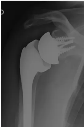
- Group A showed anatomic tuberosity healing, in which the GT was visible on the lateral part of the stem, at the level with or below (no more than 5 mm) the prosthetic head, and in con-tinuity with the diaphysis (Fig. 2).
- Group B had failed anatomic healing with malunion, non-union, or resorption (Fig. 3).
- Group C underwent excision of the tuberosities at the time of surgery (Fig. 4).
Clinical assessment
The Constant-Murley score (CMS) and Simple Shoulder Value (SSV) were used to determine the functional outcome.2,8,13For active range of motion, elevation (active anterior elevation) was measured with a goniometer (patient in a seated position) in the sagittal plane and external rotation was measured in the coronal plane with the arm by the patient’s side and in 90° of abduction. Internal rotation— measured as the highest vertebral level the patient could reach behind his or her back—was translated into a numerical value as in the CMS.
Statistical analysis
Statistical tests were performed by an independent epidemiologic department using the SAS software package (release 9.4; SAS In-stitute, Cary, NC, USA). Data are presented as number (percentage) for categorical variables and mean± standard deviation (range) for quantitative variables. The normality of distribution was checked graphically and by using the Shapiro-Wilk test. Demographic data and outcome scores between the 3 groups according to GT status were compared using theχ2or Fisher exact test for categorical
vari-ables and Kruskal-Wallis test for quantitative varivari-ables. We also performed Dwass-Steel-Critchlow-Fligner multiple-comparison anal-ysis, which is based on pair-wise 2-sample Wilcoxon comparisons. The significance threshold was set at .05.
Figure 2 Anatomic healing of greater tuberosity.
Results
Complications and revision
There were 10 postoperative infections (2.4%), 6 cases of glenoid loosening (1.4%), and 8 cases of humeral loosening (1.9%). Of the shoulders, 20 (4.7%) were unstable, with early dislocations (<1 month) in 16 and late dislocations in 4. Six were stable after closed reduction, whereas 4 others re-mained unstable but were left in their dislocated state because of an unacceptable perioperative risk of revision surgery. Reoperation was performed on 13 shoulders: 12 were suc-cessfully revised and 1 implant was removed.
Clinical results
At a mean follow-up of 28 months (range, 12-60 months), the mean CMS was 57± 15 points, the mean adjusted CMS was 86%± 23%, and the mean SSV was 70% ± 18%. Overall, mean active anterior elevation reached 115°± 30°, mean active external rotation with the arm at the side was 17°± 17°, mean external rotation in 90° of abduction was 32°± 25°, and mean internal rotation was 4± 2 points (sacrum).
Radiologic results
The patients were divided into 3 groups according to GT status as defined earlier: group A (anatomic GT healing, n= 169 [40%]), group B (GT resorption, malunion, or nonunion; n= 131 [31%]), and group C (GT excision, n = 120 [29%]). There was no significant difference in demographic charac-teristics between groups A, B, and C (Table I).
Influence of tuberosity conservation and healing
Patients in group A (tuberosity healing) had the best shoul-der function, the best range of active motion, and the highest subjective shoulder values (Table II). Overall, the mean ad-justed CMS was significantly higher in group A (93%± 22%) than in group B (82%± 22%) and group C (80% ± 24%) (P< .001), but there was no difference between group B and group C (P= .88).
At last follow-up, the mean SSV in group A (75%) out-performed the results found in groups B (69%, P< .001) and C (56%, P< .001). Anterior active elevation was signifi-cantly better in group A (127°± 27°) than in groups B (114°± 29°) and C (101° ± 25°, P < .001). Active external ro-tation was also significantly better in group A (22°± 16°) than in groups B (17°± 20°) and C (7° ± 7°, P < .001). Finally, the rate of postoperative shoulder instability was signifi-cantly higher in group C (12%, P< .0001) than in group A (n= 2) or group B (n = 3). The other complications were not correlated with the type of group.
Discussion
In this large cohort of 420 patients who underwent review and radiography with a mean follow-up period of 28 months (range, 12-60 months), we found that tuberosity reattach-ment and healing around the RSA were associated with the best functional outcomes, the best range of active motion, and the best subjective outcomes. Patients with GT malunion, non-union, or osteolysis had significantly lower active range of motion, functional results, and SSVs, whereas patients in whom the tuberosities had been excised had the worst clin-ical and subjective outcomes and the highest risk of shoulder instability (12.5%).
Instability after RSA implantation is a severe postopera-tive complication. The fact that tuberosity excision is increasing the risk of postoperative instability is not surprising: After excision of the tuberosities, the stabilizing effect of soft tissues (rotator cuff) around the ball-and-socket joint is lost. More-over, the anatomic landmark provided by the GT reduction is lost, making it difficult (if not impossible) to determine the height of the prosthesis. Without the GT landmark, the humerus can be potentially shortened (ie, the prosthesis can be im-planted too low), which can contribute to postoperative instability. Unfortunately, because we did not make postop-erative measurements of both humeri at last review, we were not able to validate this hypothesis.
RSA for the treatment of displaced PHFs in elderly pa-tients is gaining popularity among surgeons because clinical results are more reliable than those obtained with hemiarthroplasty.1,4-7,11,13,15Since RSA has been designed to
overcome cuff-deficient shoulders, some surgeons have
Table I Demographic characteristics of patients according to GT status
Total (n= 420) Group P value
A (n= 169) B (n= 131) C (n= 120)
Age, yr 77.7± 7.7 (48-97) 78± 8 (45-97) 77± 8 (58-93) 77± 8 (58-93) .19
Female 86.4% 84.0% 84.0% 93.3% .056
ASA score III or IV 32.4% 30.8% 33.0% 33.3% .86
GT, greater tuberosity; ASA, American Society of Anesthesiologists.
Values are expressed as percentage or mean± standard deviation (range). No difference was observed between groups. Group A showed anatomic healing;
proposed excising the tuberosities,5,6whereas others have
sug-gested that tuberosity reattachment is less important than in hemiarthroplasty.10,11The question that we attempted to answer
in this study was as follows: Are clinical and subjective out-comes of RSA for PHFs comparable in the case of tuberosity excision, failed fixation, or anatomic healing? To try to answer this question, we performed a multicenter study and ana-lyzed the clinical and subjective results based on the surgical tuberosity management (excision vs conservation) and the final aspect of the tuberosities on the last postoperative radio-graphs (GT healing in an anatomic position vs nonunion, malunion, or resorption).
Our study data suggest that healing of the GT in an ana-tomic position is required to achieve the best functional outcomes in patients with RSA for PHFs. As a result, sur-geons should make all efforts to perform conservation, bone grafting, and fixation of the tuberosities to obtain bone healing around the prosthesis, and they should discontinue tuberos-ity excision in RSA for acute PHFs.
The results found in this study are similar to those re-ported in the literature and confirm that tuberosity excision should not be performed in patients who receive an RSA for acute fracture. Cazeneuve and Cristofari5reported on their
first RSA cases in 47 fractures with a mean follow-up period of more than 6 years (range, 1-16 years). They did not address the tuberosities and used an implant with a bulky metaphy-sis that was not specifically designed for fracture cases. They reported a mean Constant score at the last follow-up of 53 points and found a 10% rate of postoperative instability. Gallinet et al11compared 2 cohorts of RSAs with or without
tuberosity fixation retrospectively. They concluded that pres-ervation and fixation of the tuberosities led to better clinical outcomes in terms of active range of motion, especially in external rotation in abduction with a gain of+35°. Because external rotation can be provided by only the teres minor and infraspinatus muscles, GT fixation is necessary if we expect to restore spatial control of the hand during arm elevation and activities of daily living. However, successful healing in an anatomic position after GT fixation is difficult to achieve. Gallinet et al reported that only 64% of cases had anatomic healing after use of the suture technique described by Boileau et al.1
More recently, Sebastiá-Forcada et al15
reported the same rate. The addition of a lateral autograft (from the humeral head) between the metaphysis of the implant and tuberosities results in an anatomic healing rate of 80% to 100%.10,12,14
This study has the limitations related to a multicenter, ret-rospective study. One limitation is the high number of patients who died before 1 year or were too frail to be clinically re-viewed with radiographs and therefore could not be included. Another limitation is related to the number of different im-plants used. A more lateralized design versus a medialized one may have potentially influenced the functional out-comes. Finally, we analyzed mainly the impact of GT healing and positioning, but we did not investigate the impact of the lesser tuberosity (healed or not) on clinical outcomes because the radiologic evaluation was not considered accurate enough.
Table II Functional outcomes in study groups Parameter Total (n = 420) Group P value A( n = 169) B (n = 131) C (n = 120) Constant-Murley score (out of 100 points) 56.8 ± 14.9 (12 to 93) 61.0 ± 13.5 * , †(18 to 89) 54.5 ± 15.2 * , ‡(14 to 87) 53.2 ± 15.2 † , ‡(12 to 93) <.001 Adjusted Constant-Murley score, % 85.7 ± 23.0 (19 to 142) 92.7 ± 21.5 § , ||(26 to 142) 81.7 ± 22.4 § , ¶(22 to 126) 80.1 ± 23.6 || , ¶(19 to 135) <.001 SSV, % 69.9 ± 17.9 (10 to 100) 75.5 ± 14.8 # ,** (10 to 100) 69.1 ± 18.2 # , †† (10 to 100) 56.5 ± 18.3 ** , †† (30 to 90) <.001 AAE, ° 115.4 ± 29.8 (20 to 180) 126.7 ± 27.6 ‡‡ , §§ (50 to 180) 113.8 ± 29.9 ‡‡ , || ||(30 to 180) 100.6 ± 24.9 §§ , || ||(20 to 160) <.001 ER1, ° 16.7 ± 17.1 (− 30 to 90) 22.0 ± 16.2 ¶¶ , ## (− 20 to 90) 16.7 ± 20.2 ¶¶ ,*** (− 30 to 90) 6.6 ± 6.6 ## ,*** (− 30 to 10) <.001 ER2, ° 32.4 ± 25.1 (0 to 100) 43.2 ± 26.9 ††† , ‡‡‡ (0 to 100) 33.0 ± 26.8 ††† , §§§ (0 to 100) 17.5 ± 5.9 ‡‡‡ , §§§ (0 to 20) <.001 IR 4.3 ± 2.5 (0 to 10) 4.8 ± 2.7 || || || , ¶¶¶ (0 to 10) 4.0 ± 2.4 || || || , ### (0 to 10) 4.0 ± 2.3 ¶¶¶ , ### (0 to 10) .014 SSV , Simple Shoulder Value; AAE , active anterior elevation; ER1 , external rotation with arm at side; ER2 , external rotation in 90° of abduction; IR , internal rotation. Values are expressed as mean ± standard deviation (range). Group A showed anatomic healing; group B showed failed fixation (nonunion, osteolysis, or malunion); and group C underw ent tuberosity excision. * P = .0002; † P < .0001; ‡ P = .8383; § P < .001; || P < .0001; ¶ P = .8861; # P = .0270; ** P < .0001; †† P = .0013; ‡‡ P = .0007; §§ P < .0001; |||| P = .0015; ¶¶ P = .0048; ## P < .0001; *** P < .0001; ††† P = .0101; ‡‡‡ P < .0001; §§§ P < .0001; || || || P = .0335; ¶¶¶ P = .0393; ### P = .9955.
To our knowledge, this is the largest cohort of RSAs im-planted for acute fractures reported in the literature today. Moreover, we stratified the patients and analyzed the results according to the tuberosity management and healing, and the 3 groups of patients studied (tuberosity healing vs tuberos-ity malunion or nonunion vs tuberostuberos-ity excision) were comparable in terms of age, sex, and health status (Ameri-can Society of Anesthesiologists score).
Conclusion
In elderly patients who have undergone an RSA for acute PHFs, tuberosity healing improves the clinical outcomes and decreases the risk of postoperative instability. Tuber-osity fixation failure is associated with lower functional results, whereas tuberosity excision provides the worst func-tional results and the highest risk of postoperative instability. Our results suggest that tuberosity preservation, fixation, and healing are mandatory when RSA is used to treat dis-placed PHFs in an elderly population.
Disclaimer
The authors, their immediate families, and any research foundations with which they are affiliated have not re-ceived any financial payments or other benefits from any commercial entity related to the subject of this article.
References
1. Boileau P, Krishnan SG, Tinsi L, Walch G, Coste JS, Molé D. Tuberosity
malposition and migration: reasons for poor outcomes after hemiarthroplasty for displaced fractures of the proximal humerus. J
Shoulder Elbow Surg 2002;11:401-12. https://doi.org/10.1067/
mse.2002.124527
2. Boileau P, Rumian AP, Zumstein MA. Reversed shoulder arthroplasty
with modified L’Episcopo for combined loss of active elevation and external rotation. J Shoulder Elbow Surg 2010;19(Suppl):20-30.
http://dx.doi.org/10.1016/j.jse.2009.12.011
3. Bonnevialle N, Tournier C, Clavert P, Ohl X, Sirveaux F, Saragaglia
D, et al. Hemiarthroplasty versus reverse shoulder arthroplasty in 4-part displaced fractures of the proximal humerus: multicenter retrospective
study. Orthop Traumatol Surg Res 2016;102:569-73.http://dx.doi.org/
10.1016/j.otsr.2016.02.014
4. Bufquin T, Hersan A, Hubert L, Massin P. Reverse shoulder arthroplasty
for the treatment of three- and four-part fractures of the proximal humerus in the elderly: a prospective review of 43 cases with a short-term
follow-up. J Bone Joint Surg Br 2007;89:516-20.http://dx.doi.org/
10.1302/0301-620X.89B4.18435
5. Cazeneuve JF, Cristofari DJ. The reverse shoulder prosthesis in the
treatment of fractures of the proximal humerus in the elderly. J Bone
Joint Surg Br 2010;92:535-9.http://dx.doi.org/10.1302/0301-620X.
92B4.22450
6. Cazeneuve JF, Cristofari DJ. Delta III reverse shoulder arthroplasty:
radiological outcome for acute complex fractures of the proximal humerus in elderly patients. Orthop Traumatol Surg Res 2009;95:325-9.
http://dx.doi.org/10.1016/j.otsr.2009.03.018
7. Chun YM, Kim DS, Lee DH, Shin SJ. Reverse shoulder arthroplasty
for four-part proximal humerus fracture in elderly patients: can a healed tuberosity improve the functional outcomes? J Shoulder Elbow Surg
2017;26:1216-21.http://dx.doi.org/10.1016/j.jse.2016.11.034
8. Constant CR, Gerber C, Emery RJ, Søjbjerg JO, Gohlke F, Boileau P.
A review of the Constant score: modifications and guidelines for its use.
J Shoulder Elbow Surg 2008;17:355-61.http://dx.doi.org/10.1016/
j.jse.2007.06.022
9. Court-Brown CM, Clement ND, Duckworth AD, Aitken S, Biant LC,
McQueen MM. The spectrum of fractures in the elderly. Bone Joint J
2014;96-B:366-72. http://dx.doi.org/10.1302/0301-620X.96B3.
33316
10. Formaini NT, Everding NG, Levy JC, Rosas S. Tuberosity healing after
reverse shoulder arthroplasty for acute proximal humerus fractures: the “black and tan” technique. J Shoulder Elbow Surg 2015;24:e299-306.
http://dx.doi.org/10.1016/j.jse.2015.04.014
11. Gallinet D, Adam A, Gasse N, Rochet S, Obert L. Improvement in
shoulder rotation in complex shoulder fractures treated by reverse shoulder arthroplasty. J Shoulder Elbow Surg 2013;22:38-44.
http://dx.doi.org/10.1016/j.jse.2012.03.011
12. Garofalo R, Flanagin B, Castagna A, Lo EY, Krishnan SG. Reverse
shoulder arthroplasty for proximal humerus fracture using a dedicated stem: radiological outcomes at a minimum 2 years of follow-up-case
series. J Orthop Surg Res 2015;10:129.http://dx.doi.org/10.1186/
s13018-015-0261-1
13. Gilbart MK, Gerber C. Comparison of the subjective shoulder value and
the Constant score. J Shoulder Elbow Surg 2007;16:717-21.
http://dx.doi.org/10.1016/j.jse.2007.02.123
14. Levy JC, Badman B. Reverse shoulder prosthesis for acute four-part
fracture: tuberosity fixation using a horseshoe graft. J Orthop Trauma
2011;25:318-24.http://dx.doi.org/10.1097/BOT.0b013e3181f22088
15. Sebastiá-Forcada E, Cebrián-Gómez R, Lizaur-Utrilla A, Gil-Guillén
V. Reverse shoulder arthroplasty versus hemiarthroplasty for acute proximal humeral fractures. A blinded, randomized, controlled, prospective study. J Shoulder Elbow Surg 2014;23:1419-26.