HAL Id: inserm-00404271
https://www.hal.inserm.fr/inserm-00404271
Submitted on 16 Jul 2009HAL is a multi-disciplinary open access archive for the deposit and dissemination of sci-entific research documents, whether they are pub-lished or not. The documents may come from teaching and research institutions in France or abroad, or from public or private research centers.
L’archive ouverte pluridisciplinaire HAL, est destinée au dépôt et à la diffusion de documents scientifiques de niveau recherche, publiés ou non, émanant des établissements d’enseignement et de recherche français ou étrangers, des laboratoires publics ou privés.
Study.
Chadi Yazbeck, Olivier Thiébaugeorges, Thierry Moreau, Valérie Goua,
Ginette Debotte, Josiane Sahuquillo, Anne Forhan, Bernard Foliguet,
Guillaume Magnin, Rémy Slama, et al.
To cite this version:
Chadi Yazbeck, Olivier Thiébaugeorges, Thierry Moreau, Valérie Goua, Ginette Debotte, et al.. Ma-ternal Blood Lead Levels and the Risk of Pregnancy-Induced Hypertension: The EDEN Cohort Study.: Lead and Gestational Hypertension. Environmental Health Perspectives, National Institute of Envi-ronmental Health Sciences, 2009, 117 (10), pp.1526-30. �10.1289/ehp.0800488�. �inserm-00404271�
Maternal Blood Lead Levels and the Risk of Pregnancy Induced Hypertension.
The “EDEN” Cohort Study.
Chadi Yazbeck1,2,3*, Olivier Thiebaugeorges4, Thierry Moreau1,2, Valérie Goua5, Ginette
Debotte1,2, Josiane Sahuquillo1,2, Anne Forhan1,2, Bernard Foliguet4, Guillaume Magnin5, Rémy Slama6, Marie-Aline Charles1,2, Guy Huel1,2.
Authors’ affiliations
1
INSERM Unit 780 ; IFR69 ; 94807 Villejuif, France
2
University Paris-Sud 11 ; 91400 Orsay, France
3
AP-HP; University Hospital of Bichat Claude Bernard; Obstetrics & Gynaecology Dept,
75018 Paris, France.
4
University Hospital of Nancy; Regional maternity, Obstetrics & Gynaecology Dept, 54000
Nancy, France
5
University Hospital of Poitiers; Obstetrics & Gynaecology Dept, 86000 Poitiers, France
6
Avenir Team, Inserm-Univ. J. Fourier joint Research Center, U823, Institut Albert Bonniot,
La Tronche, 38042 Grenoble, France
* Corresponding Author to whom proofs should be sent:
Dr Chadi YAZBECK
Obstetrics & Gynecology Department Bichat Claude Bernard University Hospital 46 rue Henri Huchard
75018 PARIS, FRANCE TEL: +33 6 61 48 38 63 FAX: +33 1 40 25 76 00
Email: chayazmd@hotmail.com
Running title: Lead and Gestational Hypertension
Key words: cadmium; environmental health; epidemiology; gestation; hypertension; lead;
manganese.
Acknowledgments: We are indebted to the participating families, to the midwife research
assistants (L Douhaud, S Bedel, B Lortholary, S Gabriel, M Rogeon, M Malinbaum) for data
collection and to P Lavoine for checking, coding and data entry.
Grants: The EDEN study was supported by grants from: Fondation pour la Recherche
Médicale (FRM), French Ministry of Research (IFR and cohort program), INSERM, French
Ministry of Health, French Agency for Occupational and Environmental Health Safety
(AFSSET), French National Institute for Population Health Surveillance (InVS), Univ. Paris–
Sud, French National Institute for Health Education (INPES), Nestlé, Mutuelle Générale de
l’Education Nationale (MGEN), French speaking association for the study of diabetes and
metabolism (Alfediam), and French Research Agency (ANR).
None of the authors has any competing financial interests relevant to this article.
Abbreviations:
BMI: body mass index
Cd: cadmium
CI: confidence interval
DBP: diastolic blood pressure
GM: geometric mean
Mn: manganese
OR: odds ratio
Pb: Lead
PIH: Pregnancy-induced hypertension
SBP: systolic blood pressure
SD: standard deviation
Article descriptor: Risk Assessment
Section headers: Abstract Introduction Methods Results Discussion References Tables Figure Legend Figure
ABSTRACT:
Background: Prior studies revealed associations of environmental lead exposure with
risks of hypertension and elevated blood pressure.
Objectives: To examine the effect of blood lead levels on blood pressure and the
incidence of pregnancy-induced hypertension (PIH) in the second and third trimesters of
pregnancy.
Methods: One thousand seventeen pregnant women were enrolled in two French
municipalities between 2003 and 2005 for the EDEN cohort study. Blood lead concentrations
were measured by atomic absorption spectrometry in mothers between 24 and 28 weeks of
gestation.
Results: PIH was diagnosed in 106 subjects (10.9%). Age, parity, weight gain, alcohol,
smoking habits and calcium supplementation were comparable between hypertensive and non
hypertensive women. Lead levels were significantly higher in PIH cases (2.2 µg/dl [0.11
µmol/l] SD 1.4 µg/dl) than in normotensive patients (1.9 µg/dl [0.09 µmol/l] SD 1.2 µg/dl);
p=0.02. Adjustment for potential confounders slightly attenuated but did not eliminate the
significant association between blood lead levels and the risk of PIH (adjusted OR of PIH=
3.3, 95% CI: 1.1 to 9.7). Geographic differences in lead exposure and in the incidence of PIH
were also observed. Significant correlations were found between blood lead levels and
unadjusted as well as adjusted systolic and diastolic blood pressures after 24 weeks of
gestation.
Conclusions: These findings confirm the relationship between blood lead levels at
mid-pregnancy and blood pressure, and suggest that environmental lead exposure may play an
etiologic role in PIH.
INTRODUCTION:
Lead (Pb) is one of the most extensively studied reproductive toxicants. Several
epidemiologic studies have demonstrated a positive association between blood lead levels and
blood pressure among non-pregnant adults ( Nawrot et al. 2002; Schwartz 1995). The
evidence is sufficient to infer a causal relationship of lead exposure with hypertension
(Navas-Acien et al. 2007). However the role of lead in pregnancy induced hypertension (PIH)
remains unclear.
PIH is characterized by an increase in systolic blood pressure (SBP ≥ 140 mm Hg)
and/or diastolic blood pressure (DBP ≥ 90 mm Hg) after 20 weeks of gestation. This disorder
can be complicated by proteinuria, a condition corresponding to preeclampsia. PIH is
encountered in 10% of pregnancies and is an important cause of morbidity for both mother
and fetus (NHBPEP 2000).
Environmental factors may have a role in this disease occurrence. While some studies
failed to find a relationship between lead concentrations in cord blood and preeclampsia
(Angell and Lavery 1982), several authors demonstrated higher levels of lead, cadmium (Cd),
and manganese (Mn) in blood of hypertensive or preeclampsia patients, compared to
normotensive women ( Dawson et al. 2000; Kosanovic et al. 2002; Rothenberg et al. 1999;
Vigeh et al. 2004). Other elements such as zinc and selenium were reported to be reduced in
hypertensive pregnant women (Dawson et al. 1999; Rayman et al. 2003).
Blood lead levels increase during pregnancy, from 24 weeks of gestation until
delivery, because of increased gastrointestinal absorption and because of an increase in bone
turnover in this period ( Hertz-Picciotto et al. 2000; O'Flaherty et al. 1995). Several
mechanisms may contribute to the pathogenesis of lead-induced hypertension: increases in
endothelin and thromboxane production; inhibition of vascular smooth muscle ATPases;
oxidation of endogenous nitric oxide by reactive oxygen species; and decrease in glomerular
filtration rate of the kidneys with increase in the rennin - angiotensin II - aldosterone activity
(Gonick and Behari 2002; Vaziri and Khan 2007; Vaziri and Sica 2004). Interactions between
lead and other elements are possible since oxidative stress produced by lead, cadmium or
manganese may be counterbalanced by the anti oxidative properties of manganese or
selenium ( Anastasakis et al. 2008; Campagna et al. 2000; Huel et al. 2000; Vaziri and Sica
2004; Verity 1999).
In the current study, we examined the relationship between PIH and circulating blood
lead, cadmium, manganese and selenium concentrations in a non-selected population of
pregnant women.
METHODS:
The study population included the first 1,017 pregnant women enrolled in the EDEN
mother-child cohort study (Drouillet et al. 2008). The number of subjects needed for the study
was based on a 10% prevalence of PIH and an estimated relative risk for PIH = 2.0. No other
factors influenced the inclusion process.
All women aged from 18 to 45 years who presented before 24 weeks of gestation for
prenatal care at two maternity wards in Poitiers (western France) and Nancy (eastern France)
were enrolled if they were able to read and write French, and were not planning to move out
of the region. We also excluded women who had multiple gestations or a history of diabetes.
Among women who fulfilled these criteria, 55% agreed to participate. The study was
approved by the Ethic Committee of Bicêtre Hospital (November 2002). All participants
provided informed consent consistent with policies of the Institutional Review Board.
We collected maternal blood samples between 24 and 28 weeks of gestation, just after the
recruitment was validated by the study midwife. We determined blood lead, cadmium and
manganese concentrations by electrothermal atomic-absorption spectrometry (model 4100
ZL, Perkin-Elmer) with Zeeman background correction as previously described (Fréry et al.
1993; Huel et al. 1986; Mergler et al. 1996). We measured selenium by standard fluorometric
method as described by Lee et al. (Lee et al. 1995). We calculated values as means of two
analyses of each sample expressed in µg/dl. Internal and external quality-control procedures
yielded consistently satisfactory results. The limit of detection for the blood lead
measurements was 0.5 µg/dl. We assigned a value of 0.5 divided by two for values below the
limit of detection. Of the measured values of blood lead, 2.7% were below the limit of
detection.
Gestational age was based on last menstrual period and/or ultrasound-based estimated
date of conception. We divided pregnancy into three periods: P1 before 24 weeks, P2 between
24 and 36 weeks, and P3 after 36 weeks of gestation. This choice was mainly founded on
scientific data regarding the incidence of PIH in second half of pregnancy and its frequency
with increasing gestational age (Groom et al. 2007).
We measured maternal blood pressure during routine monthly visits with the subject in supine
position, using a standard mercury sphygmomanometer. Measures also were taken by the
study midwife between 24 and 28 weeks using a different (semi-automated) device, with two
measurements averaged to determine the values for that visit. All measurements taken within
each period of gestation (P1, P2, and P3) were averaged to determine the values for SBP and
DBP assigned to each period.Women were classified as having PIH based on SBP ≥ 140mm
Hg or DBP ≥ 90mm Hg at two or more visits after the 22nd week of gestation, such that the
elevated measures could have occurred during or across any of the three periods, and no
diagnosis of PIH was made before 24 weeks.
We gathered medical and reproductive histories, clinical follow-up and delivery data from
obstetrical records. Additional risk factors for PIH were obtained using the study
questionnaires administered by trained interviewers during a structured interview. These
included basic socio-economic information, educational level, cigarette and alcohol use
before and during pregnancy. Dietary information on coffee and tea consumption, and intake
of calcium, vitamins or iron supplements was also recorded. We classified socio-economic
status based on household monthly income and categorized it into three levels (high if > 3,000
EUR; medium between 1,500 and 3,000 EUR; and low if < 1,500 EUR). We also divided
education status into two levels (high if ≥ 12 years, and low if < 12 years). For the analysis of
main effects, all variables (except for hematocrit) were categorized.
Statistical data analysis was performed using SAS software v9.1.3 (SAS Institute Inc., Cary,
NC, USA). We evaluated variables for normality and for outliers. Because of skewed
distributions, lead, cadmium, manganese and selenium blood levels were transformed into
their decimal logarithms and subsequent geometric means were calculated. Continuous
variables were summarized by calculating the mean and standard deviation. Comparisons of
means or proportions were performed by chi-square or Student t-test as appropriate.
Cochran-Armitage was used for trend analysis. We obtained adjusted odds ratios by means of
multivariable logistic regression analysis with PIH as the dependent variable. We based
selection criteria for variables on the literature regarding risk factors of PIH. Besides all
elements measured, adjustment variables included maternal age, parity, hematocrit,
body-mass index, pregnancy weight gain, gestational diabetes, educational level, socio-economic
status, geographical residence (maternity ward), smoking status and alcohol consumption
before and during pregnancy. Although we used stepwise procedure to ascertain percentage of
variance attributable to selected variables, relevant risk factors were forced into the final
model. We tested interaction terms between blood lead levels (as a continuous variable) and
other maternal variables with a significance level reduced to 0.005 (according to the
Bonferroni method). Otherwise, a P value of less than 0.05 was considered to indicate
statistical significance. Pearson partial correlations were calculated between blood lead levels
and SBP and DBP during the three periods of pregnancy.
RESULTS:
Among the 1,017 considered women, we excluded 31 records (3.0%) because of
insufficient sample volumes or analytical problems in lead measurements and 15 records
(1.5%) because of chronic hypertension under treatment before pregnancy. This left a study
group of 971 pregnant women. Mothers’ mean age was 29.3 ± 4.9 years. PIH occurred in 106
(10.9%) women and was complicated by proteinuria (preeclampsia) in 20 (2.1%) cases.
Table 1 summarizes the characteristics of the cohort in relation to PIH occurrence.
When compared with normotensive group, women who developed PIH presented with a
higher body mass index before pregnancy (21.9% versus 6.3%, p<0.001) and a higher
hematocrit (35.2% versus 34.6%, p=0.02). They were also more likely to be diagnosed with
gestational diabetes (17.0% versus 5.9%, p<0.001) and have a premature delivery (prior to
completing 36 weeks of gestation, 13.2% versus 5.0%, p<0.001). The incidence of PIH was
significantly higher in Nancy than in Poitiers (63.2 versus 36.8% of PIH patients, p<0.001;
respectively). PIH was also negatively associated with birth-weight. There were very few
missing covariate data (less than 3.8% in the PIH group and less than 5.2% among women
without PIH for most of the variables studied).
Mean blood lead concentration in the PIH group (2.2 ± 1.4 µg/dl; Range: 0.2 – 8.5
µg/dl) was significantly higher than in women without PIH (1.9 ± 1.2 µg/dl; Range: 0.2 – 6.9
µg/dl) (p=0.02). Mean manganese concentration was slightly (but not significantly) higher in
PIH women. Cadmium and selenium blood concentrations were comparable between groups
(Table 2).
The frequency of PIH was lowest among women whose blood lead concentrations
were in the lowest quartile (7.7%) and was significantly greater in the second (10.7%), third
(11.1%), and fourth exposure quartiles (13.1%) (p=0.03 for trend). The unadjusted odds ratio
(OR) of PIH associated with an increase of 1 µg/dl in maternal blood lead was 3.5 (95% CI:
1.4 to 8.9).
Adjustment for a range of characteristics and elements (cadmium, manganese and selenium
blood concentrations, hematocrit, maternal age, body-mass index, parity, gestational diabetes,
education and socio-economic level, smoking status and geographic residence) slightly
attenuated but did not eliminate the significant association between blood lead levels and the
risk of PIH (Table 3). Adjusted OR of PIH for an increase of 1 µg/dl in maternal blood lead
was estimated at 3.3 (95% CI: 1.1 to 9.7). No significant interactions were observed between
blood lead levels and any of the other elements and the maternal characteristics in predicting
the risk of PIH (all interaction p-values > 0.05). Furthermore, excluding women with
preeclampsia (n=20) did not substantially alter the association between blood lead level and
PIH (adjusted OR= 3.1; 95% CI: 1.0 to 10.0).
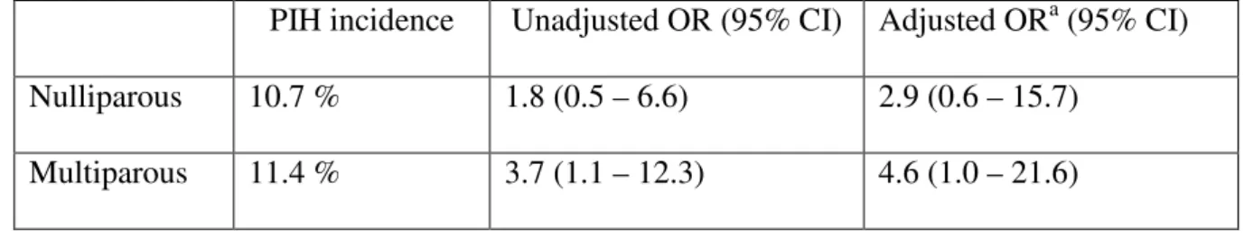
When stratified by parity (Table 4), blood lead levels were higher in multiparous
women than nulliparous women (2.0 ± 1.2 µg/dl versus 1.8 ± 1.3 µg/dl, respectively;
p=0.003). The adjusted OR for PIH also was higher in multiparous women (OR= 4.6; 95%
CI: 1.0 to 21.6) than nulliparous women (OR= 2.9; 95% CI: 0.6 to 15.7), but there was no
interaction between parity and blood lead (p=0.46).
Log-transformed blood lead at mid-pregnancy was significantly correlated with both
systolic and diastolic blood pressures at 24 – 36 weeks of gestation (r=+0.08; p=0.03 and
r=+0.07; p=0.03, respectively), and after 36 weeks of gestation (r=+0.09; p=0.03 and r=+0.08;
p=0.03, respectively). Figure 1 illustrates correlation between residuals of the linear
regression of maternal blood lead and systolic blood pressure at 24 – 36 weeks. Lead
concentrations accounted for approximately 5% of the total unexplained variance obtained by
linear models. Each decimal-log increase in blood lead was associated with an increase of 3.5
mm Hg in SBP and of 2.5 mm Hg in DBP during the second half of pregnancy.
DISCUSSION:
We found that the adjusted risk of PIH was associated with maternal blood lead levels
in mid-pregnancy. This risk was doubled in the highest quartile as compared to the lowest
quartile of lead distribution. There was no strong evidence of an interaction between lead and
cadmium, manganese, selenium, or other factors associated with the risk of PIH. The risk of
PIH increased with increasing absolute values of mid-pregnancy blood lead in a
“dose-response” pattern. A positive correlation was particularly found with systolic blood pressure
at 24 – 36 weeks that persisted after 36 weeks. All these findings suggest that blood lead level
may be one of the causal factors of PIH.
Evidence that lead increases the circulating levels of endothelin, a vaso-active
substance secreted by endothelial cells has been reported (Gonick and Behari 2002). Lead is
also reported to reduce levels of vasodilator substances such as plasma nitric oxide and
endothelial-derived relaxation factor; this reduction is due to a lead-mediated increase in
reactive oxygen species (Carmignani et al. 2000; Gonick and Behari 2002; Gonick et al.
1997). Inhibition of membrane ATPases by this metal also leads to increased intracellular
calcium ions and vasoconstriction (Moreau et al. 1988). Furthermore, a demonstrable
inhibitory effect of lead on blood enzymes such as delta-aminolevulinic acid dehydratase
activity was found at a low threshold of lead exposure, ranging from 3.2 to 4.8 µg/dl
(Campagna et al. 1999; Telisman et al. 2004). From the above arguments, it may be
hypothesized that environmental exposure to lead increases the risk of PIH by inducing
vasoconstriction and placental ischemia or by a direct toxicity on the endothelial cell and the
renal function.
Our findings are consistent with those from previous studies showing a relationship
between blood lead levels and PIH. However, most of these reports analysed lead
concentrations late in pregnancy, either from maternal prenatal red blood cells (Dawson et al.
2000), from umbilical cord (Rabinowitz et al. 1987), amniotic fluid (Dawson et al. 1999), or
even from maternal blood within 24 hours after the delivery (Vigeh et al. 2004; Vigeh et al.
2006). Our study did not involve serial measurements of blood lead levels throughout
pregnancy. However, the optimal period for such measurement would be in the second
trimester (24 weeks of gestation), at the beginning of a potential pathological change in blood
pressure. Moreover, models on lead variation during pregnancy described a constant increase
in blood lead levels from 24 weeks of gestation until delivery (O'Flaherty et al. 1995). Indeed,
longitudinal analyses showed that maternal blood lead concentrations upon entering prenatal
care (average of 13.5 weeks) were not associated with PIH, whereas higher lead levels during
the second trimester were related to PIH (Sowers et al. 2002).
Our analysis of correlations between blood lead concentrations and SBP and DBP at
24 – 36 weeks and after 36 weeks of gestation was consistent with previous studies on
essential hypertension (Batuman et al. 1983). Recent reviews suggest a weak but dependable
association between lead levels and blood pressure. The increase in SBP is estimated between
0.8 and 2.0 mm Hg for each 1 µg/dl increase in blood lead (Hertz-Picciotto and Croft 1993;
U.S.EPA 2006). Women with chronic hypertension were excluded from our study because of
risks of bias associated to prior treatment with anti hypertensive drugs. This exclusion might
have attenuated the correlation between blood lead and blood pressure.
Manganese blood level was not significantly associated with PIH in this study,
although there was some evidence of a possible weak association. The underlying
mechanisms of manganese-induced hypertension are probably independent from gestational
age. The decrease in blood manganese with intrauterine growth restriction might obscure any
other pathological effect (Vigeh et al. 2008). Cadmium also was not significantly associated
with PIH in the present model. This finding may be related to relatively low levels of
cadmium observed in this cohort. These reduced levels were probably due to the low
prevalence of smoking, which is the main environmental source of cadmium (Bonithon-Kopp
et al. 1986). Interaction between selenium and lead concentrations were not significant and
the putative protective effect of selenium through anti-oxidative properties was not confirmed
in this study.
Hematocrit levels are usually elevated in pregnancy hypertensive disorders (Goldstein
et al. 2000). This may be due to a lower water balance index correlated to stroke volume or to
a lower venous capacitance in hypertensive patients (Aardenburg et al. 2005). Hematocrit
increases automatically increase blood lead which is concentrated in red blood cells. No
statistical interaction was found between blood lead and hematocrit, but confounding can not
be ruled out.
Body-mass index was strongly associated with PIH. This finding is consistent with previous
reports (Hrazdilova et al. 2001; Vigeh et al. 2006). A “dose-response” relationship has also
been observed, thus body-mass index before pregnancy should be considered in all models
including PIH. Gestational diabetes effect on PIH was limited in the multivariate regression
analysis probably because of its association with increased body-mass index. Age and parity
were not significant covariates in this study. A trend of increasing blood pressure with age is
usually reported for diastolic blood pressure, but the restricted age range (i.e., 18 – 45 yr) and
limited number of women above 40 years in the present study may have reduced our ability to
highlight an age effect on PIH.
As to parity, high levels of blood lead observed in multiparous women of this study
may have contributed to an increase in the frequency of PIH in this group, thus reducing the
difference usually observed in the incidence of PIH and preeclampsia depending on parity.
Moreover, data obtained from systematic ambulatory monitoring in a large sample of
normotensive pregnant women indicate the lack of differences in blood pressure according to
parity (Ayala and Hermida 2001)
Finally, PIH frequency was higher in Nancy than in Poitiers. A separate analysis (not
shown) revealed higher blood lead levels in Nancy (2.0 ± 1.3 µg/dl) than in Poitiers (1.8 ± 1.1
µg/dl); p<0.001. Thus a major difference in the incidence of PIH between these two regions
might have been their history of environmental exposure to chemical pollutants. Blood lead
level primarily reflects recent exposure, although a part of lead in blood may originate from
lead stored in bone, particularly during pregnancy (Gulson et al. 1997). Since bone lead has a
half-life of years to decades, the higher blood lead levels observed in eastern versus western
region reflect differences in current exposure, but may also reflect some differences in past
exposure, or both. Hence, the relationship between blood lead and blood pressure may be
mediated by the contribution of bone lead to blood lead, placing a larger amount of
bioavailable lead into tissues and organs that affect blood pressure (Rothenberg et al. 1999).
The main sources of measurements errors of blood pressure are the inaccuracy of
measurement methods and the intra-individual variability of blood pressure. We attempted to
minimize these effects by averaging all blood pressure measurements available in the
obstetrical files.
Although the association between blood lead levels and PIH persisted in the
multivariate analysis, it is also possible that it reflects residual confounding due to
unmeasured confounders. The geographic residence is a general covariate and residual
confounding by this variable cannot be ruled out. However, if this residual confounding
explained the association found in the multivariate analysis, it is likely that an interaction
between geographic residence and blood lead levels would have been observed. Moreover,
blood lead concentration may be a better estimate of maternal environmental exposure than
external indicators, and its use as a continuous variable in the regression model limits the risk
of residual confounding.
Pregnancy-induced hypertension remains a multifactorial disease with unclear
aetiology, which could compromise maternal and newborn reproductive outcomes. We
identified a significant association between maternal blood lead levels in mid-pregnancy and
blood pressure. Our findings that lead may have an etiologic role in PIH, even at low levels of
environmental exposure, suggest that it may be appropriate for public health organizations to
consider lowering the upper limit of “acceptable” blood lead levels in pregnant women, which
is currently at 10 µg/dl.
REFERENCES:
Aardenburg R, Spaanderman ME, Courtar DA, van Eijndhoven HW, de Leeuw PW, Peeters
LL. 2005. A subnormal plasma volume in formerly preeclamptic women is associated
with a low venous capacitance. J Soc Gynecol Investig 12:107-111.
Acien P, Lloret G, Lloret M. 1990. Perinatal morbidity and mortality in pregnancy
hypertensive disorders: prognostic value of the clinical and laboratory findings. Int J
Gynaecol Obstet 32:229-235.
Anastasakis E, Papantoniou N, Daskalakis G, Mesogitis S, Antsaklis A. 2008. Screening for
pre-eclampsia by oxidative stress markers and uteroplacental blood flow. J Obstet
Gynaecol 28:285-289.
Angell NF, Lavery JP. 1982. The relationship of blood lead levels to obstetric outcome. Am J
Obstet Gynecol 142:40-46.
Ayala DE, Hermida RC. 2001. Influence of parity and age on ambulatory monitored blood
pressure during pregnancy. Hypertension 38:753-758.
Batuman V, Landy E, Maesaka JK, Wedeen RP. 1983. Contribution of lead to hypertension
with renal impairment. N Engl J Med 309:17-21.
Bonithon-Kopp C, Huel G, Grasmick C, Sarmini H, Moreau T. 1986. Effects of pregnancy on
the inter-individual variations in blood levels of lead, cadmium and mercury. Biol Res
Pregnancy Perinatol 7:37-42.
Campagna D, Huel G, Girard F, Sahuquillo J, Blot P. 1999. Environmental lead exposure and
activity of delta-aminolevulinic acid dehydratase (ALA-D) in maternal and cord
blood. Toxicology 134:143-152.
Campagna D, Huel G, Hellier G, Girard F, Sahuquillo J, Fagot-Campagna A, et al. 2000.
Negative relationships between erythrocyte Ca-pump activity and lead levels in
mothers and newborns. Life Sci 68:203-215.
Carmignani M, Volpe AR, Boscolo P, Qiao N, Di Gioacchino M, Grilli A, et al. 2000.
Catcholamine and nitric oxide systems as targets of chronic lead exposure in inducing
selective functional impairment. Life Sci 68:401-415.
Dawson EB, Evans DR, Kelly R, Van Hook JW. 2000. Blood cell lead, calcium, and
magnesium levels associated with pregnancy-induced hypertension and preeclampsia.
Biol Trace Elem Res 74:107-116.
Dawson EB, Evans DR, Nosovitch J. 1999. Third-trimester amniotic fluid metal levels
associated with preeclampsia. Arch Environ Health 54:412-415.
Drouillet P, Forhan A, De Lauzon-Guillain B, Thiebaugeorges O, Goua V, Magnin G, et al.
2008. Maternal fatty acid intake and fetal growth: evidence for an association in
overweight women. The 'EDEN mother-child' cohort (study of pre- and early postnatal
determinants of the child's development and health). Br J Nutr:1-9.
Fréry N, Girard F, Moreau T, Blot P, Sahuquillo J, Hajem S, et al. 1993. Validity of hair
cadmium in detecting chronic cadmium exposure in general populations. Bull Environ
Contam Toxicol 50:736-743.
Goldstein JD, Garry DJ, Maulik D. 2000. Obstetric conditions and erythropoietin levels. Am J
Obstet Gynecol 182:1055-1057.
Gonick HC, Behari JR. 2002. Is lead exposure the principal cause of essential hypertension?
Med Hypotheses 59:239-246.
Gonick HC, Ding Y, Bondy SC, Ni Z, Vaziri ND. 1997. Lead-induced hypertension: interplay
of nitric oxide and reactive oxygen species. Hypertension 30:1487-1492.
Groom KM, North RA, Poppe KK, Sadler L, McCowan LM. 2007. The association between
customised small for gestational age infants and pre-eclampsia or gestational
hypertension varies with gestation at delivery. BJOG 114:478-484.
Gulson BL, Jameson CW, Mahaffey KR, Mizon KJ, Korsch MJ, Vimpani G. 1997.
Pregnancy increases mobilization of lead from maternal skeleton. J Lab Clin Med
130:51-62.
Hertz-Picciotto I, Croft J. 1993. Review of the relation between blood lead and blood
pressure. Epidemiol Rev 15:352-373.
Hertz-Picciotto I, Schramm M, Watt-Morse M, Chantala K, Anderson J, Osterloh J. 2000.
Patterns and determinants of blood lead during pregnancy. Am J Epidemiol
152:829-837.
Hrazdilova O, Unzeitig V, Znojil V, Izakovicova-Holla L, Janku P, Vasku A. 2001.
Relationship of age and the body mass index to selected hypertensive complications in
pregnancy. Int J Gynaecol Obstet 75:165-169.
Huel G, Boudène C, Jouan M, Lazar P. 1986. Assessment of exposure to lead of the general
population in the French community through biological monitoring. Int Arch Occup
Environ Health 58:131-139.
Huel G, Campagna D, Girard F, Moreau T, Blot P. 2000. Does selenium reduce the risk of
threatened preterm delivery associated with placental cytochrome P450-1A1 activity?
Environ Res 84:228-233.
Kosanovic M, Jokanovic M, Jevremovic M, Dobric S, Bokonjic D. 2002. Maternal and fetal
cadmium and selenium status in normotensive and hypertensive pregnancy. Biol Trace
Elem Res 89:97-103.
Lee AM, Huel G, Godin J, Hellier G, Sahuquillo J, Moreau T, et al. 1995. Inter-individual
variation of selenium in maternal plasma, cord plasma and placenta. Sci Total Environ
159:119-127.
Mergler D, Huel G, Bellanger S, Bowler R, Truchon G, Ostiguy C, et al. 1996. Surveillance
of early neurotoxic dysfunction. Neurotoxicology 17:803-812.
Moreau T, Hannaert P, Orssaud G, Huel G, Garay RP, Claude JR, et al. 1988. Influence of
membrane sodium transport upon the relation between blood lead and blood pressure
in a general male population. Environ Health Perspect 78:47-51.
Navas-Acien A, Guallar E, Silbergeld EK, Rothenberg SJ. 2007. Lead exposure and
cardiovascular disease--a systematic review. Environ Health Perspect 115:472-482.
Nawrot TS, Thijs L, Den Hond EM, Roels HA, Staessen JA. 2002. An epidemiological
re-appraisal of the association between blood pressure and blood lead: a meta-analysis. J
Hum Hypertens 16:123-131.
NHBPEP. 2000. Report of the National High Blood Pressure Education Program Working
Group on High Blood Pressure in Pregnancy. Am J Obstet Gynecol 183:S1-S22.
O'Flaherty E, Polak J, Andriot M. 1995. Incorporation of temporal factors into physiologically
based kinetic models for risk assessment. Inhal Toxicol 7:917-925.
Rabinowitz M, Bellinger D, Leviton A, Needleman H, Schoenbaum S. 1987. Pregnancy
hypertension, blood pressure during labor, and blood lead levels. Hypertension
10:447-451.
Rayman MP, Bode P, Redman CW. 2003. Low selenium status is associated with the
occurrence of the pregnancy disease preeclampsia in women from the United
Kingdom. Am J Obstet Gynecol 189:1343-1349.
Rothenberg SJ, Manalo M, Jiang J, Khan F, Cuellar R, Reyes S, et al. 1999. Maternal blood
lead level during pregnancy in South Central Los Angeles. Arch Environ Health
54:151-157.
Schwartz J. 1995. Lead, blood pressure, and cardiovascular disease in men. Arch Environ
Health 50:31-37.
Sowers M, Jannausch M, Scholl T, Li W, Kemp FW, Bogden JD. 2002. Blood lead
concentrations and pregnancy outcomes. Arch Environ Health 57:489-495.
Telisman S, Pizent A, Jurasovic J, Cvitkovic P. 2004. Lead effect on blood pressure in
moderately lead-exposed male workers. Am J Ind Med 45:446-454.
U.S. EPA. 2006. Air Quality Criteria for Lead (Final). URL:
http://cfpub.epa.gov/ncea/cfm/recordisplay.cfm?deid=158823.
Vaziri ND, Khan M. 2007. Interplay of reactive oxygen species and nitric oxide in the
pathogenesis of experimental lead-induced hypertension. Clin Exp Pharmacol Physiol
34:920-925.
Vaziri ND, Sica DA. 2004. Lead-induced hypertension: role of oxidative stress. Curr
Hypertens Rep 6:314-320.
Verity MA. 1999. Manganese neurotoxicity: a mechanistic hypothesis. Neurotoxicology
20:489-497.
Vigeh M, Yokoyama K, Mazaheri M, Beheshti S, Ghazizadeh S, Sakai T, et al. 2004.
Relationship between increased blood lead and pregnancy hypertension in women
without occupational lead exposure in Tehran, Iran. Arch Environ Health 59:70-75.
Vigeh M, Yokoyama K, Ramezanzadeh F, Dahaghin M, Fakhriazad E, Seyedaghamiri Z, et
al. 2008. Blood manganese concentrations and intrauterine growth restriction. Reprod
Toxicol 25:219-223.
Vigeh M, Yokoyama K, Ramezanzadeh F, Dahaghin M, Sakai T, Morita Y, et al. 2006. Lead
and other trace metals in preeclampsia: a case-control study in Tehran, Iran. Environ
Res 100:268-275.
Tables
Table 1. Baseline characteristics according to PIH occurrence among 971 pregnant women
with no history of chronic hypertension.
PIH (mean or %) No PIH (mean or %) P Value Maternal age – yr < 25 19.8 17.6 25 – 34 64.2 68.2 ≥ 35 16.0 14.2 N 106 865 0.70 Years of education ≥ 12 yr 64.2 66.2 < 12 yr 30.2 29.0 Unknown 5.7 4.7 N 106 865 0.87 Socio-economic status High 25.7 25.5 Medium 52.4 55.5 Low 21.9 19.0 N 105 851 0.76 Geographic residence Poitiers (Western) 36.8 58.6 Nancy (Eastern) 63.2 41.4 N 106 865 < 0.001 Hematocrit – % a,b 35.2 (2.5) 34.6 (2.6) N 105 842 0.02
BMI before pregnancy – Kg/m2
< 25 53.3 76.2
25 – 29.9 24.8 17.5
≥ 30 21.9 6.3
N 105 835
< 0.001
Weight gain during pregnancy – kg a 13.4 (6.5) 13.4 (4.6)
N 104 834 0.92 Gestational diabetes No 83.0 94.1 Yes 17.0 5.9 N 106 851 < 0.001 Parity Nulliparous 42.5 44.3 Multiparous 57.5 55.7 N 106 848 0.71
Smoking during pregnancy – cig/day
0 72.6 71.7
≥ 1 27.4 28.3
1 – 9 19.6 22.2
7.8 6.1
0.86
N 102 820 Alcohol consumption before pregnancy
No 28.2 28.7 Yes 71.8 71.3 N 103 849 0.90 Iron/calcium supplementation No 2.8 3.9 Yes 61.3 60.1 Unknown 35.9 36.0 N 106 865 0.95
Premature delivery (before 37 wk)
No 86.8 95.1 Yes 13.2 5.0 N 106 851 <0.001 Neonate weight – gr a 3126.7 (719.9) 3299.0 (493.6) N 106 844 0.02 Neonate gender Male 58.5 52.9 Female 41.5 47.1 N 106 849 0.28 a
values are means (SD).
b
Hematocrit levels were measured concomitantly with metals between 24 and 28 gestational
weeks
Table 2. Distribution of elements levels in maternal blood at mid-pregnancy, according to PIH occurrence. PIH (n=106) No PIH (n=865) Mean (SD) GM Mean (SD) GM P Valuea Lead (µg/dl) 2.2 (1.4)b 1.9 1.9 (1.2) 1.6 0.02 Cadmium (µg/l) 0.9 (0.5) 0.8 0.9 (0.6) 0.7 0.08 Manganese (µg/l) 11.8 (6.3) 10.6 10.6 (4.5) 9.6 0.06 Selenium (µg/l) 98.8 (29.0) 93.3 98.6 (26.2) 95.5 0.76 a
t-test on the log transformed variables
b
To convert values for lead to micromoles per liter, multiply by 0.0483.
Table 3. Odds ratios for PIH, according to maternal blood lead distribution and overall
outcome characteristics.
Unadjusted Analysis Adjusted Analysisa
OR (95% CI) P value OR (95% CI) P value
Log (Pb)b 3.49 (1.37 – 8.87) 0.009 3.29 (1.11 – 9.74) 0.03
- Q1c (referent) 1.0 1.0
- Q2 1.55 (0.78 – 3.11) 0.94 1.84 (0.77 – 4.41) 0.84
- Q3 1.61 (0.78 – 3.31) 0.81 2.07 (0.83 – 5.13) 0.50
- Q4 2.19 (1.09 – 4.41) 0.06 2.56 (1.05 – 6.22) 0.09
Number of observations used in adjusted analysis: 720; Convergence criterion satisfied; R2=15.7%
a
Adjusted for maternal age, cadmium, manganese and selenium blood levels, hematocrit,
parity, body-mass index, gestational diabetes, educational level, socio-economic status,
geographical residence, and smoking status during pregnancy.
b
Log-transformed maternal blood lead used as a continuous variable
c
Quartiles of maternal blood lead distribution: Q1 (< 1.20 µg/dl); Q2 (1.20 – 1.70 µg/dl); Q3
(1.71 – 2.30 µg/dl); and Q4 (> 2.30 µg/dl).
Table 4. Odds Ratios for PIH according to parity, per unit increase in blood lead level.
PIH incidence Unadjusted OR (95% CI) Adjusted ORa (95% CI)
Nulliparous 10.7 % 1.8 (0.5 – 6.6) 2.9 (0.6 – 15.7)
Multiparous 11.4 % 3.7 (1.1 – 12.3) 4.6 (1.0 – 21.6)
a
Adjusted for maternal age, cadmium, manganese and selenium blood levels, hematocrit,
body-mass index, gestational diabetes, educational level, socio-economic status, geographical
residence, smoking status and alcohol consumption before pregnancy.
Figures
Figure 1. Scatter plot of the residuals for maternal blood Lead and systolic blood pressure
between 24 and 36 weeks (SBP2) after controlling for the effect of variables listed in the
logistic model in table 3.
This figure illustrates the correlation between residuals of the linear regression of the two
variables on the partialled variables. In the 95% and 70% prediction ellipses, the major axis
length is significantly larger than the minor axis length, indicating a partial correlation
between maternal blood lead and SBP2.