HAL Id: hal-03001918
https://hal.archives-ouvertes.fr/hal-03001918
Submitted on 27 May 2021
HAL is a multi-disciplinary open access
archive for the deposit and dissemination of
sci-entific research documents, whether they are
pub-lished or not. The documents may come from
teaching and research institutions in France or
abroad, or from public or private research centers.
L’archive ouverte pluridisciplinaire HAL, est
destinée au dépôt et à la diffusion de documents
scientifiques de niveau recherche, publiés ou non,
émanant des établissements d’enseignement et de
recherche français ou étrangers, des laboratoires
publics ou privés.
Distributed under a Creative Commons Attribution| 4.0 International License
in Beclin-1 and Beclin-2 enhance the entry of lentiviral
vectors into human cells
Saliha Majdoul, Jérémie Cosette, Ababacar Seye, Eric Bernard, Sophie Frin,
Nathalie Holic, Nathalie Chazal, Laurence Briant, Lucile Espert, Anne Galy,
et al.
To cite this version:
Saliha Majdoul, Jérémie Cosette, Ababacar Seye, Eric Bernard, Sophie Frin, et al.. Peptides derived
from evolutionarily conserved domains in Beclin-1 and Beclin-2 enhance the entry of lentiviral vectors
into human cells. Journal of Biological Chemistry, American Society for Biochemistry and Molecular
Biology, 2017, 292 (45), pp.18672-18681. �10.1074/jbc.M117.800813�. �hal-03001918�
Peptides derived from evolutionarily conserved domains in
Beclin-1 and Beclin-2 enhance the entry of lentiviral vectors
into human cells
Received for publication, June 8, 2017, and in revised form, September 7, 2017 Published, Papers in Press, September 19, 2017, DOI 10.1074/jbc.M117.800813
Saliha Majdoul‡, Jeremie Cosette§, Ababacar K. Seye‡, Eric Bernard¶, Sophie Frin‡, Nathalie Holic‡, Nathalie Chazal¶,
Laurence Briant¶, Lucile Espert¶,X Anne Galy‡1, andX David Fenard‡2
From‡Integrare Research Unit, UMR_S951, Ge´ne´thon, INSERM, University of Evry, EPHE, Evry F 91000, France,§Ge´ne´thon, 91000 Evry, France, and¶IRIM (ex-CPBS) UMR 9004, Infectious Disease Research Institute of Montpellier, University of Montpellier, CNRS, Montpellier F34293, France
Edited by Charles E. Samuel
Autophagy-related proteins such as Beclin-1 are involved in an array of complex processes, including antiviral responses, and may also modulate the efficiency of gene therapy viral vec-tors. The Tat-Beclin-1 (TB1) peptide has been reported as an autophagy-inducing factor inhibiting the replication of patho-gens such as HIV, type 1 (HIV-1). However, autophagy-related proteins are also essential for the early steps of HIV-1 infection. Therefore, we examined the effects of the Beclin-1 evolution-arily conserved domain in TB1 on viral transduction and autophagy in single-round HIV infection or with nonreplicative HIV-1– derived lentiviral vectors. TB1 enhanced transduction with various pseudotypes but without inducing the autophagy process. TB1 augmented the transduction of human CD34ⴙ hematopoietic stem/progenitor cells while maintaining their capacity to engraft in vivo into humanized mice. TB1 was as effective as other transduction additives and functioned by enhancing the adhesion and fusion of viral particles with target cells but not their aggregation. We also found that the N-termi-nal L1 loop was critical for TB1 transduction– enhancing activ-ity. Interestingly, the Tat-Beclin-2 (TB2) peptide, derived from the human Beclin-2 protein, was even more potent than TB1 in promoting viral transduction and infection. Taken together, our findings suggest that the TB1 and TB2 peptides enhance the viral entry step. Tat-Beclin peptides therefore represent a new family of viral transduction enhancers for potential use in gene therapy.
The lysosomal degradation pathway of autophagy has a dual role in HIV, type 1 (HIV-1)3replication and pathogenesis (1).
On one hand, studies have shown that some autophagy-related proteins are essential for the early steps of HIV-1 infection (2– 4), but on the other hand, autophagy induction appears to be an antiviral strategy. Indeed, Shoji-Kawata et al. (5) have described a new autophagy-inducing peptide called Tat-Beclin-1 (TB1), capable of inhibiting the replication of several pathogens, including HIV-1, in vitro. The TB1 peptide is a fusion between a cell-penetrating peptide called Tat (47–57) (6, 7) and a fragment of the evolutionarily conserved domain (ECD267–284) of Beclin-1 (8), in which three mutations have been incorporated (H275E, S279D, and Q281E) for better pep-tide solubility. ECD267–284has been shown to interact with a
newly identified negative regulator of autophagy, Golgi-associ-ated plant pathogenesis-relGolgi-associ-ated protein 1 (GAPR-1) (9). Upon TB1 treatment, a pool of Beclin-1 proteins is released from the Golgi and becomes a core component of the class III phosphati-dylinositol 3-kinase complex that induces autophagy (10). Because the early steps of HIV-1 infection are highly dependent on various autophagy-related (Atg) proteins (3), the effect of TB1 on viral replication could therefore be complex and may not be predicted for single-round infections or target cell trans-duction with non-replicative HIV-1– derived lentiviral vectors (LVs), currently used in various applications of gene therapy (11).
In this study, we evaluated the effect of TB1 on single-round infections with HIV-1 and also on LVs pseudotyped with vari-ous envelope glycoproteins; namely, the Chikungunya virus glycoprotein (CHIKV-G), the modified gibbon ape leukemia virus glycoprotein (GALVTR), the modified RD114 feline endogenous retrovirus (RD114TR), and the vesicular stomatitis virus glycoprotein (VSV-G), the latter being broadly used in gene therapy. Surprisingly, we observed that the TB1 peptide was able to strongly promote viral infection or transduction with all LV pseudotypes tested, either on cell lines or hCD34⫹ hematopoietic stem/progenitor cells (HSPCs). Consequently,
This work was supported by the Association Française contre les Myopathies (AFM/Telethon). The authors declare that they have no conflicts of interest with the contents of this article.
This article containssupplemental Figs. S1–S9, Table S1, and Materials and Methods.
1To whom correspondence may be addressed: Généthon, UMR_S951, 1bis
rue de l’Internationale, 91000 Evry, France. Tel.: 33-1-69-47-34-40; E-mail: galy@genethon.fr.
2To whom correspondence may be addressed: TxCell SA, Allée de la Nertière,
les Cardoulines, 06560 Valbonne-Sophia Antipolis, France. Tel.: 33-4-97-21-83-15 ; E-mail: david.fenard@txcell.com.
3The abbreviations used are: HIV-1, HIV, type 1; ECD, evolutionarily conserved
domain; LV, lentiviral vector; CHIKV, Chikungunya virus; GALVTR, modified gibbon ape leukemia virus glycoprotein; VSV-G, vesicular stomatitis virus
glycoprotein; RD114TR, modified RD114 feline endogenous retrovirus; HSPC, hematopoietic stem/progenitor cell; NSG mice, Nude/Scid/Gamma chain-deficient mice; BLAM,-lactamase; CFC, colony-forming cell; TS, Tat-Scrambled; GP, glycoprotein; eGFP, enhanced GFP; EBSS, Earle’s balanced salt solution; UVRAG, UV radiation resistance–associated gene protein; AZT, 3⬘-azido-3⬘-deoxythymidine; qPCR, quantitative PCR; UCB, umbilical cord blood.
cro
ARTICLE
18672
J. Biol. Chem. (2017) 292(45) 18672–18681© 2017 by The American Society for Biochemistry and Molecular Biology, Inc. Published in the U.S.A.
the potential toxicity of TB1 has been evaluated on human HSPCs either in vitro (CFC assay) or in vivo (humanized NSG mice). We also investigated which steps of the viral life cycle are targeted by TB1. Finally, this study was extended through the design of various TB1 variants and a new peptide called Tat-Beclin-2 (TB2), a fusion of the Tat (47–57) transduction pep-tide with the human Beclin-2 ECD249 –266(12).
Results
Low doses of Tat-Beclin-1 strongly improved cell line transduction with various lentiviral pseudotypes and with HIV-1
To evaluate the effect of the TB1 peptide on LV transduction, we used the human colon carcinoma cell line HCT116, which is routinely employed in our laboratory, to titer LV pseudotyped with the VSV-G envelope (VSV-G-LVs). Using a low concen-tration of LV, we found that the TB1 peptide enhanced the transduction of HCT116 cells in a dose-dependent manner and up to 8-fold compared with the control Tat-Scrambled (TS) peptide (Fig. 1A). Further increasing the TB1 peptide concen-tration was associated with a bell-shaped dose-response curve. At the same molarity, neither the Tat peptide alone nor the Beclin-1 domain alone were capable of promoting lentiviral transduction (supplemental Fig. S1). The TB1 effect was not saturable over a one-log concentration of VSV-G-LV (corre-sponding to a multiplicity of infection of 0.5 to 5), reaching up to 84% of transduction efficiency (Fig. 1B). The improvement in transduction efficiency, evaluated by transgene expression lev-els (i.e. GFP), was also confirmed by proviral DNA integration following quantification by qPCR of vector copy numbers per cell (supplemental Fig. S2).
A great advantage of LVs for gene transfer is their capacity to be pseudotyped with numerous heterologous envelope glyco-proteins for specific cell targeting (13). Certain hematopoietic-tropic LV pseudotypes (GALVTR-LV and RD114TR-LV) require the use of culture additives to promote efficient trans-duction. These additives include Polybrene, protamine sulfate, or the recently identified Vectofusin-1 peptide (14, 15). As shown in Fig. 1C, TB1 was capable to promote cell line trans-duction with GALVTR- and RD114TR-LVs to an extent com-parable with other culture additives.
The enhancing effects of TB1 on LV transduction are in apparent contradiction with reports that describe TB1 as an inhibitor of HIV-1 and Chikungunya virus replication (5). This prompted us to verify the effect of TB1 on the early phase of live virus infection using a single-round infection assay in the MAGIC 5B permissive cell line. TB1 efficiently promoted the infection of wild-type HIV-1 (pNL4.3 molecular clone) (Fig. 2A) and also of HIV-1 pseudotyped with the Chikungunya GP (CHIKV-LV), although not statistically (Fig. 2B). TB1 is there-fore defined as a lentiviral transduction enhancer of HIV-1 vec-tors or viruses bearing a large panel of envelope glycoproteins.
Tat-Beclin-1 promotes safe lentiviral transduction of hematopoietic stem/progenitor cells
An important goal for the use of LVs is to achieve clinically relevant levels of transduction of HSPCs for ex vivo gene ther-apy approaches. As shown in Fig. 3A, highly purified
VSV-G-LV particles were used to transduce human CD34⫹ HSPCs. In the presence of TB1, a 2-fold increase in lentiviral transduc-tion was observed (Fig. 3, A and B). The optimal dose of TB1 to promote HSPCs was defined to be around 10M, twice the concentration used on cell lines (data not shown). Because it has been shown previously that high doses of TB1 are triggering a specific kind of cell death called autosis (16), safety studies were performed on HSPCs to examine the effects of TB1. Incu-bation of hCD34⫹ cells with increasing concentrations of TB1 led to partial cell death only at concentrations above 20 M
Figure 1. Tat-Beclin-1 promotes cell line transduction with various lenti-viral vectors. A, HCT116 cells were transduced for 6 h with VSV-G-LVs (2⫻ 105
ig/ml) in the absence or presence of the indicated concentrations of Tat-Scrambled or Tat-Beclin-1 peptide. B, HCT116 cells were transduced with var-ious titers of VSV-G-LVs in the absence or presence of Scrambled or Tat-Beclin-1 peptide (5M). C, HCT116 cells were transduced with GALVTR-LVs or RD114TR-LVs (106TU/ml) in the absence or presence of Tat-Scrambled (5
M), Tat-Beclin-1 (5M), Vectofusin-1 (6g/ml), protamine sulfate (4 g/ml), or Polybrene (3g/ml). In all three panels, transduction efficiencies were evalu-ated after 3 to 4 days by monitoring GFP expression. All data are expressed as the average of three independent experiments performed in duplicate⫾ S.E.
(supplemental Fig. S3). A colony-forming cell (CFC) assay was used to evaluate the effect of TB1 on the hematopoietic differenti-ation of hCD34⫹ cells. As shown in Fig. 3C, TB1 did not affect the absolute number of each type of colonies compared with the scrambled control or with cells treated with RetroNectin, a fibronectin fragment peptide used in gene therapy protocols to preserve HSPCs during transduction and to enhance transduction by co-localizing viral particles and target cells (17, 18). We previ-ously reported the enhancing effects of RetroNectin in our system (19).
To extend our safety studies to an in vivo system, TB1-treated HSPCs were injected into the immunodeficient NSG mouse model, and the engraftment efficiency was evaluated after 12 weeks. As shown in Fig. 4A, the engraftment of hCD34⫹ cells in the bone marrow was comparable (around 20%) between all conditions, either in the absence or presence of TS, TB1, or the control RetroNectin. Similarly, transduced GFP⫹ hCD34⫹ cells were present in the bone marrow (Fig. 4B), with a slightly better efficiency than RetroNectin, although the difference was not statistically significant. Comparable engraftment of transduced cells was observed in the spleen, thymus, and blood (data not shown). Altogether, this encour-aging safety profile of TB1 indicates that it is compatible with clinical applications involving lentiviral gene transfer.
Tat-Beclin-1 acts on the adhesion and fusion steps of LVs with target cell membranes
In terms of timing, TB1 pretreatment was found to be ineffi-cient for viral transduction, and addition of TB1 only 3 h after the start of transduction led to a 50% drop in TB1 efficacy ( sup-plemental Fig. S4), suggesting that TB1 acts at the early steps of viral transduction. Because LV entry into target cells is a rate-limiting step, we investigated whether low doses of TB1 were capable of enhancing adhesion and fusion of LVs with target cell membranes. For that, the BLAM-LV fusion assay was used (19) and showed that TB1 promoted efficient viral fusion (Fig. 5A). Next, by quantifying the number of viral particles interact-ing with target cells at 4 °C, it was shown that the level of viral adhesion was augmented in the presence of TB1, reaching levels comparable with those of Vectofusin-1 (Fig. 5B). This
Figure 2. Tat-Beclin-1 enhances HIV-1 single-round infection and CHIKV-LV transduction. A, MAGIC 5B cells were infected with purified HIV-1 (NL4.3) in the absence or presence of different doses of Beclin-1 or Tat-Scrambled peptides for 6 h. Cells were then extensively washed and cultured in the presence of AZT for 72 h. The level of viral infection was quantified by measuring-galactosidase activity. Data are represented as the mean of three independent experiments (Student’s t test; *, p⬍ 0.05). B, HEK293T cells were challenged for 6 h with CHIKV-LVs in the presence of increasing concentrations of Tat-Scrambled or Tat-Beclin-1 peptide. Transduction efficiency was monitored after 48 h by quantification of GFP expression in the cell lysates. Data are expressed as the mean of triplicate experi-ments⫾ S.E.
Figure 3. Tat-Beclin-1 promotes lentiviral transduction of hCD34ⴙ HSPCs with no apparent cytotoxicity. A, hCD34⫹ cells were infected with highly purified VSV-G-LVs (2⫻ 107ig/ml, multiplicity of infection 240) in the
absence (None) or presence of RetroNectin (7g/cm2) or the indicated
con-centrations of Tat-Scrambled or Tat-Beclin-1 peptide. After 3 to 5 days, trans-duction efficiencies were evaluated by monitoring GFP expression. Data are expressed as the average of three independent experiments (three UCB donors) performed in duplicate⫾ S.E. B, hCD34⫹ cells (six UCB donors) were transduced in duplicate with VSV-G-LVs (2⫻ 107ig/ml) in the absence or
presence of Tat-Beclin-1 (10M). Data are represented as the average level of transduction for each UCB donor. Bars indicate the mean value of the distributions (Mann-Whitney test; **, p⬍ 0.01). C, differentiation of trans-duced hCD34⫹ cells in a CFC assay. The results represent the average number of different types of colonies obtained for 1000 cells plated after transduction with VSV-G-LVs (5⫻ 107ig/ml) in the absence (None) or
presence of RetroNectin (7 g/cm2), Tat-Scrambled (10
M), or Tat-Beclin-1 (10M) peptide. Data are the average of three independent experiments performed in duplicate⫾ S.E.
Beclin-derived peptides promote viral transduction
increase in viral adhesion was the same whether cells were pre-incubated with TB1 at 37 °C or not (data not shown). Interest-ingly, TB1 also promoted viral transduction after the adhesion of viral particles as efficiently as Vectofusin-1, described previ-ously to act on the viral fusion step (Fig. 5C). Hence, this enhancement in viral fusion is, at the same time, the conse-quence of an increase in viral adhesion and post-adhesion steps. However, this increase in viral entry was not caused by TB1-induced aggregation of viral particles, contrary to Vectofusin-1 (Fig. 5D). The effect was also not caused by an increase in cell surface expression of retroviral receptors (supplemental Fig. S5).
The effect of TB1 on lentiviral transduction seems to be independent of the autophagy process
To better understand the mechanism of action of TB1, we sought to investigate whether it involved the activation of the autophagy process. To monitor autophagy at the single-cell level, HEK293T cells were transfected with a plasmid express-ing the mCherry-eGFP-LC3 fusion protein to mark the autophagolysosomes in the cells (supplemental Fig. S6). Next, cells were incubated with TB1 (5M) or EBSS starvation solu-tion and analyzed with an imaging flow cytometer. As expected, EBSS increased the number of autophagolysosomes (mCherry spots) per cell (Fig. 6A). On the contrary, the number of
autophagolysosomes in TB1-treated HEK293T cells was com-parable with that under control conditions in the absence of peptide (Fig. 6A, None). The autophagy level was also deter-mined by monitoring LC3 lipidation using immunoblot exper-iments. As expected, an increase in the LC3-II form was observed in the presence of EBSS, but the results obtained with TB1-treated cells and control TS-treated cells were compara-ble. Consistently, the level of p62/SQSTM1, which is known to be degraded by autophagic flux, decreased under starvation conditions (EBSS) but did not vary between cells treated with TS or the TB1 peptide (Fig. 6B). In parallel, we confirmed that TB1 is able to induce autophagic flux at high concentration, as described previously (5) (supplemental Fig. S7). Finally, the use of various PI3K inhibitors, which are known to block the autophagy process, did not impact TB1-induced lentiviral transduction (supplemental Fig. S8). Altogether, these data suggest that the improvement of lentiviral transduction observed in the presence of TB1 is certainly not the conse-quence of an induction of autophagy flux.
Critical role of the N-terminal L1 loop of Beclin-1 for efficient promotion of viral transduction
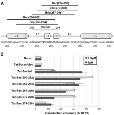
To better define the critical molecular determinants in the TB1 peptide necessary for efficient promotion of viral trans-duction, numerous peptides overlapping the human Beclin-1 protein from position 250 to 300 were designed (Fig. 7A). As a reference, the Beclin-1 domain of TB1 (267–284) is also repre-sented. All of these peptides were tested for their capacity to promote the infectivity of VSV-G-LV pseudotypes. As shown in Fig. 7B, the more potent TB1 variant is Tat-Bec (250 –282), corresponding to the␣ helix1/loop1/1 region. This peptide is three times more efficient than TB1 at only 2.5M. Interest-ingly, a gradual loss in viral transduction efficiency is observed when the peptide variants are less and less encompassing the N-terminal region of the L1 loop, suggesting that this short domain is critical for viral infectivity improvement.
The Tat-Beclin-2 peptide derived from human Beclin-2 is more efficient than TB1 for lentiviral transduction enhancement
In 2013, He et al. (12) identified a new mammal-specific pro-tein called Beclin-2 (12). Beclin-2 behaves in autophagy like Beclin-1 but also plays a major role in an additional lysosomal degradation pathway. Sequence alignment of the human Beclin-1 and Beclin-2 proteins shows a high degree of homo-logy between the ECDs. Therefore, the Tat-Beclin-2 (TB2) pep-tide, a fusion of the Tat (47–57) peptide with human Beclin-2 ECD249 –266, was designed (Fig. 8A). Because TB1 contains
three mutations (H275E, S279D, and Q281E), the Tat-BecWT peptide corresponding to the fusion of the Tat (47–57) peptide with wild-type human Beclin-1 ECD267–284was also designed
and is represented in the sequence alignment (Fig. 8A and sup-plemental Table S1). These three peptides were tested for their capacity to promote lentiviral transduction over a large range of concentrations. As shown in Fig. 8B, all peptides promoted len-tiviral transduction but with a large variability in their optimal doses: 3Mfor TB1, 1Mfor Tat-BecWT, and 500 nMfor TB2. As few as 100 nM TB2 increased lentiviral transduction by 3-fold, from 10% to 30%, whereas TB1 and Tat-BecWT had no
Figure 4. Evaluation of the safety of Tat-Beclin-1 in the immunodeficient NSG mouse model. A and B, hCD34⫹ cells were transduced for 6 h with VSV-G-LVs (5⫻ 107ig/ml) in the absence or presence of RetroNectin (7
g/cm2), Tat-Scrambled (10M), or Tat-Beclin-1 (10M) and injected into NSG
mice. Thirteen weeks post-injection, the engraftment of human cells in the bone marrow was monitored by flow cytometry using fluorescent anti-hCD45 and anti-hCD34 antibodies. Each point represents a mouse.
effect at this concentration (Fig. 8B). Similarly tested as in Fig. 2, TB2 efficiently promoted the infection of wild-type HIV-1 (Fig. 8C) and also of CHIKV-LV (Fig. 8D). Tat-Scr2, a scrambled version of TB2, behaved as a negative control in the concentra-tion range for TB2 activity. At higher concentraconcentra-tion, Tat-Scr2 promoted lentiviral transduction, but only in HCT116 cells (Fig. 8B), not in MAGIC 5B (Fig. 8C) or HEK293T cells (Fig. 8D), possibly as a result of interactions between this peptide and specific membrane components of HCT116 cells. Interestingly, like TB1 and Tat-Bec(250 –282), TB2 is capable of promoting viral transduction after viral adhesion (supplemental Fig. S9), excluding a simple nonspecific effect of TB2 on viral adhesion. In conclusion, the TB2 peptide is a potent enhancer of lentiviral transduction at very low doses.
Discussion
Our results show that the TB1 peptide, at low doses, can be a potent enhancer of the entry of LV into target cells without inducing apparent autophagy in the cells. These results are not
inconsistent with the more complex effects TB1 can exert as a potent inducer of autophagy and as an efficient antiviral agent on replicative viruses at higher doses (5), considering that dif-ferent conditions are involved. Here we observed that a short exposure of cells to TB1 efficiently promoted the transduction of cell lines and HSPCs with various non-replicative HIV-1– derived lentiviral pseudotypes (VSV-G-LV, RD114TR-LV, GALVTR-LV, and CHIKV-LV) as well as HIV-1 infection in
vitroin single-round assays. Such findings are compatible with the notion that the replication of various enveloped viruses (i.e. HIV-1, VSV, CHIKV, and influenza virus) requires the expres-sion of autophagy-related factors (3, 20 –24). Our new findings regarding TB1 peptide properties are compatible with applica-tions in gene therapy. The safety profile of TB1 in HSPCs, the lack of effect on differentiation of hCD34⫹ cells in vitro, and the possibility to engraft TB1-exposed hCD34⫹ cells into the bone marrow of humanized NSG mice support the use of TB1 as a transduction additive to promote CD34⫹ cell transduction with LV in ex vivo gene therapy protocols. Additional
preclini-Figure 5. Tat-Beclin-1 promotes the adhesion and fusion of lentiviral particles with target cells. A, viral fusion assay. Cells were incubated for 2.5 h at 37 °C with VSV-G-BLAM-LVs in the absence (None) or presence of Tat-Scrambled or Tat-Beclin-1 at 10M. Next, viral fusion efficiency was estimated by monitoring the percentage of cleaved CCF2 substrate in the target cells using flow cytometry. Data are represented as the average of three independent experiments (three UCB donors for hCD34⫹ cells) performed in duplicate ⫾ S.E. B, adhesion assay. HCT116 cells were preincubated for 30 min at 37 °C in the absence or presence of Tat-Scrambled or Tat-Beclin-1 (5M). Next, cells were incubated for 2.5 h at 4 °C with VSV-G-LVs (75 ng of p24) in the absence or presence of Tat-Scrambled or Tat-Beclin-1 (5M). As a positive control, HCT116 cells were incubated with a cold solution of VSV-G-LVs mixed with the aggregating peptide Vectofusin-1 (12g/ml). Data are represented as the average of three independent experiments performed in duplicate ⫾ S.E. C, HCT116 cells were incubated for 2.5 h at 4 °C with VSV-G-LVs (2⫻ 107ig/ml). Unbound LVs were washed out with cold PBS, and cells were transferred at 37 °C in the absence or presence of
the indicated peptides as in B. Transduction efficiencies were evaluated after 3 days by monitoring GFP expression. Data are represented as the average of two independent experiments performed in duplicate⫾ S.E. D, viral pulldown assay. VSV-G-LV particles were mixed either with Tat-Scrambled (10M), Tat-Beclin-1 (10M), or the positive control Vectofusin-1 (10M). After a short centrifugation (15,000⫻ g), the percentage of pelleted viral particles was quantified using an HIV-1 p24 ELISA kit. Data are normalized to the level of p24 input and are represented as the average of three independent experiments performed in duplicate⫾ S.E.
Beclin-derived peptides promote viral transduction
cal evaluation, including toxicity studies, will be necessary in this regard.
The use of numerous TB1 variants, encompassing the human Beclin-1 protein from position 250 to 300 (␣1-L1-1-2-L2), suggests that the N-terminal region of the L1 loop is critical for viral infectivity improvement. This critical domain is described as the docking site of HIV-1 Nef, allowing the virus to modulate autophagy through this specific interaction with Beclin-1. The L1 loop is also the target of the Golgi-associated protein GAPR-1 (5). GAPR-1 belongs to the cysteine-rich secretory proteins, antigen 5, and pathogenesis-related 1 proteins (CAP) superfamily, found in a remarkable range of organisms (25). GAPR-1 could be seen as an innate immune effector, triggered upon viral infection. However, we never observed any negative impact of GAPR-1 overexpression on transduction efficiencies (data not shown). It cannot be excluded that other cellular fac-tor(s), critical for transduction enhancement, interact with the L1 loop of Beclin-1.
In the viral cycle, TB1 acts on the adhesion and post-adhe-sion step, leading to an increase in viral fupost-adhe-sion, a phenomenon that is not the consequence of an increase in the surface expres-sion or the receptor density of various retroviral receptors (supplementalFig. S5). Furthermore, contrary to transduction enhancers forming peptide nanofibers (26), TB1 does not
pro-mote viral entry through the induction of viral particle aggre-gation (Fig. 5D). It is unlikely that TB1 or TB2 exert some non-specific cationic effect capable to improve viral adhesion. Indeed, neither the Tat peptide alone nor the Beclin-1 domain alone was capable of promoting lentiviral transduction. Fur-thermore, TB2 is a lot more efficient than TB1 in promoting lentiviral transduction, and Tat-Scrambled is inefficient, but all of these peptides contain the same number of cationic charges. All of these features are rather reminiscent of a recently described transduction enhancer called-Deliverin (27). -De-liverin seems to promote viral adhesion/fusion by modulating endosomal maturation and trafficking. In the past, some host factors related to the autophagy process have been described as modulators of viral entry. For instance, the UV radiation resis-tance–associated gene protein (UVRAG) has been shown to regulate the VSV entry by promoting the formation of a fuso-genic SNARE complex involving vesicle-associated membrane protein 8 (24). However, that study exclusively described a mechanism that allowed enveloped viruses to interact with late acidic endosomes. In our study, Tat-Beclin peptides efficiently promote not only lentiviral pseudotypes that are pH-depen-dent, like VSV-G-LVs or CHIK-LVs (28), but also pH-indepen-dent pseudotypes (e.g. GALVTR-LVs (29) or RD114TR-LVs (30)), for which fusion occurs at the cell surface and/or in early endosomes. Further investigations will be needed to determine whether the relief of viral entry exerted by Beclin ECD-derived peptides could be a consequence of the modulation of endo-somal maturation and trafficking.
Figure 6. The optimal dose of Tat-Beclin-1 to promote lentiviral trans-duction does not induce autophagy. A, HEK293T cells expressing the mCherry-eGFP-LC3 fusion protein were incubated in the absence or presence of Tat-Beclin-1 (5M) (TB1) or in EBSS for 6 h. Next, cells were analyzed using an imaging flow cytometer. Images of cells were acquired in a bright field, in mCherry fluorescence, and in an SSC channel. Data are represented as the number of mCherry spots observed in each individual cell. Bars indicate the mean value of the distributions obtained from three independent experi-ments. The p values were determined using Mann-Whitney tests. n.s., not statistical. *** indicates highly significant values. B, HEK293T cells were incu-bated in the absence or presence of Tat-Beclin-1 (5M) (TB1), Tat-Scrambled (5M) (TS), or mock control-treated (CT), or in EBSS for 6 h. The levels of LC3-II and p62/SQSTM1 were monitored by Western blotting. CT, control-treated cells; EBSS, EBSS-cultured cells; TS, tat-scramble-treated cells.
Figure 7. Viral transduction efficiency of Tat-Beclin-1 variants. A, primary sequence of the human Beclin-1 protein from residue 246 to 322 (Homo sapi-ens, NP_003757). Mutations H275E, S279D, and Q281E are highlighted in bold italic. Secondary structural elements are indicated above the sequence (␣ helix (␣), loop (L), and  sheet ()). Above the structural elements, dark lines represent the protein sequence coverage of each Beclin-1 peptide variant. B, HCT116 cells were transduced as described in Fig. 1A in the absence (None) or presence of the indicated peptides either at 2.5M(light gray histogram) or 5 M(black histogram). For all panels, data are expressed as the average of three independent experiments performed in duplicate⫾ S.E.
In conclusion, autophagy-related factors, like Beclin-1 or UVRAG proteins, play a crucial role not only in the autophagy process but also in cellular endocytic membrane trafficking and fusion events. In fact, many host autophagy molecular factors have to be seen in a broader way as endomembrane traffick-ing regulators. Therefore, modulation of Beclin-1 and 2 functions using Beclin ECD-derived peptides could be of high interest for the development of more efficient vaccines
or gene therapy protocols through an optimal virus and viral vector entry.
Experimental procedures
Peptides and reagents
Tat-Beclin-1 and Tat-Beclin-2 peptides and their variants were obtained from Genecust Europe (Dudelange, Luxem-bourg) (see supplemental Table S1 for primary sequences). Vectofusin-1 was obtained from Genecust and Miltenyi Biotec (Paris, France). 7-amino-actinomycin D, trypan blue, 3 ⬘-azido-3⬘-deoxythymidine (AZT), protamine sulfate, bafilomycin A1, and Triton X-100 were obtained from Sigma-Aldrich (Saint-Quentin-Fallavier, France). RetroNectin was obtained from Ozyme (Saint-Quentin-en-Yvelines, France).
Cell line culture
HCT116 cells (derived from a human colorectal carcinoma, CCL-247, ATCC, Manassas, VA), HEK293T cells (31), and MAGIC 5B cells (a cell line expressing CXCR4, CCR5, and -galactosidase under the control of the HIV LTR, obtained from T. Masashi (Tokyo, Japan)), were cultured at 37 °C, 5% CO2in DMEM⫹ Glutamax supplemented with 10%
heat-in-activated FCS (Thermo Fisher Scientific, Courtabœuf, France).
Viral vector production and titering
LVs were generated as described previously (32). Briefly, HEK293T cells were transiently transfected using calci-um phosphate transfection with four plasmids: the gagpol (pKLgagpol) and rev (pKrev) expression plasmids, a transfer plasmid (pCCLsin.cPPT.hPGK.eGFP.WPRE), and a plasmid encoding either the VSV-G (pMDG) envelope glycoprotein (GP), the GALVTR GP (pBA.GALV/Ampho-Kana), or the RD114TR GP (pHCMV-RD114TR). After 24 h of production, raw viral supernatants were harvested, filtered (0.45m), and frozen at⫺80 °C. The purification of GFP-expressing VSV-G-LVs, through several membrane-based and chromatographic steps, has been described previously (31). Infectious titers were determined in HCT116 cells using either detection of GFP by flow cytometry (FACSCalibur, BD Biosciences, Le Pont de Claix, France), with titers expressed as transducing units (TU) per milliliter (33), or using qPCR with titers expressed as infec-tious genome (ig) per milliliter (31). The same qPCR approach was used to measure integrated vector copy numbers per cell following transduction. CHIKV-LVs were produced by cotransfection of the self-inactivating transfer plasmid pHR ⬘S-INcPPT-SGW (34), the lentiviral packaging plasmid psPAX2 (a gift from Didier Trono, Addgene, 12259), and the pCAGGS-EnvCHIKV plasmid encoding CHIKV GP (35). After 48 h of production, viral supernatants were harvested, filtered (0.22 m), and stored at ⫺80 °C. Viral inputs were calibrated to infect around 10% of the cell culture. Transduction by CHIKV-LVs was monitored by quantification of GFP expression in total cell lysates using an Infinite F200PRO fluorometer (Tecan, Sal-zburg, Austria), and values were normalized according to pro-tein content (BCA assay, Pierce).
HIV-1 stocks were prepared from the culture supernatant of HEK293T cells 48 h after transfection with the pNL4.3 provirus
Figure 8. Beclin-2 derived peptide promotes cell line transduction with viral vectors and viruses at very low doses. A, primary peptide sequence alignment of Beclin-1, Beclin-1 wild-type (BecWT), and Tat-Beclin-2. B, HCT116 cells were transduced for 6 h with VSV-G-LVs (2⫻ 105
TU/ml) in the absence or presence of the indicated concentrations of Tat-Scrambled (Tat-Scr), Tat-Beclin-1, Tat-BecWT, Tat-Tat-Scrambled 2 (Tat-Scr2), and Tat-Beclin-2 peptides. All data are expressed as the average of three indepen-dent experiments performed in duplicate⫾ S.E. C, MAGIC 5B cells were infected for 6 h with purified HIV-1 (NL4.3) in the absence or presence of different doses of Tat-Beclin-2 or Tat-Scr2 peptides. Cells were then exten-sively washed and cultured in the presence of AZT for 72 h. The level of viral infection was quantified by measuring-galactosidase activity. The data pre-sented are the mean of three independent experiments. D, HEK293T cells were transduced with CHIK-LVs in the presence of the indicated concentra-tions of Tat-Scr2 or Tat-Bec2WT peptide. Transduction efficiency was moni-tored after 48 h by monitoring GFP expression. Data are expressed as the mean of triplicate experiments⫾ S.E. *, p ⬍ 0.05; **, p ⬍ 0.01.
Beclin-derived peptides promote viral transduction
using TurboFect reagent (Thermo Fisher Scientific). After fil-tration (0.45 m), viral supernatants were concentrated by ultracentrifugation (25,000 rpm for 2 h at 4 °C). HIV-1 and LV physical titers were obtained by measuring HIV-1 p24 capsid contents using commercial ELISA kits from Fujirebio/Innoge-netics and PerkinElmer Life Sciences. The levels of viral infec-tion were determined by quantifying-galactosidase activity using the Galacto-Star-galactosidase reporter gene assay sys-tem (Thermo Fisher Scientific) according to the instructions of the manufacturer. Briefly, 0.2.106 MAGIC 5B cells were
infected in the presence of different peptides for 6 h. Cells were then washed five times and cultured for 72 h in DMEM and 10% FCS containing 1MAZT. Cells were then lysed in 200l of lysis buffer provided by the manufacturer. 50l of cell lysate was used in each assay in duplicate. Results were normalized by quantifying the total protein content using a Bradford assay (Sigma-Aldrich).
Human CD34ⴙ cell culture and transduction
Umbilical cord blood (UCB) samples were obtained after uncomplicated births and in accordance with international ethical principles and French national law under declaration DC-201-1655 to the French Ministry of Research and Higher Studies. Human CD34⫹ cells were isolated by immunomag-netic selection (Miltenyi Biotec). The survival rate of fresh or frozen hCD34⫹ cells was evaluated using the trypan blue exclu-sion method. Next the preactivation of hCD34⫹ cells was per-formed overnight as described previously (19). Preactivated cells were plated in 96-well plates, and transduction was initi-ated by adding the desired amount of LV particles mixed with or without the peptides of interest. At 6 h post-transduction, reactions were diluted by adding differentiation medium to each well. After 4 – 6 days, cellular mortality and transduction efficiency were evaluated, respectively, by 7-amino-actinomy-cin D labeling and measurement of GFP expression using flow cytometry (FACSCalibur, BD Biosciences).
Viral pulldown assay
The pulldown of LV particles in presence of culture additives was adapted from a protocol described previously (36). Briefly, the VSV-G-LV supernatant was diluted to 100 ng/ml of p24 with X-Vivo20 medium equilibrated at room temperature. Next, 1.5-ml tubes were loaded with 500l of LV suspension in the absence or presence of the indicated culture additive (10 m). After homogenization, samples were centrifuged at low speed (15,000⫻ g) for 5 min at room temperature. Then the supernatant was discarded, and the pellet was suspended in 100 l of fresh medium and frozen at ⫺20 °C. For each condition, the amount of pelleted p24 was evaluated using a commercial HIV-1 p24 ELISA kit as described above.
Adhesion, post-adhesion transduction, and BLAM-LV fusion assay
The protocol for LV adhesion to target cells has been described previously (37). Briefly, viral supernatants were incu-bated for 3 h at 4 °C in HCT116 cells in the absence or presence of culture additives. Next, cells were washed three times with cold 1⫻ PBS and lysed in 1⫻ PBS containing 1% Triton X-100
and a protease mixture inhibitor, Complete (Roche Diagnos-tics). p24 contents in lysates were evaluated using a commercial HIV-1 p24 ELISA kit, and data were normalized to total protein content using the DC protein assay (Bio-Rad). For the post-adhesion transduction assay, after incubation of 2.5 h at 4 °C in HCT116 cells, viral supernatants were washed twice with cold PBS. Next, the indicated peptides were added to the wells, and transduction was performed at 37 °C for 6 h. Transduction effi-ciencies were evaluated by monitoring GFP expression after 3 days. The BLAM-LV assay has been described extensively pre-viously (19).
Western blot
Cell lysates were loaded in 4 –20% precast gels (Bio-Rad) and transferred to PVDF membranes. After a blocking step for 1 h at room temperature in PBS containing 0.5% casein, membranes were incubated overnight at 4 °C with anti-LC3B antibody (Sig-ma-Aldrich) or anti-p62/SQSTM1 antibody (Ozyme) in block-ing buffer. After three washes with PBS supplemented with 0.05% Tween, the membranes were incubated for 1 h at room temperature with peroxidase-coupled secondary antibody. Upon extensive washes, membranes were incubated with Luminata Western HRP substrate (Millipore, Molsheim, France). Proteins were detected by chemiluminescence and imaged with a G-box camera (Syngene imaging system, Cam-bridge, UK). The expression level of GAPDH was used as a loadingcontrol.TheformofLC3conjugatedtophosphatidyletha-nolamine, named LC3-II, presents a higher electrophoretic mobility in gels and is used to determine the autophagy level. Autophagic flux can be monitored by quantification of the p62/ SQSTM1 level.
Autophagy assay based on imaging flow cytometry (ImageStream)
Using the transient calcium phosphate transfection method, HEK293T cells were transfected with the pBABE-puro-mCherry-eGFP-LC3B expression plasmid (38), a gift from Jay-anta Debnath (Addgene plasmid 22418). Next, cells (5⫻ 105
cells/well) were incubated in the absence or presence of Tat-Beclin-1 (5M) or in Earle’s balanced salt solution (EBSS) for 6 h at 37 °C, 5% CO2. Next, cells were washed in 1⫻ PBS, fixed
(1.2% paraformaldehyde), and analyzed using ImageStream (Amnis Corp., Seattle, WA). Images of cells were acquired in a bright-field channel, in an mCherry-fluorescence channel, and finally in the SSC channel (742 nm) using a⫻40 objective and the lowest flow velocity to optimize sensitivity. After acqui-sition, images were treated with Ideas威 software. Focused images were gated on a histogram displaying Gradient_RMS feature values in the bright-field channel between 40 and 90. Then a scatterplot of area versus aspect ratio (in the bright-field channel) was used to gate on single cells and remove doublets of cells. When gated on single cells, the spots on each cell were counted in the mCherry channel using the automatic spot counting wizard included in the Ideas威 soft-ware. Data were exported as text files and processed with GraphPad Prism 5.
CFC assay
The CFC assay was performed by plating 1000 human CD34⫹ cells per milliliter of Methocult medium, a methyl-cellulose medium enriched with human recombinant cy-tokines (H4434, StemCell Technologies, Vancouver, CA). After 15 days of culture, the burst-forming units–erythroid (BFU-E), colony-forming units–granulocyte-monocyte (CFU-GM), and colony-forming units–granulocyte erythrocyte, myeloid, megakaryocyte (CFU-GEMM) colonies were visu-alized and counted using an inverted light microscope.
Conditioning and reconstitution of NSG mice
Mice were housed in an accredited facility (CERFE, Evry, France) in a specific pathogen-free environment (French National Agreement C91.228.101). Experiments were per-formed according to the institutional and international guide-lines for animal care and use. Experimental procedures were performed in accordance with French and European directives, approved by the local ethical committee (C2EA-64) and Minis-try of Research for Gene-modified Organism Studies (Agree-ment 5244-CAI). NSG mice 3– 4 weeks of age were conditioned intraperitoneally with two doses of busulfan (25 mg/kg). Human CD34⫹ HSPCs from cord blood transduced with VSV-G-LV were injected intravenously into mice 24 h after the last dose of busulfan. Twelve weeks post-injection, the levels of engraftment and hematopoietic reconstitution were evaluated by immunophenotyping of human cells.
Author contributions—D. F. supervised the project. S. M., J. C., N. H., N. C., L. B., L. E., A. G., and D. F. participated in research design. S. M., J. C., A. K. S., E. B., S. F., and L. E. conducted the exper-iments. J. C. contributed new analytic tools. S. M., J. C., A. K. S., N. H., L. B., L. E., and D. F. performed data analyses. S. M., J. C., N. H., N. C., L. B., L. E., A. G., and D. F. wrote or contributed to the writing of the manuscript.
Acknowledgments—We thank the staff of the bioexperimentation group of Genethon and the animal facility of Genopole (CERFE, Evry, France) for support and care of our animals. We also thank Genethon collaborators for providing a highly purified lentiviral vector batch. We thank Joanna Benayoun for technical assistance. We also thank the consenting mothers and Dr. Rigonnot and staff of the Maternity Unit at the Centre Hospitalier Sud-Francilien (Evry, France) for pro-viding umbilical cord blood samples.
References
1. Killian, M. S. (2012) Dual role of autophagy in HIV-1 replication and pathogenesis. AIDS Res. Ther. 9, 16
2. Campbell, G. R., and Spector, S. A. (2011) Hormonally active vitamin D3 (1␣,25-dihydroxycholecalciferol) triggers autophagy in human macro-phages that inhibits HIV-1 infection. J. Biol. Chem. 286, 18890 –18902 3. Eekels, J. J., Sagnier, S., Geerts, D., Jeeninga, R. E., Biard-Piechaczyk, M.,
and Berkhout, B. (2012) Inhibition of HIV-1 replication with stable RNAi-mediated knockdown of autophagy factors. Virol. J. 9, 69
4. Kyei, G. B., Dinkins, C., Davis, A. S., Roberts, E., Singh, S. B., Dong, C., Wu, L., Kominami, E., Ueno, T., Yamamoto, A., Federico, M., Panganiban, A., Vergne, I., and Deretic, V. (2009) Autophagy pathway intersects with HIV-1 biosynthesis and regulates viral yields in macrophages. J. Cell Biol. 186,255–268
5. Shoji-Kawata, S., Sumpter, R., Leveno, M., Campbell, G. R., Zou, Z., Kinch, L., Wilkins, A. D., Sun, Q., Pallauf, K., MacDuff, D., Huerta, C.,
Virgin, H. W., Helms, J. B., Eerland, R., Tooze, S. A., et al. (2013) Identification of a candidate therapeutic autophagy-inducing peptide. Nature 494,201–206
6. Mishra, A., Lai, G. H., Schmidt, N. W., Sun, V. Z., Rodriguez, A. R., Tong, R., Tang, L., Cheng, J., Deming, T. J., Kamei, D. T., and Wong, G. C. (2011) Translocation of HIV TAT peptide and analogues induced by multiplexed membrane and cytoskeletal interactions. Proc. Natl. Acad. Sci. U.S.A. 108, 16883–16888
7. Nagahara, H., Vocero-Akbani, A. M., Snyder, E. L., Ho, A., Latham, D. G., Lissy, N. A., Becker-Hapak, M., Ezhevsky, S. A., and Dowdy, S. F. (1998) Transduction of full-length TAT fusion proteins into mammalian cells: TAT-p27Kip1 induces cell migration. Nat. Med. 4, 1449 –1452 8. Huang, W., Choi, W., Hu, W., Mi, N., Guo, Q., Ma, M., Liu, M., Tian, Y.,
Lu, P., Wang, F. L., Deng, H., Liu, L., Gao, N., Yu, L., and Shi, Y. (2012) Crystal structure and biochemical analyses reveal Beclin 1 as a novel mem-brane binding protein. Cell Res. 22, 473– 489
9. Eberle, H. B., Serrano, R. L., Füllekrug, J., Schlosser, A., Lehmann, W. D., Lottspeich, F., Kaloyanova, D., Wieland, F. T., and Helms, J. B. (2002) Identification and characterization of a novel human plant pathogenesis-related protein that localizes to lipid-enriched microdomains in the Golgi complex. J. Cell Sci. 115, 827– 838
10. Funderburk, S. F., Wang, Q. J., and Yue, Z. (2010) The Beclin 1-VPS34 complex: at the crossroads of autophagy and beyond. Trends Cell Biol. 20, 355–362
11. Kaufmann, K. B., Büning, H., Galy, A., Schambach, A., and Grez, M. (2013) Gene therapy on the move. EMBO Mol. Med. 5, 1642–1661
12. He, C., Wei, Y., Sun, K., Li, B., Dong, X., Zou, Z., Liu, Y., Kinch, L. N., Khan, S., Sinha, S., Xavier, R. J., Grishin, N. V., Xiao, G., Eskelinen, E. L., Scherer, P. E., et al. (2013) Beclin 2 functions in autophagy, degradation of G pro-tein-coupled receptors, and metabolism. Cell 154, 1085–1099
13. Frecha, C., Szécsi, J., Cosset, F. L., and Verhoeyen, E. (2008) Strategies for targeting lentiviral vectors. Curr. Gene Ther. 8, 449 – 460
14. Fenard, D., Ingrao, D., Seye, A., Buisset, J., Genries, S., Martin, S., Kichler, A., and Galy, A. (2013) Vectofusin-1, a new viral entry enhancer, strongly promotes lentiviral transduction of human hematopoietic stem cells. Mol. Ther. Nucleic Acids 2,e90
15. Majdoul, S., Seye, A. K., Kichler, A., Holic, N., Galy, A., Bechinger, B., and Fenard, D. (2016) Molecular determinants of Vectofusin-1 and its deriv-atives for the enhancement of lentivirally mediated gene transfer into hematopoietic stem/progenitor cells. J. Biol. Chem. 291, 2161–2169 16. Liu, Y., Shoji-Kawata, S., Sumpter, R. M., Jr., Wei, Y., Ginet, V., Zhang, L.,
Posner, B., Tran, K. A., Green, D. R., Xavier, R. J., Shaw, S. Y., Clarke, P. G., Puyal, J., and Levine, B. (2013) Autosis is a Na⫹,K⫹-ATPase-regulated form of cell death triggered by autophagy-inducing peptides, starvation, and hypoxia-ischemia. Proc. Natl. Acad. Sci. U.S.A. 110, 20364 –20371 17. Moritz, T., Patel, V. P., and Williams, D. A. (1994) Bone marrow
extracel-lular matrix molecules improve gene transfer into human hematopoietic cells via retroviral vectors. J. Clin. Invest. 93, 1451–1457
18. Hanenberg, H., Xiao, X. L., Dilloo, D., Hashino, K., Kato, I., and Williams, D. A. (1996) Colocalization of retrovirus and target cells on specific fi-bronectin fragments increases genetic transduction of mammalian cells. Nat. Med. 2,876 – 882
19. Ingrao, D., Majdoul, S., Seye, A. K., Galy, A., and Fenard, D. (2014) Con-current measures of fusion and transduction efficiency of primary CD34⫹ cells with human immunodeficiency virus 1-based lentiviral vectors reveal different effects of transduction enhancers. Hum. Gene Ther. Methods 25, 48 –56
20. Brass, A. L., Dykxhoorn, D. M., Benita, Y., Yan, N., Engelman, A., Xavier, R. J., Lieberman, J., and Elledge, S. J. (2008) Identification of host proteins required for HIV infection through a functional genomic screen. Science 319,921–926
21. Judith, D., Mostowy, S., Bourai, M., Gangneux, N., Lelek, M., Lucas-Hourani, M., Cayet, N., Jacob, Y., Prévost, M. C., Pierre, P., Tangy, F., Zimmer, C., Vidalain, P. O., Couderc, T., and Lecuit, M. (2013) Species-specific impact of the autophagy machinery on Chikungunya virus infec-tion. EMBO Rep. 14, 534 –544
Beclin-derived peptides promote viral transduction
22. Krejbich-Trotot, P., Gay, B., Li-Pat-Yuen, G., Hoarau, J. J., Jaffar-Bandjee, M. C., Briant, L., Gasque, P., and Denizot, M. (2011) Chikungunya triggers an autophagic process which promotes viral replication. Virol. J. 8, 432 23. Pirooz, S., He, S., O’Connell, D., Khalilzadeh, P., Yang, Y., and Liang, C.
(2014) Viruses customize autophagy protein for efficient viral entry. Au-tophagy 10,1355–1356
24. Pirooz, S. D., He, S., Zhang, T., Zhang, X., Zhao, Z., Oh, S., O’Connell, D., Khalilzadeh, P., Amini-Bavil-Olyaee, S., Farzan, M., and Liang, C. (2014) UVRAG is required for virus entry through combinatorial interaction with the class C-Vps complex and SNAREs. Proc. Natl. Acad. Sci. U.S.A. 111,2716 –2721
25. Gibbs, G. M., Roelants, K., and O’Bryan, M. K. (2008) The CAP superfam-ily: cysteine-rich secretory proteins, antigen 5, and pathogenesis-related 1 proteins: roles in reproduction, cancer, and immune defense. Endocr. Rev. 29,865– 897
26. Meier, C., Weil, T., Kirchhoff, F., and Münch, J. (2014) Peptide nanofibrils as enhancers of retroviral gene transfer. Wiley Interdiscip. Rev. Nanomed. Nanobiotechnol. 6,438 – 451
27. Timberlake, N., Ozog, S., D’Souza, S., Slukvin, I., Catz, S. D., Snyder, S., and Torbett, B. E. (2016)-Deliverin: a small molecule for improving gene transfer to hematopoietic stem cells and probing mechanisms of lentiviral vector restriction. Mol. Ther. 24, S2-S3
28. Bernard, E., Solignat, M., Gay, B., Chazal, N., Higgs, S., Devaux, C., and Briant, L. (2010) Endocytosis of chikungunya virus into mammalian cells: role of clathrin and early endosomal compartments. PLoS ONE 5, e11479 29. Liang, M., Morizono, K., Pariente, N., Kamata, M., Lee, B., and Chen, I. S. (2009) Targeted transduction via CD4 by a lentiviral vector uses a clath-rin-mediated entry pathway. J. Virol. 83, 13026 –13031
30. McClure, M. O., Sommerfelt, M. A., Marsh, M., and Weiss, R. A. (1990) The pH independence of mammalian retrovirus infection. J. Gen. Virol. 71,767–773
31. Merten, O. W., Charrier, S., Laroudie, N., Fauchille, S., Dugué, C., Jenny, C., Audit, M., Zanta-Boussif, M. A., Chautard, H., Radrizzani, M., Vallanti, G., Naldini, L., Noguiez-Hellin, P., and Galy, A. (2011) Large-scale manu-facture and characterization of a lentiviral vector produced for clinical ex vivogene therapy application. Hum. Gene Ther. 22, 343–356
32. Holic, N., and Fenard, D. (2016) Production of retrovirus-based vectors in mildly acidic pH conditions. Methods Mol. Biol. 1448, 41– 48
33. Kutner, R. H., Zhang, X. Y., and Reiser, J. (2009) Production, concentration and titration of pseudotyped HIV-1-based lentiviral vectors. Nat. Protoc. 4,495–505
34. Demaison, C., Parsley, K., Brouns, G., Scherr, M., Battmer, K., Kinnon, C., Grez, M., and Thrasher, A. J. (2002) High-level transduction and gene expression in hematopoietic repopulating cells using a human immuno-deficiency virus type 1-based lentiviral vector containing an internal spleen focus forming virus promoter. Hum. Gene Ther. 13, 803– 813 35. Salvador, B., Zhou, Y., Michault, A., Muench, M. O., and Simmons, G.
(2009) Characterization of Chikungunya pseudotyped viruses: identifica-tion of refractory cell lines and demonstraidentifica-tion of cellular tropism differ-ences mediated by mutations in E1 glycoprotein. Virology 393, 33– 41 36. Yolamanova, M., Meier, C., Shaytan, A. K., Vas, V., Bertoncini, C. W.,
Arnold, F., Zirafi, O., Usmani, S. M., Müller, J. A., Sauter, D., Goffinet, C., Palesch, D., Walther, P., Roan, N. R., Geiger, H., et al. (2013) Peptide nanofibrils boost retroviral gene transfer and provide a rapid means for concentrating viruses. Nat. Nanotechnol. 8, 130 –136
37. Fenard, D., Genries, S., Scherman, D., Galy, A., Martin, S., and Kichler, A. (2013) Infectivity enhancement of different HIV-1-based lentiviral pseu-dotypes in presence of the cationic amphipathic peptide LAH4-L1. J. Vi-rol. Methods 189,375–378
38. N’Diaye, E. N., Kajihara, K. K., Hsieh, I., Morisaki, H., Debnath, J., and Brown, E. J. (2009) PLIC proteins or ubiquilins regulate autophagy-depen-dent cell survival during nutrient starvation. EMBO Rep. 10, 173–179