HAL Id: hal-01863381
https://hal-univ-rennes1.archives-ouvertes.fr/hal-01863381
Submitted on 5 Oct 2018
HAL is a multi-disciplinary open access
archive for the deposit and dissemination of
sci-entific research documents, whether they are
pub-lished or not. The documents may come from
teaching and research institutions in France or
abroad, or from public or private research centers.
L’archive ouverte pluridisciplinaire HAL, est
destinée au dépôt et à la diffusion de documents
scientifiques de niveau recherche, publiés ou non,
émanant des établissements d’enseignement et de
recherche français ou étrangers, des laboratoires
publics ou privés.
Marie de Charette, Roch Houot
To cite this version:
Marie de Charette, Roch Houot. Hide or defend, the two strategies of lymphoma immune evasion
potential implications for immunotherapy. Haematologica, Ferrata Storti Foundation, 2018, 103 (8),
pp.1256-1268. �10.3324/haematol.2017.184192�. �hal-01863381�
Received: February 1, 2018. Accepted: April 24, 2018. Pre-published: July 13, 2018.
©2018 Ferrata Storti Foundation
Material published in Haematologica is covered by copyright. All rights are reserved to the Ferrata Storti Foundation. Use of published material is allowed under the following terms and conditions:
https://creativecommons.org/licenses/by-nc/4.0/legalcode. Copies of published material are allowed for personal or inter-nal use. Sharing published material for non-commercial pur-poses is subject to the following conditions:
https://creativecommons.org/licenses/by-nc/4.0/legalcode, sect. 3. Reproducing and sharing published material for com-mercial purposes is not allowed without permission in writing from the publisher.
Correspondence:
roch.houot@chu-rennes.fr Ferrata Storti FoundationHaematologica
2018
Volume 103(8):1256-1268
doi:10.3324/haematol.2017.184192Check the online version for the most updated information on this article, online supplements, and information on authorship & disclosures: www.haematologica.org/content/103/8/01256
Introduction
Since the hypothesis of “cancer immunosurveillance” proposed by Burnet and Thomas about 60 years ago,1our knowledge about the interactions between cancer cells and the host immune system has dramatically increased. These interactions, referred to as “immunoediting”, are summarized in the three “Es” theory: Elimination, Equilibrium and Escape.2Because of: i) genetic instability and tumor heterogeneity; and ii) immune selection pressure, tumor cells become progressively capable of avoid-ing immune destruction duravoid-ing carcinogenesis. This property of cancer cells is now recognized as a hallmark of cancer.3
The generation of an antitumor immune response requires several steps, elegantly summarized in the “cancer immunity cycle”.4It consists of the release of tumor anti-gens (Ag), their capture by professional antigen-presenting cells (APC), and the priming of T cells. Then, effector T cells traffic to the tumor site, and recognize and kill cancer cells. To be effective, the priming of T cells needs two signals: i) the recognition of the MHC-Ag complex by the T-cell receptor (TCR) (signal 1); and ii) the co-stimulation by the CD80/CD86 molecules of CD28 (signal 2). Signal 1 without signal 2 leads to T-cell anergy.5Only professional APC express both class I (MHC-I) and class II (MHC-II) major histocompatibility complex, and co-stimulatory molecules. All nucleated cells present endogenous Ag to CD8 T cells through MHC-I. Professional APC present exogenous Ag to CD4 T cells through MHC-II, but also exogenous Ag to CD8 T cells through MHC-I, a process called cross-presentation.6B-cell lymphomas are unique among cancers because the tumor cells themselves are professional APC.7With the advent of new immunotherapies including checkpoint inhibitors, bispecific antibodies and CAR T cells, understanding lymphoma immunity and immune evasion may be crucial to determine the optimal treatment and/or combinations for a given patient.
Here, we review the different immune escape strategies of lymphoma and classify them into two main mechanisms. First, lymphoma cells may “hide” to become invis-ible to the immune system. Second, lymphoma cells may “defend” themselves to become resistant to immune eradication. Finally, we discuss how the understanding of
E
vading immune eradication is a prerequisite for neoplastic
progres-sion and one of the hallmarks of cancer. Here, we review the
differ-ent immune escape strategies of lymphoma and classify them into
two main mechanisms. First, lymphoma cells may “hide” to become
invisible to the immune system. This can be achieved by losing or
down-regulating MHC and/or molecules involved in antigen presentation
(including antigen processing machinery and adhesion molecules),
there-by preventing their recognition there-by the immune system. Second,
lym-phoma cells may “defend” themselves to become resistant to immune
eradication. This can be achieved in several ways: by becoming resistant
to apoptosis, by expressing inhibitory ligands that deactivate immune
cells and/or by inducing an immunosuppressive (humoral and cellular)
microenvironment. These immune escape mechanisms may have
thera-peutic implications. Their identification may be used to guide
“personal-ized immunotherapy” for lymphoma.
Hide or defend, the two strategies of lymphoma
immune evasion: potential implications for
immunotherapy
Marie de Charette1and Roch Houot1,2
1CHU Rennes, Service Hématologie Clinique, F-35033 and 2INSERM, U1236, F-35043,
France
these immune escape mechanisms may be used to deter-mine the optimal immunotherapy for patients with lym-phoma.
How lymphoma may hide from the immune
system
In order to evade immune eradication, tumor cells may first become “invisible”. This can be achieved by the loss or downregulation of molecules involved in antigen pres-entation (MHC), co-stimulation (CD80, CD86), and/or adhesion (CD54),8thereby preventing their recognition by the immune system.
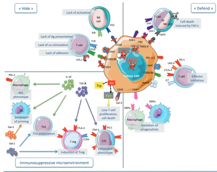
Two types of mechanisms may be responsible for the loss of these molecules: i) “hard lesions” which consist of irreversible genetic alterations of the gene of interest or genes implicated in their transcriptional regulation; and ii) “soft lesions” which are reversible epigenetic changes that repress gene expression9(Figure 1, "hide").
Prevention of antigen presentation
MHC-I loss/downregulation
Loss of MHC-I at the surface of lymphoma cells (total loss or miss-localization) occurs in 55-75% of diffuse large B-cell lymphoma (DLBCL)10,11 and 63% of Hodgkin lym-phomas (HL).11Most frequently, this results from mutations of the Beta2-microglobulin (β2M) gene which occurs in 29% of DLBCL,1050% of primary mediastinal B-cell lym-phoma (PMBCL),12and at least 50% of classical HL (cHL).13 In immune-privileged lymphomas, MHC-I loss was found in 18% of primary central nervous system lymphomas (PCNSL) but not in primary testicular lymphomas (PTL).11 In HL, MHC loss is preferentially observed in EBV-negative rather than in EBV-positive HL (83% vs. 27%).11 Patients whose Reed Stenberg cells (RS) are negative for MHC-I or β2M have a shorter progression-free survival (PFS).14 Interestingly, 9p24.1 amplification (leading to PD-L1 over-expression, as discussed below) adversely impacts survival only in HL patients in whom RS have lost MHC-I.15Loss of MHC-I is also observed in 30% of Burkitt lymphomas (BL) and 20% of follicular lymphoma (FL)16with rare β2M muta-tions.17 In FL, the frequency of β2M mutations is higher after histological transformation18and is associated with a lower infiltration of the tumor by CD8 T cells.19
Other irreversible mechanisms leading to MHC-I loss include alterations in MHC-I gene.16,20Unlike non-hemato-logic cancers, epigenetic mechanisms do not seem to be frequently responsible for MHC-I loss/downregulation in lymphoma.7
Importantly, natural killer (NK) cells are activated in the absence of MHC-I and in the presence of CD58 (which stimulates NK cells through CD2). Interestingly, 67% of DLBCL lack CD58 surface expression, and 61% lack both CD58 and MHC-I expression, thereby preventing NK-cell activation.10Of note, genetic alterations of CD58 are also found in transformed FL but not in FL.18 Genetic lesions disrupting the CD58 gene have been found only in 10-21% of DLBCLs, suggesting alternative mechanisms.10,21,22 MHC- II loss/downregulation
Transcriptional regulation
Expression of MHC-II is regulated, through epigenetic mechanisms. CREBBP regulates CIITA by catalyzing
H3K27 acetylation at its promoter/enhancer in normal GC B cells and lymphoma cell lines.23-25CREBBP may undergo loss-of-function mutation in the histone acetyl transferase domain. Thus, in FL and DLBCL, mutations of CREBBP prevent CIITA transcription, which in turn prevent MHC-II transcription.
HLA-DR expression is lost in 20% of DLBCL26 and is associated with a reduced T-cell infiltrate within the tumor27and a poor outcome.27,28Moreover, 19% of DLBCL have MHC-II intra-cytoplasmic aberrant localization which is associated with a worse outcome. This mislocal-ization is preferentially seen in BCL-2 and c-MYC double expresser lymphomas. Of note, c-MYC down-regulates enzymes implicated in the antigen presentation machinery (cf 2.1.3).29 The mechanisms of MHC-II downregulation remain incompletely understood but seem to occur at tran-scriptional level independently of genetic lesions on MHC-II gene.30Indeed, genes implicated in epigenetic regulation, including HMTs and HATs, are the most frequently altered genes in DLBCL (approx. 50% of GC-DLBCL and 30% of ABC-DLBCL).31Moreover, DLBCL frequently harbor inac-tivating mutations of CREBP (19% of all DLBCL, 31% of GC-DLBCL and 6% of ABC-DLBCL)12and CIITA (10% of DLBCL).12 CIITA is a target of somatic hypermutation (SHM) caused by AID.12 Finally, expression of CIITA and CREBP may be repressed through epigenetic silencing (i.e. independent of genetic alterations). Reduced expression of CIITA and CREBP is frequently found in DLBCL, leading to MHC-II downregulation and poor outcome.32-35In some cases, MHC-II may be restored by lifting the repression of CIITA with HDAC inhibitors.33MHC-II downregulation in DLBCL may also result from an overexpression of the tran-scription factor FOXP1 through a mechanism which, although not clearly elucidated, seems to be independent of CIITA.36FOXP1 expression is associated with the non-GC phenotype (48% of DLBCL vs. 71% of non- non-GC-DLBCL)37 and a poor prognosis.38 The underlying mecha-nisms responsible for FOXP1 overexpression remain large-ly unknown. Genetic alterations on chromosome 3p lead-ing to FOXP1 overexpression are found in a small subset of DLBCL.38FOXP1 translocations are found in 5% of DLBCL and are associated with extra-nodal localizations and high proliferative index.39Bea et al. also reported 15% of trisomy 3 and 31% of copy number gains of the chromosome 3p in ABC-DLBCL (versus 1% in GC-DLBCL), associated with MHC-II downregulation.40
In PMBCL, MHC-II downregulation also occurs at the transcriptional level and CIITA alterations is the most common mechanism:41CIITA breaks are found in 38-56% of PMBCL and correlate with poor outcome;12,42 CREBP mutations are present in 11% of cases12and abnormalities on chromosome 3 can be found, although rarely.40 However, loss of expression of MHC-II is found only in 12% of PMBCL.43This is associated with poor survival.43
In FL, there is no evidence for mutation in MHC-II genes17but CREBBP is mutated in 32-68% of cases17,34 and CIITA in 35%44suggesting a downregulation at the tran-scriptional level. Furthermore, CREBBP mutation is an early event and a driver mutation in FL development.45
In HL, lack of MHC-II on RS occurs in 41% of cases and represents an independent prognosis factor.46In 37.2% of cases, RS show aberrant localization in their cytoplasm.46 The mechanisms responsible for MHC-II loss in HL is not completely known but genomic CIITA break is found in 15% of HL42and FOXP1 is not implicated.47
Genetic alterations
Direct, genetic alterations leading to MHC-II loss are mostly seen in DLBCL of immune-privileged sites. PTL and PCNSL have lost HLA-DR in 61% and 46% of cases, respectively.48 In contrast with other types of DLBCL, genetic lesions of MHC-II genes represent the main mech-anism of HLA-DR loss:48,49 MHC-II is mutated in 78% of PTL and 50% of PCNSL.49Transcription factors seem to be rarely implicated in HLA-II loss in PTL: CIITA and FOXP1 rearrangements are present in only 10% and 7% of cases, respectively.50
It is noteworthy that, when expressed, MHC-II may drive inhibitory signals. Indeed, lymphocyte-activation gene 3 (LAG-3), a member of immunoglobulin superfami-ly expressed on tumor infiltrating superfami-lymphocytes (TILs),51 binds to MHC-II with greater affinity than CD4, leading
to the inhibition of TCR signaling, proliferation and cytokine secretion by antigen-specific T cells. Exhausted LAG-3 positive TILs are present in the immune infiltrate of FL, DLBCL and HL (mostly in EBV positive cases, mixed cellularity and rich lymphocyte subtypes).52,53 Furthermore, circulating CD4 T cells from HL patients with active disease express LAG-3 at higher levels than healthy donors or patients in long-term remission.53
Antigen processing machinery alterations
GILT and HLA-DM are enzymes of the antigen process-ing machinery (APM), located in the endocytic compart-ment of APC and B cells. Both are down-regulated by cMYC, leading to a defective antigen presentation that can be restored in vitro by cMYC inhibitors.54
GILT generates epitopes to be loaded on MHC-II. In
Figure 1. Lymphoma immune evasion mechanisms. (Top left panel) "Hide". Tumor cells may become “invisible” to the immune system by down-regulating MHC, co-stimulatory (CD80 and CD86) and/or adhesion (CD54) molecules. Downregulation of CD58 allows tumor cells to escape killing by natural killer (NK) cells, which are activated by self-missing signal (loss of MHC-I). (Right panel) "Defend". Tumor cells are seen by the immune system but avoid destruction through resistance to apop-tosis signals and/or expression of inhibitory receptors. Tumor cells may resist apopapop-tosis by different means: loss of FAS and/or TRAIL receptors (extrinsic pathway), hyperexpression of anti-apoptotic molecules such as BCL-2 (intrinsic pathway) or PI9 (Granzyme pathway). T cells can be inhibited by inhibitory ligands which are expressed by lymphoma cells or cells from their microenvironment such as PD-L1 or PD-L2/PD-1, LAG-3/MHC-II, CTLA-4/CD80 or CD86 and HLA-G/ILT. CD47 sends a “don’t eat me” signal to macrophages and DCs by interacting with its ligand SIRPa. Tumor cells may also express FAS-L to induce death of immune cells. Some mol-ecules expressed by lymphoma cells may have dual roles: expression of MHC-II allows antigen presentation but also binds to the inhibitory receptor LAG-3; CD80 and CD86 stimulate T cells through CD28 but may also inhibit T cells through CTLA-4. (Bottom left panel) Immunosuppressive microenvironment. The tumor cells interact with their microenvironment to contribute to lymphoma immune evasion. IL-10 is a potent immunosuppressive cytokine that inhibits priming by dendritic cells (DC), promotes Th2 and Treg differentiation and M2 macrophages; TGF-β induces exhausted phenotype of CTL and Treg differentiation; IDO suppresses cytotoxic T lympho-cyte (CTL) and NK immune response through degradation of tryptophan and production of kynurenine. Trp: tryptophan; Kyn: kynurenine; Gal: galectin; Ag: antigen.
DLBCL patients treated with CHOP or rituximab-CHOP, Phipps-Yonas et al. identified lower GILT expression as an adverse prognostic factor for OS.55 Once formatted by GILT, peptides are loaded on MHC-II instead of CLIP frag-ment of invariant chain. This exchange is performed by HLA-DM. In absence of HLA-DM, antigens cannot be exposed and MHC-II present CLIP at the cell surface.11 HLA-DM is lost in 49% of cHL, 14% of DLBCL, and 2.9% of PTL and PCNSL.11
Prevention of co-stimulation: B7 molecule
downregulation
CD80 and CD86 are members of the B7 co-stimulatory family and are expressed on professional APC, including B cells. They have a dual specificity: they can bind to the stimulatory receptor CD28 promoting T-cell activation and to the inhibitory receptor CTLA-4 (with a much high-er affinity than CD28) leading to T-cell inhibition.56
In B-cell lymphomas, CD80 and CD86 may be expressed on tumor cells and/or on cells from their microenvironment.57 CD80 is expressed in 97% of FL, 91% of marginal zone lymphomas (MZL), 90% of DLBCL, and 75% of mantle cell lymphomas (MCL).58 Interestingly, T and non-T cells present in the microenvi-ronment of these tumors also express CD80.58 Loss of CD86 was found to be associated with decreased TIL infiltration in DLBCL.59However, the prognostic value of CD80 and CD86 expression in lymphoma remains unclear, maybe because of their dual activity.
Prevention of adhesion
Intercellular adhesion molecule 1 (ICAM-1 or CD54) plays a crucial role in cell-to-cell interaction, especially in the immune synapse and tumor cell adhesion and dissem-ination.8 Lower expression of CD54 compromises the interaction between tumor and immune cells. In DLBCL, lymphocyte infiltration is decreased in tumors which have lost CD54.59 However, in aggressive NHL, lower expres-sion of CD54 correlates with more advanced stage of the disease, higher bone marrow infiltration and worse prog-nosis.60
Expression of CD54 is lost in 50%60 of non-Hodgkin lymphomas (NHL), but only 7% in DLBCL.59
How lymphoma may defend itself against the
immune system
Lymphoma cells may “defend” themselves to become resistant to immune eradication. This can be achieved in several ways: by becoming resistant to apoptosis and/or by expressing inhibitory ligands that deactivate immune cells (Figure 1, "defend").
Resistance to apoptosis
Three apoptopic pathways may induce cell death: i) the perforin/granzyme pathway which results from the release of cytotoxic granules from NK cells or CTL activat-ed through their TCR; ii) the extrinsic pathway, activatactivat-ed by T and NK cells through FAS or TRAIL death receptors; iii) the intrinsic pathway, involving BCL-2 family proteins and activated by intrinsic stress signals.61
By apoptopic gene profiling, Muris et al. identified two subsets of DLBCL with poor overall survival.62The acti-vated apoptosis cascade group (mostly ABC-DLBCL) was
characterized by high expression level of many pro- and anti-apoptotic genes of the intrinsic pathway, suggesting that these lymphoma cells are “primed for death” and their survival depends on the high expression level of anti-apoptotic genes. The cellular cytotoxic response group was characterized by the expression of apoptosis-induc-ing effector molecules from CTL and NK cells (granzyme, TRAIL, FASL and other) and a high resistance to chemotherapy.63The large immune cell infiltration in this subset suggests a selection of resistant lymphoma cells under the pressure of a strong cellular immune response. Inhibition of granzyme
The protease inhibitor 9 (PI9) was found to inhibit granzyme B and therefore to protect against apoptosis.64 PI9 is expressed in DLBCL, BL and HL (in RS), but is seems to be rarely found in low-grade lymphomas.57 Of note, few studies have analyzed PI9 expression in B-cell lym-phomas and there is no evidence of relationship between PI9 expression and CTL infiltration or clinical outcome.65
To our knowledge, there is no mechanism of perforin inhibition in lymphoma.
Inactivation of death receptor extrinsic pathway: FAS/TRAIL-R FAS (CD95) belongs to the TNF receptor family and lig-ation of FASL (CD95L) induces apoptosis through its intra-cellular death domain and caspase activation. This mech-anism plays a crucial role in affinity selection during the GC reaction.66 Immune cells also use this mechanism to kill cancer cells.67
In normal B cells, FAS is expressed on activated B cells from the GC and is absent in mantle zone or circulating B cells. CD95 is lost in 17% of FL68and 27% of MALT lym-phomas.69In DLBCL, CD95 is lost in 51% of extra-nodal cases69but rarely in cutaneous cases.70CD95 expression on lymphoma cells is associated with improved survival and response to R-CHOP therapy in DLBCL.69-72In HL, CD95 is rarely lost.73
Mutations in the CD95 gene are more commonly found in post-GC lymphomas, including 20% of DLBCL, and 44% of extra-nodal lymphomas (all types).74,75Surprisingly, although derived from GC, no mutation of CD95 were found in BL.75CD95 mutations are rare in FL (6%) and in pre-GC lymphomas (<2%) such as MCL.74,75Only 5% of HL are associated with FAS mutation in RS.73Müschen et al. hypothesized that FAS mutations are mostly found in post-GC lymphomas because CD95 mutations are target errors in the SHM process during the GC reaction.74 However, FAS mutations do not share features of AID-mediated activity and their underlying mechanism remains unclear. In some cases, lymphoma cells express-ing CD95 are resistant to apoptosis, suggestexpress-ing the exis-tence of other mechanisms. For instance, HL resist to FAS-induced apoptosis by expressing c-FLIP which is located at the cell membrane where it binds to the death domain of CD95.73High levels of soluble CD95 are associated with poor outcome,76-78supposedly because it binds to CD95L and prevents apoptosis. As discussed below, Galectin 3 also protects tumor cells from FAS-induced death.
TRAIL is also a member of TNF receptor family, which triggers the extrinsic apoptotic pathway after ligation to death receptors (TRAIL receptors 1 and 2). The role of TRAIL in B-cell lymphomagenesis has been suggested by the association between TRAIL polymorphisms and high-er risk of lymphoma79and the rapid development of
spon-taneous lymphoid malignancies in mice with TRAIL defi-ciency.80 Loss of TRAIL receptor was found in 6.8% of NHL.81 It is mainly caused by mutations of TRAIL death domain on chromosome 8p21.3 but may also occur at the transcriptional level by mutation of p53.82 Mutations of TRAIL receptor are found in 26% of MCL (55% of leukemic MCL vs. 19% of nodal MCL) and have a more aggressive phenotype.83
Inhibition of the stress-induced intrinsic pathway: BCL-2 overex-pression
BCL-2 family molecules are crucial regulators of the intrinsic pathway of mitochondrial apoptosis.84 BCL-2 itself is an anti-apoptotic protein but other members of the BCL-2 family are pro-apoptotic.
BCL-2 is one of most commonly mutated genes in NHL, notably in DLBCL (37% of cases, particularly in GC sub-type) and FL (54% of cases),85-87whereas it is a rare event in peripheral T-cell lymphomas, MCL and PMBL.86
The t(14;18), present in almost all FL45and 34% of GC-DLBCL88 (vs. 17% of non-GC DLBCL), juxtaposes the BCL-2 gene and the enhancer of the heavy chain immunoglobulin. Thus, it induces a constitutive overex-pression of BCL-2 and exposes BCL-2 oncogene to somat-ic hyper-mutations in the GC.84Other mechanisms may explain genetic variations of the BCL-2 gene in t(14;18) negative DLBCL.84
In DLBCL, BCL-2 expression (but not mutation nor translocation) were historically associated with a worse prognosis but this negative impact seems to be overcome by the addition of rituximab to CHOP chemotherapy.86,89,90 Nevertheless, BCL-2 protein expression remains the strongest independent prognostic factor in primary cuta-neous DLBCL.91In FL, Correia et al. found that the pres-ence of BCL-2 mutation at diagnosis was an independent risk factor of transformation and death, but patients were mostly treated without rituximab.92This observation was not confirmed in another study in which FL patients were treated with a rituximab-containing regimen.87
Inhibition / killing of immune cells
PD-L1/L2 expression
PD-L1 and PD-L2 are members of the CD28 family and inhibit T cells through ligation to PD-1 receptor.56Most FL contain a rich immune infiltrate of PD1+cells, mostly in the inter-follicular areas, but tumor cells do not express PD-L1 (PD-L2 is weakly expressed in some rare tumor cells).52In contrast, DLBCL often express PD-L1 and PD-L2 on tumor cells and in their microenvironment.52PD-L1 and PD-L2 are more frequently expressed on tumor cells of ABC-DLBCL (36% and 60%, respectively) than GC-DLBCL (4% and 26%, respectively).93 PD-L1 is also fre-quently expressed on tumor cells of PMBL (71% of cases)94 and HL (97% of cases).14 In immune-privileged lym-phomas, level of PD-L1 protein expression is unknown in PTL and reported in a small study of PCNS lymphomas.95 The mechanisms responsible for PD-L1 and/or PD-L2 overexpression include: i) genetic alteration in 9p24; and ii) Epstein-Barr virus (EBV) infection. In the first case, the 9p24 amplicon contains the PD-L1 and PD-L2 genes that are directly amplified and over-expressed. It also contains the JAK2 gene that, indirectly, induces the transcription of
the PD-L1 and PD-L2 genes. 9p24 alterations are found in all cases of HL,14in most cases of PMBL (9p24 amplifica-tion in 63% of cases and translocaamplifica-tion in 20% of cases),96,97 in 54% of PTL, and 52% of PCNSL (mainly due to copy number gain, whereas translocations are rare),98 and in 19% of DLBCL (mainly due to copy number gains) partic-ularly in the non-GC subset.99 Structural variations dis-rupting the 3’ region of the PD-L1 gene are also implicated in 8% of DLBCL.100Notably, immunoglobulin locus and CIITA are common partners of PD-L1 translocation.42,98,99 Finally, EBV infection (which is present in approx. 40% of HL tumors) also induces PD-L1 expression via the viral protein LMP1.101
PD-L1 expression in the tumor is an adverse prognostic factor for HL,14 PMBL,94 and DLBCL.93 Soluble PD-L1, although not correlated with PD-L1 expression by the tumor, is also associated with a poor prognosis in DLBCL.102,103In these studies, high level of PD-L1 was asso-ciated with the clinical and histological aggressiveness of the disease.14,52,93,102
HLA-G expression
HLA-G is a non-classical MHC-I molecule transcribed in membrane-bound or soluble (sHLA-G) isoforms. HLA-G binds to the inhibitory receptors ILT2 (on lymphoid cells, including B cells, and myeloid cells) and ILT4 (on myeloid cells). HLA-G also binds to CD8 co-receptor and induces FAS-mediated apoptosis of T and NK cells.104
HLA-G is expressed in 24% of DLBCL105and 67% of cHL (on RS) at a higher level than healthy controls.73,106In HL, HLA-G expression is associated with the loss of MHC-I on RS and the absence of EBV.107
sHLA-G is increased in lymphoproliferative disorders and contributes to immune escape.108,109 Indeed, sHLA-G purified from plasma of patients with lymphoproliferative disorders inhibits T-cell proliferation in vitro.108However, there is no correlation between the level of sHLA-G and clinical or pathological characteristics of the disease108or its prognosis.110
Thus, HLA-G may have ambivalent effects in lym-phoma: on one hand, sHLA-G may inhibit the prolifera-tion of tumor B cells through ILT2 receptor whereas, on the other hand, HLA-G expressed in the tumor may pro-mote immune escape by inhibiting NK and CTL.104 CD47 expression
CD47, the expression of which is ubiquitous, interacts with the inhibitory receptor SIRPα expressed by myeloid cells and macrophages. CD47-SIRPα interaction delivers a “don’t eat me” signal to the phagocytic cells which prevents phagocytosis.111Thus, CD47 may lead to immune evasion in two ways: i) by inhibiting phagocytosis;112,113and ii) by inhibiting cross-presentation by dendritic cells (DC).114
In NHL, CD47 is expressed at a higher level on tumor B cells compared to normal B cells.112 Additionally, CD47 expression is increased on lymphoma cells circulating in the blood compared to lymphoma cells in lymph nodes supporting the role of CD47 in lymphoma dissemina-tion.113Finally, high expression of CD47 is associated with poor prognosis in DLBCL and MCL.112
FASL expression
Tumor cells may also “counter-attack” immune effector cells by expressing FASL in order to kill them.115FASL was found to be strongly expressed in aggressive B-cell
lym-HL: Hodgkin lymphoma; BL: Burkitt lymphoma; DLBCL: diffuse large B-cell lymphoma; PMBL: primary mediastinal B-cell lymphoma; PTL: primary testicular lymphoma; PCNS: primary central nerv-ous system lymphoma; MCL: mantle cell lymphoma; FL: follicular lymphoma; MZL: marginal zone lymphoma; MALT: mucosal associated lymphoid tissue; PTCL-NOS: primary T-cell lymphoma not otherwise specified; AITL: angio-immunoblastic T-cell lymphoma; ALCL: anaplastic large cell lymphoma; CTCL: cutaneous T-cell lymphoma; MF: mycosis fungoid; SS: Sezary syndrome; ATLL: acute T-cell lymphoma/leukemia; ENKTL: extranodal NK/T lymphoma; MDSC: myeloid-derived suppressor cell; TAM: tumor associated macrophage; APM: antigen processing machinery. °Refers to GILT. °°Refers to HLA-DM. *Refers to mycosis fungoid. #Refers to Sezary syndrome.
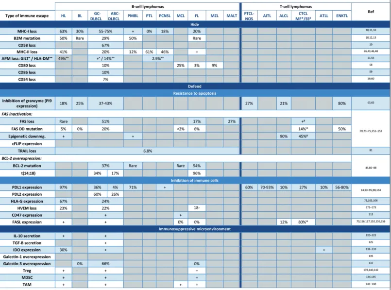
Table 1.Overview of lymphoma immune escape mechanisms. The respective contribution of each immune escape mechanism according to lymphoma subtype.
phomas,116 secondary cutaneous DLBCL, primary cuta-neous leg-type DLBCL,70and HL,117but seems to be weak in non-aggressive lymphomas (such as small lymphocytic lymphoma, lymphoplasmacytic lymphoma, and grade 1 FL) and MCL.116In DLBCL, FASL expression is an adverse prognostic marker.69-72
Immunosuppressive microenvironment
Lymphoma cells may evade immune eradication by inducing an immunosuppressive (humoral and cellular) microenvironment. Interactions between the lymphoma cells and their microenvironment have been reviewed in detail by Scott and Gascoyne.118Here, we highlight the main immunosuppressive components present in the lym-phoma microenvironment (Figure 1, "immunosuppressive microenvironment").
Cytokines
IL-10 secretion
IL-10 is an immunosuppressive cytokine which inhibits myeloid effector cells and priming functions of DC, pro-motes Th2 immune responses, induces Treg, and stimu-lates growth and differentiation of B cells.119Thus, IL-10 may promote lymphoma in two ways: i) by stimulating the growth of tumor B cells; ii) by inducing an immuno-suppressive environment. IL-10 serum level is higher in lymphoma patients than in healthy subjects and is associ-ated with poor prognosis.120,121Moreover, high levels of IL-10 before treatment is associated with treatment failure and a worse outcome.120-122
TGF-β secretion
immuno-suppressive environment in several ways: i) it induces an exhausted phenotype in CTL (mostly on memory T cells) with a high PD-1 and TIM-3 expression;123ii) it leads to FOXP3 expression, mostly in naïve T CD4+ cells123 and induces the differentiation of Treg; and iii) represses the expression of CD95, perforin, granzyme and cytokines.124 Because TGF-β suppresses lymphoma growth by inhibit-ing proliferation and apoptosis, lymphoma cells may first acquire resistance or aberrant response to TGF-β.124This may be achieved by several mechanisms including down-regulation of TGF-β receptor on lymphoma cells125 through epigenetic mechanisms,126abnormal signal trans-duction127and expression of CD109, a negative regulator of TGF-β signaling.128 Thus, there is no clear prognostic impact of TGF-β in lymphoma.
IDO expression
IDO is an enzyme, expressed by lymphoma cells and cells from the microenvironment, which suppresses CTL and NK immune responses and induces Treg through
degradation of tryptophan. The most important metabo-lite of tryptophan is kynurenine which inhibits antigen specific proliferation and induces T-cell death.129
IDO protein is expressed in stromal cells of HL130and approximately 30% of NHL express IDO, and intra-tumoral levels are significantly higher than in reactive lymph nodes.131-133In DLBCL131-133and HL,130,134IDO activity is associated with a more aggressive disease and a worse outcome. Upregulation of IDO is associated with Treg infiltration in both DLBCL and HL.130,133
Galectins expression
Galectins (Gal) are key regulators of inflammation. These molecules act in the extra-cellular milieu by inter-acting with glycosylated receptors and, at the intra-cellular level, by modulating signalization and splicing.135 Among the 15 different galectins identified, types 1 and 3 have been implicated in lymphoma immune escape. Gal-1 is known to suppress Th1 responses and promote secretion of Th2 cytokines and expansion of Treg. Gal-1 is
over-Treg: regulatory T cells; mAb: monoclonal antibody; DC: dendritic cell. *No result available in lymphoma patients. #Pre-clinical data.
expressed in EBV-associated lymphoma cells and is associ-ated with an increased secretion of Th2 cytokines and infiltration by Tregs.135
Gal-3 can positively or negatively regulate T-cell sur-vival, cytokine profiles and DC function. Gal-3 protects tumor cells from death induced by FAS,136 possibly through interaction with CD45.137Gal-3 is over-expressed in 66% of DLBCL136(but not in BL nor in FL).
Cells
Regulatory T cells
Tregs, which are characterized by the expression of CD4, FOXP3 and CTLA-4, are responsible for the preven-tion of autoimmunity.138 Tregs suppress immune cells through direct contact-dependent mechanisms, including induction of effector cell death, and indirect mechanisms by secreting inhibitory cytokines (IL-10, TGF-β) or inter-fering with effector T-cell metabolism.138
Tregs are more numerous in lymphoma tumors than in reactive lymph nodes139 and in the blood of lymphoma patients compared to healthy controls or cured patients.139,140Tregs are recruited by CCR4 ligands (notably in cutaneous DLBCL, HL and EBV-associated lym-phomas141) or converted from a conventional into a regula-tory phenotype within the tumor microenvironment by modulation of tryptophan catabolism. Interestingly, Liu et al. demonstrated that Tregs found within the tumor microenvironment of FL are highly clonal.142In this study, the diversity of Treg TCR repertoire inversely correlated with the TCR repertoire of CD8 T cells, suggesting an antigen-specific suppression of CTL by Tregs. High level of circulating Tregs at diagnosis is an adverse prognostic factor in DLBCL and correlates with elevated LDH, advanced stage of the disease,139and poor survival.138,143 Myeloid-derived suppressor cells
Myeloid-derived suppressor cells (MDSC) were recently described and remain poorly characterized. While their immunosuppressive properties are well established, only few mechanisms have been explored in lymphoma.144 Immunosuppressive functions of MDSC include: i) secre-tion of immunomodulatory factors and Treg expansion; ii) modulation of amino-acid metabolism and decrease of T-cell proliferation; iii) oxidative stress; iv) inhibition of T- or NK-cell viability and homing into the lymph nodes; and v) induction of T-cell apoptosis. In B-cell lymphoma, MDSC are involved in T-cell defect through PDL-1 expression, IL-10 secretion, Treg expansion, and modulation of amino-acid metabolism.144MDSC are increased in various B-cell lymphomas (including HL, DLBCL, FL) and correlate with poor prognosis.144,145
Macrophages
Macrophages are divided into M1 (pro-inflammatory, CD163-) and M2 (anti-inflammatory, CD163+) subsets. M2 macrophages are recruited into the tumor or differenced in situ (notably by IL-10) and promote tumor progression.146
In HL, a meta-analysis of 22 studies showed that a high density of CD68+/CD163+ macrophages was associated with poor survival.147 In DLBCL146 and MCL,148 CD163+ macrophages correlates with poor clinical outcome. In FL, a high density of CD68+ macrophages was associated with a poor prognosis in the pre-rituximab era while it was associated with a good prognosis in the
post-ritux-imab era.146This may be due to the antitumor activity of macrophages through phagocytosis of rituximab-coated tumor B cells.149 This observation was further supported by the GELA-GOELAMS study showing that macrophages were associated with adverse outcome only in patients treated without rituximab while there was no difference in survival in patients treated with rituximab.150 Finally, macrophages may also promote immune evasion by expression of PDL-1.146
Immune escape mechanisms in T-cell lymphomas
Mechanisms of immune evasion in T-cell lymphomas are less well characterized. Best described mechanisms result from resistance to apoptosis and from PD-L1 expression.PI9 granzyme inhibitor is expressed in 21% of anaplas-tic large cell lymphoma (ALCL), 27% of peripheral T-cell lymphoma not otherwise specified (PTCL-NOS), 80% of NK-/T-cell nasal type lymphoma (ENKTL), and 89% of enteropathy-type NHL.63 A defect in the extrinsic apopto-sis (i.e. FAS) pathway is observed in many T-cell lym-phomas which may be caused by three distinct mecha-nisms: i) FAS mutations, which are present in 50% of ENKTL151and in some cases of MF (<20% of cases);152ii) decreased expression of FAS through epigenetic mecha-nisms such as promoter methylation (45% of Sezary Syndrome) or splicing (43% of MF, 50% of CD30-CTCL);152 iii) expression of c-FLIP inhibitory protein, which is seen in 90% of ALCL153(although the underlying mechanism is not completely elucidated).
Both PD1 and PD-L1 may be expressed in T-cell lym-phomas, both on tumor cells and in their microenviron-ment. PD-L1 is expressed on tumor cells in less than 10% of ALCL and adult T-cell lymphoma / leukemia (ATLL), 27% of cutaneous T-cell lymphoma (CTCL), approxi-mately 60% of PTCL-NOS, 56-80% of ENKTL and 70-93% of angio-immunoblastic T-cell lymphoma (AITL).154 In both ALK negative and positive ALCL, and in CTCL, PD-L1 overexpression occurs through the STAT3 path-way.154Like in B-cell lymphomas, structural variations dis-rupting the 3’ region of the PD-L1 gene (27% of ATLL) and EBV infection (particularly in ENKTL) are also responsible for PDL-1 expression.
FAS-L is expressed in 12% of ALCL,15381% of mycosis fungoid (MF),155and a majority of CTCL156which may lead to the elimination of CTL (through FAS-induced death) and to a worse outcome.155,156
Finally, IDO may also contribute to immune escape in ATLL and is associated with a worse outcome.157
Implications for immunotherapy
Restoring antigen recognition
When tumor cells hide from the immune system by pre-venting Ag presentation, strategies to circumvent this escape mechanism depend on the type of lesions (Table 1). If antigen presentation deficiency results from genetic irreversible lesions, then immunotherapies that are MHC-independent may bypass the lack of antigen presentation. This can be achieved with bi-specific T-cell engager anti-bodies (BiTE) or CAR T cells which target surface antigens
without the need for MHC.158,159
If antigen presentation deficiency results from epigenet-ic reversible lesions, then one may use therapies whepigenet-ich can induce re-expression of MHC, co-stimulatory or adhe-sion molecules, such as epigenetic drugs, chemotherapy, radiotherapy or certain immunotherapies (e.g. CD40 ago-nists, CpG, IFN).7,160 Notably, the addition of histone deacetylase inhibitor (HDACI) to R-CHOP restored MHC-II expression161and erased the negative prognostic value associated with MHC-II loss in DLBLC.162
Restoring cell death
BCL-2 inhibitors, such as venetoclax, may sensitize tumor cells to death induced through the intrinsic path-way. They have a strong efficacy in CLL and, to a lesser extent, in some NHL (MCL, FL, DLBCL).163 Surprisingly, despite the pathophysiological importance of BCL-2 translocation in FL, venetoclax demonstrated only poor efficacy in this disease.
In pre-clinical models, Gal-3 inhibitor can disturb CD45/Gal-3 interaction and restore apoptosis.137
Blocking inhibitory signals
Immune checkpoint (ICP) blockade releases inhibition of effector cells but requires an intact antigen presentation and a pre-existing anti-tumor immune response. Blockade of CTLA4, PD1 and PD-L1 have demonstrated efficacy in solid tumors and hematologic malignancies.158Surprisingly, anti-PD1 mAbs were found to be particularly efficient in HL despite the fact that MHC expression was lost in most cases, suggesting an alternative mechanism of action.
Phagocytosis may be blocked by CD47 signaling. Blocking antibodies against CD47 or SIRPα can disrupt CD47-SIRPα interaction and restore phagocytosis. Blocking CD47 signaling may also potentiate the efficacy of anti-CD20 mAb by increasing antibody-dependent cel-lular phagocytosis (ADCP).112-114
Modulating the tumor microenvironment
Immunosuppressive macrophages may be depleted by chemotherapy164 or anti-CSF-1 receptor mAb.165 Treg
depletion may be achieved with anti-CTLA4 mAbs (such as ipilimumab)166,167or mAbs against CCR-4 (such as moga-mulizumab) which is preferentially expressed by Th2 and Tregs.141,168Treg infiltration may also be decreased by low doses of cyclophosphamide through downregulation of FOXP3.160 IDO enzyme may be down-regulated using IDO inhibitors or fludarabine.169,170
Conclusion
The recent success of ICP blocking antibodies in cancer patients confirmed the hypothesis of “cancer immuno-surveillance” and demonstrated the potency of immunotherapy for the treatment of cancer. The goal of immunotherapy is to re-educate the immune system and to reverse the immune escape mechanisms to destroy the tumor cells.
B-cell lymphoma is unique because tumor cells are pro-fessional APC and therefore can present their own anti-gens to the immune system. Immune escape in lymphoma may occur at the priming or at the effector phase. It may result from defects in antigen presentation (which may prevent the priming of T cells or the recognition of tumor cells at the effector phase), from resistance to immune killing, or from immunosuppressive mechanisms (either directly by the tumor cells or indirectly by their microen-vironment).
The advent of new classes of immunotherapies (includ-ing checkpoint inhibitors, bispecific antibodies and CAR T cells) offers novel opportunities to mobilize the immune system against lymphoma.159However, we need to deter-mine which of these immunotherapies will be optimal for a given patient. Furthermore, some immune escape mech-anisms may dampen the efficacy of these immunothera-pies and may require combination with other theraimmunothera-pies to sensitize tumor cells to immune eradication. The charac-terization of immune escape mechanisms may be used to guide “personalized immunotherapy”, i.e. determine the optimal immunotherapy and/or combination in a given lymphoma patient.
References
1. Burnet M. Cancer: a biological approach. III. Viruses associated with neoplastic condi-tions. IV. Practical applicacondi-tions. Br Med J. 1957;1(5023):841-847.
2. Dunn GP, Bruce AT, Ikeda H, Old LJ, Schreiber RD. Cancer immunoediting: from immunosurveillance to tumor escape. Nat Immunol. 2002;3(11):991-998.
3. Hanahan D, Weinberg RA. Hallmarks of Cancer: The Next Generation. Cell. 2011;144(5):646-674.
4. Chen DS, Mellman I. Oncology Meets Immunology: The Cancer-Immunity Cycle. Immunity. 2013;39(1):1-10.
5. Goodnow CC, Sprent J, de St Groth BF, Vinuesa CG. Cellular and genetic mecha-nisms of self tolerance and autoimmunity. Nature. 2005;435(7042):590-597.
6. Heath WR, Carbone FR. Cross-Presentation in Viral Immunity and Sefl-Tolerance. Nat Rev Immunol. 2001;1(2):126-134.
7. de Charette M, Marabelle A, Houot R.
Turning tumour cells into antigen presenting cells: The next step to improve cancer immunotherapy? Eur J Cancer. 2016;68134-68147.
8. Dustin ML. The Immunological Synapse. Cancer Immunol Res. 2014;2(11):1023-1033. 9. Garrido F, Cabrera T, Aptsiauri N. “Hard” and “soft” lesions underlying the HLA class I alterations in cancer cells: Implications for immunotherapy. Int J Cancer. 2010;127249-127256.
10. Challa-Malladi M, Lieu YK, Califano O, et al. Combined Genetic Inactivation of 2-Microglobulin and CD58 Reveals Frequent Escape from Immune Recognition in Diffuse Large B Cell Lymphoma. Cancer Cell. 2011;20(6):728-740.
11. Nijland M, Veenstra RN, Visser L, et al. HLA dependent immune escape mechanisms in B-cell lymphomas: Implications for immune checkpoint inhibitor therapy? OncoImmunology. 2017;6(4):e1295202. 12. Dubois S, Viailly P-J, Mareschal S, et al.
Next-Generation Sequencing in Diffuse Large B-Cell Lymphoma Highlights
Molecular Divergence and Therapeutic Opportunities: a LYSA Study. Clin Cancer Res. 2016;22(12):2919-2928.
13. Reichel J, Chadburn A, Rubinstein PG, et al. Flow sorting and exome sequencing reveal the oncogenome of primary Hodgkin and Reed-Sternberg cells. Blood. 2015;125(7): 1061-1072.
14. Roemer MGM, Advani RH, Ligon AH, et al. PD-L1 and PD-L2 Genetic Alterations Define Classical Hodgkin Lymphoma and Predict Outcome. J Clin Oncol. 2016;34(23):2690-2697.
15. Roemer MGM, Advani RH, Redd RA, et al. Classical Hodgkin Lymphoma with Reduced B2M/MHC Class I Expression Is Associated with Inferior Outcome Independent of 9p24.1 Status. Cancer Immunol Res. 2016;4(11):910-916. 16. Fangazio M, Dominguez-Sola D, Tabbò F, et
al. Genetic Mechanisms of Immune Escape in Diffuse Large B Cell Lymphoma. Blood. 2014;124(21):1692.
17. Green MR, Kihira S, Liu CL, et al. Mutations in early follicular lymphoma progenitors are
associated with suppressed antigen presen-tation. Proc Natl Acad Sci USA. 2015;112 (10): E1116-E1125.
18. Pasqualucci L, Khiabanian H, Fangazio M, et al. Genetics of Follicular Lymphoma Transformation. Cell Rep. 2014;6(1):130-140. 19. Kridel R, Chan FC, Mottok A, et al. Histological Transformation and Progression in Follicular Lymphoma: A Clonal Evolution Study. PLOS Med. 2016;13(12):e1002197. 20. Drenou B, Tilanus M, Semana G, et al. Loss
of heterozygosity, a frequent but a non-exclusive mechanism responsible for HLA dysregulation in non-Hodgkin’s lym-phomas. Br J Haematol. 2004;127(1):40-49. 21. Lohr JG, Stojanov P, Lawrence MS, et al.
Discovery and prioritization of somatic mutations in diffuse large B-cell lymphoma (DLBCL) by whole-exome sequencing. Proc Natl Acad Sci USA. 2012;109(10):3879-3884. 22. Cao Y, Zhu T, Zhang P, et al. Mutations or copy number losses of CD58 and TP53 genes in diffuse large B cell lymphoma are independent unfavorable prognostic factors. Oncotarget. 2016;7(50):83294–83307. 23. Jiang Y, Ortega-Molina A, Geng H, et al.
CREBBP Inactivation Promotes the Development of HDAC3-Dependent Lymphomas. Cancer Discov. 2017;7(1):38-53.
24. Zhang J, Vlasevska S, Wells VA, et al. The CREBBP Acetyltransferase Is a Haploinsufficient Tumor Suppressor in B-cell Lymphoma. Cancer Discov. 2017;7(3):322-337.
25. Hashwah H, Schmid CA, Kasser S, et al. Inactivation of CREBBP expands the germi-nal center B cell compartment, down-regu-lates MHCII expression and promotes DLBCL growth. Proc Natl Acad Sci USA. 2017;114(36):9701-9706.
26. Tada K, Maeshima AM, Hiraoka N, et al. Prognostic significance of HLA class I and II expression in patients with diffuse large B cell lymphoma treated with standard chemoimmunotherapy. Cancer Immunol Immunother. 2016;65(10):1213-1222. 27. Rimsza LM. Loss of MHC class II gene and
protein expression in diffuse large B-cell lymphoma is related to decreased tumor immunosurveillance and poor patient sur-vival regardless of other prognostic factors: a follow-up study from the Leukemia and Lymphoma Molecular Profiling Project. Blood. 2004;103(11):4251-4258.
28. Rosenwald A, Wright G, Chan WC, et al. The use of molecular profiling to predict sur-vival after chemotherapy for diffuse large-B-cell lymphoma. N Engl J Med. 2002;346(25):1937-1947.
29. Kendrick S, Rimsza LM, Scott DW, et al. Aberrant cytoplasmic expression of MHCII confers worse progression free survival in diffuse large B-cell lymphoma. Virchows Arch. 2017;470(1):113-117.
30. Rimsza LM. Loss of major histocompatibili-ty class II expression in non-immune-privi-leged site diffuse large B-cell lymphoma is highly coordinated and not due to chromo-somal deletions. Blood. 2005;107(3):1101-1107.
31. Pasqualucci L, Trifonov V, Fabbri G, et al. Analysis of the coding genome of diffuse large B-cell lymphoma. Nat Genet. 2011;43(9):830-837.
32. Cycon KA, Rimsza LM, Murphy SP. Alterations in CIITA constitute a common mechanism accounting for downregulation of MHC class II expression in diffuse large B-cell lymphoma (DLBCL). Exp Hematol.
2009;37(2):184-194.e2.
33. Cycon KA, Mulvaney K, Rimsza LM, Persky D, Murphy SP. Histone deacetylase inhibitors activate CIITA and MHC class II antigen expression in diffuse large B-cell lymphoma. Immunology. 2013;140(2):259-272.
34. Pasqualucci L, Dominguez-Sola D, Chiarenza A, et al. Inactivating mutations of acetyltransferase genes in B-cell lymphoma. Nature. 2011;471(7337):189-195.
35. Autio M, Jäntti K, Cervera A, Hautaniemi S, Leppä S. Low Expression of the CIITA Gene Predicts Poor Outcome in Diffuse Large B-Cell Lymphoma. Blood 2016;128(22):2948. 36. Brown PJ, Wong KK, Felce SL, et al. FOXP1
suppresses immune response signatures and MHC class II expression in activated B-cell-like diffuse large B-cell lymphomas. Leukemia. 2016;30(3):605-616.
37. Hans CP. Confirmation of the molecular classification of diffuse large B-cell lym-phoma by immunohistochemistry using a tissue microarray. Blood. 2004;103(1):275-282.
38. Koon HB, Ippolito GC, Banham AH, Tucker PW. FOXP1: a potential therapeutic target in cancer. Expert Opin Ther Targets. 2007;11(7):955-965.
39. Haralambieva E, Adam P, Ventura R, et al. Genetic rearrangement of FOXP1 is pre-dominantly detected in a subset of diffuse large B-cell lymphomas with extranodal presentation. Leukemia. 2006;20(7):1300-1303.
40. Bea S. Diffuse large B-cell lymphoma sub-groups have distinct genetic profiles that influence tumor biology and improve gene-expression-based survival prediction. Blood. 2005;106(9):3183-3190.
41. Mottok A, Woolcock B, Chan FC, et al. Genomic Alterations in CIITA Are Frequent in Primary Mediastinal Large B Cell Lymphoma and Are Associated with Diminished MHC Class II Expression. Cell Rep. 2015;13(7):1418-1431.
42. Steidl C, Shah SP, Woolcock BW, et al. MHC class II transactivator CIITA is a recurrent gene fusion partner in lymphoid cancers. Nature. 2011;471(7338):377-381.
43. Roberts RA. Loss of major histocompatibili-ty class II gene and protein expression in pri-mary mediastinal large B-cell lymphoma is highly coordinated and related to poor patient survival. Blood. 2006;108(1):311-318. 44. Loeffler M, Kreuz M, et al; on behalf of the HaematoSys-Project. Genomic and epige-nomic co-evolution in follicular lymphomas. Leukemia. 2015;29(2):456-463.
45. Green MR, Gentles AJ, Nair RV, et al. Hierarchy in somatic mutations arising dur-ing genomic evolution and progression of follicular lymphoma. Blood. 2013;121(9): 1604-1611.
46. Diepstra A, van Imhoff GW, Karim-Kos HE, et al. HLA Class II Expression by Hodgkin Reed-Sternberg Cells Is an Independent Prognostic Factor in Classical Hodgkin’s Lymphoma. J Clin Oncol. 2007;25(21):3101-3108.
47. Brown P, Marafioti T, Kusec R, Banham AH. The FOXP1 Transcription Factor is Expressed in the Majority of Follicular Lymphomas but is Rarely Expressed in Classical and Lymphocyte Predominant Hodgkin’s Lymphoma. J Mol Histol. 2005;36(4):249-256.
48. Riemersma SA, Jordanova ES, Schop RF, et al. Extensive genetic alterations of the HLA region, including homozygous deletions of
HLA class II genes in B-cell lymphomas aris-ing in immune-privileged sites. Blood. 2000;96(10):3569-3577.
49. Mottok A, Steidl C. Genomic alterations underlying immune privilege in malignant lymphomas. Curr Opin Hematol. 2015;22(4):343-354.
50. Twa DD, Mottok A, Chan FC, et al. Recurrent genomic rearrangements in pri-mary testicular lymphoma: Genomic rearrangements in primary testicular lym-phoma. J Pathol. 2015;236(2):136-141. 51. He Y, Rivard CJ, Rozeboom L, et al.
Lymphocyte-activation gene-3, an impor-tant immune checkpoint in cancer. Cancer Sci. 2016;107(9):1193-1197.
52. Laurent C, Charmpi K, Gravelle P, et al. Several immune escape patterns in non-Hodgkin’s lymphomas. OncoImmunology. 2015;4(8):e1026530.
53. Gandhi MK. Expression of LAG-3 by tumor-infiltrating lymphocytes is coincident with the suppression of latent membrane anti-gen-specific CD8+ T-cell function in Hodgkin lymphoma patients. Blood. 2006;108(7):2280-2289.
54. God JM, Cameron C, Figueroa J, et al. Elevation of c-MYC Disrupts HLA Class II– Mediated Immune Recognition of Human B Cell Tumors. J Immunol. 2015;194(4):1434-1445.
55. Phipps-Yonas H, Cui H, Sebastiao N, et al. Low GILT Expression is Associated with Poor Patient Survival in Diffuse Large B-Cell Lymphoma. Front Immunol. 2013;4:425. 56. Sharpe AH, Freeman GJ. THE B7–CD28
SUPERFAMILY. Nat Rev Immunol. 2002; 2(2):116-126.
57. Greaves P, Gribben JG. The role of B7 family molecules in hematologic malignancy. Blood. 2013;121(5):734-744.
58. Dakappagari N, Ho SN, Gascoyne RD, Ranuio J, Weng AP, Tangri S. CD80 (B7.1) is expressed on both malignant B cells and nonmalignant stromal cells in non-Hodgkin lymphoma. Cytometry B Clin Cytom. 2012;82B(2):112-119.
59. Stopeck AT, Gessner A, Miller TP, et al. Loss of B7. 2 (CD86) and intracellular adhesion molecule 1 (CD54) expression is associated with decreased tumor-infiltrating T lympho-cytes in diffuse B-cell large-cell lymphoma. Clin Cancer Res. 2000;6(10):3904-3909. 60. Terol MJ, López-Guillermo A, Bosch F, et al.
Expression of the adhesion molecule ICAM-1 in non-Hodgkin’s lymphoma: relationship with tumor dissemination and prognostic importance. J Clin Oncol. 1998;16(1):35-40. 61. Muris JJ, Meijer CJ, Ossenkoppele GJ, Vos
W, Oudejans JJ. Apoptosis resistance and response to chemotherapy in primary nodal diffuse large B-cell lymphoma. Hematol Oncol. 2006;24(3):97-104.
62. Muris JJF, Ylstra B, Cillessen SAGM, et al. Profiling of apoptosis genes allows for clini-cal stratification of primary nodal diffuse large B-cell lymphomas. Br J Haematol. 2007;136(1):38-47.
63. Bladergroen BA, Meijer CJLM, ten Berge RL, et al. Expression of the granzyme B inhibitor, protease inhibitor 9, by tumor cells in patients with non-Hodgkin and Hodgkin lymphoma: a novel protective mechanism for tumor cells to circumvent the immune system? Blood. 2002;99(1):232-237. 64. Bird CH, Sutton VR, Sun J, et al. Selective
Regulation of Apoptosis: the Cytotoxic Lymphocyte Serpin Proteinase Inhibitor 9 Protects against Granzyme B-Mediated Apoptosis without Perturbing the Fas Cell
6387-6398.
65. Muris JJF, Meijer CJLM, Cillessen SAGM, et al. Prognostic significance of activated cyto-toxic T-lymphocytes in primary nodal dif-fuse large B-cell lymphomas. Leukemia. 2004;18(3):589-596.
66. van Eijk M, Defrance T, Hennino A, de Groot C. Death-receptor contribution to the germinal-center reaction. Trends Immunol. 2001;22(12):677-682.
67. Afshar-Sterle S, Zotos D, Bernard NJ, et al. Fas ligand–mediated immune surveillance by T cells is essential for the control of spon-taneous B cell lymphomas. Nat Med. 2014;20(3):283-290.
68. Kondo E, Yoshino T, Yamadori I, et al. Expression of Bcl-2 protein and Fas antigen in non-Hodgkin’s lymphomas. Am J Pathol. 1994;145(2):330.
69. Chatzitolios A, Venizelos I, Tripsiannis G, Anastassopoulos G, Papadopoulos N. Prognostic significance of CD95, P53, and BCL2 expression in extranodal non-Hodgkin’s lymphoma. Ann Hematol. 2010;89(9):889-896.
70. Zoi-Toli O, Meijer CJ, Oudejans JJ, de Vries E, van Beek P, Willemze R. Expression of Fas and Fas ligand in cutaneous B-cell lym-phomas. J Pathol. 1999;189(4):533-538. 71. Eser B, Sari I, Canoz O, et al. Prognostic
sig-nificance of Fas (CD95/APO-1) positivity in patients with primary nodal diffuse large B-cell lymphoma. Am J Hematol. 2006;81(5): 307-314.
72. Markovic O, Marisavljevic D, Cemerikic V, et al. Clinical and prognostic significance of apoptotic profile in patients with newly diagnosed nodal diffuse large B-cell lym-phoma (DLBCL): Apoptosis in nodal diffuse large B-cell lymphoma. Eur J Haematol. 2011;86(3):246-255.
73. Poppema S. Immunobiology and patho-physiology of Hodgkin lymphomas. Hematology Am Soc Hematol Educ Program. 2005;2005:231-238.
74. Müschen M, Rajewsky K, Krönke M, Küppers R. The origin of CD95-gene muta-tions in B-cell lymphoma. Trends Immunol. 2002;23(2):75-80.
75. Grønbaek K, Straten PT, Ralfkiaer E, et al. Somatic Fas mutations in non-Hodgkin’s lymphoma: association with extranodal dis-ease and autoimmunity. Blood. 1998;92(9): 3018-3024.
76. Niitsu N, Sasaki K, Umeda M. A high serum soluble Fas/APO-1 level is associated with a poor outcome of aggressive non-Hodgkin’s lymphoma. Leukemia. 1999;13(9):1434-1440.
77. Hara T, Tsurumi H, Takemura M, et al. Serum-soluble fas level determines clinical symptoms and outcome of patients with aggressive non-Hodgkin’s lymphoma. Am J Hematol. 2000;64(4):257-261.
78. Hara T, Tsurumi H, Goto N, et al. Serum sol-uble Fas level determines clinical outcome of patients with diffuse large B-cell lymphoma treated with CHOP and R-CHOP. J Cancer Res Clin Oncol. 2009;135(10):1421-1428. 79. Heredia-Galvez B, Ruiz-Cosano J,
Torres-Moreno D, et al. Association of polymor-phisms in TRAIL1 and TRAILR1 genes with susceptibility to lymphomas. Ann Hematol. 2014;93(2):243-247.
80. Zerafa N, Westwood JA, Cretney E, et al. Cutting edge: TRAIL deficiency accelerates hematological malignancies. J Immunol. 2005;175(9):5586-5590.
81. Lee SH, Shin MS, Kim HS, et al. Somatic mutations of receptor 1 and
TRAIL-phoma. Oncogene. 2001;20(3):399. 82. Young KH, Weisenburger DD, Dave BJ, et al.
Mutations in the DNA-binding codons of TP53, which are associated with decreased expression of TRAILreceptor-2, predict for poor survival in diffuse large B-cell lym-phoma. Blood. 2007;110(13):4396-4405. 83. Rubio-Moscardo F, Climent J, Siebert R, et
al. Mantle-cell lymphoma genotypes identi-fied with CGH to BAC microarrays define a leukemic subgroup of disease and predict patient outcome. Blood. 2005;105(11):4445-4454.
84. Singh K, Briggs JM. Functional Implications of the spectrum of BCL2 mutations in Lymphoma. Mutat Res Mutat Res. 2016;769:1-18.
85. Morin RD, Mendez-Lago M, Mungall AJ, et al. Frequent mutation of histone-modifying genes in non-Hodgkin lymphoma. Nature. 2011;476(7360):298.
86. Schuetz JM, Johnson NA, Morin RD, et al. BCL2 mutations in diffuse large B-cell lym-phoma. Leukemia. 2012;26(6):1383. 87. Huet S, Szafer-Glusman E, Tesson B, et al.
BCL2 mutations do not confer adverse prog-nosis in follicular lymphoma patients treated with rituximab. Am J Hematol. 2017;92(6): 515-519.
88. Iqbal J, Sanger WG, Horsman DE, et al. BCL2 translocation defines a unique tumor subset within the germinal center B-cell-like diffuse large B-cell lymphoma. Am J Pathol. 2004;165(1):159-166.
89. Mounier N, Briere J, Gisselbrecht C, et al. Rituximab plus CHOP (R-CHOP) over-comes bcl-2-associated resistance to chemotherapy in elderly patients with dif-fuse large B-cell lymphoma (DLBCL). Blood. 2003;101(11):4279-4284.
90. Akyurek N, Uner A, Benekli M, Barista I. Prognostic significance of MYC , BCL2 , and BCL6 rearrangements in patients with dif-fuse large B-cell lymphoma treated with cyclophosphamide, doxorubicin, vincristine, and prednisone plus rituximab: MYC, BCL2, BCL6 Rearrangements in DLBCL. Cancer. 2012;118(17):4173-4183.
91. Grange F, Petrella T, Beylot-Barry M, et al. Bcl-2 protein expression is the strongest independent prognostic factor of survival in primary cutaneous large B-cell lymphomas. Blood. 2004;103(10):3662-3668.
92. Correia C, Schneider PA, Dai H, et al. BCL2 mutations are associated with increased risk of transformation and shortened survival in follicular lymphoma. Blood. 2015;125(4): 658-667.
93. Kiyasu J, Miyoshi H, Hirata A, et al. Expression of programmed cell death ligand 1 is associated with poor overall survival in patients with diffuse large B-cell lymphoma. Blood. 2015;126(19):2193-2201.
94. Bledsoe JR, Redd RA, Hasserjian RP, et al. The immunophenotypic spectrum of pri-mary mediastinal large B-cell lymphoma reveals prognostic biomarkers associated with outcome: Immunophenotypic Prognostic Markers in PMBL. Am J Hematol. 2016;91(10):E436-E441.
95. Berghoff AS, Ricken G, Widhalm G, et al. PD1 (CD279) and PD-L1 (CD274, B7H1) expression in primary central nervous sys-tem lymphomas (PCNSL). Clin Neuropathol. 2014;33(1):42-49.
96. Green MR, Monti S, Rodig SJ, et al. Integrative analysis reveals selective 9p24.1 amplification, increased PD-1 ligand expres-sion, and further induction via JAK2 in nodular sclerosing Hodgkin lymphoma and
Blood. 2010;116(17):3268-3277.
97. Twa DDW, Chan FC, Ben-Neriah S, et al. Genomic rearrangements involving pro-grammed death ligands are recurrent in pri-mary mediastinal large B-cell lymphoma. Blood. 2014;123(13):2062-2065.
98. Chapuy B, Roemer MG, Stewart C, et al. Targetable genetic features of primary testic-ular and primary central nervous system lymphomas. Blood. 2016;127(7):869-881. 99. Georgiou K, Chen L, Berglund M, et al.
Genetic basis of PD-L1 overexpression in diffuse large B-cell lymphomas. Blood. 2016;127(24):3026-3034.
100. Kataoka K, Shiraishi Y, Takeda Y, et al. Aberrant PD-L1 expression through 3 -UTR disruption in multiple cancers. Nature. 2016;534(7607):402-406.
101. Green MR, Rodig S, Juszczynski P, et al. Constitutive AP-1 activity and EBV infection induce PD-L1 in Hodgkin lymphomas and posttransplant lymphoproliferative disor-ders: implications for targeted therapy. Clin Cancer Res Off J Am Assoc Cancer Res. 2012;18(6):1611-1618.
102. Rossille D, Gressier M, Damotte D, et al. High level of soluble programmed cell death ligand 1 in blood impacts overall survival in aggressive diffuse large B-Cell lymphoma: results from a French multicenter clinical trial. Leukemia. 2014;28(12):2367-2375. 103. Rossille D, Azzaoui I, Feldman AL, et al.
Soluble programmed death-ligand 1 as a prognostic biomarker for overall survival in patients with diffuse large B-cell lymphoma: a replication study and combined analysis of 508 patients. Leukemia. 2017;31(4):988. 104. Carosella ED, Rouas-Freiss N, Roux DT-L,
Moreau P, LeMaoult J. HLA-G. In: Arun K Shukla, eds. Advances in Immunology. Elsevier; 2015; p.33-144.
105. Jesionek-Kupnicka D, Bojo M, Prochorec-Sobieszek M, et al. HLA-G and MHC Class II Protein Expression in Diffuse Large B-Cell Lymphoma. Arch Immunol Ther Exp (Warsz). 2016;64(3):225-240.
106. Caocci G, Greco M, Fanni D, et al. HLA-G expression and role in advanced-stage classi-cal Hodgkin lymphoma. Eur J Histochem. 2016;60(2):2606.
107. Diepstra A, Poppema S, Boot M, et al. HLA-G protein expression as a potential immune escape mechanism in classical Hodgkin’s lymphoma. Tissue Antigens. 2008;71(3): 219-226.
108. Sebti Y, Le Maux A, Gros F, et al. Expression of functional soluble human leucocyte anti-gen-G molecules in lymphoproliferative dis-orders. Br J Haematol. 2007;138(2):202–212. 109. Sebti Y, Le Friec G, Pangault C, et al. Soluble HLA-G molecules are increased in lympho-proliferative disorders. Hum Immunol. 2003;64(11):1093-1101.
110. Yong P, Kim SJ, Lee SJ, Kim BS. Serum level of soluble human leukocyte antigen-G mol-ecules in non-Hodgkin lymphoma: Does it have a prognostic value? Leuk Lymphoma. 2008;49(8):1623-1626.
111. Barclay AN, van den Berg TK. The Interaction Between Signal Regulatory Protein Alpha (SIRP-a) and CD47: Structure, Function, and Therapeutic Target. Annu Rev Immunol. 2014;32(1):25-50.
112. Chao MP, Alizadeh AA, Tang C, et al. Anti-CD47 Antibody Synergizes with Rituximab to Promote Phagocytosis and Eradicate Non-Hodgkin Lymphoma. Cell. 2010;142(5):699-713.
113. Chao MP, Tang C, Pachynski RK, Chin R, Majeti R, Weissman IL. Extranodal