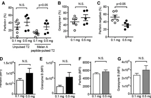
Enhanced cytotoxicity and decreased CD8 dependence of human cancer-specific cytotoxic T lymphocytes after vaccination with low peptide dose
10
0
0
Texte intégral
Figure

Documents relatifs