The Journal of Laryngology and Otology January 1990, Vol. 104, pp. 20-23
Resection of palatal tumours with the CO
2laser
R. SUTTER and R. GROSSENBACHER, (St Gallen, Switzerland)
Abstract
On the basis of the authors' experience with 20 patients, CO2 laser resection of palatal tumours has proved
to be a good alternative to conventional surgical resection. The CO2 laser beam permits precise resection,
due to only slight intra-operative bleeding coupled with use of the operating microscope. Wound healing is good and post-operative pain remarkably little.
Introduction
The CO2 laser beam has acquired increasing importance
in recent years as a surgical cutting instrument in various specialist fields of medicine. It is particularly suited to operations on the upper respiratory and digestive tracts because its energy output is confined to the surface and unaffected by pigmentation, and because it can conse-quently be well controlled (Grossenbacher, 1979,1985). As early as the end of the 1970's, the advantages of CO2
laser surgery were recognized for the treatment of local-ized tumours of the tongue and floor of the mouth, since it facilitated precise resection with minimal intra-oper-ative bleeding, slight post operintra-oper-ative oedema and pain, and good preservation of function (Strong et al., 1979, Rhys Evans and Frame, 1988).
More recently, we have also employed the CO2 laser
beam increasingly for tumours of the hard and soft palate, and report our experience below.
Materials and methods
During the period from May 1985 to December 1988, a total of 21 laser surgical procedures were performed on 20 patients with palatal tumours at the Department of Oto-Rhino-Laryngology, Head and Neck Surgery, Can-tonal Hospital, St Gallen. The treated cases were retro-spectively analysed with regard to age, sex, histology, tumour localization, complications, healing period and recurrence rate.
The apparatus employed is a CO2 laser system
(Cooper, model 250Z) coupled with an operating micro-scope (Zeiss OP Mi 1) (Fig. 1). For easily accessible palatal tumours, it is also suitable for direct incision using the handpiece, with which the microscope is also employed. The smallest possible focus (0.8 mm dia-meter) is used for continuous working with a power out-put between 10 and 20 watts. Most operations take place under general anaesthesia with nasotracheal intubation. After exposing the operative field (Fig. 2a) with
Accepted for publication: 4 October 1989.
Mclvor's tonsillar spatula, the nasotracheal tube and all exposed skin and mucosal sites are covered with moist towels to protect from stray radiation. For protection of the cornea, the operation room personnel wear pro-tective spectacles.
The line of incision is first marked with the CO, laser
FIG. 1
The CO, laser system employed, with operating microscope attached.
beam 5-10 mm peripheral to the microscopically-visible tumour boundary (Fig. 2b). The margin of the tumour specimen is now grasped at one point with a clamp and, under continuous tension, the tumour can be cut around step by step in healthy tissue, working with the CO, laser beam in the gaping incisional fissure. The tissue struc-tures can be readily differentiated because of the virtual absence of bleeding. Larger vessels with a diameter over 0.5 mm must occasionally be coagulated with bipolar forceps. The periosteum of the hard palate is left intact if this permits sufficiently radical excision. The wound sur-face is left uncovered to heal by secondary granulation
20
https:/www.cambridge.org/core/terms. https://doi.org/10.1017/S0022215100111685
RESECTION OF PALATAL TUMOURS WITH THE CO2 LASER 21
a. Operation site defined. b. Incision line marked with CO, laser beam.
c. Wound surface after tumour removal by CO, laser surgery. d. Resected tumour with typical cut surface. FIG. 2a-d
Resection of a pleomorphic adenoma.
and epithelization (Fig. 2c). The pathologist can assess the cut edges of the resected tumour without difficulty, as with the conventional procedure (Fig. 2d).
Results
With the technique described above, a total of 21 laser surgery operations on the palate were performed on 20 patients. The 12 female and eight male patients were aged between 16 and 78. The distribution of the histolog-ical morphology of the treated lesions is shown in Table 1. Among the 12 benign tumours most commonly repre-sented are pleomorphic adenomata, and among the eight malignant tumours squamous cell carcinomata. In all, seven tumours can be traced by their fine structure to the small salivary glands. One-third of the tumours were
localized on the hard palate, one-third on the soft palate and one-third in the transitional zone.
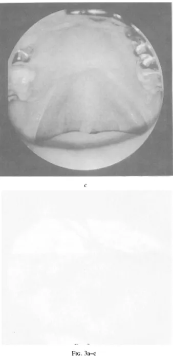
A further example is presented to illustrate the method described (Fig. 3a-c).
TABLE I
HISTOLOGY OF THE 2 0 PALATAL TUMOURS Squamous cell carcinoma
Pleomorphic adenoma Squamous cell papilloma Acantho-hyperkeratosis Haemangioma Monomorphic adenoma Mucoepidermoid tumour Adenoidcystic carcinoma Malignant mesenchymal tumour Fibroma
https:/www.cambridge.org/core/terms. https://doi.org/10.1017/S0022215100111685
22 R. SUTTER AND R. GROSSENBACHER
After resection of a large, superficially ulcerated monomorphic adenoma from a 35-year-old woman, the wound measured over 4 cm in diameter, with exposure of bone. Even this resection defect healed without prob-lems within 2J months.
A striking feature was the slight post-operative pain even among patients with such extensive resections. Neither wound infection nor post-operative bleeding was observed. According to the size of the defect, com-plete epithelialization took between three weeks and 2£ months. Exposed bone, in particular, delayed granu-loma formation but, astonishingly, this caused the patients scarcely any discomfort. In two patients the operation produced a small perforation in the soft palate, which however closed spontaneously within a short time. Two further patients with extensive resec-tions of the soft palate for squamous cell carcinomata
FIG. 3a-c Monomorphic adenoma of palate:
a. Superficially ulcerated tumour of palate, before operation.
b. Large resected area, over 4 cm in diameter, with exposed bone. c. Condition one year post-operatively.
had post-operative problems with eating because of velo-pharyngeal deficiency, which could be improved with an obturator prosthesis. During the follow-up period between five months and four years, one of the 20 patients developed a tumour recurrence, and two cases treated palliatively had persistent tumours.
Discussion
In the light of our experience, CO2 laser surgery
appears to be as valuable for palatal tumours as repeat-edly reported for other sites in the oral cavity (Duncav-age and Ossoff, 1986; Flynn etal, 1988; Grossenbacher, 1985; Guerry etal, 1986; Nagorsky and Sessions, 1987; Rhys Evans and Frame, 1988). The main advantage of the method is the excellent visualization due to only slight intraoperative bleeding, especially when using the
https:/www.cambridge.org/core/terms. https://doi.org/10.1017/S0022215100111685
RESECTION OF PALATAL TUMOURS WITH THE CO2 LASER 23
operating microscope. With the relatively common sal-ivary gland tumours, primary radical resection without injuring the tumour capsule is known to be particularly important. In comparison to electrosurgical resection, the significantly smaller depth of damage at the margin of the incision with CO2 laser-surgical resection leads to
better wound healing and improved histological assess-ment of the cut edge (Duncavage and Ossoff, 1986; Guerry et ai, 1986). Even larger defects with exposed bone healed without problems in all our cases with mini-mal post-operative complaints. The method described does not consist of uncontrolled tumour vaporization, but tumour resection with consistent histological assess-ment of the cut edge. In no case should the employassess-ment of the CO2 laser for treating malignancy lead to neglect
the generally recognized oncological principles of tum-our resection. The uncontrolled vaporization of malig-nant tumours performed elsewhere should, in our view, be decisively rejected. Among squamous cell carcino-mata, only tumours with minimal deep spread and no lymph-node metastases are suitable for curative ther-apy. For palliation, the CO2 laser can occasionally be
employed in incurable conditions for tumour resection. Whether local recurrence, or with malignant tumours metastasis formation, occurs less commonly after CO2
laser-surgical resection are speculative questions that need to be settled by large controlled studies.
Conclusion
CO2 laser-surgery for palatal tumours offers a good
alternative in comparison to conventional resection with
the scalpel or electrotome. The main advantage of the method is that it facilitates precise resection with opti-mal visibility. Striking features are miniopti-mal post-oper-ative complaints with good wound healing.
References
Duncavage, J. A., Ossoff, R. H. (1986). Use of the CO2 laser for
malignant disease of the oral cavity. Lasers in Surgery and
Medi-cine, 6: 442^44.
Flynn, M. B., White, M., Tabah, R. J. (1988). Use of the carbon dioxide laser for the treatment of premalignant lesions of the oral mucosa. Journal of Surgical Oncology, 37: 232-234. Grossenbacher, R. (1979). Erfahrungen mit der CO2
-Laser-chi-rurgie in der Otorhinolaryngologie. HNO, 27: 403.
Grossenbacher, R. (1985). Laserchirurgie in der Oto-Rhino-Laryngologie. Georg Thieme Verlag, Stuttgart.
Guerry, T. L., Silverman, S., Dedo, H. H. (1986). Carbon dioxide laser resection of superficial oral carcinoma: Indications, tech-nique and results. Annals of Otology, Rhinology and
Laryngol-ogy, 95: 547-555.
Nagorsky, M. J., Sessions, D. G. (1987). Laser resection for early oral cavity cancer: Results and complications. Annals of
Otol-ogy, Rhinology and LaryngolOtol-ogy, 96: 556-560.
Rhys Evans, P. H., Frame, J. W. (1988). CO2 Laser surgery in the
oral cavity. In Lasers in Otolaryngology (Carruth, J. A. S. and Simpson, G. T., eds) pp. 101-132. Chapman and Hall Ltd., London.
Strong, M. S., Vaughan, C. W., Healy, G. B., Shapshay, S. M., Jako, G. J. (1979). Transoral management of localized car-cinoma of the oral cavity using the CO2 laser. Laryngoscope, 89:
897-905.
Address for correspondence: PD Dr R. Grossenbacher,
Department of Otorhinolaryngology, Head and Neck Surgery, Cantonal Hospital,
CH-9007, St Gallen (Switzerland).
https:/www.cambridge.org/core/terms. https://doi.org/10.1017/S0022215100111685