Case report
A fully echo-guided trans-apical aortic valve implantation
Enrico Ferrari
a,*
, Christopher Sulzer
b, Elena Rizzo
c, Ludwig Karl von Segesser
aa
Department of Cardio-Vascular Surgery, University Hospital of Lausanne (CHUV), 46 Rue du Bugnon, CH-1011 Lausanne, Switzerland
bDepartment of Cardiac Anaesthesia, University Hospital of Lausanne (CHUV), 46 Rue du Bugnon,
CH-1011 Lausanne, Switzerland
cDepartment of Radiology, University Hospital of Lausanne (CHUV), 46 Rue du Bugnon,
CH-1011 Lausanne, Switzerland
Received 28 April 2009; received in revised form 15 June 2009; accepted 18 June 2009; Available online 14 August 2009
Abstract
The trans-apical aortic valve implantation (TA-AVI) is an established technique for high-risk patients requiring aortic valve replacement. Traditionally, preoperative (computed tomography (CT) scan, coronary angiogram) and intra-operative imaging (fluoroscopy) for stent-valve positioning and implantation require contrast medium injections. To preserve the renal function in elderly patients suffering from chronic renal insufficiency, a fully echo-guided trans-catheter valve implantation seems to be a reasonable alternative. We report the first successful TA-AVI procedure performed solely under trans-oesophageal echocardiogram control, in the absence of contrast medium injections.
#2009 European Association for Cardio-Thoracic Surgery. Published by Elsevier B.V. All rights reserved.
Keywords: Echo-guided; Trans-apical aortic valve replacement; Aortic valve stenosis; Renal insufficiency
1. Introduction
The trans-apical aortic valve implantation (TA-AVI) is a valid technique for aortic valve replacement (AVR) in high-risk patients. The stent-valve positioning and implantation are normally based on landmarks (annulus diameter, pattern of calcifications, distance between the annulus and the coronary ostia, coronary anomalies) preoperatively identi-fied by a cardiac computed tomography (CT) scan and a coronary angiogram, and confirmed intra-operatively by fluoroscopy with contrast medium injections [1]. Unfortu-nately, following extensive use of contrast medium, patients selected for trans-catheter AVR and suffering from chronic renal insufficiency are exposed to a higher risk of acute postoperative renal failure [2]. Consequently, we believe that the quantity of contrast medium administered intra-operatively should be decreased or completely avoided: following our experience on endovascular aorta repair without contrast medium [3,4], we identified a valid alternative in the fully echo-guided trans-catheter valve replacement.
2. Case report
An 82-year-old female patient suffered from severe respiratory disease, coronary artery disease (stent in the left anterior descending (LAD)), diabetes, hypertension, periph-eral vascular disease and chronic renal insufficiency (pre-operative creatinine level of 170 mg dl 1). She was also suffering from a severe and symptomatic aortic valve stenosis (aortic orifice area of 0.43 cm2m 2), with moderate left ventricular hypertrophy, good left ventricular function (ejec-tion frac(ejec-tion of 60%) and pulmonary hypertension (systolic pulmonary pressure of 55 mmHg). The logistic EuroSCORE was 42% and the patient accepted a TA-AVI procedure. To identify the landmarks and to calculate the perspective of the aortic valve plane for the C-arm fluoroscopy orientation (108 caudal and 108 left, as shown inFig. 1), a cardiac CT scan with a low dose of contrast medium (50 ml) was performed: no coronary anomalies were found and the distance between the aortic annulus and the coronary ostia was 10.5 mm left and 11 mm right, respectively. The calculated aortic annulus diameter was 23.5 mm. Under general anaesthesia and in the operative theatre, the patient underwent a TA-AVI procedure: the guide-wires in the femoral vessels (CPB stand-by), the left mini-thoracotomy and two reinforced 2/0 prolene purse-string sutures in the apex were prepared in the standard way. After heparinisation (100 U kg 1), a pig-tail catheter was placed in
the ascending aorta (kept ready for contrast medium www.elsevier.com/locate/ejcts European Journal of Cardio-thoracic Surgery 36 (2009) 938—940
* Corresponding author. Address: Department of Cardiovascular Surgery, Centre Hoˆpitalier Universitaire Vaudois (CHUV), 46 Rue du Bugnon, CH-1011 Lausanne, Switzerland. Tel.: +41 79 310 1386; fax: +41 21 314 2278.
E-mail address:[email protected](E. Ferrari).
1010-7940/$ — see front matter # 2009 European Association for Cardio-Thoracic Surgery. Published by Elsevier B.V. All rights reserved. doi:10.1016/j.ejcts.2009.06.030
injection, if necessary) and guide-wires were inserted through the apex and towards the aortic valve, under C-arm fluoroscopy and trans-oesophageal echocardiogram (TEE control). The two-dimensional TEE (2D-TEE) measurements confirmed the annulus diameter (23 mm) and landmarks (specifically with regard to the coronary ostia and the aortic annulus position). The valvuloplasty was performed under direct TEE-guided balloon positioning plus fluoroscopy control, and, successively, a 26-mm Sapien stent valve (Edwards Lifesciences Inc., Irvine, CA, USA) was implanted under fully
TEE-guided valve positioning and simultaneous fluoroscopy control (Fig. 2). The 2D-TEE confirmed the correct valve placement with a peak gradient of 6 mmHg and absence of paravalvular leakage. Postoperatively, the recovery was uneventful with a fast-track extubation and 2 days in the intensive care unit. The postoperative serum creatinine level did not increase (153 mg dl 1at day 1 and 145 mg dl 1at day 2)
and a pre-discharge echocardiogram (day 8) showed an aortic peak and mean gradient of 12 mmHg and 7.5 mmHg, respectively, without paravalvular leakage.
E. Ferrari et al. / European Journal of Cardio-thoracic Surgery 36 (2009) 938—940 939
Fig. 1. (A) Selective three-dimensional aortic root reconstruction from the preoperative cardiac CT-scan: the perspective of the aortic valve plane is identified and measured as number of degrees in cranio-caudal and lateral orientation. The procedure is helpful to orientate the C-arm fluoroscopy in the operative room (in this case: 108 caudal and 108 on the left). (B) A picture taken from the C-arm fluoroscopy after the stent-valve deployment: the optimal preoperative C-arm fluoroscopy orientation is confirmed by the stent-valve
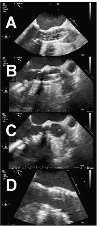
plane. Fig. 2. The sequence shows the full echo-guided trans-apical valve implanta-tion. (A) Aortic valvuloplasty; (B) stent-valve ballooning; (C) balloon deflation; (D) sapien stent-valve in place.
3. Discussion
The fully echo-guided TA-VI seems a valid alternative to the standard TA-VI procedure: the recent published reports have shown an incidence of postoperative renal failure reaching 28% followed, sometimes, by transitory or permanent dialysis[2]. This severe complication is reasonably due to the impaired preoperative renal function coupled with an extensive use of contrast medium: the average of contrast injected during a standard TAVI procedure is around 100—300 ml[5,6], and the total amount increases rapidly when we take into considera-tion the doses necessary for the preoperative CT scan and the coronary angiogram. In order to diminish the risk of renal failure, we recommend a fully echo-guided TA-AVI without contrast medium: the landmarks identification, the balloon positioning and the stent-valve implantation can be easily performed with a 2D-TEE (3D-TEE is under evaluation) and the fluoroscopy remains essential for guide-wire placement, valvuloplasty control, and stent-valve deployment[7]. This first case report confirms that this technique allows TA-VI procedures in intubated high-risk patients and empathise the role of the intra-operative 2D-TEE as well as the role of specialised doctors performing it (anaesthetists or cardiolo-gists). Limitations to this procedure are the presence of artefacts in the TEE images (concomitant mechanical heart valve prosthesis in mitral position) and low-quality preopera-tive CT scans (i.e., presence of cardiac arrhythmias). There-fore, further reports are required to finalise this technique.
References
[1] Walther T, Dewey T, Borger MA, Kempfert J, Linke A, Becht R, Falk V, Schuler G, Mohr FW, Mack M. Transapical aortic valve implantation: step by step. Ann Thorac Surg 2009;87(1):276—83.
[2] Aregger F, Wenaweser P, Hellige GJ, Kadner A, Carrel T, Windecker S, Frey FJ. Risk of acute kidney injury in patients with severe aortic valve stenosis undergoing transcatheter valve replacement. Nephrol Dial Transplant 2009;24(7):2175—9.
[3] Marty B, Tozzi P, Ruchat P, Haesler E, von Segesser LK. Systematic and exclusive use of intravascular ultrasound for endovascular aneurysm repair—the Lausanne experience. Interact Cardiovasc Thorac Surg 2005;4 (3):275—9.
[4] von Segesser LK, Marty B, Ruchat P, Bogen M, Gallino A. Routine use of intravascular ultrasound for endovascular aneurysm repair: angiography is not necessary. Eur J Vasc Endovasc Surg 2002;23(6):537—42.
[5] Covello RD, Maj G, Landoni G, Maisano F, Michev I, Guarracino F, Alfieri O, Colombo A, Zangrillo A. Anesthetic management of percutaneous aortic valve implantation: focus on challenges encountered and proposed solu-tions. J Cardiothorac Vasc Anesth 2009;23(3):280—5.
[6] Bleiziffer S, Ruge H, Mazzitelli D, Schreiber C, Hutter A, Laborde JC, Bauernschmitt R, Lange R. Results of percutaneous and transapical trans-catheter aortic valve implantation performed by a surgical team. Eur J Cardiothorac Surg 2009;35(4):615—20.
[7] Vahanian A, Alfieri OR, Al-Attar N, Antunes MJ, Bax J, Cormier B, Cribier A, De Jaegere P, Fournial G, Kappetein AP, Kovac J, Ludgate S, Maisano F, Moat N, Mohr FW, Nataf P, Pierard L, Pomar JL, Schofer J, Tornos P, Tuzcu M, van Hout B, von Segesser LK, Walther T. Transcatheter valve implantation for patients with aortic stenosis: a position statement from the European Association of Cardio-Thoracic Surgery (EACTS) and the European Society of Cardiology (ESC), in collaboration with the European Association of Percutaneous Cardiovascular Interventions (EAPCI). Eur J Cardiothorac Surg 2008;34(1):1—8.
E. Ferrari et al. / European Journal of Cardio-thoracic Surgery 36 (2009) 938—940 940