Thymic stroma is required for the
development of human T cell lineages in
vitro
Antonio de la Hera, Wendy Marston, Crlsanto Aranda1, Marie-Lulsa Toribio2, and
Carlos Martinez-A.2
Basel Institute for Immunology, Grenzacherstrasse 487, Basel 4005, Switzerland 'Coulter Cientifica, Mostoles, Madrid, Spain
2lnstituto de Biologia Molecular, CSIC, Universidad Autonoma de Madrid, Cantoblanco, Madrid 28049, Spain
Key words: a/3 and yd T cells, natural killer cells, interleukins 1, 2, 3, and 4, hematopoietic progenitors,
differentiation pathways
Abstract
Development of the T cell lineage Is characterized by the homing of hematopoietic precursors to thymus, followed by their acquisition of receptors for antigen. T cell receptors are a/3 or >5 heterodimers associated with CD3 (TCR - CD3). Very early T cell precursors in humans have been characterized as CD7+45+ cells which lack the T cell differentiation antigens CD1, CD2, CD3, CD4, and CD8. A phenotypically equivalent early thymocyte population also occurs In postnatal life, and we have previously shown that interleukin 2 (IL2) promotes the development in vitro of both the a/3 and the y5 T cells from these early thymocytes. Here we have analyzed the
requirements of the induction of the IL2 pathway In early thymocytes, and their developmental potential. We show that: (I) thymic stromal cells, which are present In thymocyte suspensions, are necessary to induce the IL2 pathway and the development of a/3 or 7<5 T cell lineages from early thymocytes in vitro; and (II) when removed from the In vivo environment, early thymocytes can develop In vitro into T C R - C D 3 - cells of the natural killer (NK) lineage. We conclude that CD7+45+, CD1 - 2 - 3 - 4 - 8 - early thymocytes are multipotential progenitors that, at least, have the capacity to develop into a/3 or yd T cell and NK lineages. The analysis of the mechanisms of generation and selection of human T and NK cell diversity, not feasible In bone marrow cultures, Is now possible.
Introduction
T cell precursors develop apart from the other hematopoietic lineages within the thymic microenvironment (1,2). The importance of the thymus for T cell precursor differentiation is underscored by the severe T cell immunodeficiency, but not by B or natural killer (NK) lymphocyte defects, suffered by athymic individuals. The defects in the thymic microenvironment reside in non-T lineage 'thymic stroma' cells, as shown by the reconstitution of T cells after transplants of thymic stroma (3,4). The recent characterization of monoclonal antibodies specific for thymic stromal components (i.e. the distinct types of epithelial and hematopoietic stroma; 5 - 7 ) provides the tools necessary to study the role of the thymus in human T cell development in
vitro.
Experiments ex vivo have shown that T cell progenitors colonize the human thymus at week 7 of gestation, before
development of any mature T cell. The colonizing cells are hematopoietic cells (i.e. CD45+) that express an early T cell marker, CD7, but lack other T cell differentiation antigens (5) In postnatal life an equivalent CD7 + 45 + , CD1 2 3 4 8 -mtrathymic population has been demonstrated (2). Hereafter, we will refer to this population as early thymocytes, according to the nomenclature originally proposed for human thymocyte subpopulations by Reinherz and Schlossman (8).
Experiments in vitro have shown that distinct subpopulations of human CD4-8- 'double-negative' thymocytes, such as early thymocytes, are able to grow and differentiate into T cells by culture in recombinant interleukin-2 (rll_2; 2,7,9,10). In the mouse, development of mature T cells from double-negative thymocytes is apparently not feasible in vitro (11). However, double-negative thymocytes develop into mature T cells upon transfer in vivo if
Correspondence to Antonio de la Hera, as above
a thymus is present (11,12). Here we have analyzed whether thymic stroma is required for the IL2-dependent in vitro development of human early thymocytes. We have also studied whether human early thymocytes are irreversibly committed to the T cell lineage. We show that thymic stroma cells are present in low numbers in populations of early thymocytes isolated by negative selection of CD1, CD2, CD3, CD4, and CD8 antigen-positive thymocytes. Such thymic stroma cells, absent from early thymocytes which are carefully purified by positive selection, are required both for IL2-dependent growth and the differentiation into a£ and 76 T cells. We further show that early thymocytes are a multipotential population able to develop into NK cells in
vitro.
Methods
Isolation of thymocyte populations
Single-cell suspensions were prepared from thymic fragments that had been removed from patients 2 months-6 years old during corrective cardiac surgery. Thymocytes expressing CD1, CD2, CD3, CD4, or CD8 differentiation antigens were eliminated by incubation with cytotoxic antibodies and rabbit complement (Behringwerke, Marburg, FRG), as described elsewhere (10). Viable thymocytes were isolated by Ficoll - Hypaque density centrifugation. CD7 + , CD1 - 2 - 3 - 4 - 8 - early thymocytes were separated from non-T lineage, stromal components using M-450 beads coupled with anti-CD7 antibodies and a sintered magnetic alloy. Early thymocytes were then detached from the beads by incubation at 37°C, before use in the experiments reported. CD1 - 2 - 3 - 4 - 7 - 8 - thymocytes were used as a source of fresh, autologous stromal cells. We followed the manufacturer's recommendations (Dynal, Oslo, Norway) throughout the method. The same protocol was used to enrich for mature thymocyte from the total population, using M-450 beads coated with either Na1/34 (CD1, negative selection) or F10-44-2 (CD44, positive selection). NK cells derived from early thymocyte cultures were isolated by either cytotoxic elimination of TCR-CD3+ cells with OKT3 antibody and complement, or by positive selection of CD16 + cells using M-450 magnetic beads coated with B73.1 antibody.
Monoclonal antibodies
CD1, Na1/34 (13); CD2, Leu-5 (Becton-Dickinson, Mountainview, CA, USA) and CoulterClone T11 (Coulter, Hialeah, FL, USA), CD3, OKT3 (American Type Culture Collection, ATCC, Bethesda, MD, USA) and CoulterClone T3; CD4, HP2/6 (14) and CoulterClone T4; CD7, 3A1 (ATCC) and RFT2 (15); CD8, B9.4 (16) and CoulterClone T8; CD11b, CoulterClone Mo1; CD14, CoulterClone Mo2; CD16, Ieu11 (B73.1); CD20, CoulterClone B1; CD45, D3/9 (17); a/3TCR/CD3, WT31 (Sanbio, Leiden, The Netherlands); anti-7/61, TCR7/6 (18), DA4-4, IgM (ATCC); HNK-1, Leu7; Coulter NKH1 and PTL-1; CD44 F10-44-2 (19); RFD1 (6); and TE3A and TE4 (20, Serotec, Bicester, UK) were used.
Cell cultures
Cultures were maintained in RPMI 1640 medium supplemented with 2 mM glutamine, 10 mM HEPES, and 10% human serum (complement-depleted and pooled male AB donor), hereafter referred to as complete medium. Cells (106) were cultured in Cluster24 plates (Costar no. 3524, Cambridge, MA, USA) in
complete medium supplemented with ML2 (Hoffmann-LaRoche Ltd, Basel, Switzerland) and/or autologous thymic stroma at 1:8 ratio of thymic stroma to early thymocytes or mature thymocytes. Stroma cells were irradiated (2500 rad) to prevent their pro-liferation in these complementation experiments Where indicated, stroma cells were placed in the inner chamber of a Transwell system (Costar no. 3408), separated from thymocytes by a 0.4 /tm pore, polycarbonate membrane. Cells in replicate cultures were harvested after the indicated period of time, and aliquots were used in immunofluorescence and cytotoxicity assays after determination of the cellular recoveries. For proliferative assays, 10s cells were cultured for 4 days in the presence of the indicated interleukins (50 U/ml) and [methyPHJthymidine (Radiochemical Centre, Amersham, UK) was added at 1 /iCi/well for the last 12 h of culture. rlL1a and rlL1/3, rlL3, and rlL4 were generously provided by Hoffmann-LaRoche (Basel), Nippon-Roche, (Kumamoto, Japan), and Sandoz (Basel) respectively
Quantitative flow cytometry
The procedure for indirect immunofluorescence surface staining of the cells has been described (21) Staining of cytoplasmic antigens was performed as described by Shoroff etal. (22). Briefly, cell membranes were permeabilized with 10 /tg/ml lysolecithin (Sigma, St Louis, MO, USA) for 2 mm at 4°C Thereafter, they were processed as for cells undergoing surface immunofluorescence. Fluorescein-conjugated goat anti-mouse Ig (Southern Biothechology, Birmingham, AL, USA) was used as a second step reagent. For two-color analyses, antibodies were either directly labeled with fluorescein isothiocyanate (FITC, green) or phycoerythrin (PE, orange-red), or biotin-conjugated In the latter case, a second layer of avidin - FITC was added to reveal the binding (2,23). Quantitation of the staining of 104 (one-color) or 5x1CH (two-colors) viable cells was performed with a Coulter EPICS-C cell sorter or a FACScan analyzer, as detailed elsewhere (23).
Cytotoxicity assays
Putative effector cells were tested for cytotoxicity against 5 x 10³ 5'Cr-labeled K562 target cells at various effector/target ratios in a 4 h siCr-release assay. Cytotoxic activity was calculated as described elsewhere (24). Assays were carried out either in the absence or presence of 75 ng/ml anti-CD3 antibody OKT3, using heat-inactivated human AB sera.
Results
Phenotypic analyses of CD1 -2-3-4-8- thymocytes reveals CD7+45+ early precursors and thymic stromal components
Our interest was to analyze the requirements for growth and differentiation of the postnatal CD7+45+, CD1 - 2 - 3 - 4 - 8 - early thymocytes. Therefore we treated total thymocytes with a mixture of cytotoxic anti-T cell antibodies, specific for CD1, CD2, CD3, CD4, and CD8, and complement. The phenotype of the remaining population (0.3 ± 0.1%, n = 6) was further analyzed in order to characterize the surface molecules potentially expressed on these cells, as well as to quantify the percentage of contaminating thymic stroma or hematopaetic cells from other lineages. The thymocyte subset isolated contained < 1 % of
CD1+, CD2 + , CD3 + , CD4 + , or CD8+ cells (Fig. 1b). Three distinctive cell types were present,
(i) Most, but not all, cells (93%, Fig. 1c) expressed the leukocyte
Log F l u o r e s c e n c e I n t e n s i t y
Fig. 1. Early thymocytes and thymic stroma are co-enriched after
negative selection of CD1, CD2, CD3, CD4, and CD8-posrtive thymocytes. Thymocytes were treated with cytotoxic anti-T cell antibodies and com-plement, and the phenotype of the surviving population was analyzed by indirect immunofluorescence and quantitative flow cytometry in a FACScan analyzer, as indicated in Methods Histograms show the fluorescence profiles of viable cells labeled with (a) normal mouse serum; (b) a cocktail of CD1, CD2, CD3, CD4, and CD8, (c) CD45, (d) CD7; (e) TE3A plusTE4; (f) CD14, (g) CD16 plus NKH1 and HNK1, and (h) CD20 plus anti-ji Vertical dotted lines indicate the limit chosen for background fluorescence.
common antigen (LCA, CD45), which is normally present on nucleated hematopoietic cells but not on other lineages, and the majority of them wereCD7+ (~81%3A1 +, Fig. 1d). Thus, their phenotype resembles that of the human fetal prethymic T cell progenitor.
(n) Approximately 6% are epithelial thymic stroma cells, as indicated by the binding of antibodies TE4 and TE3A to a minor subset of the CD1 - 2 - 3 - 4 - 8 - cells (Fig. 1e).
(lii) Myelomonocytic CD14+ (Mo2) cells constitute a minor, but sizeable (— 7%, Fig. 1f), portion of hematopoietic origin. At least some of these cells are likely to represent thymic stroma hematopoietic cells because of the detection of some positive cells by the antibody RFD1, a reliable marker of interdigitating, dendritic stroma cells (6, see below). Other hematopoietic lineages, such as CD16 + , NKH-1+, and HNK-1 + NK cells (Fig. 1g), or PLT-1 + platelets are undetectable (<1%). The finding of a few B cells (Fig. 1h), as estimated by antibodies specific for CD20 (B1) and surface IgM, is not surprising in view of the existence of a small subset of B cells in normal thymus (25; J. L. Andreu, personal communication).
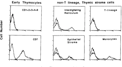
The demonstration of thymic stromal components points to an unsuspected shortcoming of negative selection approaches for the isolation of T cell progenitors from postnatal 'thymocyte' suspensions We therefore isolated cells bearing CD7 by positive selection using magnetic beads coated with antj-CD7 antibodies. Immunofluorescence and flow cytometry analyses of the purified cells demonstrated that these positively selected cells are a homogeneous population of CD7 + , CD1 - 2 - 3 - 4 - 8 - cells (Fig. 2, left panels). In contrast, the CD1 2 3 4 7 8 -thymocytes are a heterogeneous population that contains, at least, the following thymic stroma cell types: TE4+/TE3A + thymic epithelial cells, RFD1 + interdigitating cells, and CD11b/Mac-1+ monocytes (Fig. 2, right panels). A clear demonstration that CD1 - 2 - 3 - 4 - 7 - 8 - thymocyte suspensions
Early Thymocytes non-T lineage, Thymic stroma cells
ID
SI
E
o
Log Fluorescence Intensity
Fig. 2. Early thymocytes can be separated from thymic stroma using anti-CD7 antibodies. Early thymocytes were sorted from non-T lineage, stroma
cells present in CD1 - 2 3 - 4 - 8 - thymocyte suspensions using magnetic beads coated with anti-CD7 antibodies, and the phenotype of the separated populations was analyzed by indirect immunofluorescence and flow cytometry, as described in Methods The profiles of immunofluorescence for background staining (left) and for the indicated antigens (right) are superimposed for better comparison. Early thymocytes were labeled with either CD7 or a mixture of CD1, CD2, CD3, CD4, and CD8 antibodies. Non-T lineage cells were labeled with RFD1 antibody, specific for interdigitating cells, TE3A plus TE4 antibodies, specific for epithelial cells (20); CD11 b, staining monocytes; and a cocktail (CD1, CD2, CD3, CD4, CD7, and CD8) of antibodies to the T lineage panel
Early Thymocytes Mature Thymocytes
CD44+
CD1-rlL4 rlL3 rll_2 rILiB rlL1o ^Gdtum Cp .
11
r
1 ])
rlL4 rlL3 rlL2 rILiB rlL1a Medium 20 40 60 80100 0 1 2 3 4 5 6 7 8 9 0 1 2 3 4 5 6 7 8 93H-thymidine uptake (cpm x 10-3)
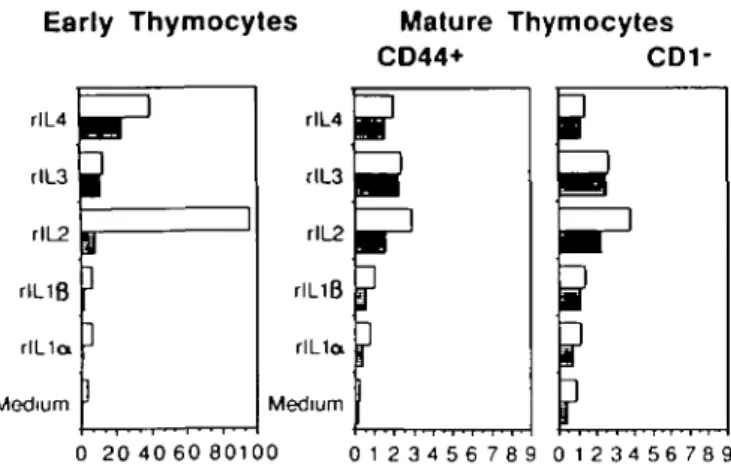
Fig. 3. Thymic stroma selectively induces IL-2 responsiveness in early
human thymocytes. Early thymocytes, and CD44+ or CD1 - thymo-cytes, were sorted using magnetic beads coated with the specific anti-bodies, as described in Fig 2 and Methods. 105 cells were cultured for
4 days in 96-well, flat-bottomed mcrotiter plates in the presence of 50 U/ml of the indicated interieukins and either in the presence ( • ) or absence ( • ) of irradiated stroma cells Results show the uptake of [3H]thymidine
during the last 12 h of culture The experiment is representative of three independent ones Standard deviations were < 1 1 % in the triplicate cultures Note the different scale used for early and mature thymocytes
contain thymic stroma comes from the establishment of epithelial cell lines from the suspensions reported here (A de la Hera, unpublished observations). In view of these results, we wish to stress that single-cell suspensions prepared from thymus contain not only 'thymocytes' but, albeit in very low numbers, epithelial and hematopoietic stromal cells.
Requirements for early thymocyte growth
In previous studies in the human system, different subsets of C D 4 - 8 - thymocytes isolated by negative selection have been shown to produce and use IL2 to grow autonomously and differentiate into T cells (7,9,10,21,24). Considering the central role played by the thymic microenvironment in the development of T cells, rt seemed unwarranted to assume that the requirements for growth and differentiation of mixed populations of early thymocytes and stroma cells are the same as those for isolated intrathymic T cell progenitors. We therefore studied the proliferative responses of early thymocytes to recombinant interieukins (from IL1a to IL4) in the presence or absence of autologous thymic stroma.
Purified early thymocytes proliferate in rlL4 but not in rlL1o or IL1/3. rlL2and rlL3 promoted some proliferation in these cells, though to a much lesser degree than rlL4 (Fig. 3, filled bars). The failure of rll_2 to promote proliferation of positively selected earty thymocytes contrasts with the proliferative responses of early thymocytes enriched by negative selection (2). In comple-mentation experiments, mixing purified early thymocytes and irradiated stroma cells, a strong proliferative response to rll_2 was observed, showing that the IL2 unresponsiveness of early thymocytes is not due to a non-specific suppression caused by the binding of CD7 antibodies (Fig. 3, open bars). Since stroma cells fail to induce significant IL2 responsiveness in either
CD44+ or CD1 - thymocytes (enriched in mature T cells, refs 2, 5), this complementation is not due to a non-specific enhancement of proliferation by stromal cells. The reduced proliferation of early thymocytes of IL2 after their separation from stroma does not reflect a non-specific 'filler defect' since their proliferation to rlL4 did not require the addition of stroma. IL4 responsiveness might thus precede IL2 responsiveness in the human, as it appears to with mouse prethymic PRO-T cells (1) We conclude that thymic stroma cells selectively promote IL2 responsiveness of early thymocyte in vitro
Early thymocytes can develop into either a/3 and yd T cell or NK cell lineages
A requisite for a putative intrathymic T cell precursor is the ability to differentiate into mature cells (1). In our study on human T cell precursors we took advantage of the inducibility of the IL2 pathway in early human thymocytes (2) and the existence of monoclonal antibodies to framework determinants of a/3 and y& TCRs. Here we explored the differentiation potential of positively selected early thymocytes cultured in IL2 in either the presence or absence of stroma cells. As a readout for differentiation, two-color immunofluorescence and flow cytometry were used to distinguish and charactenze the putative progeny separately from their precursors The CD7 + 45 + , CD1 - 2 - 3 - 4 - 8 - phenotype was maintained by the cultured population during the initial 3 - 4 days of culture without any detectable T cell progeny. After this lag period, early thymocytes started to develop into T cells. Remarkably, the TCR - CD3 complex, whether ajS or y8, was first expressed in CD2 + , CD4-8- double-negative thymocytes at days 3 - 4 . Single-positive CD4+ or CD8+ cells first appeared in the cultures at day 5, and accumulated thereafter. Either a/3 or y5 TCR-CD3 was expressed in these single-positive thymocytes. These results are illustrated in Fig. 4 with two color contour plots obtained at day 7, when all subsets were present in significant numbers. Direct contact between early thymocytes and stroma cells was required for T cell development, since if stroma cells were present in the well but separated from the thymocytes by a micropore membrane, no TCR-CD3 expression was induced in early thymocytes cultured in 50 U rll_2 for 1 week (data not shown). From these results, we conclude that human early thymocytes exposed in vitro to the influence of thymic stromal elements and rlL2 have the potential to develop into the T cell lineage expressing both types of CD3-associated TCR.
In addition, populations of CD11b+ (Mac-1), CD16+ (FcR7), HNK1+, and NKH1+ cells also developed in these cultures. This combination of differentiation antigens is currently used to define NK lymphocytes (23,26) The small amounts of CD11 b/CD14+ monocytes, as well as the B cells initially present in the mixed population of early thymocytes and stroma cells, were gradually diluted by the expanding population of early thymocytes and their progeny, and represented ^ 1 % after 1 week of culture. The distribution of 'NK markers' and CD3 in the progeny of early thymocytes is illustrated in Fig 5. Most CD16+ lymphocytes were CD3- cells, and thus not TCR-CD3+ T cells The finding of some CD3+ cells express-ing either HNK1 or NKH1 antigens is not unique to this in vitro development system. It further reflects the existence in vivo of minor subpopulations of lymphocytes within the T cell lineage
00
Q
O
s
-O ^
Q
O -I
CD3
CD8
O) 00
O Q
-» o
Q
O
QpTCR
COa
o
[e
SS:CO
Q
o
-;D4
+
f
^ :
r* • ' .apTCR
Log green fluorescence intensity
y 6 TCR
Fig. 4. Development of a/3 and y6 T cells from human early thymocytes. Early thymocytes were cultured for 7 days in the presence of 50 U/ml
rlL2 and irradiated stroma cells Aiiquots of the cultures were stained for two-color analyses of the indicated antigens using antibodies labeled with either FITC (green) or PE (red) fluorochromes Antibodies specific for CD antigens were directly conjugated (Coulter), TCRafS WT31 and TCR>5 anti-y/51 were biotinylated and the binding revealed by FITC-avidin (Becton-Dtckinson) Contour plots depict the two color immunoflorescence distribution recorded by an EPICS-C cell-sorter equipped with a three-decade logarithmic amplifier. Dotted lines indcate background immunofluorescence for each color obtained with isotype-matched irrelevant, control antibodies (Coulter), as indicated elsewhere (10,22)
CD16 —>
CD3 —>
CD3 —>
f
* • ' * - •R g . 5. Human early thymocytes have the potential to develop into phenotypically defined NK cells. Early thymocytes were cultured for 7 days and labeled for two-color immunofluorescence, as indicated in Fig. 4. Directly labeled antibodies were obtained from Coulter (CD3 and NKH1) or Becton-Dickinson (CD16 and NKH1). Three dimensional isometnc displays show the distribution of the green fluorescence in the x-axis (CD16, CD3 and CD3, respectively), of the red fluorescence in the y-axis (CD3, NKH1 and HNK1), and the number of cells accumulated is depicted in the vertical z-axis Plots were generated using an EPICS-C cytometer and V2.2 software (Coulter).
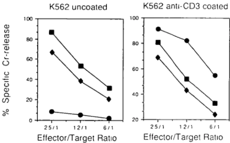
exhibiting NK activity (27). Consistent with the view that the CD3-16+ cells may represent NK but not T lymphocytes (23,26), both CD3- and CD16+cultured early thymocytes kill-ed K562 target cells in a 4 h 51Cr-release assay. Their
cytotox-icity was not modified by anti-CD3 antibodies, which induce strong cytotoxic activity in a CD3+16- T cell clone (Fig 6)
Discussion
These analyses of the requirements for the growth and the differentiation potential of CD7 + 45 + , CD1 - 2 - 3 - 4 - 8 - early thymocytes in vitro have revealed that: (i) their IL2 responsiveness requires the presence of thymic stroma cells; and (ii) at a minimum
K562 uncoated K562 anti-CD3 coated ro CD O u IS) 25/1 12/1 6/1 Effector/Target Ratio 2 5 / 1 12/1 6/1 Effector/Target Ratio
Fig. 6. The CD3 -16 + progeny of early thymocytes have the functional
features of NK cells. Aliquots of CD16+ ( • ) or CD3" ( • ) cells were sorted from the cultures of early thymocytes described in Figs 4 and 5, using magnetic beads coated with CD16 antibody or 0KT3 antibody and complement respectively. Their cytotoxic activity was measured against
51Cr-labeled K562 target cells, either uncoated or coated with anti-CD3
antibody OKT3 A CD3 + 16- T cell clone ( • ) was used as an internal control in these experiments. It was expanded from peripheral blood using PHA, rll_2, and irradiated peripheral blood thymocytes, according to standard protocols (18) Displays show the specific 51Cr release in a 4
h assay, at the indicated effector-to-target ratios. Spontaneous release was < 1 4 % in both cases
they have the potency to develop into a/3 or yb T cells or T C R - C D 3 - NK cells. Two controversial issues merit discussion before considering the relevance of these results to the proliferation and differentiation of T cell progenitors in vivo: (i) whether early thymocytes use IL2 for their development, and (ii) what are the precursor-product relationships among early thymocytes and a/3 T cells, yd T cells, and the NK cell lineage. The first controversy concerns the question of whether the T cell development is promoted by IL2. Our studies in vitro suggest tht the I L 2 - I L 2 receptor pathway might be implicated in the proliferation of functional a/3 or y8 TCR-CD3+ T cell precursors (2) after the contact of the early thymocyte progenitor with stroma cells, as it appears to with the ontogeny of mouse single-positive a/3TCR + thymocytes in vivo (28). We have shown that when stroma cells are not deliberately removed, different subsets of human double-negative, but not mature, thymocytes autonomously use the IL2 pathway for their growth and differentiation into T cells (10,21). The discrepant observations regarding the use of the IL2 pathway by double-negative thymocytes (reviewed in ref. 2) might thus be explained by the concomitant enrichment in some experiments for thymic stroma accessory cells (7,9,10,21). In addition there are thought to be distinct subsets of double-negative thymocytes in terms of their IL2 responsiveness (11,42). Enrichment for the 'early' CD7+45 + , CD1 - 2 - 3 - 4 - 8 - thymocytes may well allow the IL2 responsiveness of this population to be expressed in vitro. In the mouse, phagocytic cells of thymic stroma but not spleen stroma have functional IL2 receptors (29). Thymic stromaJ cells were not the source of proliferating cells in our assays because they were irradiated before they were used in complementation experi-ments. Additional experiments are underway to study whether the effects of IL2 in T cell development (2,27,29) are mediated
directly on the early thymocyte, indirectly via thymic stroma, or both.
Our studies in vitro suggest that T cell differentiation requires early thymocyte-to-stroma contact; what might the requirement for cell-tc-cell contact be due to7 In the absence of thymic stroma, some combinations of lymphokines can promote mouse PRO-T cell growth without T cell differentiation. PRO-T cells will differentiate into TCR-CD3+ T cells upon intrathymic injection (1). Using this approach we have recently established clones of human CD7 + , CD1 - 2 - 3 - 4 - 8 - thymocytes which do not further differentiate into T cells despite the fact that they respond to IL2 with proliferation (our unpublished observations). Thus, the activation of IL2 or other known lymphokine pathways does not itself promote T cell development (1). There are some, not exclusive, possible mechanisms that could account for this. T cell precursor - stroma interaction may trigger surface receptors that raise second messages to activate the T cell development program, or promote the production of soluble 'differentiation' factors by stromal cells to which T cell precursors become responsive after the cell-to-cell contact
The second controversy concerns the precursor-product relationships in the thymus Our data suggest that the early thymocytes are the precursors for both the T and NK cells detected in vitro and that CD4+8+ double-positive thymocytes are not an obligatory intermediate in the development of a/3 TCR-CD3+ T cells. It could be argued that the T and NK cells detected in vitro are actually an outgrowth of residual mature lymphocytes but we have seen that mature thymocytes fail to proliferate under conditions that allow the early thymocytes to develop (Fig. 3). Moreover, recent analyses of the plating efficiency and phenotype of the progeny of single early thymocytes are in general agreement with the results reported here for bulk cultures (10). The possibility that contaminant blood NK cells are the source of NK cells in the culture can also be ruled out. NK cells do not proliferate under these culture conditions In fact they are completely overgrown by TCR-CD3+ cells after day 7 of culture in 50 U rlL2/ml (23,30). We find that a/S TCR is expressed before CD4 or CD8 (Fig 4b, d and e). This is thought to be unusual because comparative analyses of TCRa/3 ontogeny in normal mouse have detected V08-CD3+4-8- cells in newborns but not in fetuses (28,31). These cells were considered to be late arrivals with no precursor potential. However, such V08-CD3+4-8- thymocytes are readily evident in female fetuses from anti-HY a/3 TCR transgenic mice before CD4 and CD8 expression (H. von Boehmer, personal communication). Thus, if a/3 TCR-CD3 + 4 - 8 - thymocytes behave in vivo as a rapidly switching transit population for CD4 or CD8 cells, as they appear to do in vitro (2), their existence in fetuses might have been difficult to detect. Of course the possibility still exists that an accelerated rate of surface expres-sion for TCR chains, due to inductive microenvironment (i.e. thymic stroma + rll_2 or the rearranged transgene respectively), allows a() TCR acquisition before CD4/CD8 expression.
Yet another odd finding is the production of mature 76 bearing T cells that express either CD4 or CD8 antigens (Fig 4b, d and f). However, these are not an artefact from development in vitro, but have also been recently identified ex vivo in humans (18; P. Aparicio et al., submitted). In view of these results, the role of these accessory molecules in the yd T cell lineage needs to be reassessed
Our studies suggest that T and NK cells may have a common precursor. Other authors have also suggested the existence of a common precursor for T and NK cells, to explain a common T and NK defect in a type of human SCID (32). Moreover, studies of human thymomas have revealed that treatment of lymphomas of early thymocytes can cause the remission of the thymoma but can also be accompanied by the onset of clonally related myelad leukemias in the patient (33,34). In addition, erythro- and myelopoiesis occurs in normal human thymus (35). In the mouse, recent in vivo transfer studies further support the view that negative selected Ly1duS Lyt2- L3T4- thymocytes can act as precursors for both T and NK cells (36). Also a subset of mouse Lyt2- L3T4- la- Mac-1 - thymocytes contains stem cells and differentiates in vitro into macrophage/dendritic cells (37). Thus, early thymocytes have the potential to be precursors, at least, for distinct lymphoid lineages: a/3 or yS T and NK cells, and perhaps for other hematopoietjc cell lineages. Two non-exclusive possibilities may account for these results: (i) pluripotential stem cells or committed precursors for every lineage reach the thymus; and (ii) the early thymocyte population contains a common precursor for the T and NK cell lineages. Whereas the expression of CD34 (a putative differentiation antigen in human hematopoietic stem cells) in a portion of early T cell precursors from bone marrow and thymus or the finding of CD7 in NK cells are consistent with those possibilities (38, our unpublished observations), quantitative analyses for these progenitors, conducted at the single cell level, would be required to definitively solve this issue.
Concluding remarks
Cell lineage studies are a fundamental step in the analyas of every developmental system. They delineate the basic differentiation events in the ontogeny of a cell type A knowledge of the normal fate of a progenitor cell allows one to assess its potency outside the normal environment, thus ascertaining when cells are irreversibly committed to a given fate (reviewed in ref. 39). Signals, as yet unknown, provided by thymic stromal components, are required both for the growth of early thymocytes in rlL2 and their acquisition of TCR -CD3. In retrospect, this is not surprising in view of the need for a thymus for T cell development and the
in vivo fate of early thymocytes. Along the above lines, we take
the development of NK cells in vitro as an indication that a subset of early thymocytes is not irreversibly committed to T cell lineage, and we are providing an in vitro microenvironment 'leaky' for the development of this distinct lineage.
The present study points out that, besides soluble factors, cell-to-cell contacts with accessory cells are necessary for the developmental decisions that determine commitment to a given lineage during human thymocyte differentiation. Dexter and Whitlock - Witte cultures support development in vitro of erythrad, myeloid, and B cell but not T cell lineages (40,41). This report indicates that complementary culture systems may be developed for studying the mechanisms for the generation and selection of diversity in the T and NK cell lineages.
Acknowledgements
We thank Drs C Mackay and P Matzirtger for their critical reading of the manuscript, Dr R. Palacios for his helpful suggestions, Drs M. Bofill,
R Bolhuis, R. Dalchau, G Janossy, C. Mawas, C. Miyamoto, M O de Landazun, F. di Padova, R Peck, F. Sanchez-Madnd, and F. Sinigaglia for the kind gifts of their valuable reagents, and the Pediatnc Cardiosurgery Unit of Ramon y Cajal (Madrid) and Toracic Surgery Team of KantonSpital (Basel), who provided the thyme samples We are grateful to A R Bernabe, E Leonardo, M A Sanz, and D. Thorpe for technical assistance, to H P. Stalhberger for the artwork, and to the staff of Coulter Cientrfica (Mostoles, Madrid) for continued support. The Basel Institute for Immunology was founded and is supported by F Hoffmann-LaRoche Ltd, Cia, Basel, Switzerland. This work was partially supported by Comision Investigation Cientifica y Tecnica (CICyT) and Fondo de Investigaciones Sanitanas (FISS). A H is on leave of absence from the Centra de Investigaciones Biologicas (CSIC)
Abbreviations CD FITC NK PE rIL TCR
cluster of differentiation antigens fluorescein isothiocyanate natural killer phycoerythnn recombmant interleukin T cell receptor References
1 Palacios, R and Pelkonen, J. 1988. Prethymic and intrathymic T-cell progenitors Immunol Rev 104:5
2 Tonbio, M L., Alonso, J. M., Barcena, A , Gutierrez, J C , de la Hera, A., Marcos, M. A. R., Marquez, C , and Martinez-A C 1988. Human T-cell precursors' involvement of the interleukin 2 pathway in the generation of mature T cells Immunol. Rev. 10455 3 Cooper, M D , Lawton, A. R , Miescher, P. A , and
Mueller-Eberhard, H J. 1979. In Immunodeficiency Springer, Berlin 4 WHO Report on Immunodeficiency. 1979. Clin Immunol.
Immunopathol 13.296.
5 Lobach, D. F and Haynes, B F. 1987. Ontogeny of the human thymus during fetal development. J Clin Immunol 7.81. 6 Bofill, M., Janossy, G , Willcox, N., Chilosi, M , Trejdosiewicz, L. K ,
Bagott, M , and Newson-Davis, J 1985 Microenwonments in normal and myasthenia gravis thymus Am J. Pathol. 119462.
7 Denning, S D., Kurtzenberg, J , Le, P T , Tuck, D.T., Singer, K H , and Haynes, B F. 1988 Human thymic epithelial cells directly induce activation of autologous immature thymocytes Proc Natl. Acad Sci USA 85.3125.
8 Ranherz, E L and Schlossman, S. F. 1980 Discrete stages of human intrathymic differentiation analysis of normal thymocytes and leukemia lymphoblasts of T cell lineage. Cell 19821.
9 Piantelli, M , Larocca, L. M., AieJIo, F. B , Maggiano, N , Carbone, A., Ranelletti, E. 0 , and Mussiani, P 1986. Proliferation of phenotypically immature human thymocytes with and without interleukin 2 receptors. J Immunol. 136.3204
10 Toribio, M. L, de la Hera, A , Borst, J., Marcos, M A. R , Marquez, C , Alonso, J. M , Barcena, A., and Martinez-A C. 1988. Involvement of the interleukin 2 pathway in the rearrangement and expression of both a/3 and y& T cell receptor genes in human T cell precursors J. Exp. Med 1682231
11 MacDonald, H. R., Howe, R C , Pedrazim, T., Lees, R K., Budd, R. C, Schneider, R., Liao, N. S., Zinkernagel, R. M., Louis, J A., Raulet, D. H , Hengartner, H , and Miescher, G 1988 T cell lineages, repertoire selection and tolerance induction. Immunol. Rev 104:157
12 Fowlkes, B. J., Edison, L, Mathieson, B J , and Chused, T 1985 Early thymocytes. Differentiation in vivo of adult intrathymic precursor cells J. Exp. Med. 162 802.
13 McMichael, A. J , Pilch, J R, Gatfre, G., Masson, D. Y., Fabre, J. W., and Mastein, C 1979. A human thymocyte antigen defined by a hybrid myeloma monoclonal antibody Eur J. Immunol. 9 205.
14 Carrera, A. C , Sanchez-Madrid, F., Lopez-Botet, M , Bernabeu, C , and O de Landazuri, M. 1987. Involvement of the CD4 molecule in a postactivation event in T-cell proliferation Eur. J. Immunol. 17.179. 15 Campana, D. and Janossy, G. 1986. Leukemia diagnosis and testing
of complement fixing antibodies for bone marrow purging in ALL Blood 681264
16 Malissen, B., Rebai, N , Liabeuf, A , and Mawas, C 1982 Human cytotoxic T cell structures associated with the expression of cytolysis Eur J. Immunol. 15.88.
17 Cebrian, M., Carrera, A C , O. de Landazuri, M, Acevedo, A , Bernabeu, C , and Sanchez-Madnd, F 1987. Three different anbgenc specificities within the leukocyte common antigen or T200 complex In McMchae), A J., ed , Leukocyte Typing III Oxford University Press, Oxford
18 Borst, J., van Dongen, J. J M , Bolhuis, R L H , Peters, P. J., and van de Griend, R J 1988 Distinct molecular forms of the T cell receptor 76 detected on viable T cells by a monoclonal antibody. J. Exp. Med 167 1625
19 Dalchau, R , KirkJey, J., and Fabre, J W. 1980. Monoclonal antibody to a human-brain-granulocyte-T lymphocyte antigen probably homologous to the W3/13 antgen of the rat. Eur J Immunol 10 745 20 Haynes, B F , Scearce, R M , Lobach, D. F., and Hensley, L L.
1984. Phenotypic characterization and ontogeny of mesodermal-derived and endocrine epithelial components of human thymic microenvironment J Exp Med 1591149
21 de la Hera, A , Tonbio, M L., Marcos, M A R , Marquez, C , and Martinez-A., C. 1987 Interleukin 2 pathway is autonomously activated in human T11 + 3 - 4 - 6 - 8 - thymocytes. Eur. J. Immunol 17-683 22 Schoroff, R. W., Bucana, C D , Klein, R A., Farrel, M M , and
Morgan, A C 1984 Detection of cytoplasmic antigens by flow cytometry J Immunol Methods 70 167
23 Ales-Martinez, J. E., Mon, M A., Menno, F , Bonilla, F , Martinez-A , C, Durantez, A , and de la Hera, A 1988 Decreased TCR/CD3+ T-ceJIs in healthy aged humans Eur J. Immunol. 18 1827
24 de la Hera, A , Tonbio, M. L., Marquez, C , and Martinez-A , C 1985 Interleukin 2 promotes tha growth and cytdytic activity in human T3 + 4 - 8 - thymocytes. Proc. Natl Acad. Sci USA 826268 25 Miyama-lnaba, M , Kuma, S-l , Inaba, K., Ogata, H., Iwai, H ,
Yasumizu, R., Maramatsu, S., Steinman, R M , and Ikehara, S 1988 Unusual feature of thymic B cells J Exp Med. 168:811. 26 Hercend, T and Schmidt, R E. 1988. Characteristics and uses of
natural killer cells. Immunol Today 9 291
27 Ritz, J , Schmidt, R E , Michon, J., Hercend, T , and Schlossman, S F 1988. Characterization of functional structures on human natural killer cells Adv Immunol 42 181.
28 Tenton, L , Longo, D L., Zuniga, J C , Wing, C , and Kruisbeek, A
M. 1988. Essential role of the interleukin 2-mterleukin 2 receptor pathway in the thymocyte maturation/n vivo J Exp Med 168 1741 29 Rocha, B , Lehuen, A., and Papiernik, M. 1988 IL-2 dependent
proliferation of thymic accessory cells. J Immunol., 140-1076. 30 de la Hera, A 1986. Diferenciacion de celulas T humanas. Ph D
Thesis, Universidad Autonoma de Madrid, Madrid
31 Fowlkes, B. J , Kruisbeek, A M., TonThat, H , Weston, M.A , Coligan, J. E , Schwartz, R. H., and Pardoll, D M 1987 A novel population of a/3-beanng thymocytes which predominantly expresses a single V3 family Nature 329 251.
32 Peter, H. H. 1983. The origin of human NK cells An ontogenic model derived from the studies in patients with immunodeficiencies Blut 46-239
33 Kejdsberg, C R., Nathwani, B N , and Rappaport, H 1979 Acute myeloblastic leukemia developing in patients with mediastinal lymphoblastic lymphoma. Cancer 44 2316
34 Nosaka, T , Ohno, H , Doi, S , Fukuhara, S , Miwa, H., Kita, K , Shirakawa, S, Honjo, T, and Hatanaka, M. 1988 Phenotypic conversion of T lymphoblastic lymphoma to acute biphenotypic leukemia composed of lymphoblasts and myeloblasts J Clin Invest 81.1824.
35 Taylor, C R and Skinner, J M. 1976 Evidence for significant hematopoiesis in the human thymus Blood 47 305
36 Ortaldo, J R., Mathieson, B J , and Wiltrout, R H 1988 Characterization and function of natural killer cells Ann Immunol 139 444
37 Papiernik, M., Lepault, F , and Pontoux, C 1988 Synergistic effect of colony-stimulating factors on prothymocyte proliferation linked to maturation of macrophage/dendntic cells within L3T4 Lyt2 l a -Mac- cells J Immunol 140 1431
38 Berenson, R J., Andrews, R. G , Bensmger, W I , Kalamasz, D., Knitter, G , Buckner, C. D, and Bernstein, I. D 1988. Antigen CD34+ marrow cells engraft lethally irradiated baboons. J Clin Invest. 81-951
39 Rossant, J. 1987 Cell lineage analysis in mammalian embryogenesis Curr Top Dev Bid. 23.115.
40 Dexter, T M , and Testa, N G. 1980 In vitro methods in haematopoiesis J. Immunol. Methods 91 335
41 Witlock, C A. and Witte, O. N. 1982. Long-term culture of B thymocytes and their precursors from murme bone marrow. Proc Natl Acad. Sci USA 79 3608