658 Cardiovascular Research 1995;29:658-663
Bradykinin improves postischaemic recovery in the
rat heart: role of high energy phosphates, nitric
oxide, and prostacyclin
Peili Zhu, Christian E Zaugg, David Simper, Pius Hornstein, Peter R Allegrini,
and Peter T Buser
Objective: The aim was to define: (1) whether bradykinin administration during reperfusion improves postischaemic myocardial recovery; (2) whether high energy phosphate compounds are involved in the protective effects of bradykinin; and (3) whether bradykinin-induced release of prostacyclin and nitric oxide mediate the protective effects of bradykinin. Methods: In the Langendorff rat heart preparation, coronary flow, left ventricular developed pressure, and, using 3’P magnetic resonance spectroscopy, the high energy phosphate compounds phosphocreatine and B-ATP were assessed during 15 min of global ischaemia and 30 min of reperfusion. Administration of 10m7 M bradykinin was started before ischaemia and maintained throughout the experiment (BK-pre). This was compared to 10e7 M bradykinin given exclusively with reperfusion (BK-post). Then 10e7 M bradykinin was given simultaneously with lOi’ M Nw-nitro-L-arginine-methyl ester (BK-LNAME) or 10m5 M indomethacin (BK-indo). Results: In comparison to control hearts, BK-pre exerted a significant protective effect on the postischaemic recovery of coronary flow [71(5)% v 43(4)%, P < 0.051, left ventricular pressure [81(S)% v 42(5)%, P< 0.051, phosphocreatine [105(4)% v 67(g)%, P <0.05], and B-ATP [78(9)% v 48(7)%, P<O.O5]. With BK-post, recovery of coronary flow [71(4)% v 43(4)%, P<O.O5] and left ventricular pressure [78(4)% v 42(5)%, P < 0.051 significantly improved; however the recovery of phosphocreatine [70(4)% v 67(g)%, NS] and B-ATP [58(2)% v 48(7)%, NS] was not different from control. When bradykinin and L-NAME or indomethacin was given the beneficial effects of bradykinin on ischaemic hearts were abolished. Conclusions: (1) Bradykinin improved postischaemic myocardial recovery when given before ischaemia or starting exclusively with reperfusion; (2) this was only partially related to a protective action on the high energy phosphate compounds during ischaemia; (3) the beneficial effects of bradykinin on ischaemic hearts are dependent from an unrestrained action of prostacyclin and nitric oxide.
Cardiovascular Research 1995;29:658-663
C
urrent therapy of acute myocardial infarction is aimed at early reperfusion by means of thrombolysis, angioplasty, and coronary bypass surgery in order to rescue myocardial tissue and to reduce ischaemic and reperfusion damage. A variety of drugs has been investigated for their ability to attenuate ischaemic myocardial damage and to improve postischaemic recovery. There is experi- mental evidence that angiotensin converting enzyme (ACE) inhibitors exert protective effects on the ischaemically threatened myocardium.’ ’ Three properties of ACE inhibitors may contribute to this protective effect. First, ACE inhibitors reduce the conversion of angiotensin I to angiotensin II, one of the most potent vasoconstrictive peptides.” Second, it has been proposed that some ACE inhibitors scavenge free oxygen radicals by the sulphydryl groups4 Third, ACE inhibitors reduce the bradykinin breakdown.5 The potential role of bradykinin in the pro- tective effect of ACE inhibitors in the ischaemic heart was recently supported by the abolition of this protection after coadministration of a bradykinin antagonist6 ’The kinin precursors are (Y globulins called kininogens.* It has been shown that acute myocardial ischaemia is associated with a decrease in plasma kininogen and an
increase in plasma kinins in rats.’ ‘O In humans, kinin levels in peripheral blood were found to increase soon after myocardial infarction. It was suggested that kinins released during myocardial infarction may have cardioprotective effects ” Furthermore, administration of bradykinin before L . the onset of ischaemia has been shown to improve postischaemic recovery of left ventricular performance and to attenuate the severity of reperfusion arrhythmias in rat hearts6 However, it is not known whether the administration of bradykinin exclusively during reperfusion also leads to a beneficial effect. Furthermore, the mechanism behind the protective effects of bradykinin in ischaemic hearts is still unclear. It has been proposed that high energy phosphate compounds like ATP or phosphocreatine are involved in the protective effect of bradykinin. On the other hand, nitric oxide or prostacyclin may contribute to the protective effect of bradykinin because bradykinin could release these compounds.
Our first objective was therefore to determine whether bradykinin administration during reperfusion improves the recovery of the ischaemic heart. Our second was to de- termine whether high energy phosphate compounds are involved in the protective effect of bradykinin. Thirdly, we Division of Cardiology, DIM, University Hospital, Petersgraben 4, 4031 Basel, Switzerland: P Zhu, C E Zaugg, D Simper, P Homstein, P T Buser; Biology Research Laboratories, CIBA Ltd, Base1 Switzerland: P R Allegrini. Correspondence to Dr Buser.
Bradykinin in the repe@sed myocardium 659
wanted to show whether bradykinin-induced release of prostacyclin or nitric oxide may mediate the protective effect of bradykinin.
Petfused heart model
Methods
Male Sprague-Dawley rats weighing 250-330 g were anaesthetised by injection of sodium pentobarbitone (30 mg.kg-’ intraperitoneally) and heparinised (1000 IU.kg-’ intraperitoneally). The hearts were excised ranidlv throueh a midline stemotomv and Derfused at 37°C according to-a modified Langendozf method.” All hearts were perfused with a constant perfusion pressure of 90 cm HzO. As perfusate a modified Krebs-Henseleit solution was used containing (in mmollitre-‘): NaCl 117, KC1 4.3, CaCl, 2.0, MgClz 1.2, KH,PO, 0.1, NaHCO, 25, EDTA 0.5, and glucose 15, with insulin 10 unitslit&, pH 7.4, and gassed with 95% OZ and 5% C02.” The heart was maintained at 37°C by a thermostatic system throughout the experiment. A Grass SD 5 pulse generator was used throughout the experiment to pace the hearts at a constant frequency which was 20% above the spontaneous heart rate [264(SEM 34) beatsmin~‘].‘3 Eleven percent of all hearts were rejected because of delaved nrenaration resulting in a denressed left ventricular pressure development L during the eqmlibrium phase or persistent ventricular fibrillation following nostischaemic renerfusion. The number of rejected hearts was no’t significantly different between the treatment groups. All animal experiments were performed according to the guidelines of the ethics committee on animal research of the University Basel, Switzerland.
Physiological measurements
Left ventricular pressure was obtained from a cannula inserted through the left atria1 appendage into the left ventricular cavity and connected to a pressure transducer and a multitrace recorder. Left ventricular developed pressure was calculated as left ventricular systolic pressure minus left ventricular diastolic pressure. Coronary flow was determined by collecting the effluent from the right ventricular outflow tract.14 “‘Phosphorus magnetic resonance spectroscopy (MRS)
“P MRS of the isolated beating rat heart was obtained on a 4.7 Tesla horizontal 300 mm bore magnet (Bruker Spectrospin B-C 47/30). “P spectra were obtained at 8 I .O MHz with a spectral width of 5000 Hz. The pulse angle was 45” and the pulse duration was 50 ps with a repetition time of 1.0 s. Three hundred transients were accumulated at 5 min intervals. The signal to noise ratio was 20: 1 for uhosnhocreatine. For each spectrum, the-characteristic peaks for phosphocreatine, the (Y, B, and y phosphate group of ATP, inorganic phosphate, and sugar monophosphates were identified.” After definition of the baseline, each spectrum and the corresponding areas were numerically integrated. Chemical shifts were referred to the resonance nosition of PCr.16 The intracellular pH (pHi) was calculated from the&chemical shift of the inorganic phosphate peak relative to phosphocreatine.”
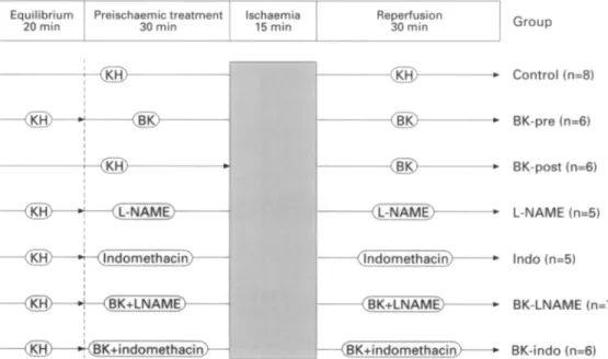
Experimental protocol
Following 20 min drug-free perfusion under control conditions, allowing the hearts to stabilise (equilibrium period), the drugs were added to the perfusate for 30 min (preischaemic treatment period). Global ischaemia was then induced by stopping perfusion for 15 min (ischaemic period). The stopcock was re-opened and the heart reperfused under control condition for 30 min (reperfused period) (fig 1). Left ventricular pressure was continuously monitored, coronary flow was measured at 5 min intervals, and “P spectra were recorded durine 5 min neriods.
The’ rats were randomly assigned to seven groups: (1) untreated control eroun (control. n= 8): (2) lo-’ M bradvkinin eiven nre- ischaemizally’ throughout the experiment (BK-pre, *n = 6); T3) lo-’ M bradykinin given postischaemically at the beginning of reperfusion (BK-post, n=6); (4) lo-’ M Nw-nitro-L-arginine methyl ester (L- NAME) given throughout the experiment (L-NAME, n = 5); (5) 10m5 M indomethacin eiven throughout the exneriment (Indo. n = 5): (6) lo-’ M bradykinin and lOA M L-NAME given throughout the experiment (BK- LNAME. n = 7): (7) lo-’ M bradvkinin and lo-’ M indomethacin given throughout the’experiment (BK-ado, n = 6) (fig 1).
Drugs
Bradykinin, L-NAME, and indomethacin were from Sigma. Statistical analysis
Results are reported as mean(SEM). The percent recovery was obtained compared to the values before ischaemia. Statistical analvsis between groups was performed with the one factor ANOVA Scheffe F test. Statistical analysis in the same group between equilibrium and preischaemic treatment period was performed with a paired Student’s
t test. For all statistical analysis, the null hypothesis was rejected at the 95% level, considering P < 0.05 significant.
Results
Absolute values
At the end of the equilibrium phase, during which the hearts were allowed to stabilise, no significant changes were observed for coronary flow, left ventricular pressure, phosphocreatine, P-ATP, and pHi in any of the groups. There were no significant differences between the control group and all treatment groups. In the table absolute values of coronary flow and left ventricular pressure at the end of the equilibrium phase, the effects of bradykinin, L-NAME, and indomethacin on these variables, and the values after 30 min of postischaemic reperfusion are presented for all groups.
Equilibrium Preischaemic treatment lschaemia Reperfusion
20 min 30 min 15 min 30 min Group
-a-
- *L-NAME)--- ---(L-NAME* <ndomethacin)- (BK+indomethacin> ---, - ---) ----c - - - Control (n=8) BK-pre (n=6) BK-post (n=6) L-NAME (n=5) lndo (n-5) BK-LNAME (n=7) BK-indo (n=6) *-+@K+LNAME> I I *&-#@K+indomethacin>Figure 1 The experimental protocol. Control group: Krebs-Henseleit solution throughout the experiment; BK-pre: bradykinin given preischaemically throughout the experiment; BK-post: bradykinin given postischaemically at the beginning of reperfusion; L-NAME: L-NAME given throughout the experiment; Indo: indomethacin given throughout the experiment: BK-LNAME: bradykinin and L-NAME given throughout the experiment; BK-indo: bradykinin and indomethacin given throughout the experiment.
660 Zhu, Zaugg, Simper, Hornstein, Allegrini, Buser
Effect of bradykinin, L-NAME, and indomethacin on haemodynamic variables after 20 min of equilibrium, 30 min of preischaemic treatment, and 30 min of repefusion. Values are means(SEM).
Coronary flow (ml.min-‘)
Equilibrium Drug Repelfusion
LVP (mm Hg)
Equilibrium Drug Reperjiision Control (n = 8) lO.O(O.3) lO.O(O.3) 4.3(0.4) 8 l(2.6) 80(3.0) 33(3.7)
BK-post (n = 6) lO.O(O.2) lO.O(O.2) 7.1(0.2)* 75(1.4) 74(1.6) 58(3.6)*
BK-pre (n = 6) lO.O(O.2) 13.1(0.8)*t 9.4(1.2)* 76(1.1) 91(3.3)*t 75(8.8)*
L-NAME (n = 5) 10.4(0.3) 5.5(0.2)*t 3.8(0.2) 80(0.9) 80(0.8) 19(4.7)*
Indo (n = 5) 10.3(0.5) 10.6(0.4) 5.4(0.2) 82(0.5) 82(0.7) 40(6.8)
BK-LNAME (n = 7) 10.2(0.6) 9.5(0.6) 5.6(0.2) 79(1.6) 89(2.1)*t 33(7.6)
BK-indo (n = 6) 10.6(0.7) 12.0( 1.0) 5.5(0.7) 81(0.4) 93(0.8)*t 24(7.4)
LVP = left ventricular developed pressure; BK-post = postischaemic administration of bradykinin; BK-pre = preischaemic administration of bradykinin; L-NAME = administration of L-NAME; Indo = administration of indomethacin; BK-LNAME = simultaneous administration of bradykinin and L-NAME; BK-indo = simultaneous administration of bradykinin and indomethacin
*P < 0.05 v control group; tP < 0.05 v respective equilibrium period.
Percent changes during ischaemia and repetfusion
During the 20 min equilibrium period no significant changes were observed for coronary flow, left ventricular pressure, phosphocreatine, B-ATP, and pHi in any group. There were no significant differences between control group and all treatment groups.
CONTROL GROUP
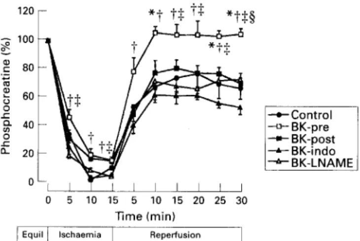
After 15 min of ischaemia and 30 min of reperfusion, coronary flow, left ventricular pressure, phosphocreatine and B-ATP gradually recovered to 43(SEM 4)%, 42(5)%, 67(8)%, and 48(7)%, respectively (figs 2-5).
PREI~~HAEMI~ ADMINISTRATION OF BRADYHNIN (BK-PE)
After 30 min of preischaemic treatment with bradykinin, the absolute values of coronary flow and left ventricular pressure in BK-pre group were significantly increased compared to the equilibrium period and to control (P < 0.05).
After 15 min of ischaemia and 30 min of reperfusion, percent recovery of coronary flow [71(5)% v 43(4)%, P~O.051, left ventricular pressure [81(8)% v 42(5)%, P < 0.051, phosphocreatine [ 105(4)% v 67(g)%, P < 0.051, and B-ATP[78(9)% v 48(7)%, P~O.051 was significantly improved in BK-pre compared to control. Recovery from
100 4 ; 80- a- 3 o 60- ;F t 2 40- : v zo- : 0 5 10 15 5 10 15 20 25 30 Time (min)
I Eauil I lschaemia Rerxrfusion
+Control
Figure 2 Relative changes of coronary flow during ischaemia and reperfusion in control (n = S), and with preischaemic administra- tion of bradykinin (BK-pre, n = 6), postischaemic administration of bradykinin (BK-post, n = 6) simultaneous administration of
bradykinin and indomethacin (BK-indo, n = 6), and simultaneous administration of bradykinin and L-NAME (BK-LNAME, n = 7) in rat hearts.
*P < 0.05 v control; tP < 0.05 v BK-indo.
intracellular acidosis, as assessed by pHi, was not different between BK-pre and control (figs 2-5).
PosxscH~~MIc ADMINISTRATION OF BRADYKININ (BK-POST)
Preischaemic values of coronary flow and left ventricular pressure were not different between BK-post and control. After 15 min of ischaemia and 30 min reperfusion, percent recovery of coronary flow [71(4)% v 43(4)%, P<O.O5] and left ventricular pressure [78(4)% v 42(5)%, P~O.051 was improved in BK-post compared to control. However, recovery of phosphocreatine [70(4)% v 67(8)%, NS] and B-ATP [58(2)% v 48(7)%, NS] was similar. There was no difference for recovery of coronary flow [71(5)% \-I 71(4)%, NS] and left ventricular pressure [81(8)% v 78(4)%, NS] between BK-pre and BK-post. However, recovery of phosphocreatine [105(4)% v 70(4)%, P < 0.051 and B-ATP [78(9)% v 58(2)%, P <0.05] was improved with BK-pre (figs 2-5).
PREISCHAEMIC ADMINISTRATION OF INDOMETHACIN (INDO)
Preischaemic values of coronary flow and left ventricular pressure were not different between Indo and control. After 15 min of ischaemia and 30 min reperfusion, percent recovery of coronary flow [51(3)% v 43(4)%, NS] and left
0 5 10 15 5 10 15 20 25 30
Time (min)
Equil lschaemka Reperfusion
Figure 3 Relative changes of left ventricular developed pressure during ischaemia and reperjusion in control (n = 8) and with preischaemic administration of bradykinin (BK-pre, n = 6), post-
ischaemic administration of bradykinin (BK-post, n = 6), simul- taneous administration of bradykinin and indomethacin (BK-indo, n = 6) and simultaneous administration of bradykinin and L-NAME (BK-LNAME, n = 7) in rats hearts.
*PC 0.05 v control; tP< 0.05 v BK-indo; $P< 0.05 v BK- LNAME.
Bradykinin in the reperfused myocardium 661 g '00 7 ? 80 ‘i; 2 ; 60 E ,a 40 0 if 20 0 11 0 5 IO 15 5 10 15 20 25 30 Time (min)
Equil lschaemia Reperfusion
Figure 4 Relative changes of phosphocreatine during ischaemia and repetjiision in control (n = S), and with preischaemic administration of bradykinin (BK-pre, n = 6j, postischaemic ad- ministration of bradykinin (BK-post, n = 6), simultaneous adminis- tration
qf
bradykinin and indomethacin (BK-indo, n = 6), and simultaneous administration of bradykinin and L-NAME (BK- LNAME, n = 7) in rat hearts.*PC 0.05 v control; iP < 0.05 v BK-indo; $P< 0.05 v BK- LNAME; pSP < 0.05 v BK-post.
ventricular pressure [48(S)% v 42(5)%, NS] was similar to control (figs 2-5).
SIMULTANEOUS ADMINISTRATION OF BRADYKININ AND INDOMETHACIN (BK-INDO)
Preischaemic values of coronary flow showed a non- significant trend to be increased in BK-indo compared to control. However, preischaemic values of left ventricu- lar pressure were higher in BK-indo than in control. (P < 0.05).
After 15 min of ischaemia and 30 min reperfusion, recovery of coronary flow [45(4)% v 43(4)%, NS], left ventricular pressure [29(9)% v 42(5)%, NS], phospho- creatine [53(5)% v 67(8)%, NS], and B-ATP [38(4)% v 48(7)%, NS] with BK-indo was not different from control. Thus the improvement of recovery with bradykinin was
120 I
O--. I I II I I I 1 0 5 IO 15 5 10 15 20 25 30
Time (min)
Equil lschaemia IReperfusion
Figure 5 Relative changes of adenosine triphosphate (B-ATP) during ischaemia and reperjirsion in control (n = 8), and with preischaemic administration of bradykinin (BK-pre, n = 6), post- ischaemic administration of bradykinin (BK-post, n = 6), simul- taneous administration of bradykinin and indomethacin (BK-indo, n = 6), and simultaneous administration of bradykinin and L-NAME (BK-LNAME, n = 7) in rat hearts.
*P < 0.05 v control: +Pc 0.05 v BK-indo; $P< 0.05 v BK- LNAME. 3 5 i- 60 a 20 - 1
L
o- / , , , ,I I
0 5 IO 15 5 10 15 20 25 30 Time (min) lEquil1 lschaemia ReperfusionFigure 6 Relative changes of intracellular pH (pH,) during ischaemia and reperfusion in control (n = 8), and with pre- ischaemic administration of bradykinin (BK-pre, n = 6), postischae- mic administration of bradykinin (BK-post, n = 6), simultaneous administration of bradykinin and indomethacin (BK-indo, n = 6), and simultaneous administration of bradykinin and L-NAME (BK-
LNAME, n = 7) in rat hearts.
totally abolished with the combination of bradykinin and indomethacin (figs 2-5).
PREISCHAEMI~ ADMINISTRATION OF L-NAME (L-NAME)
Preischaemic administration of L-NAME caused a signifi- cant decrease of coronary flow, but left ventricular pressure remained unchanged.
After 15 min of ischaemia and 30 min reperfusion, percent recovery of left ventricular pressure was 23(6)% (NS v control). Percent recovery of coronary flow was higher than in control (P < 0.05), but absolute values [3.8(0.2) ml.min’] indicated a persistent low flow ischaemia (figs 2-5). SIMULTANEOUS ADMINISTRATION OF BRADYKININ AND L-NAME (BK-LNAME)
Preischaemic values of coronary how with BK-LNAME were not different from control. However, a slight increase in left ventricular pressure (P < 0.05) was observed.
After 15 min of ischaemia and 30 min reperfusion, recovery of coronary flow [59(2)% v 43(4)%, NS], left ventricular pressure [40(9)% v 42(5)%, NS], phospho- creatine [73(4)% v 67(8)%, NS], B-ATP [58(6)% v 48(7)%, NS], and pH, [99(0.4)% v 99(0.4)%, NS] with BK-LNAME were not different from control. Thus the improvement of recovery with bradykinin was totally abolished with the combination of bradykinin and L-NAME (figs 2-5).
Discussion
In the present study, we first show that bradykinin ad- ministration during reperfusion improves the recovery of coronary flow and myocardial performance; second that high energy phosphate compounds are not directly involved in the protective effect of bradykinin; and third that bradykinin- induced release of nitric oxide and probably prostacyclin mediate the protective effect of bradykinin.
Evidence for the first finding arises from the improved recovery of coronary flow and left ventricular pressure after postischaemic administration of bradykinin. In the present study, the effect of bradykinin on the postischaemic recovery was compared when bradykinin was given before ischaemia and exclusively after ischaemia, starting with reperfusion.
662 Zhu, Zaugg, Simper, Hornstein, Allegrini, Buser
Both BK-pre and BK-post improved the postischaemic recovery of coronary flow and myocardial performance. It has previously been reported that preischaemic administra- tion of bradykinin significantly reduced myocardial infarct size in the canine and rabbit model.6 7 Increased coronary flow, reduced enzymatic activities, a decreased incidence and duration of postischaemic reperfusion arrhythmias, and improved cardiodynamic indices in isolated rat hearts were also reported. I8 I9 In a pig model, bradykinin improved electrical stability two weeks after experimental myocardial infarction.” However, in all these previous studies bradykinin was given before the onset of ischaemia.
Our results strongly support the hypothesis that bradykinin has protective effects on ischaemic and reperfused hearts.
Second, we showed that high energy phosphate com- pounds are not directly related to the protective effect of bradykinin. In our study, an improved postischaemic recovery of mechanical performance concomitant with preservation of high energy phosphate compounds following preischaemic administration of bradykinin was observed. In contrast postischaemic administration of bradykinin resulted in an improved recovery of mechanical performance and coronary flow while the recovery of the high energy phosphate compounds was not different from control hearts. Therefore the cardioprotective effect of bradykinin was not directly related to the preservation of the high energy phosphate compounds. Some investigators previously pro- posed that an improvement of energy metabolism in ischaemic hearts may contribute to protective effects of bradykinin in the ischaemic heart. Following ischaemia and reperfusion, myocardial tissue levels of glycogen, ATP, and creatine phosphate were increased in bradykinin pretreated isolated rat hearts.5 I8 Our results in BK-pre group correspond very well with previously published data.27 However, postischaemic administration of bradykinin re- sulted in an improved recovery of mechanical performance and coronary flow, while the recovery of the high energy phosphate compounds was not different from control hearts. Because of this discrepancy we speculate that the impaired breakdown of high energy phosphate compounds during ischaemia, when bradykinin is given preischaemically, may not be critical for the bradykinin related improvement of postischaemic recovery. On the other hand, in BK-indo and BK-LNAME there was faster depletion of ATP during ischaemia compared to BK-pre, suggesting that an un- restrained action of both nitric oxide (NO) and prostacyclin may contribute to the protective effect of bradykinin on ATP during ischaemia.
Third, we showed that bradykinin-induced release of a cyclooxygenase product, probably prostacyclin, mediated the protective effect. Evidence for this finding arises from the results that administration of indomethacin alone did not change the postischaemic recovery but simultaneous ad- ministration of bradykinin and indomethacin completely abolished the beneficial effect of bradykinin on the recovery of coronary flow, left ventricular pressure, phosphocreatine, and B-ATP in ischaemic hearts. Consequently the recovery of these variables did not differ significantly from control but did differ from BK-pre. Some investigators previously proposed that the beneficial effect of bradykinin in myocardial ischaemia is probably mediated by stimulation of endothelial Bz receptors. This results in an increase in the intracellular calcium concentration and in the release of prostacyclin.’ The ability of bradykinin to stimulate prostacyclin generation from cultured endothelial cells is well recognised25 and this effect was inhibited in a dose
dependent manner by the bradykinin receptor antagonist.26 Prostacyclin given to isolated working rat hearts reduced ischaemia-reperfusion injury.27 Our results support these observations. It is suggested that cyclooxygenase products, probably prostacyclin, are important mediators involved in the protective effects of bradykinin on ischaemic hearts. Thus bradykinin-induced release of vasodilator cyclo- oxygenase products contributes to the cardioprotective effects of bradykinin.
In addition, we showed that bradykinin-induced release of NO mediated the protective effect of bradykinin. Evidence for this arises from the finding that administration of L-NAME alone worsened the postischaemic recovery, and simultaneous administration of bradykinin and L-NAME abolished the beneficial effect of bradykinin on the recovery of coronary flow, left ventricular pressure, phosphocreatine, and B-ATP, which did not differ from control. Previous studies have shown that bradykinin is a potent endothelium dependent vasodilator in many vessels.‘l It can stimulate the release of NO from cultured endothelial cells*’ as well as from isolated guinea pig hearts.23 NO released from endothelial cells activates the haem protein soluble guanylyl cyclase in vascular smooth muscle cells and in platelets. The resulting increase in cyclic guanosine monophosphate inhibits smooth muscle contraction and platelet activation by reducing the intracellular concentration of free calcium. A recent study showed that this effect was blocked by the bradykinin B2 antagonist HOE140.24 In the present study, the improved postischaemic recovery of coronary flow reflects the dilator action of bradykinin, and the improved recovery of left ventricular pressure may result from the improved tissue perfusion. With simultaneous administration of brady- kinin and L-NAME the beneficial effects of bradykinin on recovery of coronary flow, left ventricular pressure, phosphocreatine, and B-ATP in ischaemic hearts were abolished. Therefore an unrestrained action of nitric oxide contributes to the cardioprotective effect of bradykinin.
Our conclusions are: (1) bradykinin improves postischae- mic myocardial recovery when given before ischaemia or starting with reperfusion; (2) this beneficial effect is only partially related to the preservation of the high energy phosphate compounds; and (3) the beneficial effects of bradykinin on postischaemic myocardial recovery are dependent from an unrestrained action of nitrix oxide and prostacyclin.
PZ is supported by the Swiss Foundation for Cardiology. CEZ is sunuorted bv a grant from Swiss National Science Foundation. PTB is a recipient of acareer development grant (SCORE) from the Swiss National Science Foundation.
Key terms: ischaemia; reperfusion; myocardium; bradykinin; nitric oxide; prostacyclin; magnetic resonance spectroscopy; cardioprotective effect.
Received 8 August 1994; accepted 20 December 1994. Time for primary review 28 days.
1 Scholkens BA, Linz W. Cardiourotective effects of ACE inhibitors: experimental proof and clinical perspectives. Clin
Physiol Biochem 1990;8(suppl 1):33-43.
2 Gavras I, Gavras H. Cardioprotective potential of angiotensin- converting enzyme inhibitors. Clin Car&o1 1991;14(suppl IV): IV-68-71.
3 Ondetti MA, Cushman D. Enzymes of the renin-angiotensin system and their inhibitions. Annu Rev Biochem 198251: 283-308.
4 Westlin W, Mullane K. Does captopril attenuate reperfusion- induced myocardial dysfunction by scavenging free radicals?
Bradykinin in the repe@sed myocardium 663 5 6 7 8 9 10 II 12 13 14 15
Linz W, Scholkens BA, Influence of local converting enzyme inhibition on angiotensin and bradykinin effects in ischemic rat hearts. J Curdiovusc Pharmacol 1987;1O(suppl VII): S75-82.
Linz W, Scholkens BA. Role of bradykinin in the cardiac effects of angiotensin-converting enzyme inhibitors. J Cardiovasc Phur-
macol 1992;2O(suppl IX):S81-90.
Hartman JC, Wall TM, Hullinger TG, Shebuski RJ. Reduction of myocardial infarct size in rabbits by ramiprilat: reversal by the bradykinin antagonist HOE 140. J Curdiovasc Pharmucol 1993; 21:9961003.
Muller-Ester1 W. Kininogens, kinins, and kinships. J Curdiovusc Phurmucol 1990;15(suppl VI):Sl-6.
Mats&i T, Yoshida S. Sympathetically induced myocardial ischaemia causes the heart to release olasma kinin. Cardiovusc Res 1987;21:428-32.
Baumgarten CR, Linz W, Kunkel G, Scholkens BA, Wiemer G. Rami@ilat increases bradykinin outflow from isolated hearts of rat. Br J Pharmacol 1993;108:293-5.
Hashimoto K, Hamamoto H, Honda Y, Hirose M, Furukawa S, Kimura E. Changes in components of kinin system and hemodynamics in acute myocardial infarction. Am Heart J
1978;95:619-26.
Buser PT, Wagner S, Wu ST, et al. Verapamil preserves myocardial performance and energy metabolism in left ventricular hypertrophy following ischemia and reperfusion: phosphorus 31 magnetic resonance spectroscopy study. Circulation 1989;80: 183745.
Zaugg CE, Zhu P, Simper D, Liischer TF, Allegrini PR, Buser PT. Differential effects of endothelin-1 on normal and postischemic reperfused myocardium. J Curdiovusc Phurmucol 1993;22(suppl VIII):S367-70.
Buser PT, Wagner S, Wu S, Higgins CB, Wikman-Coffelt J. Protective effects of calcium antagonists on energy and substrate metabolism during ischemia and reperfusion. J Curdiovasc Pharmacol 1991;18(suppl X):S87-92.
Buser PT, Auffermann W, Wu ST, Jasmin G, Parmley WW, W&man-Coffelt J. Dobutamine potentiates amrinone’s beneficial effects in moderate but not in advanced heart failure: “P-MRS in isolated hamster hearts. Circ Res 1990:66:747-53.
16 17 18 19 20 21 22 23 24 25 26 27
Buser PT, Wikman-Coffelt J, Wu ST, Derugin N, Parmley WW, Higgins CB. Postischemic recovery of mechanical performance and energy metabolism in the presence of left ventricular hypertrophy: a “p-MRS study. Circ Res 1990;66:73546.
Gaddian DG, Radda GK, Richards RE, Seeley PJ. Biological applications of magnetic resonance. New York: Academic Press, 1979.
Linz W, Martorana PA, Scholkens BA. Local inhibition of bradykinin degradation in ischemic hearts. J Curdiovusc Phurmu-
col 199O;lS(suppl VI):S99-109.
Chahine R, Adam A, Yamaguchi N, Gaspo R, Regoli D, Nadeau R. Protective effects of bradykinin on the ischemic heart: implication of the B, receptor. Br J Pharmucol 1993;108: 318-22.
Tobe TJ, de LC, Tio RA, Be1 KJ, Mook PH, Wesseling H. In vivo effect of bradykinin during ischemia and reperfusion: improved electrical stability two weeks after myocardial infarction in the pig. J Curdiovusc Phurmucol 1991;17:60&7.
Leipert B, Becker BF, Gerlach E. Different endothelial mechan- isms involved in coronarv resoonses to known vasodilators. Am J Physiol 1992;262:Hl6761-83. ’
Palmer RMJ, Ferrige AG. Nitric oxide release accounts for the biological activity of endothelium-derived relaxing factor. Nature 1987;327:524d.
Kelm M. Schrader J. Control of coronarv vascular tone bv nitric
oxide. Ckc Res 1990;66:1561-75. ’ ,
Wiemer G, Schiilkens BA, Becker RHA, Busse R. Ramiprilat enhances endotbelial autacoid formation by inhibiting breakdown of endothelium-derived bradvkinin. Hwertension 1991:18:558-63. McIntyre TM, Zimmerman GA, S.M. P Cultured endothelial cells synthesize both platelet-activating factor and prostacyclin in response to histamine, bradykinin and adenosine triphosphate. J Clin Invest 1985;76:271-80.
Hoffmann G, Pietsch R. Converting enzyme inhibition and vascular prostacyclin synthesis: effect of kinin receptor antagon- ism. Eur J Pharmacol 1990;178:79-83.
Linz W, Martorana PA, Grotsch H, Bei-Yin Q. Antagonizing bradykinin obliterates the cardioprotective effects of bradykinin and angiotensin-converting enzyme (ACE) inhibitors in ischemic hearts. Drug Dev Res 1990;19:393-408.