Virus Burden in Lymph Nodes and Blood of Subjects with Primary Human
Immunodeficiency Virus Type 1 Infection on Bitherapy
Luc Perrin, Sabine Yerly, Francis Marchal, Laboratory of Virology, AIDS Centre, Division of Infectious Diseases, Department of Otho-Rhino-Laryngology, Geneva University Hospital, Ge´rard A. Schockmel, Bernard Hirschel, Cecil H. Fox,
Geneva, and Laboratory of Immunopathology of AIDS, Division of and Giuseppe Pantaleo
Infectious Diseases, Hoˆpital de Beaumont, Lausanne, Switzerland; Molecular Histology Labs, Inc., Gaithersburg, Maryland At present, it is not known whether undetectable plasma viremia corresponds to an absence of
human immunodeficiency virus type 1 (HIV-1) replication in lymphoid tissues. This issue has been explored in 11 subjects with primary HIV-1 infection treated with zidovudine plus didanosine by evaluating virologic markers in blood and lymphoid tissues 9 –18 months after initiation of treatment. These markers include plasma viremia, measured with a sensitive assay with a detection limit of 20 HIV-1 RNA copies/mL, infectious virus titers and proviral DNA in lymph node mononuclear cells, and HIV-1 RNA in lymphoid tissue. Five subjects had plasma viremiaõ20 copies/mL and showed no evidence of viral replication in lymphoid tissue. Six subjects had both detectable plasma viremia and evidence of HIV-1 RNA in lymphoid tissue. The results indicate that absence of detectable HIV RNA in lymphoid tissue is associated with viremia levels of HIV-1 RNAõ20 copies/mL.
The primary goal of combined antiviral therapy is to achieve lymphoid tissue biopsies were obtained from lymph nodes, tonsils, and adenoids, and viral replication was assessed in complete suppression of viral replication and possibly
eradica-tion of human immunodeficiency virus type 1 (HIV-1). Re- parallel in blood and lymphoid tissues. cently, several studies showed a dramatic reduction of viremia
to levels below the detection limit of currently used assays
(i.e., 500 – 200 HIV-1 RNA copies/mL of plasma) in HIV-1 – Patients and Methods infected subjects receiving combined antiviral therapy [1 – 3].
Study population. All subjects with primary HIV-1 infection However, the recent development of a sensitive assay for the
referred to us between January 1995 and March 1996 were offered determination of plasma viremia, with a lower detection limit
an open-label therapeutic regimen with zidovudine (250 mg twice of 20 copies/mL, demonstrated that 45% of plasma samples
a day) and didanosine (200 mg twice a day) [8]. All subjects with viremiaõ200 copies/mL, had ú20 HIV-1 RNA copies/
experienced an acute retroviral syndrome at the time of or in the mL of plasma [4].
month preceding initiation of antiviral therapy, except 1 subject To date, it is difficult to assess whether the absence of detect- who was included 2 months after the onset of the acute retroviral able HIV-1 RNA in plasma reflects the situation in other ana- syndrome. The subjects were Caucasians; 7 were male and 4 fe-tomic compartments, such as lymphoid tissue, which is the male, with a mean age at study entry of 37 years (range, 26 – 54). primary site for viral spreading and replication [5 – 7]. There- HIV-1 infection was acquired through homosexual contacts in 6 subjects and heterosexual contacts in 5. At 9 – 18 months after fore, it seems crucial to compare the levels of viral replication
initiation of treatment, excisional lymph node biopsies were per-in blood and lymphoid tissue and to identify plasma viremia
formed in the inguinal area in 11 subjects; tonsil and adenoid levels that correspond to undetectable viral replication in
biopsies were collected in parallel in 3 of them. lymphoid tissue.
HIV-1 RNA assay. Viremia levels were determined in batches To address these issues, 11 subjects with primary HIV-1
using EDTA-treated plasma samples, stored in aliquots at0757C, infection receiving combination therapy [8] were enrolled in a
using the Amplicor HIV-1 Monitor test (Roche, Basel, Switzer-prospective study. At 9 – 18 months after treatment initiation,
land) according to the manufacturer’s instructions, with a detection limit of Ç200 HIV-1 RNA copies/mL. Samples with viremia õ200 copies/mL were retested using a ‘‘boosted’’ modification of the Amplicor HIV Monitor test with a detection limit of 20 Received 16 September 1997; revised 8 January 1998. HIV-1 RNA copies/mL [4].
Presented in part: Antiviral Academy Meeting, Cannes, France, 21 March
Quantification of proviral DNA. The proviral DNA concentra-1997.
tion associated with lymph node mononuclear cells (LNMC) and Informed consent was obtained from all patients in the study, and the
proto-col was approved by the Institutional Review Board. peripheral blood mononuclear cells (PBMC) was measured in du-Financial support: Swiss National AIDS Research Program (3239.041951.94). plicate as previously reported [9]. The input of cellular DNA was Reprints or correspondence: Dr. Luc Perrin, Laboratory of Virology, Geneva
2.5 mg/assay, corresponding to 375,000 cells. The limit of detection University Hospital, 1211 Geneva 14, Switzerland ([email protected]).
of the assay was 1 – 3 HIV-1 DNA copies/2.5 mg of DNA. Samples The Journal of Infectious Diseases 1998; 177:1497 – 501
withõ3 copies were retested in triplicate using the same
proce-q 1998 by The University of Chicago. All rights reserved.
In situ hybridization. In situ hybridization for the detection of Results
HIV-1 RNA was performed on paraffin-embedded tissue sections HIV-1 replication in blood. Viral replication in blood was using RNA probes that comprised 90% of the HIV-1 genome, as
evaluated, at the time of lymph node collection, via the determi-previously described [5]. The number of individual cells
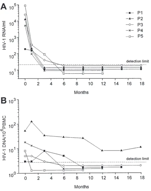
express-nation of plasma viremia. Five of 11 subjects (subjects 1 – 5; ing HIV-1 RNA were quantified by first determining the area of
table 1) had levels of plasma viremiaõ20 HIV-1 RNA copies/ the biopsy section, using computerized planimetry, followed by
mL. Subjects 6 – 8 had levels of plasma viremia just above the counting the number of HIV-1 RNA – expressing cells in the biopsy
detection limit, ranging between 22 and 25 copies/mL, whereas section, using a darkfield microscope at1100 magnification. The
plasma viremia wasú200 copies/mL in subjects 9 – 11 (table sensitivity of the in situ hybridization procedure was determined
1). The sensitive assay allowed the detection of viremia in by constructing a standard containing a known number of HIV-1
virions and was found to correspond to 13 virus particles (5 silver subjects 6 – 10, in whom viremia levels were below the detec-grains). HIV-1 RNA – expressing cells were reported as the number tion limit (500 to 200 HIV-1 RNA copies/mL) of the commer-of cells per 10 mm2
. cial assay. Figure 1A shows the kinetics of plasma viremia in Infectious HIV-1 titers. Suspensions of LNMC were ob- the 5 subjects withõ20 HIV-1 RNA copies/mL at the time of tained after mincing lymph node tissue with a scalpel, teasing lymph node collection. All 5 had plasma viremia ofõ20 cop-out the cells, and isolating them by ficoll-hypaque gradient
cen-ies/mL at 6 months after initiation of combination therapy. trifugation. Aliquots of 51 106
cells in 20% fetal calf serum and
HIV-1 replication in lymph nodes. Viral replication in 10% dimethyl sulfoxide were stored in liquid nitrogen. Cellular
lymph nodes was evaluated by in situ hybridization of cell-infectious titers were determined using quantitative cell
micro-associated HIV-1 RNA and by determination of infectious virus culture [10]. Six dilutions of LNMC, depleted of CD8 cells,
titers in suspensions of LNMC (table 1). Subjects 1 – 6 showed starting at a concentration of 106 LNMC, were cocultivated in
no evidence of cells expressing HIV-1 RNA. Cells expressing duplicate with 106
phytohemagglutinin (PHA)-stimulated CD8
HIV-1 RNA were, however, detected in subjects 7 – 11 (figure cell – depleted PBMC from HIV-negative donors. Half of the
culture medium was replaced with fresh medium twice a week. 2). A second lymph node biopsy was performed in subjects 1 Fresh uninfected PHA-stimulated PBMC were added to the cul- and 2 at 7 months after the first biopsy. Like the first lymph ture once a week. On days 14 and 21, culture fluid from each node analyzed, the second had no detectable cells expressing well was tested for HIV-1 p24 antigen level using a commercial HIV-1 RNA. Infectious virus was recovered from subjects 6, ELISA (Abbott Laboratories, North Chicago). Results are ex- 7, and 9 – 11. The titers of infectious virus were very low in pressed as infectious units per million cells.
subjects 6 and 7 and higher in subjects 9 – 11 (table 1). No Immunologic parameters. CD3, CD4, and CD8 lymphocyte
evidence of active viral replication as assessed by coculture counts were determined on fresh material in lymph nodes and
was found in subjects 1 – 5, whereas virus was isolated in sub-blood by flow cytometry (Coulter EPICS IV; Instrumente
Gesell-jects 6 – 11 (table 1). Failure to detect HIV-1 replication in schaft, Basel, Switzerland) using fluoresceinated DAKO-CD3,
lymph nodes was confined to subjects 1 – 5, who had plasma DAKO-CD8, and R-Phycoerythrin DAKO-CD4 (Dako,
Glos-viremia ofõ20 copies/mL. trup, Denmark). To minimize variations linked to fluctuations
HIV-1 RNA associated with follicular dendritic cells (FDC).
in total lymphocyte counts, results were expressed as the CD4/
CD8 ratio. Previous studies showed that the diffuse HIV-1 RNA
hybrid-Table 1. Virus burden and CD4/CD8 cell ratio in blood and lymph nodes afterú9 months of antiviral therapy.
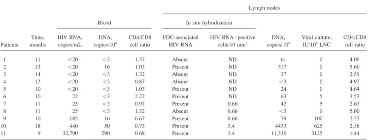
Lymph nodes
Blood In situ hybridization
Time, HIV RNA, DNA, CD4/CD8 FDC-associated HIV RNA – positive DNA, Viral culture, CD4/CD8 Patients months copies/mL copies/106 cell ratio HIV RNA cells/10 mm2 copies/106 IU/106LNC cell ratio
1 11 õ20 õ3 1.57 Absent ND 61 0 4.00 2 13 õ20 16 1.03 Present ND 317 0 5.60 3 14 õ20 õ3 1.32 Absent ND 37 0 2.59 4 12 õ20 õ3 0.87 Absent ND õ3 0 4.92 5 10 õ20 õ3 1.03 Present ND 24 0 4.64 6 10 22 õ3 2.22 Present ND 63 5 3.53 7 11 25 õ3 0.97 Present 0.66 42 5 2.63 8 11 25 õ3 1.32 Absent 0.66 õ3 0 5.00 9 10 185 16 0.67 Present 0.66 79 100 2.32 10 18 440 50 0.73 Present 3.4 4433 625 2.38 11 9 32,790 290 0.68 Present 3.4 11,336 3125 1.44
Figure 1. Viremia (A) and proviral DNA (B) levels in 5 subjects treated with zido-vudine plus didanosine with no detectable viral replication in lymph nodes. Patient nos. (P1 – P5) indicated in A correspond to those reported in table 1. Detection limit in dashed line corresponds to 20 HIV-1 RNA copies/mL of plasma (A) and 3 proviral DNA copies/106
peripheral blood mononu-clear cells (PBMC) (B).
ization signal associated with germinal centers corresponded ever, only traces of HIV-1 trapped in the FDC network were observed in tonsil tissue (data not shown) compared with the to HIV-1 virions trapped in the FDC network [5 – 7]. In our
study, trapping of HIV-1 in the FDC network was either absent presence of both trapped virions and individual cells expressing HIV-1 RNA in lymph node tissue (table 1).
(subjects 1, 3, and 4) or present in traces (subjects 2 and 5) in
subjects with plasma viremia ofõ20 copies/mL (table 1, figure Proviral HIV-1 DNA. Proviral HIV-1 DNA concentrations in blood wereõ3 copies/106PBMC in 4 of 5 subjects with
2). In subject 2, trapping of HIV-1 was no longer detected
in the second lymph node biopsy, performed 7 months later. plasma viremia ofõ20 HIV-1 RNA copies/mL. Among the subjects with viremia of ú20 copies/mL, proviral DNA in Trapping of HIV-1 in the FDC network was present in all but
1 of the subjects with plasma viremia ofú20 copies/mL (ta- blood was below the detection limit in subjects 6 – 8, with viremia of 20 – 30 copies/mL, whereas the highest levels were ble 1).
HIV-1 distribution in lymph nodes, tonsils, and adenoids. observed in subjects 9 – 11, with viremia ofú30 copies/mL (table 1). Proviral DNA was detected in lymph nodes of all Biopsies of tonsils and adenoids were performed in subjects 4
and 9 at the same time as the lymph node biopsy and in subject subjects except 4 and 8 (table 1). Proviral DNA was signifi-cantly higher (6- to 39-fold) in LNMC than in PBMC (table P2 at the time of the second lymph node biopsy. No differences
in HIV-1 distribution, including trapping of virions and cell 1). Kinetics of proviral DNA decay in PBMC from subjects 1 – 5 are shown in figure 1B. Four of 5 subjects had proviral expression of HIV-1 RNA, were observed in subjects 2 or 4
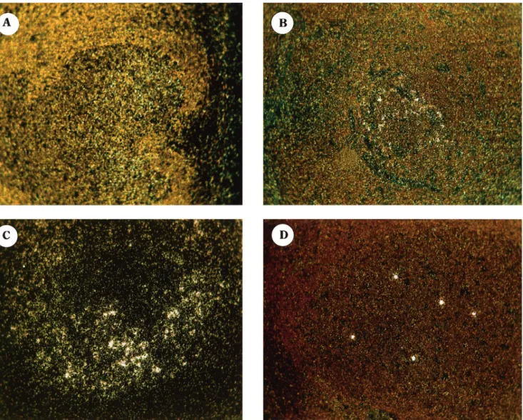
Figure 2. Sections of lymph node tissue from 4 subjects who received antiviral therapy with zidovudine plus didanosine at time of primary HIV infection and who had evidence of persistent viral replication and/or follicular dendritic cell (FDC) – associated deposits. Darkfield images of in situ hybridization for HIV RNA are shown. Location of HIV RNA is indicated by silver grains, which appear as white dots. A – C, Hybridization signal is localized over germinal centers; typical examples of different degrees of virus trapping in FDC are represented in A (very low), B (low), and C (intermediate). D, Typical example of hybridization signal localized in 5 isolated cells expressing HIV-1 RNA.
decrease of proviral DNA was slower than that of viremia HIV-1 RNA copies/mL of plasma [1 – 3]. However, HIV-1 replication in lymphoid tissue was detected in subjects with (figure 1).
plasma viremia below 500 to 200 copies/mL [11, 12]. The
Lymphocyte markers. CD4/CD8 ratios in lymph nodes and
results of the present study, performed by using a sensitive blood are reported in table 1. All subjects, regardless of plasma
assay with a detection limit of 20 HIV-1 RNA copies/mL, viremia levels, had a higher CD4/CD8 ratio in lymph nodes
indicate that the plasma viremia levels corresponding to un-than in blood. However, no significant correlation was observed
detectable viral replication in lymphoid tissue, including between the CD4/CD8 ratios in lymph nodes and blood.
Sub-lymph nodes, adenoids, and tonsils, are generallyõ20 cop-jects with no evidence of viral replication in lymph nodes
ies/mL. When detectable, viral replication in lymphoid tissue tended to have a higher CD4/CD8 ratio in lymphoid tissue
appears to be associated with plasma viremia ú20 copies/ (median, 4.64 vs. 2.51; PÅ .07).
mL. It is important to underscore, however, that levels of plasma viremiaõ20 copies/mL and undetectable viral repli-Discussion
cation in lymphoid tissue do not necessarily reflect complete Viremia levels in blood are routinely measured by com- suppression of viral replication, since the techniques used mercial assays with a lower detection limit of 500 to 200 are limited by their sensitivity and the size of the sample
References
analyzed [13]. Moreover, the use of frozen LNMC might
decrease the recovery of infectious virus. 1. Hammer SM, Squires KE, Hughes MD, et al. A controlled trial of two In situ hybridization analysis using lymph node sections nucleoside analogues plus indinavir in persons with human immunode-presents advantages in terms of the surface area analyzed com- ficiency virus infection and CD4 cell counts of 200 per cubic millimeter
or less. N Engl J Med 1997; 337:725 – 33. pared with tonsil biopsies, but the screening of a limited number
2. Gulick RM, Mellors JW, Havlir D, et al. Treatment with indinavir, zido-of tissue sections for the presence zido-of virus-expressing cells
vudine, and lamivudine in adults with human immunodeficiency virus does not exclude the possibility of low viral replication in other
infection and prior antiretroviral therapy. N Engl J Med 1997; 337:734 – lymph nodes or lymphoid tissues [12]. Furthermore, although 9.
virus distribution (trapping of virions by FDC) and viral repli- 3. Myers M, Montaner J, Group IS. A randomized, double-blinded compara-cation levels appear to be quite homogeneous in different tive trial of the effects of zidovudine, didanosine, and nevirapine combi-nations in antiviral naive, AIDS-free, HIV-infected patients with CD4 lymphoid tissues in the absence of antiviral therapy,
heteroge-counts 200 – 600/mm3
[abstract]. In: Program and abstracts: XI Interna-neity was observed in 1 subject in comparing lymph nodes,
tional Conference on AIDS (Vancouver, Canada). Vancouver: XI In-tonsils, and adenoid biopsies.
ternation Conference on AIDS Society, 1996.
Virions trapped within the FDC network might be a source 4. Schockmel G, Yerly S, Perrin L. Detection of low HIV-1 RNA levels in of viral spreading and replication [7, 2]. Similarly, proviral plasma. J Acquir Immune Defic Syndr 1997; 14:179 – 83.
5. Fox CH, Tenner-Racz K, Racz P, Firpo A, Pizzo PA, Fauci AS. Lymphoid HIV-1 DNA may allow reactivation of the latent viral genome
germinal centers are reservoirs of human immunodeficiency virus type leading to viral replication. Although most of the proviral DNA
1 RNA. J Infect Dis 1991; 164:1051 – 7. corresponds to defective virus, recombination of defective virus
6. Pantaleo G, Graziosi C, Butini L, et al. Lymphoid organs function as and generation of replication-competent virus in vivo may po- major reservoirs for human immunodeficiency virus. Proc Natl Acad tentially contribute to maintain HIV-1 infection [14, 15]. Eradi- Sci USA 1991; 88:9838 – 42.
cation of HIV-1 in infected individuals may require that viral 7. Haase AT, Henry K, Zupancic M, et al. Quantitative image analysis of HIV-1 infection in lymphoid tissue. Science 1996; 274:985 – 9. replication, viral trapping, and proviral DNA are completely
8. Perrin L, Yerly S, Charvier A, Figueras G, Schockmel GA, Hirschel B. eliminated from all body tissues. In the present study, in only
Zidovudine plus didanosine in primary HIV-1 infection. Antiviral Ther subject P4 were those virologic measures below the limit of
1997; 2:9 – 15.
detection of the assays used.
9. Baumberger C, Kinloch-de Loe¨s S, Yerly S, et al. High level of circulating The slower decay of proviral DNA in PBMC compared with RNA in patients with symptomatic HIV-1 infection. AIDS 1993; 7(suppl plasma viremia in our study is consistent with the recent mathe- 2):59 – 64.
10. Fiscus SA, DeGruttola V, Gupta P, et al. Human immunodeficiency virus matical model indicating a half-life of 1 – 3 weeks for
chroni-type 1 quantitative cell microculture as a measure of antiviral efficacy cally infected cells [16]. In the present study, proviral DNA in
in a multicenter clinical trial. J Infect Dis 1995; 171:305 – 11. PBMC was not detectable in most subjects with undetectable
11. Tamalet C, Lafeuillade A, Fantini J, Poggi C, Yahi N. Quantification of viremia after 9 – 18 months of treatment. In contrast, proviral HIV-1 viral load in lymphoid and blood cells: assessment during four-DNA in PBMC is consistently detected in subjects with chronic drug combination therapy. AIDS 1997; 11:895 – 901.
HIV infection treated with combination therapy and in whom 12. Cavert W, Notermans DW, Staskus K, et al. Kinetics of response in lymphoid tissues to antiretroviral therapy of HIV-1 infection. Science viremia was undetectable [13] (unpublished data). Treatment
1997; 276:960 – 4.
at the time of primary infection might thus reduce spreading
13. Finzi D, Hermankova M, Pierson T, et al. Identification of a reservoir for of the infection and the size of the pool of latently HIV-1 –
HIV-1 in patients on highly active antiretroviral therapy. Science 1997;
infected cells. 278:1295 – 300.
In conclusion, in this investigation there was a good correla- 14. Li Y, Kappes JC, Conaway JA, Price RW, Shaw GM, Hahn BH. Molecular characterization of human immunodeficiency virus type 1 cloned di-tion between viremia and HIV-1 replicadi-tion in lymphoid tissues
rectly from uncultured human brain tissue: identification of replication-when viremia levels were assessed by a sensitive assay in
competent and defective viral genomes. J Virol 1991; 65:3973 – 85. subjects with primary HIV infection treated forú9 months.
15. Sanchez G, Xu X, Chermann JC, Hirsch I. Accumulation of defective viral genomes in peripheral blood mononuclear cells of human im-munodeficiency virus type 1 – infected individuals. J Virol 1997; 71: Acknowledgments
2233 – 40.
We are indebted to Elbe Ramirez, Chantal Gaille, and Wanda 16. Perelson AS, Essunger P, Cao Y, et al. Decay characteristics of HIV-1 – Caveng for excellent technical assistance and Christine Brown for infected compartments during combination therapy. Nature 1997; 387:
188 – 91. preparing the manuscript.