Sex-dependent differences in left ventricular function and
structure in chronic pressure overload
B. VILLARI*, S. E. CAMPBELL")", J. SCHNEIDER, G. VASSALLI, M. CHIARIELLO* AND O. M. HESS
Department of Internal Medicine, Cardiology and Institute of Pathology, University Hospital, Zurich, Switzerland; * Division of Cardiology, Federico II University of Naples; ^Division of Cardiology, University of
Missouri-Columbia, Columbia, MO, U.S.A.
KEY WORDS: Aortic stenosis, myocardial function, collagen network, left ventricular hypertrophy.
To evaluate gender-related differences in left ventricular (LV) structure and function in aortic stenosis, LV biplane cineangiography, micromanometry and endomyocardial biopsies were carried out in 56 patients with aortic stenosis and normal coronary arteries. Patients were divided into males (M: n=35), and females (F: n=21). Sixteen normal subjects 8 M, 8 F) served as haemodynamic controls. Control biopsy data were obtained from six pre-transplantation donor hearts (3 M and 3 F). LV systolic function was evaluated by ejection fraction and its relationship to mean systolic circumferential wall stress, diastolic function by the time constant of LV pressure decay, peak filling rates and passive myocardial stiffness constant. Biopsy samples were evaluated for interstitial fibrosis, muscle fibre diameter and volume fraction of myofibrils. In a subset of 27 consecutive patients, biopsy samples were evaluated with a
morphometric-morphological method, for total collagen volume fraction, endocardia! fibrosis and the extension and thickness of orthogonal collagen fibres (cross-hatching).
In patients with aortic stenosis, aortic valve area, aortic valve resistance and mean aortic pressure gradient were comparable in males and females, whereas end-systolic and end-diastolic volumes were larger in males than females. Ejection fraction was lower (56%) in males than females (64%) (P<0-05); 20 of 35 males and four of 21 females had depressed systolic contractility when assessed with regard to the relationship ejection fraction-mean systolic stress (P<00I). Myocardial stiffness constant was higher in males than in females (P<001). Nine of 14 males and two of 13 females had endocardia! fibrosis (P<0009), whereas increased cross-hatching (> 1-5 grade) was present in 11 males and four females with aortic stenosis (¥<001). An abnormal collagen architecture was present in 13114 males and 5113 females (?<0002).
In aortic stenosis, males have a depressed systolic function and abnormal passive elastic properties when compared to females with valve lesions of similar severity. Changes in collagen architecture may account, at least in part, for these
differences.
Introduction
The adaptation of the left ventricle to chronic pressure overload has been studied extensively in the past, but only recently have gender-related differences been found to be important'1"51. Female rats develop hypertrophy in response to physical training, while the male counterpart does not'1'; spontaneously hypertensive rats show less hypertrophy in males than in similarly aged and equally hypertensive females'21. In humans, Carroll et a/.'3' and Douglas et a/.'4' observed gender-associated differences in left ventricular (LV) adaptation to systolic pressure overload in aortic stenosis of the elderly. Topol et a/.'51 reported in a group of 21 elderly patients with hyperten-sive hypertrophic cardiomyopathy that 16 were females. Several factors have been discussed which could be
Submitted for publication on 1 September 1994, and accepted 21 December 1994.
This paper is dedicated to the memory of Hans Pietcr Krayenbuehl.
Correspondence: Otto M. Hess, MD, Division of Cardiology, Unhersity
Hospital, Raemistrasse 100. 8091 Zurich. Switzerland
responsible for these gender-related differences including mechanical, hormonal and cellular mechanisms'1 5!.
The purpose of the present study was to evaluate possible gender-related differences in LV adaptation in a control group and in patients with severe aortic stenosis. Special attention was paid to differences in systolic and diastolic function as well as to differences in cellular hypertrophy and collagen network.
Patients and methods
PATIENT POPULATION
A group of 72 patients was studied, which comprised 16 controls (eight males and eight females) and 56 patients with severe aortic stenosis (35 males and 21 females). Six pre-transplantation donor hearts (three males and three females) were used for control biopsy data. Patients with significant aortic regurgitation (>30%) or with coronary artery disease (>50% coronary artery narrowing) were excluded form the present analy-sis. Patient characteristics and baseline haemodynamics are given in Table 1.
Table I Patient characteristics Age (years) NYHA AVA ( c m2. m -2) AVR (dyne. s. c m -5) A? (mmHg) fao (%) CI ( l . m i n . m -2) Dur sympt (months)
Controls Male (n = 8) 49 ± 8 — — — — — 4-3 ±0-5 — Female (n = 8) 49 ± 8 — — — — — 4 0 ±0-9 — Aortic Male (n = 35) 51 ± 12 2-6 ±0-5 0-4 ± 0-2 622 ±137 70 ± 17 13± 12 3-1 ±0-7*** 21 ±20 stenosis Female (n=21) 53 ±15 2-3 ±0-6 0-4 ±0-2 649 ±217 73 ±25 10± 10 3O±O-5ttt 28 ±27 AVA = aortic valve area index; AVR = aortic valve resistance; NYHA = New York Heart Associ-ation functional class; ,dP=mean aortic gradient; fao = aortic regurgitant fraction; CI = cardiac index; Dur sympt = duration of symptoms.
•••/><0001 vs control males, t t t ^<0 0 0 1 vs control females.
CARDIAC CATHETERIZATION
Informed consent was obtained from all patients. Cardiovascular medications were withheld 12 to 24 h prior to the procedure. Pre-medication consisted of
10 mg chlordiazepoxide given orally 1 h prior to cath-eterization. Right and left heart catheterization were carried out in all patients. Biplane left ventriculography was performed in the right anterior oblique (30°) and left anterior oblique (60°) projection at a filming rate of 50 frames per second. LV pressure was measured simulta-neously with ventriculography by means of a Millar 7F micromanometer-catheter introduced transseptally into the left ventricle via an 11-5F Brockenbrough guiding catheter. In controls, a pigtail-Millar 8F micromanom-eter was introduced retrogradely into the left ventricle. Central aortic pressure was measured through a fluid-filled 8F pigtail catheter. All pressures were recorded at a paper speed of 250 mm . s~ ' with a standard lead of the ECG; the first derivative of LV high-fidelity pressure (dP/dt) and the time markers corresponded with the digital numbers on the angiographic images'6'71. Cardiac index was measured by the Fick method.
LV angiographic silhouettes were drawn manually from an adequately opacified sinus beat, excluding extrasystolic and post-extrasystolic beats. LV volumes were determined on a frame-by-frame basis using the area-length method18'. Dimensional and volume data were filtered using the moving average technique16'71. The LV pressure tracing was digitized for one cardiac cycle using an electronic digitizer, interfaced to a com-puter. Pressure and volume data were analysed every 20 ms for one cardiac cycle. End-diastole was defined as the time point of the rapid upstroke of the first deriva-tive of the left ventricular pressure (dP/dt). LV mass was determined according to the method of Rackley et alP\ Circumferential wall stress was calculated from a simpli-fied version of Mirsky's thick wall mode1101. Mean systolic circumferential wall stress was defined as the mean wall stress occurring during the systolic ejection period.
The end-diastolic ratio of the short axis to wall thickness was computed and used as a parameter for assessing the type of chamber hypertrophy'"1.
ASSESSMENT OF LEFT VENTRICULAR FUNCTION
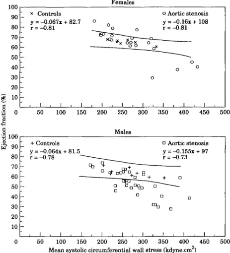
Systolic function was assessed from biplane ejection fraction and from the ejection fraction-mean circumfer-ential wall stress relationship112'. The normal range and the 95% prediction limits are given by the 16 control patients (Fig. 2). Patients with values below this range were considered to have depressed contractile function. Diastolic function was evaluated from isovolumic re-laxation, peak diastolic filling rates and passive elastic properties.
LV relaxation was assessed from the time constant of isovolumic pressure decline which was calculated as the negative reciprocal of the slope of the linear relationship between left ventricular pressure and negative dP/dt'13'. The isovolumetric relaxation period was defined as the time interval beginning immediately after maximal nega-tive dP/dt and ending when pressure had decreased to 5 mmHg above LV end-diastolic pressure1'3'. From this time interval, seven to 14 points were available for calculation of the time constant of isovolumic pressure decline.
Peak diastolic filling rate was defined as the largest value of diastolic inflow (ml. m ~2. s~ ') during the first half (early) and the second half of diastole (late peak filling rate). The filling phase was considered to begin 20 ms before the first frame showing the entry of un-opacified blood into the left ventricle and to end at end-diastole114'. Instantaneous diastolic filling rates were calculated every 20 ms after mitral valve opening. To minimize error due to random noise, raw data were filtered using the fifth grade moving average161.
Diastolic passive elastic properties were determined during the period from minimum ventricular pressure to end-diastole. LV myocardial properties were evaluated from the diastolic stress-strain relationship using an
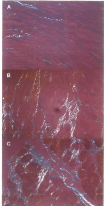
Figure 1 Photomicrographs (magnification x 300, and
repro-duced here at 80%) showing the different patterns of collagen network in controls, males and females with aortic stenosis. (A) Normal human myocardium: there are only few collagen fibres within the myocardium. (B) Female with aortic stenosis: there is an increase in collagen fibres which are mostly parallel to each other and to the muscular fibres. (C) Male with aortic stenosis: an increase in collagen fibres orthogonally oriented (cross-hatching) to each other (white-yellow and green-blue) is evident.
elastic model with shifting asymptote: S=aeb*+c or dS/d£=b (S-c) where S = LV circumferential wall stress (kdyne . cm ~ 2), a=elastic constant (kdyne . cm ~ 2), b = constant of myocardial stiffness, c=asymptote of the stress-strain relationship (kdyne. cm ~2), dS/ de=instantaneous myocardial stiffness (kdyne . cm"2). The three constants, a, b and c were determined by an iteration procedure171. The constant of myocardial stiff-ness is mathematically represented by the slope of the
stress-strain curve, and the tangent to this function is defined as the operative instantaneous myocardial stiff-ness dS/ds*7'151. End-diastolic muscle fibre stretch was also calculated as a measure for preload. It reflects relative sarcomere length'161.
ENDOMYOCARDIAL BIOPSIES
LV endomyocardial biopsies were performed with the King's College bioptome, which was introduced into the left ventricle through the I1-5F Brockenbrough catheter1'71. In each patient, two to four biopsy samples were obtained from the antero-lateral wall of the left ventricle. Immediately after biopsy, two specimens were fixed in glutaraldehyde, cut in semi-thin sections and evaluated by light microscopy. In a subset of 27 consecu-tive patients with aortic stenosis (14 males and 13 females, Table 4) two further biopsy specimens were taken, fixed in buffered formalin and prepared for light microscopy as reported previously118'191 for evaluation of the collagen network (Fig. 1).
Assessment of cellular hypertrophy and interstitial non-muscular tissue
Morphometric analyses were carried out in glutaraldehyde-fixed specimens'171. The following four parameters were determined: (1) Muscle fibre diameter (MFD): a mechanical-optical pen was used to take at least 100 measurements at the level of the nucleus from several randomly chosen cross-sections, and from these the average fibre diameter was calculated. (2) Volume fraction of myofibrils (VFM): myofibrils were evaluated at a magnification of 1000:1 with oil immersion and phase contrast microscopy. From four randomly chosen sections of each patient and at least three random test areas from each section, the intra-myocyte volume frac-tion of myofibrils was determined with a counting grid with 36 intersection points'201. (3) Interstitial non-muscular tissue (IF): interstitial non-non-muscular tissue was determined with the point-counting system excluding areas with arterioles and perivascular tissue, as previ-ously described'171. The term 'interstitial fibrosis' was used — as it was by others — because fibrous tissue is the predominant component of the interstitial space117'211. (4) Fibrous content (FC): fibrous content of the left ventricle was calculated as FC = LV muscle mass x IF/100 and expressed in g . m"21171.
Assessment of left ventricular collagen structure Paraffin-embedded endomyocardial biopsy specimens were sectioned at thicknesses of 5 mm and stained with Sinus Red F3BA dissolved in a saturated picric acid solution (picrosirius red; PSR). PSR is a collagen-specific stain and enhances the natural birefringence of collagen fibrils under polarized light'18'191. The number of fields analysed per tissue section (range 7-23) varied depending on the size of the biopsy specimens. The following variables were determined'221: (1) Total area of the biopsy specimens was determined as the sum of the
100 r— 90 80 70 60 50 40 30 20 10 -•S 0 Females x Controls y = -0.067x + 82.7 r = -0.81 o Aortic stenosis y = -0.16x+108 r = -0.81 J , L J i I i L i i i _L 50 100 150 200 250 300 350 400 450 500
g
5? 90 -A n LrtJ 80 70 60 50 40 30 20 10 + - y — r — -Controls = - 0 . 0 6 4 x + 81.5 = - 0 . 7 8 "~ ———_ I . I . I . Males S- " D qT 0 ^ 0 ^ + ft n D I . I . I a y r — • a 1 Aortic stenosis = -0.155x + 97 = -0.73 o • a I . I . 50 100 150 200 250 300 350 400 450 500Mean systolic circumferential wall stress (kdyne.cm )
Figure 2 Relationship between ejection fraction and mean systolic circumferential wall
stress in females (upper panel) and males (lower panel). The lines represent the 95% prediction limits of the controls. Four of 21 females with aortic stenosis and 20 of 35 males with aortic stenosis are below the prediction limits of the controls indicating a depressed systolic ejection performance.
areas of all fields of the sections evaluated for each patient. (2) Total collagen volume fraction was calcu-lated as the sum of all interstitial connective tissue areas of the biopsy specimen, including interstitial and perivascular fibrosis and microscopic scars, divided by the sum of all connective tissue and muscle areas in all fields of the section. This method has been previously validated and was shown to be closely related to hydroxyproline concentrations of the left ventricle1221. (3) Interstitial collagen volume fraction was calculated from the total collagen volume fraction by subtracting perivascular fibrosis and the area of myocardial scars. (4) Perivascular fibrosis was evaluated as the ratio of perivascular collagen area to vessel luminal area and was measured for all vessels included in the specimen. (5) Focal accumulation of collagen or microscopic scars. (6) Presence and amount of endocardial fibrosis, which was defined as abnormal fibrillar collagen accumulation in the endocardium; endocardial fibrosis was expressed as percent of the total area of the biopsy specimen. The endocardium was seen in all patients and controls. Normally it consisted of a thin surface where collagen
fibres were very rare. (7) Orthogonal collagen fibre meshwork (cross-hatching)'22'23' was evaluated by the picrosirius-polarization technique using a first-order red compensator filter and graded as: 0=no cross-hatching, 0-5 = minimal, 10=moderate, 1-5 = fair, 20=extensive and 3-0 = massive cross-hatching (Fig. 1).
In brief, sections were analysed by rotating a first order red filter tint plate through 90° under polarized light. This allowed for delineation of fibres perpendicu-lar to one another by distinguishing variations in colour. Yellow fibres were oriented perpendicular to green fibres. The extent of the collagen meshwork was determined and semiquantitatively defined using the aforementioned scale. The sectioning angle of the biopsy specimens might have an influence on the evaluation of cross-hatching because in one plane the collagen fibres might be arranged parallel to the muscle fibres but in the next plane orthogonally to them. Thus, for an optimal evaluation of hatching, several different cross-sections were used where the orthogonally running collagen fibres were optimally seen in the presence of cross-hatching. All morphological-morphometric
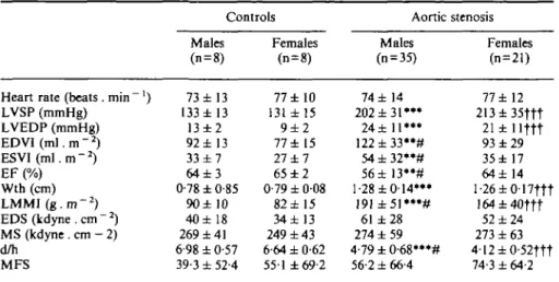
Table 2 Systolic function parameters
Heart rate (beats. min ') LVSP (mmHg) LVEDP (mmHg) EDVI(m].m-2) ESVl(m\.m-2) EF (%) Wth (cm) LMMI ( g . m ~2) EDS(kdyne.cm"2) MS (kdyne . cm - 2) d/h MFS Controls Males (n = 8) 73 ±13 133 ±13 13 ± 2 92 ± 13 33 ± 7 6 4 ± 3 0-78 ±0-85 90± 10 40±18 269 ± 41 6-98 ± 0-57 39-3 ± 52-4 Females (n = 8) 77 ± 10 131 ± 15 9 ± 2 77 ± 15 27 ± 7 65 ± 2 0-79 ± 008 82 ±15 34± 13 249 ± 43 6-64 ± 0-62 551 ±69-2 Aortic stenosis Males (n = 35) 74 ± 14 202 ± 3 1 " * 24 ±11*** 1 2 2 ± 3 3 " # 54±32**# 56±13**# 1-28 ±0-14*** 191±51*"# 61 ±28 274 ± 59 4-79±0-68***# 56-2 ± 66-4 Females (n = 21) 77 ±12 213±35ftt 21 ±llftt 93 ±29 35±17 64 ±14 1 -26 ± 0-17ftt 164±40ttt 52 ±24 273 ± 63 4-12±0-52ttt 74-3 ± 64-2 LVSP=left ventricular peak systolic pressure; LVEDP=left ventricular end-diastolic pressure; EDVI = end-diastolic volume; ESVI=end-systolic volume; EF=ejection fraction; Wth=end-diastolic wall thickness; LMMI = left ventricular mass; EDS=end-Wth=end-diastolic circumferential wall stress; MS = mean systolic circumferential wall stress; d/h=ratio of the end-diastolic short axis to wall thickness; MFS = end-diastolic muscle fibre stretch.
** = /><0-01, •**P<000\ vs control males; t t t = /3<0001 vs control females; # = P=005 vs aortic stenosis females.
analyses of myocardial collagen were carried out at the University of Missouri-Columbia; they were performed in a blinded fashion without knowledge of the haemo-dynamic data. Two observers looked at the biopsy samples for the evaluation of cross-hatching and the data of these two observers were averaged to reduce sampling error.
Reproducibility of the cross-hatch grading was deter-mined by correlating the cross-hatch grade of the two observations in the two biopsy specimens from each patient. The relationship between the two measurements of cross-hatching was r=0-94, /><00001 with a standard error of 0063.
The relationship between left ventricular function and structure was also assessed for left ventricular ejection fraction, the constant of myocardial stiffness and cross-hatching (Fig. 3).
STATISTICS
Comparisons between the four groups were per-formed by a one-way analysis of variance. When the analysis was significant, the Tukey's test was applied. Correlations between structural and haemodynamic data were carried out with linear regression analysis using the least squares method. Comparisons of systolic contractile function and collagen network were per-formed by the chi-square procedure. In all tables mean values ± 1 standard deviation are given.
Results
Age was comparable in the four groups. The severity of aortic valve stenosis, (aortic valve area, valve
resistance, aortic valve gradient and mean aortic pressure gradient) was also comparable between males and females with aortic stenosis. Cardiac index was reduced in patients with aortic stenosis when com-pared to controls. Functional classification according to the New York Heart Association was similar in males and females in patients with aortic stenosis. The duration of symptoms was also comparable in males and females with aortic stenosis (Table 1).
SYSTOLIC FUNCTION DATA
Haemodynamic data are given in Table 2; LV end-diastolic and end-systolic volumes were significantly larger in males than in females in patients with aortic stenosis. However, ejection fraction was reduced in males with aortic stenosis, but LV mass as well as the end-diastolic ratio of LV short axis to wall thickness were increased when compared to females. Systolic ejection performance, as assessed from the ejection fraction-mean systolic wall stress relationship, was depressed in 20/35 males and 4/21 females (P<001) (Fig. 2).
DIASTOLIC FUNCTION DATA
Diastolic function data are listed in Table 3. No significant differences were found between males and females in the control group. In males with aortic stenosis a significant increase in myocardial stiffness constant was observed when compared to females. End-diastolic muscle fibre stretch was comparable in the four groups (Table 2).
Table 3 Diaslolic function parameters
Relaxation T (ms) Filling
PFRI ( m l . m -2. s " ' )
P F R 2 ( m l . m -2. s - ' )
Passive elastic properties b Controls Males (n=8) 49 ±13 287 ±61 230 ± 80 9 ± 3 Females (n = 8) 43 ±10 246 ±98 185 ±36 10±3 Aortic Males (n=35) 73±24**» 351 ±93 240 ±118 31 ± 14***## stenosis Females (n = 21) 73±18ftt 293 ± 97 274 ± 119 17± 11
b=constant of myocardial stiffness; PFRI and PFR2=peak filling rate during the first and second half of diastole; r=time constant of left ventricular pressure decay.
*** = /><0001 vs control males; t t t = ^<OOOl vs control females; ## = P<001 vs aortic stenosis
females.
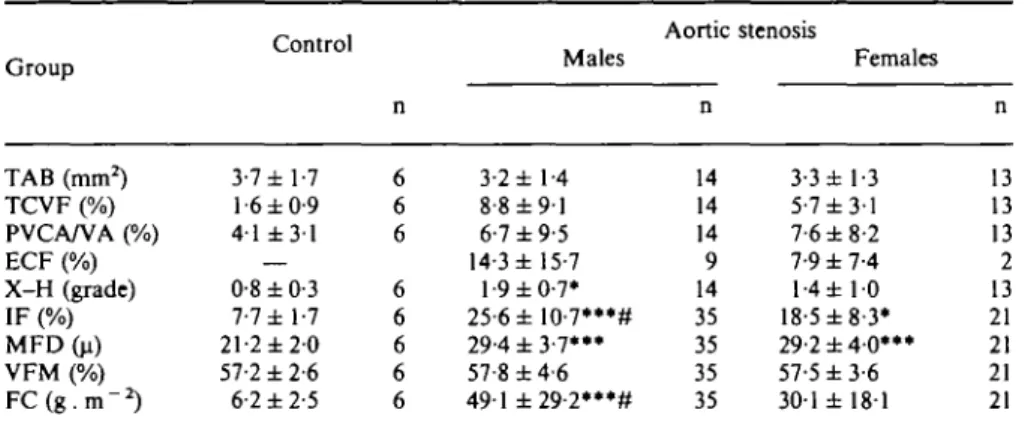
Table 4 Morphological-morphometric data
Group TAB (mm2) TCVF (%) PVCA/VA (%) ECF (%) X-H (grade) IF (%) MFD(u) VFM (%) F C ( g . m -2) Control 3-7 ±1-7 1-6 ±0-9 41 ± 3 1 — 0-8 ±0-3 7-7 ± 1-7 21-2 ± 2 0 57-2 ±2-6 6-2 ±2-5 n 6 6 6 6 6 6 6 6 Males 3-2 ± 1-4 8-8 ± 9 1 6-7 ±9-5 14-3 ± 15-7 1-9 ±0-7* 25-6±10-7***# 29-4 ± 3 - 7 ' " 57-8 ± 4 6 491 ±29-2***# Aortic n 14 14 14 9 14 35 35 35 35 stenosis Females 3-3 ± 1-3 5-7 ±3-1 7-6 ±8-2 7-9 ±7-4 l-4± 10 18-5 ±8-3* 29-2±40»»* 57-5 ±3-6 301 ± 181 n 13 13 13 2 13 21 21 21 21 TAB = total area of the biopsy sample; TCVF = total collagen volume fraction; PVCA/VA = perivascular collagen area/vessel area; ECF = endocardial fibrosis; X-H = cross-hatching; IF = interstitial non-muscular space; MFD = muscle fibre diameter; VFM = volume fraction of myofibrils; FC=fibrous content; —=not present.
• = /'<005> •** = /><001 vs controls; # = P<005 vs females.
STRUCTURAL DATA
Structural data are given in Table 4. Muscle fibre diameter was increased in males and females with aortic stenosis when compared to controls (/><0-001), whereas volume fraction of myofibrils was comparable. Inter-stitial fibrosis (non-muscular tissue) was significantly larger in patients with aortic stenosis than in controls (/><0001 and /><005 in males and females, respec-tively) and was significantly larger in male than female aortic patients (Z'<005).
In a subset of 27 consecutive patients with aortic stenosis the total amount of collagen and endocardial fibrosis was larger in males than females or in controls. Perivascular collagen did not differ between patients with aortic stenosis and controls; the total area of the biopsy specimens was comparable in the three groups. Cross-hatching was enhanced in males with aortic stenosis when compared to controls
Eleven of 14 males and 11 of 13 females with aortic stenosis showed an increased total collagen volume fraction (>2-7%, i.e. upper limit of controls). Nine of 14 males and only two of 13 females showed endocardial fibrosis (/)<0009). Eleven males and four females demonstrated increased cross-hatching (>l-5 grade, i.e. upper limit of controls) (/)<001).
An abnormal collagen architecture, defined as in-creased total collagen volume fraction and presence of endocardial fibrosis and/or increased cross-hatching of collagen fibres'221, was present in 13/14 males and 5/13 females (i><0-002).
LEFT VENTRICULAR FUNCTION AND STRUCTURE
No correlations were found between total collagen volume fraction and left ventricular ejection fraction, the time constant of relaxation and peak filling rate, respec-tively. A weak relationship between total collagen
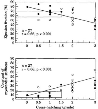
«" -• _ -7 n - r = 27 = 0.66, 1 , _j o o p< 0.001 1 , 1 , o 1 * o g i i t i , 0 0.5 1.5 n = 27 r = 0.68, p < 0.001 0 0.5 1 1.5 2 Cross-hatching (grade)
Figure 3 Influence of cross-hatching on constant of myocardial
stiffness (lower panel) and ejection fraction (upper panel). An increased degree of cross-hatching was associated to higher con-stant of myocardial stiffness and decreased ejection fraction. • = males, O = females.
volume fraction and the constant of myocardial stiffness was observed (n=27, r=0-39, /><005). Cross-hatching was correlated inversely with ejection fraction (n = 27, r= -0-66, /)<00002) and directly with the constant of myocardial stiffness (n = 27, r=0-68, /><0-0001) (Fig. 3).
In presence of endocardial fibrosis (11 of 27 patients with aortic stenosis), ejection fraction was significantly lower (51% vs 65%, /><001) and the constant of myo-cardial stiffness higher (32 vs 19, P<00\) than in the patients without endocardial fibrosis, whereas the time constant of relaxation and peak filling rate were com-parable in the two subgroups. A weak correlation was observed between interstitial fibrosis and the constant of myocardial stiffness (n = 56, r=0-31, P<005) and between muscle fibre diameter and ejection fraction
(n = 56, r= -0-41, /><001).
Discussion
Gender-associated differences in left ventricular func-tion have been reported in animals'1'21 and in man'3"51. Normal females have smaller left ventricles with lower mass than normal men'3'4' but comparable ejection parameters. Previous reports have shown gender-related differences in adaptation of the left ventricle in elderly patients with chronic pressure overload due to aortic stenosis: women tended to have a smaller, more concen-tric hypertrophied left venconcen-tricle with supernormal ejec-tion performance, whereas men more often showed a
larger, more eccentric hypertrophied ventricle with depressed ventricular performance'3"51. Recently, we observed that significant cross-hatching and endocardial fibrosis were present in 50% and 60% of 32 patients with aortic valve disease, and that these collagen network abnormalities influence left ventricular systolic and diastolic function'221. Thus, the aim of the present study was to evaluate possible gender-related differences in structure and function in aortic stenosis.
ADAPTATION OF THE LEFT VENTRICLE TO AORTIC STENOSIS The left ventricle responds to chronic pressure over-load by an increase in the number of contractile units (hypertrophy but no hyperplasia). This compensatory response permits, at least initially, maintenance of ventricular performance, but when pressure overload persists and hypertrophy progresses ventricular decom-pensation eventually supervenes'241. Previously it has been shown in patients with aortic valve disease that in the presence of severe left ventricular hypertrophy pro-nounced abnormalities in systolic and diastolic function can occur, which are accompanied by structural altera-tions as well'17'22'251. However, in 50% of patients with aortic stenosis, diastolic dysfunction precedes systolic dysfunction and thus, systolic function may be normal, although important changes in diastolic ventricular function have occurred at this stage'151. Parallel to the changes in the functional properties of the left ventricle, abnormalities in the non-muscular compartments of the left ventricular wall were shown to occur117'18'22'25'261. Recently we have demonstrated that in aortic valve disease changes in collagen network are associated with altered systolic function and passive elastic prop-erties, and that the sole increase in collagen volume fraction, without a change in collagen network, leaves systolic and diastolic function unaltered'221. These structural abnormalities may influence ventricular function and, hence, postoperative outcome of patients with severe aortic stenosis'17'251. A direct correlation was found between cross-hatching and left ven-tricular ejection fraction (Fig. 3), suggesting a direct influence of the collagen network on myocardial performance117'22'251.
GENDER AND LEFT VENTRICULAR FUNCTION
Recently, gender has been identified as a factor re-sponsible for adaptation of the left ventricle to aortic stenosis'3'41. Carroll et a/.'3' have demonstrated in a group of elderly patients with aortic stenosis that cardiac performance is more often depressed in men than in women. Furthermore, women commonly demonstrated a distinctive pattern of left ventricular function with normal to supernormal ejection performance and a small thick-walled left ventricular chamber. However, these authors recognized that it is unknown whether the sex-associated differences documented in their study are also present in patients younger than 60 years. It was postulated that a combination of ageing and sex might
be involved in these differences of left ventricular adaptation to systolic pressure overload. In fact an increase in left ventricular mass is well known during normal life in women1271, whereas men show a slight decrease1271. In elderly patients with aortic stenosis the degree of left ventricular hypertrophy was similar in males and females with comparable ejection perform-ance, suggesting that the increase in mass was not responsible for the decrease in systolic function131. How-ever, previous studies from our group have suggested that a more excessive increase in left ventricular mass might still be responsible for a decrease in ejection performance16-151.
The present study clearly demonstrates a similar be-haviour in a younger age group of patients with aortic stenosis, as in the previously reported elderly group'31: a female population with severe aortic stenosis (ranging between 26 and 78 years of age) showed a smaller, more concentric hypertrophied left ventricle than an age-matched male group with valve lesions of comparable severity. The latter showed, however, a larger increase in left ventricular mass and a larger reduction in systolic contractility than females. Thus, impaired myocardial contractility and increased left ventricular mass appear to be responsible for the observed depression of left ventricular function in males with aortic stenosis. Moreover, the presence of comparable end-diastolic muscle fibre stretch in the four groups, as a measure of preload at the sarcomere level'"^, seems to suggest that the slightly higher left ventricular end-diastolic pressure and volume are counterbalanced by the increased myocardial stiffness in men.
Several studies have demonstrated that structural factors, such as the geometry of the left ventricle, the degree of left ventricular hypertrophy and the collagen network of the ventricular wall are important determinants of left ventricular systolic and diastolic function in patients with pressure overload hyper-trophy117-19-22'25-261. Previously it has been shown'151 that the ratio of end-diastolic radius at the equator to wall thickness is a determinant of the passive elastic stiffness of the left ventricle, because the latter tend to increase when the former is decreased and vice versa. In the present study, males showed an increased ratio of end-diastolic radius to wall thickness as well as an increased myocardial stiffness constant, whereas females showed decreased values for both.
Gender-related differences in the development of left ventricular hypertrophy in the presence of pressure overload have been reported1'"51. In the present study, the degree of cellular hypertrophy, expressed as volume fraction of myofibrils and muscle fibre diameter, was comparable in men and women, although males with aortic stenosis had a larger left ventricular mass than females. Thus, a difference in the degree of left ventricular hypertrophy was evident not at the microscopic (cellular) but rather at the macroscopic level.
The collagen network, as previously demonstrated in animals and human studies, plays an important role
in the regulation and adaptation of left ventricular function to aortic valve disease117"l9'22a5-261. An abnor-mal coHagen architecture, (i.e. increased total collagen volume fraction, the presence of endocardial fibrosis and increased cross-hatching), may influence adversely myo-cardial function'221. In a subset of 27 patients with aortic stenosis, an abnormal collagen architecture was ob-served in 93% of all males and in 38% of all females (/><0002). Thus, it appears likely that differences in collagen network may account for the observed gender-related differences in left ventricular function in patients with aortic stenosis. Mechanical or hormonal factors may have contributed to these findings.
Mechanical factors
The duration and the time of the onset of left ven-tricular pressure overload influence left venven-tricular adaptation to pressure overload'28-291. In the present study, the exact duration of the pressure overload could not be determined due to obvious reasons. However, the duration of symptoms was comparable in males and females.
Physical working capacity or daily physical activities might have influenced the degree of left ventricular hypertrophy. However, physical working capacity, as determined by bicycle ergometry (60 vs 57%, ns), did not differ in the two groups. Another important factor is the severity of the valve lesion, which was comparable in the two groups.
Hormonal factors
Although hormonal influences on myocardial hyper-trophy have been discussed'1^*-30"331, no human data are available. Several studies suggest that circulating angio-tensin and tissue renin-angioangio-tensin may be involved in the remodelling of the muscular and non-muscular compartments of the ventricular wall"8-30-311 in left ventricular hypertrophy1181 and in the myocardium after infarction'3'1. Simpson'321 has demonstrated that nore-phinephrine induces hypertrophy in cultured neonatal rat myocytes by an alpha-receptor-mediated mechanism. However, in humans the exact role of the hormonal influences in the remodelling of the left ventricle during hypertrophy needs further investigation. Unfortunately, in the present study we have no direct measure of the ACE activity or of the norepinephrine level. However, the serum sodium and potassium concentrations were comparable in males and females, as were heart rate and New York Heart Association functional class. These parameters are influenced by those hormones.
In animal models, conflicting results have been re-ported in regard to the role of sex hormones in the regulation of cardiovascular adaptation'1"2"24-3^3*1. Koenig et a/.'351 have shown that testosterone exerts an anabolic effect on the ventricular myocardium. In rats, differences in myocardial function have been observed which are modified by testosterone administration'361. Pfeffer and co-workers'241 observed that spontaneously hypertensive male rats developed heart failure earlier
than the female counterpart in the presence of a compa-rable degree of pressure overload. No data are available in the present study concerning the hormone profile of patients with aortic stenosis.
Coronary perfusion
Subendocardial ischaemia in patients with severe pres-sure overload may promote the development of struc-tural remodelling with changes in the collagen structure. However, subendocardial coronary perfusion could not be measured in the present study and, thus, it remains open whether there were differences in myocardial per-fusion between males and females. The cross-sectional area of the proximal coronary arteries was determined in 12 males and seven females and no difference in the size of the arteries was noted either in absolute units (mm2) or after normalization per 100 g of mass'37'38'. Appar-ently, the structural differences of the myocardium in males and females with severe aortic stenosis are not reflected by changes in coronary artery size.
References
[1] Schaible TF, Penpargkul S, Scheuer J. Cardiac responses to exercise training in male and female rats. J Appl Physiol 1981; 50: 112-17.
[2] Pfeffer J, Pfeffer M, Fletcher P, Braunwald E. Alterations of cardiac performance in rats with established spontaneous hypertension. Am J Cardiol 1979; 44: 994-8.
[3] Carroll JD, Carroll EP, Feldman T et al. Sex-associated differences in left ventricular function in aortic stenosis of the elderly. Circulation 1992; 86: 1099-1107.
[4] Douglas PS, Otto CM, Mickel MC, Reid CL, Devis KB. Gender is a determinant of left ventricular hypertrophy and function in aortic stenosis. Circulation 1992; 86: 1-538. [5] Topol EJ, TraiU TA, Fortuin NJ. Hypertensive hypertrophic
cardiomyopathy of the elderly. N Engl J Med 1985; 312: 277-83.
[6] Murakami T, Hess OM, Gage JE, Grimm J, Krayenbuehl HP. Diastolic filling dynamics in patients with aortic valve disease. Circulation 1986; 73: 1162-74.
[7] Corin WJ, Murakami T, Monrad ES, Hess OM, Krayenbuehl HP. Left ventricular diastolic properties in chronic mitral regurgitation. Circulation 1991; 83: 797-807.
[8] Dodge HT, Sandier H, Baxley WA, Hawley RR. Usefulness and limitations of radiographic methods for determining left ventricular volume. Am J Cardiol 1966; 18: ;10-24.
[9] Rackley CE, Dodge HT, Coble YD, Hay RE. A method for determining left ventricular mass in man. Circulation 1964; 29: 666-71.
[10] Mirsky I. Left ventricular stresses in the intact human heart. Biophys J 1969; 9: 189-208.
[11] Gaasch WH. Left ventricular radius to wall thickness ratio. Am J Cardiol 1979; 43: 1189-94.
[12] Huber D, Grimm J, Koch R, Krayenbuehl HP. Determinants of ejection performance in aortic stenosis. Circulation 1981; 64: 126-34.
[13] Eichhorn P, Grimm J, Koch R, Hess OM, Carroll J, Krayenbuehl HP. Left ventricular relaxation in patients with left ventricular hypertrophy secondary to aortic valve disease. Circulation 1982; 65: 1395-1404.
[14] Carroll JD, Hess OM, Hirzel HO, Krayenbuehl HP. Dynamics of left ventricular filling at rest and during exercise. Circulation 1983; 68: 59-67.
[15] Villari B, Hess OM, Kaufmann Ph, Krogmann ON, Grimm J, Krayenbuehl HP. Effects of aortic valve stenosis (pressure overload) and regurgitation (volume overload) on left ven-tricular systolic and diastolic function. Am J Cardiol 1992; 69: 927-34.
[16] Gaash WA, Battle WE, Oboler AA, Banas JS, Levine HJ. Left ventricular stress and compliance in man. Circulation 1972; 45: 746-62.
[17] Krayenbuehl HP, Hess OM, Momad SE, Schneider J, Mall G, Turina M. Left ventricular myocardial structure in aortic valve disease before, intermediate and late after aortic valve replacement. Circulation 1989; 79: 744-55.
[18] Brilla CG, Janicki JS, Weber KT. Cardioreparative effects of lisinopril in rats with genetic hypertension and left ventricular hypertrophy. Circulation 1991; 83: 1771-9.
[19] Doering CW, Jalil JE, Janicki JS et al. Collagen network remodelling and diastolic stiffness of the rat left ventricle with pressure overload. Cardiovasc Res 1988; 22: 686-95. [20] Mall G, Schwarz F, Derks H. Clinicopathologic correlations
in congestive cardiomyopathy: A study on endomyocardial biopsies. Virchows Arch 1982; 397: 67-82.
[21] Schwarz F, Schaper J, Kittstein D, Flameng W, Walter P, Schaper W. Reduced volume fraction of myofibrils in myo-cardium of patients with decompensated pressure overload. Circulation 1981; 63: 1299-1304.
[22] Villari B, Campbell SE, Hess OM, Mall G, Vassalli G, Weber KT, Krayenbuehl HP. Influence of collagen network on left ventricular systolic and diastolic function in aortic valve disease. J Am Coll Cardiol 1993; 22: 1147-54.
[23] Weber KT, Janicki JS, Shroff S. Mechanical and structural aspects of the hypertrophied human myocardium. In: Tarazi RC, Dunbar JB, eds. Cardiac Hypertrophy in Hypertension, Perspectives in Cardiovascular Research Series, vol. 8. New York, Raven Press Publishers, 1983: 201-10.
[24] Pfeffer JM, Pfeffer MA, Fishbein MC, Frohlich ED. Cardiac function and morphology with ageing in the spontaneously hypertensive rat. Am J Physiol 1979; 237: 461-8.
[25] Hess OM, Ritter M, Schneider J, Grimm J, Turina M, Krayenbuehl HP. Diastolic stiffness and myocardial structure in aortic valve disease before and after valve replacement. Circulation 1984; 69: 855-65.
[26] Weber JT, Janicki JS, Shroff SG, Pick R, Chen RM, Bashey RI. Collagen remodelling of the pressure-overloaded, hyper-trophied non-human primate myocardium. Circ Res 1988; 62: 757-65.
[27] Dannenberg AL, Levy D, Garrison RJ. Impact of age on echocardiographic left ventricular mass in a healthy popu-lation (The Framingham Study). Am J Cardiol 1989; 64: 1066-8.
[28] Sasayama S, Ross J Jr, Franklin D, Bloor C, Bishop S, Dilley RB. Adaptations of the left ventricle to chronic pressure overload. Circ Res 1976; 38: 172-9.
[29] Assey ME, Wisenbaugh T, Spann JF, Gillette PC, Carabello BA. Unexpected persistence into adulthood of low wall stress in patients with congenital aortic stenosis: Is there a fundamental difference in the hypertrophic response to a pressure overload present from birth? Circulation 1989; 75: 973-9.
[30] Weber KT, Brilla CG. Pathological hypertrophy and cardiac interstitium. Fibrosis and renin-angiotensin-aldosterone system. Circulation 1991; 83: 1849-65.
[31] Pfeffer MA, Lamas GA, Vaughan DE, Parisi AF, Braunwald E. Effect of captopril on progressive ventricular dilatation after anterior myocardial infarction. N Engl J Med 1988; 319: 80-6.
[32] Simpson P, Stimulation of hypertrophy of cultured neonatal rat heart cells through an alpha-1- and beta-1-adrenergic receptor interaction. Circ Res 1985; 56: 884-9.
[33] Scheuer J, Malhotra A, Schaible TF, Capasso J. Effects of gonadectomy and hormonal replacement on rat hearts. Circ Res 1987; 61: 12-19.
[34] Schaible TF, Malhotra A, Ciambrone G, Scheuer J. The effects of gonadectomy on left ventricular function and car-diac contractile proteins in male and female rats. Circ Res
1984; 54: 38-49.
[35] Koenig H, Goldstone A, Lu CY. Testosterone-mediated sexual dysmorphism of the rodent heart. Circ Res 1982; 50: 782-7.
[36] Malhotra A, Buttrick P, Scheuer J. Effects of sex hormones on development of physiological and pathological cardiac hyper-trophy in male and female rats. Am J Physiol 1990; 259: H866-H871.
[37] Villari B, Hess OM, Meier C et al. Regression of coronary artery dimensions after successful aortic valve replacement. Circulation 1992; 85: 972-8.
[38] Villari B, Hess OM, Moccetti D, Vassalli G, Krayenbuehl HP. Effect of progression of left ventricular hypertrophy on coro-nary artery dimensions in aortic valve disease. J Am Coll Cardiol 1992; 20: 1073-9.