The Developmental Role and Regulation of the Anaphase-Promoting Complex/Cyclosome in Drosophila Meiosis
by
Jillian Annice Pesin
B.A. Biology and Russian Williams College Williamstown, MA, 2000
Submitted to the Department of Biology
in Partial Fulfillment of the Requirements for the Degree of Doctor of Philosophy in Biology
at the
Massachusetts Institute of Technology Cambridge, MA
February 2008
© 2007 Jillian Annice Pesin. All rights reserved.
The author hereby grants to MIT permission to reproduce or distribute publicly paper and electronic copies of this thesis document in whole or in part
in any medium now known or hereafter created.
Signature of Author...
. .
...=...
t. t.
:..-..
...
SignatureofAuthor. October 2, 2007 Certified by... . .. ... ... Terry L. Orr-Weaver Professor of Biology Thesis Supervisor A ccepted by ... - ... 6 .... . ' . ... Stephen P. BellOF TEOHMNO Y Chair, Committee on Graduate Students
Department of Biology ARCHIVES
The Developmental Role and Regulation of the Anaphase-Promoting Complex/Cyclosome in Drosophila Meiosis
by
Jillian Annice Pesin
Submitted to the Department of Biology on October 2, 2007 in Partial Fulfillment of the Requirements for the Degree of
Doctor of Philosophy in Biology
ABSTRACT
Meiosis is a modified cell cycle in which two round of chromosome segregation occur without an intervening DNA synthesis phase. As in mitosis, these cell divisions are driven in part by proteolysis mediated by the Anaphase-Promoting Complex/ Cyclosome (APC/C). During oogenesis of multicellular organisms, molecular control of meiosis has an added complexity because these divisions must be coordinated with the growth and development of the oocyte as well as its fertilization. The Drosophila gene cortex (cort), a putative female meiosis-specific APC/C activator, provides a unique opportunity to study the role of the APC/C in metazoan meiosis. We demonstrate that CORT protein associates with the core APC/C in ovaries, confirming its function as an activator of the APC/C. CORT triggers the sequential degradation of mitotic Cyclins in meiosis, as well as causing the degradation of PIMPLES, the Drosophila homolog of Securin. Both post-transcriptional and post-translational regulation of cort result in expression of CORT protein being restricted to the meiotic divisions. Cytoplasmic polyadenylation of cort mRNA is tightly correlated with appearance of the protein in mature oocytes. At the end of meiosis, CORT is rapidly degraded in an APC/C and D-box-dependent manner. We initiated a genetic screen to identify substrates of APC/CCORT using grauzone (grau) mutants, a gene encoding a transcriptional activator of cort. We have identified at least one deficiency on the third chromosome, Df(3R)p-XT103, that dominantly suppresses
grau. Levels of BEL, a protein encoded within this deficiency, are elevated in cort
mutants. Our analysis of the regulation of CORT protein levels reveals one mechanism that may be important for developmental control of meiotic progression and the transition from meiosis to embryonic mitotic divisions. In addition, our screen to identify meiosis-specific substrates of APC/CCORT has the potential to uncover novel regulators of meiosis in a multicellular organism.
Thesis Supervisor: Terry L. Orr-Weaver Title: Professor of Biology
Dedicated to my parents, Madgelyn and Richard Pesin
and
In loving memory of my grandmother, Ethel Pesin
Acknowledgements
Thank you to all of my wonderful colleagues in the Orr-Weaver lab, past and present. I am lucky to have shared the daily routines of lab life with them and am grateful for the many friendships this has fostered. I would especially like to thank Laurie Lee, who patiently taught me molecular biology and protein biochemistry when I first joined the lab, Julie Wallace, for answering my endless questions from across the bench, Leah Vardy, for her enthusiasm and experimental advice, and Astrid Clarke, for being a good listener and sounding board for ideas.
I am very grateful to my advisor, Terry Orr-Weaver, for her guidance and support.
Through my many interactions with Terry over the years and by her example, I have truly learned how to be a scientist. She is an excellent teacher and committed to her graduate students. I am also grateful to her for her skill and thoughtfulness in creating a nurturing lab environment.
I shared many lunches, conversations, and laughs with my classmates and friends, Tamar Resnick, Jessica Alfoldi, and Teresa Holm, which made getting through graduate school more fun. I'm glad to have made this journey with them. I also cannot imagine going through graduate school without the support of my friends Kimberly Hartwell, Megan Gustafson, and Anu Seshan, with whom I shared an apartment for my first years of living in Boston.
My family has been a tremendous source of support throughout the past six years. My grandparents Victor and Thelma Jacque LaVallee never fail to encourage me and make me feel proud of what I've accomplished. My brother, Isaac, and his wife, Mariana, and my sister, Sarah, have given me much needed perspective and reminded me of life outside of Boston through all of our phone calls and visits to see each other. Most of all, I am deeply grateful to my parents, whose love and encouragement has been the greatest influence on me as a student. They have instilled in me a love of learning and a belief that I can accomplish whatever I set my mind to with hard work and determination. Without these two things, I could not have made it to where I am today.
Finally, thank you to my husband, Daniel. His unconditional love and unflagging patience have seen me through the many challenges that graduate school has presented. He has inspired me to be a more confident scientist and a more confident person in many areas of my life. I'm so glad we have had each other to share the moments of both exhilaration and disappointment that come with being a scientist.
TABLE OF CONTENTS Chapter One:
Introduction 7
The Role of Proteolysis in Mitotic Events 8
The Anaphase-Promoting Complex/Cyclosome in Mitosis 9
The Ubiquitin-Proteasome Pathway 9
Discovery of the APC/C 10
Structure of the APC/C 11
APC/C Activators and Substrate Adaptors: Cdc20/Fzy and Cdhl/
Fzr 15
Inhibition of APC/CCdc2o by the Spindle Checkpoint 32
Role of the APC/C in Female Meiosis 37
Requirement for APC/C in Meiosis 40
Meiosis-Specific APC/C Activators 41
Regulation of the APC/C in Meiosis 44
Summary of Thesis 54
References 56
Chapter Two:
Developmental Role and Regulation of cortex, a Meiosis-Specific APC/C
Activator 83
Abstract 84
Introduction 85
Results 89
CORT protein is associated with the APC/C in vivo 89
cortex cannot provide fizzy function in the early embryo 93 Cortex triggers degradation of Cyclin A early in meiosis I and additional
substrates later 94
Developmental regulation of appearance of CORT protein in female
meiosis 98
cort mRNA becomes deadenylated in early embryogenesis 102 CORT protein becomes a target of the APC/C after the completion of
meiosis 105
CORT is likely to be targeted for degradation by APC/CFzY 109
Discussion 110
Materials and Methods 119
Acknowledgements 124
References 125
Supplemental Table and Figures 131
Chapter Three:
A Genetic Screen for Substrates of the Anaphase-Promoting Complex in
Meiosis 136
Abstract 137
Introduction 138
cortex mutants are not suppressed by candidate APC/CCORT substrates 141
A screen for dominant suppressors of grauzone 145
Df(3R)p-XTI03 dominantly suppresses grauzone 148 Testing of smaller deficiencies within the Df(3R)p-XT103 region 150
Candidate substrates within Df(3R)XT103 152
Discussion 154
Materials and Methods 160
Acknowledgements 162
References 162
Chapter Four:
Conclusions and Perspectives 171
Function of Putative Meiosis-Specific APC/C Activators in Drosophila 172
Meiosis-Specific APC/C Substrates 174
Developmental Regulation of CORT Protein Levels 175
Inhibitors of APC/CCORT 177
Localization of CORT in Meiosis 179
References 181
Appendix One: Attempts to Generate Recombinant CORT Protein 185
Appendix Two: Role of Spindle Checkpoint and Phosphorylation in
Chapter One
I. The Role of Proteolysis in Mitotic Events
Mitosis is a highly regulated process by which a cell divides to produce two daughter cells, each containing the same chromosomal complement as the parent cell. Proper mitotic cell division is crucial for the development of a multicellular organism, and its regulation is key, as deregulation of mitosis and chromosome segregation can lead to aneuploidy and unrestrained proliferation, hallmarks of cancer. Mitosis can be
separated into several discrete stages based on the changing morphology of the cell as it divides. First, in prophase the newly replicated chromosomes condense and the nuclear envelope breaks down. During prometaphase "search and capture" events occur as microtubules bind to kinetochores and create an attachment between chromosomes and the spindle. Once stable bipolar attachments of kinetochores lead to alignment of chromosomes on the metaphase plate, metaphase begins. During metaphase a particular set of proteins is degraded (see below) that allows for rapid separation of sister
chromatids in anaphase. Telophase begins once the sister chromatids have reached opposite ends of the spindle. The chromatids start to decondense, the nuclear envelope re-forms, and the spindle disassembles. During anaphase and telophase, the cell goes through cytokinesis to divide its cytoplasm into two cells. All of these events contribute to solving a critical problem for the cell when it divides: each daughter cell must receive an equal and identical set of genetic material.
Many studies of the regulation of the cell cycle have revealed that proteolysis is a key mechanism that drives the events of mitosis. Cyclin-dependent kinases and their activating subunits, cyclins, control many cell cycle events. Kinase activity is controlled by the availability of cyclin subunits, whose levels fluctuate during the cell cycle. Tim Hunt and colleagues discovered and gave cyclin its name after observing a protein in sea
urchin eggs that accumulates prior to mitosis and is destroyed each time the cell divides (Evans et al. 1983). Their hypothesis that the synthesis of cyclin drives entry into mitosis and its destruction drives exit from mitosis has turned out to be true (Murray and
Kirschner 1989; Murray et al. 1989). In addition to cyclin, another key target of proteolysis, Securin, must be destroyed for anaphase to be initiated (Cohen-Fix et al.
1996). Thus, proteolysis is crucial for the progression of mitosis and provides an irreversible and directional switch to restart the cell cycle.
In this chapter, we will review the role of the anaphase-promoting complex/ cyclosome during mitosis and meiosis. We will focus on the activating subunits of the complex and discuss both their mechanism of action and their regulation. We will consider the specific regulation of Cdc20 by the spindle checkpoint, and look in depth at unique function and regulation of the APC/C in the developmental context of meiosis.
II. The Anaphase-Promoting Complex/ Cyclosome in Mitosis A. The Ubiquitin-Proteasome Pathway
The ubiquitin-proteasome pathway is a widely used mechanism that conjugates ubiquitin to proteins for various purposes, the best understood of which is degradation by the proteasome (for review, see Pickart 2001). Three enzymes act sequentially to add ubiquitin and polyubiquitin chains onto substrates. In an ATP-dependent reaction, El, the ubiquitin-activating enzyme, forms a high energy thioester bond between an active-site cysteine on El with a C-terminal glycine on ubiquitin. The activated ubiquitin is then transferred to a cysteine in E2, the ubiquitin-conjugating enzyme. Finally, the E3 ligase transfers the ubiquitin from the E2 to a lysine residue on the substrate. This 3-step reaction is repeated multiple times to attach subsequent ubiquitins to specific lysines on
the ubiquitin already bound to the substrate to generate polyubiquitin chains. A chain length of at least four ubiquitins is a strong signal to target a substrate for degradation by the 26S proteasome (Thrower et al. 2000).
The number of enzymes available for each step of the reaction increases as the reaction proceeds. Most organisms contain just one El enzyme that acts for all downstream ubiquitin-conjugating events, although recently a second El enzyme was identified in vertebrates and sea urchin (Jin et al. 2007). There are a limited number of E2 enzymes, and each E2 may act with several different E3 enzymes. Each E3
cooperates with one or more E2s and targets a specific set of substrates. In S. cerevisiae the APC/C E3 seems to act with both Ubc4 and Ubcl, two different E2s that have
sequential roles in chain assembly (Rodrigo-Brenni and Morgan 2007). Ubc4 acts in monoubiquitination of target proteins that provides a substrate for assembly of
polyubiquitin chains by Ubcl. In vertebrates the APC/C E3 can act with two E2 enzymes, UBCH5 and UBCH10/ UbcX/ E2-C in vitro, but their in vivo role is not entirely clear (Aristarkhov et al. 1996; Yu et al. 1996; Kirkpatrick et al. 2006;). However, in Drosophila, genetic and RNAi analysis has revealed a role for vihar, the gene encoding the E2-C homolog, in spatiotemporal control of Cyclin B degradation at spindle poles (Milth6 et al. 2004).
B. Discovery of the APC/C
Biochemical and genetic studies converged for discovery of the enzyme that catalyzes the degradation of cyclin. Fractionation of extracts from clam oocytes led to the identification of three components that are required for ubiquitination of Cyclin B, an El ubiquitin-activating enzyme, an E2 ubiquitin-conjugating enzyme specific to cyclin,
and a possible E3 ubiquitin ligase specific to cyclin (Hershko et al. 1994). The E3 fraction was only active when isolated from mitotic extracts but not from interphase extracts. This E3 activity was found to be associated with a 1500 kDa complex called the "cyclosome" (Sudakin et al. 1995). At the same time, a 20S complex was identified from fractionation of Xenopus egg extracts and called the "anaphase-promoting complex" (APC) (King et al. 1995). The APC was able to trigger ubiquitination of a cyclin B substrate when complemented with interphase extract or a mixture of recombinant El and an E2, UBC4.
In parallel, a genetic screen was carried out in S. cerevisiae to identify genes required for proteolysis of Cyclin B (Irniger et al. 1995). Conditional mutants of two essential genes, cdcl6 and cdc23, were isolated, and these genes were shown to be required for entering and exiting anaphase. The identification of two of the proteins in the Xenopus APC complex as homologs of S. cerevisiae cdc16 and cdc23 linked the biochemical and genetic studies together (King et al. 1995). Finally, antibodies were raised against the human homologs of Cdcl6 and Cdc27, a protein that had been shown to associate with Cdcl6 and Cdc23 (Lamb et al. 1994; Tugendreich et al. 1995). These two proteins cosedimented as a 20S complex from mammalian cell extracts, and when antibodies against Cdc27 were injected into HeLa cells, the cell cycle arrested in metaphase, suggesting a conservation of function for these proteins in mammals.
D. Structure of the APC/C
The APC/C is a large multisubunit complex comprised of at least 12 core subunits (Table 1-1). An understanding of the structure of the APC/C has been hindered due to its large and complex nature, lack of a crystal structure, and difficulty in reconstituting the
Table 1-1. APC/C Subunits*
S. cerevisiae Drosophila Vertebrates Structural Motif
Core subunits Shattered Morula Makos APC4/CG327 Ida cdcl 16/ CG67 APC7/ CG1 cdc23/ CG25 Docl/ApclO Lemming APC1/TSG24 APC2 CDC27/APC3 '07 APC4 APC5 759 CDC16/APC6 1444 APC7 08 CDC23/APC8 DOC1/APC10O APC11 homology to Rpnl/2, subunits of the 26S proteasome
Cullin homology TPRs WD40 repeats TPRs TPRs TPRs TPRs
Doe domain, IR motif RING-H2 finger CDC26 SWM1/APC13 Swml/Apcl3 Apc9 Mnd2 Co-activators Fizzy Fizzy-related Cortex Fizzy-related 2
* See text for references
CDC20/FZY C-box, WD40 repeats, IR-tail CDH1/FZR C-box, WD40 repeats, IR-tail C-box, WD40 repeats, IR-tail C-box, WD40 repeats, IR-tail C-box, WD40 repeats, IR-tail
Apcl Apc2 Cdc27 Apc4 Apc5 Cdcl6 Cdc23 Apcll Cdc26 Cdc20 Cdhl Amal
complex from purified subunits. Purification of the APC/C from budding yeast and from vertebrate cells and tissues has facilitated the development of in vitro assays to elucidate the mechanisms of APC/C activity (Carroll and Morgan 2005; Herzog and Peters 2005). A budding yeast strain that renders the APC/C nonessential has allowed for a detailed analysis of the architecture of the complex (Thornton and Toczyski 2003; Thornton et al. 2006). Additionally, electron microscopy studies have revealed important structural information and generated a 3-dimensional model of the enzyme (Gieffers et al. 2001; Passmore et al. 2005; Dube et al. 2005). The APC/C can be divided into four parts: an arm containing tetratricopeptide repeat (TPR) proteins, Cdc23, Cdc16, Cdc27, and
Swml; a scaffolding complex, Apc1, Apc4, and Apc5; a catalytic arm, Apc2, Apc 11, and Doc 1; and the non-core substrate adaptor subunits, Cdc20 and Cdhl (Figure 1-1).
The TPR subunits form a separable subcomplex, which is thought to interact with substrate adaptors to facilitate ubiquitination (discussed below) (Passmore et al. 2003; Vodermaier et al. 2003). The TPR arm contains several copies of each TPR-containing protein, Cdcl6, Cdc23, and Cdc27 (Lamb et al. 1994; Dube et al. 2005; Passmore et al. 2005). Vertebrate cells contain an additional TPR-containing protein, Apc7. The TPR arm also contains non-TPR proteins Cdc26, Swml/Apcl3, and Apc9 (only in S.
cerevisiae), small, nonessential subunits that are important for the stability of this
subcomplex (Zachariae et al. 1998; Schwickart et al. 2004).
The TPR arm is tethered to Apc1, a large scaffolding subunit, through stabilizing associations with Apc4 and Apc5 (Thornton et al. 2006). Cdc23 appears to hold the TPR arm to Apcl by being most closely associated with the scaffold. Cdcl6 is bound to Cdc23, and Cdc27 is bound to Cdcl6 and farthest away from the scaffold (Schwickart et al. 2004; Kraft et al. 2005; Thornton et al. 2006).
Ub- Ub-Ub
Ub
E2
Figure 1-1. Architectural Map of the Anaphase-Promoting Complex/ Cyclosome
A map of the subunits of the APC/C based on binding and association data. Subunits of the TPR arm
are represented in shades of blue, those of the scaffolding complex in shades of brown, and those of the catalytic arm in green. Adapted from Thornton and Toczyski (2006).
The APC/C is a member of the Cullin-RING finger family of E3 ligases. The catalytic arm of the APC/C that contains the Cullin and RING finger proteins binds the other side of the scaffold. Apc2 is a member of the cullin family of proteins (Yu et al.
1998; Zachariae et al. 1998). The cullin domain of Apc2 binds the RING finger domain of Apcl 1 (Tang et al. 2001; Vodermaier et al. 2003). The RING finger domain of Apcl 1 recruits E2 enzymes to the complex. Together, these two proteins can catalyze the
ubiquitination of substrates in the absence of any other APC/C subunits, although with little substrate specificity (Gmachl et al. 2000; Leverson et al. 2000; Tang et al. 2001). Docl is a small globular protein that is thought to contribute to processivity of
ubiquitination through enhancing substrate recognition by the APC/C (Carroll and Morgan 2002; Carroll et al. 2005).
The APC/C has an overall asymmetric triangular shape with two flexible arms and an inner cavity (Gieffers et al. 2001; Passmore et al. 2005; Dube et al. 2005). Although the cavity was once thought to be the site of ubiquitination, cryo-electron microscopy has mapped the locations of Apc2 and Cdhl to the same outside wall of the complex, suggesting that ubiquitination of substrates occurs in this location (Dube et al. 2005).
E. APC/C Activators and Substrate Adaptors: Cdc20/Fzy and Cdhl/FzrF 1. Interactions with Core APC/C and Substrates
Cdc20/Fzy and Cdhl/Fzr comprise the non-core subunits of the APC/C and activate and confer substrate specificity to the complex (Dawson et al. 1995; Schwab et al. 1997; Visintin et al. 1997; Sigrist and Lehner 1997). Cdc20 directs the ubiquitination
of securin, mitotic cyclins, and other substrates for anaphase onset, while Cdhl targets mitotic cyclins and additional substrates for degradation in mitotic exit and G1. Cdc20 and Cdhl are members of the Cdc20 protein family whose members contain seven WD-40 repeats in their C-terminus (for a review, see Smith et al. 1999). These repeats form a seven-bladed propeller structure that mediates protein-protein interactions.
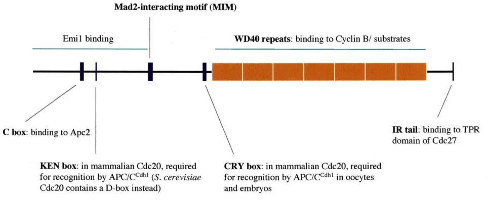
Substrate adaptors bind directly to the APC/C, although this association is not as stable as the binding of APC/C core subunits to each other (Fang et al. 1998a; Yamano et al. 2004). Direct binding to the APC/C appears to occur through two motifs that are conserved among substrate adaptor proteins (Figure 1-2). The C-box is a short motif, DR(F/Y)IPXRX~45-75(K/R)XL, in the N-terminal half of these proteins and is required for association of Cdhl with the APC/C (Schwab et al. 2001; Pfleger et al. 2001;
Thornton et al. 2006). Analysis of the ability of C-box mutant Cdhl to bind and activate wild-type and mutant APC/C complexes has suggested that the C-box mediates an interaction between Cdhl and Apc2 (Thornton et al. 2006). Additionally, substrate adaptor proteins, as well as Doc 1, contain a conserved isoleucine arginine (IR) tail in the C-terminus that is proposed to bind directly to the TPR domain of Cdc27 (Wendt et al. 2001; Vodermaier et al. 2003). C-terminal peptides of Cdc20 and Cdhl specifically bind Cdc27, and the TPR domain of Cdc27 is sufficient for this binding (Vodermaier et al. 2003). Furthermore, Cdhl specifically crosslinks to Cdc27 through its IR tail, and an APC/C complex that is missing Cdc27 fails to bind Cdhl (Kraft et al. 2005; Thornton et al. 2006). In summary, both the C-box and IR tail conserved regions of the substrate adaptor proteins contribute to their binding of the APC/C, and Cdc27 and Apc2 are thought to be the APC/C subunits that are directly bound.
Mad2-interacting motif (MIM)
Emil binding
C box: binding to Apc2
VD40 repeats: binding to Cyclin B/ substrates
---IR tail: binding to TPR domain of Cdc27
KEN box: in mammalian Cdc20, required
for recognition by APC/CCdhl (S. cerevisiae Cdc20 contains a D-box instead)
CRY box: in mammalian Cdc20, required
for recognition by APC/CCdhl in oocytes and embryos
Figure 1-2. Domains and motifs of Cdc20 and Cdhl
A summary of the domains and motifs in Cdc20 and Cdhl that are crucial for their interactions with core APC/C,
substrates, and inhibitors/ regulators. See text for details and references. l
Phosphorylation plays a role in regulating the association of substrate adaptors with the APC/C and contributes to the differential timing of Cdc20 and Cdhl binding and activation of the complex. Association of Cdc20 with the APC/C requires mitotic
phosphorylation of the APC/C (Fang et al. 1998a; Kramer et al. 2000). This
phosphorylation is thought to be carried out by Cdkl-Cyclin B and Plkl/ Polo-like kinase on Apcl and TPR proteins (Shteinberg et al. 1999; Rudner and Murray 2000; Golan et al. 2002; Kraft et al. 2003).
In contrast, mitotic phosphorylation acts to inhibit association of Cdhl with the APC/C. Cdhl itself is phosphorylated by cyclin-dependent kinases during S, G2, and M-phase, and only upon dephosphorylation in GL is it able to bind and activate APC/C (Zachariae et al. 1998; Jaspersen et al. 1999; Lukas et al. 1999; Blanco et al. 2000;
Kramer et al. 2000; Yamaguchi et al. 2000; Huang et al. 2001; Sorensen et al. 2001; Keck et al. 2007). Thus, mitotic phosphorylation directs the temporal differences in activity of
APC/Ccdc2o and APC/CCdhl. As cells enter mitosis and mitotic kinases become active,
APC/C subunits are phosphorylated and allow for Cdc20 binding and activation. At the same time Cdhl is phosphorylated and inhibited from binding APC/C. Once APC/Ccdc20 targets mitotic cyclins for degradation, a decrease in mitotic Cdk-cyclin activity leads to a decrease in inhibitory phosphorylation of Cdhl, as well as dephosphorylation of Cdhl by Cdcl4 phosphatase, allowing for binding to and activation of the APC/C by Cdhl in G1 of the cell cycle (Jaspersen et al. 1999).
In addition to binding directly to the APC/C, Cdc20 and Cdhl are thought to bind directly to APC/C substrates. Two conserved motifs in substrates act as the main
destruction box (D-box), R-x-x-L-x-x-x-x-N, which is sufficient to target a protein for degradation, whereas APC/CCdh' recognizes substrates containing an additional motif, K-E-N, called the KEN box (Glotzer et al. 1991; Pfleger and Kirschner 2000).
A long-standing hypothesis has been that Cdc20 and Cdhl recruit substrates to the APC/C by binding them directly. Many groups have demonstrated binding of Cdc20 and Cdh 1 to substrates by yeast two-hybrid analysis, co-immunoprecipitation, and in vitro binding assays, although the details of these results are sometimes contradictory. It is clear that the D-box and KEN box are required for binding of substrates to activators (Ohtoshi et al. 2000; Pfleger et al. 2001; Burton and Solomon 2001; Hilioti et al. 2001; Kraft et al. 2005). However, three groups reported a dependence on the WD repeat region of Cdc20 and Cdhl for binding to substrates, whereas Pfleger et al. showed a requirement of the C-box for this interaction (Ohtoshi et al. 2000; Sorensen et al. 2001; Pfleger et al. 2001; Hilioti et al. 2001).
A more recent study using a photocrosslinking approach may offer a more definitive answer (Kraft et al. 2005). Peptides from human Securin and Cyclin B containing the D-box were incubated with Xenopus extracts and crosslinked to
interacting proteins to identify D-box receptors. Cdc20 and Cdhl crosslinked to Cyclin B and Securin peptides in a D-box dependent manner, and crosslinking of mutant Cdhl to Cyclin B peptides revealed that a conserved surface on the WD40 propeller of Cdhl is required for this interaction. Furthermore, Cdhl mutants unable to crosslink to Cyclin B peptides were also unable to activate APC/C in an ubiquitination assay. The C-box and IR tail of Cdhl were not required for crosslinking to Cyclin B peptides (Figure 1-2).
Studies in the past few years have questioned the direct binding of activators with substrates and revealed a role for the core APC/C in substrate binding. Yamano et al. ran Xenopus egg extracts over a D-box affinity column and found that the column depleted the majority of core APC/C in the extract but only a small fraction of Fzy, suggesting that the APC/C core binds substrate more tightly than the activator (Yamano et al. 2004). Doc 1 appears to be the core subunit that may be responsible for an interaction with substrates. It is required for D-box dependent substrate recognition by APC/CCdhl and APC/CCdc20, although a direct interaction between Doc 1 and substrates has not been shown (Passmore et al. 2003; Carroll et al. 2005; Burton et al. 2005; Passmore and Barford 2005). Finally, "isotope trapping" assays demonstrated that both core APC/C and Cdc20 are required for functional substrate binding (Eytan et al. 2006). A model is emerging in which substrates may bind to both core APC/C and Cdc20 or Cdhl at once or only after a conformational change in APC/C has been induced by activator binding for a functional interaction between all three components (Dube et al. 1995).
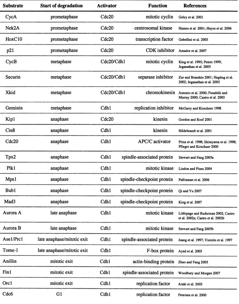
The APC/C recognizes a range of substrates during mitosis, and, certainly, many remain to be identified (Table 1-2). In prometaphase and metaphase, APC/CCdcO 2 targets
mitotic cyclins and securin for degradation to allow for anaphase onset and chromosome disjunction. In anaphase, APC/CCdhl takes over to target Cdc20, mitotic cyclins, and mitotic regulators such as Polo kinase, Aurora A, and Aurora B. In late anaphase, APC/CCdhl targets proteins that are involved in spindle function and cytokinesis.
Cdc20 generally recognizes substrates containing a D-box, whereas Cdhl
generally recognizes substrates with both a D-box and KEN box. However, in addition to these key recognition sequences, additional motifs have been identified. In many cases,
Table 1-2. Mitotic Substrates of APC/C
Start of degradation prometaphase prometaphase prometaphase prometaphase metaphase Activator Cdc20 Cdc20 Cdc20 Cdc20 Cdc20/Cdhl Function mitotic cyclin centrosomal kinase transcription factor CDK inhibitor mitotic cyclin References Geley et al. 2001Hamies et al. 2001; Hayes et al. 2006
Gabellini et al. 2003
Amador et al. 2007
King et al. 1995; Peters 1999; Jeganathan et al. 2005
Securin metaphase Cdc20/Cdhl separase inhibitor Zur and Brandeis 2001; Hagting et al.
2002; Jeganathan et al. 2005
Xkid metaphase Cdc20/Cdhl chromokinesin Antonio et al. 2000; Funabiki and
Murray 2000; Castro et al. 2003
Geminin metaphase Cdhl replication inhibitor McGarry and Kirschner 1998
Kipl anaphase Cdc20 kinesin Gordon and Roof 2001
Cin8 anaphase Cdh 1 kinesin Hildebrandt et al. 2001
Cdc20 anaphase Cdhl APC/C activator Prinz et al. 1998; Shirayama et al. 1998;
Pfleger and Kirschner 2000
anaphase anaphase anaphase anaphase anaphase late anaphase late anaphase late anaphase/mitotic exit late anaphase/mitotic exit
mitotic exit mitotic exit mitotic exit G1 Cdhl Cdhl Cdhl Cdhl Cdhl Cdhl Cdhl Cdhl Cdhl Cdhl Cdhl Cdhl Cdhl spindle-associated protein mitotic kinase spindle-checkpoint protein spindle-checkpoint protein spindle-checkpoint protein mitotic kinase mitotic kinase spindle-associated protein F-box protein actin-binding protein spindle-associated protein replication factor replication factor
Stewart and Fang 2005a
Lindon and Pines 2004
Palframan et al. 2006
Qi and Yu 2007
King et al. 2007
Littlepage and Ruderman 2002; Castro et al. 2002a; Castro et al. 2002b
Stewart and Fang 2005b
Juang et al. 1997; Visintin et al. 1997
Ayad et al. 2003
Zhao and Fang 2005
Woodbury and Morgan 2007
Araki et al. 2003 Petersen et al. 2000 Substrate CycA Nek2A HoxC10 p21 CycB Tpx2 Plkl Mpsl1 Bubl Mad3 Aurora A Aurora B Asel/Prcl Tome-i Anillin Finl Orcl Cdc6 ~ ... .. .. II ·II 11 1111 .._.._..II~ -... ..._ ..._.11111111 1--11~--111- ~1 ~ I I Illt I~ ~·---·~1~ II --- ~-3*1(
I
-~-.I~
I--I
~
I
D-boxes or KEN boxes that are present in the APC/C substrate sequence are actually not required for their degradation. Xkid, a chromokinesin in Xenopus, contains 5 putative D-boxes, none of which are required for its degradation. Instead, a novel recognition
sequence, GxEN, in Xkid's C-terminus is recognized by both Cdc20 and Cdhl and is sufficient to cause degradation (Castro et al. 2003). Both Aurora A and Aurora B contain an A box, QrxL, which is required for their degradation. The KEN box in Xenopus Aurora A is not used, but one of the D-boxes in Xenopus Aurora B contributes to its degradation (Littlepage and Ruderman 2002; Crane et al. 2004). In contrast, the D-boxes in human Aurora B are not used, whereas the KEN box contributes to its degradation along with its A-box (Nguyen et al. 2005). Mammalian Cdc20 contains another novel recognition sequence, the CRY box, CRYxPS, which is required for its degradation in oocytes and embryos (Reis et al. 2006). Drosophila Orcl contains a KEN box and 3 D-boxes, all of which are dispensable for its degradation by the APC/C. A sequence termed the O-box is used instead (Araki et al. 2003).
2. Regulation of Cdc20/Fzy
One important way in which APC/C activity is governed is through the regulation of the substrate adaptors. The activity of APC/Ccdc20 generally parallels its protein
expression profile. Cdc20 accumulates in S-phase, peaks in mitosis, and drops in G1. A combination of transcriptional upregulation in mitosis and protein degradation in Gi contributes to this profile (Fang et al. 1998a; Prinz et al. 1998). Control of Cdc20 transcription is best understood in S. cerevisiae where Cks , a small Cdk-associated protein, binds to the Cdc20 promoter and recruits Cdc28 and the proteasome to induce
Cdc20 transcription (Morris et al. 2003). It is not known whether this mechanism controls Cdc20 transcription in higher eukaryotes.
Cdc20 protein is one of several targets of APC/Ccdhl when APC/Cca' becomes
active in Gi (Prinz et al. 1998). Degradation of S. cerevisiae Cdc20 is dependent on the presence of a D-box in its N-terminus (Prinz et al 1998). Vertebrate Cdc20 sequences do not contain a D-box, and analysis of human Cdc20 led to the identification of the KEN box motif (Figure 1-2) (Pfleger and Kirschner 2000). As previously noted, in
mammalian oocytes and embryos, the degradation of Cdc20 is mediated through an additional motif called the CRY box (Reis et al. 2006).
Another major player contributing to the presence of functional Cdc20 in cells is the CCT chaperonin (chaperonin-containing TCP1). In S. cerevisiae CCT is required for the proper folding of Cdc20 in an ATP-dependent manner. CCT-dependent folding of
Cdc20 is required for its associations and activity with the APC/C and for its regulation by the spindle checkpoint (Camasses et al. 2003). A strong dependence on CCT for the generation of functional Cdc20 may be why it has been difficult to produce large amounts of purified recombinant Cdc20 for biochemical and in vitro assays. For example, in the production of in vitro transcribed and translated Cdc20 in reticulocyte lysate, the bulk of Cdc20 remains associated with CCT, perhaps because it is not folded efficiently by the rabbit CCT (Passmore et al. 2003).
In addition to regulation of levels of Cdc20 protein in the cell, the activity of APC/Ccdc20 is controlled by several inhibitors, which are discussed below.
Xenopus Emil was isolated in a yeast two-hybrid screen for Skpl binding
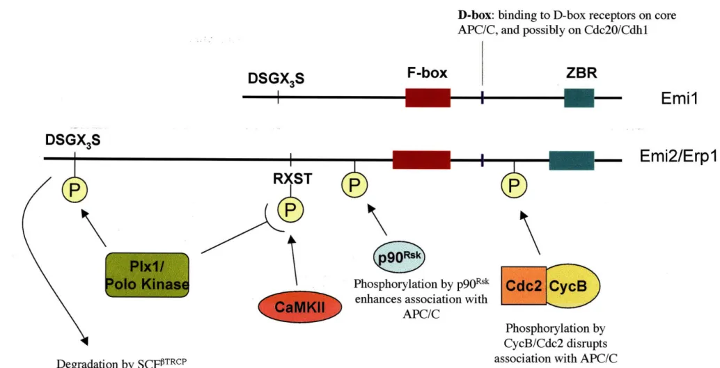
proteins, the F-box protein of the E3 SCF complex (Reimann et al. 2001a). It contains an F-box and a C-terminal Zn2 binding region (ZBR) (Figure 1-3). In cycling embryos,
levels of Emil increase in S-phase and decrease in mitosis, and addition of recombinant Emi 1 to cycling egg extracts stabilizes APC/C substrates and prevents their
ubiquitination. Emil associates with Cdc20 in interphase egg extracts, and inhibition of APC/CCdc2O-mediated ubiquitination of mitotic substrates requires the ZBR domain in
Emil (Reimann et al. 2001a).
The levels of Emil itself are regulated in a cell-cycle dependent manner by several mechanisms. Emil is a substrate of the E3 ubiquitin ligase SCF-TrcP
(Guardavaccaro et al. 2003; Margottin-Goguet et al. 2003). Phosphorylation by both Cdk and Plkl contribute to recognition and ubiquitination of Emi I by SCfT rcP in late
prophase (Margottin-Goguet et al. 2003; Moshe et al. 2004; Hansen et al. 2004). Two mechanisms protect Emi 1 from SCF-mediated degradation at other stages of the cell cycle. In S/G2 phase, the Evi5 oncogene binds directly to Emil and blocks
phosphorylation of Emil by Plkl, thus preventing its recognition by SCF-TrcP (Eldridge
et al. 2006). Emil's degradation also seems to be inhibited by Pinl, a peptidyl-prolyl
cis/trans isomerase during G2. Emil associates with Pinl in vivo during G2 and is
stabilized by Pinl in an isomerization-dependent pathway (Bernis et al. 2007).
Emil degradation in late prophase immediately precedes Cyclin A degradation by the APC/C, suggesting a requirement for Emil destruction for APC/C activation in mitosis. In addition, overexpression of non-degradable Emi 1 in mammalian cells causes a prometaphase block (Margottin-Goguet et al. 2003). These results, combined with
D-box: binding to D-box receptors on core APC/C, and possibly on Cdc20/Cdhl
ZBR
Emil
-
Emi2/Erpl
Phosphorylation by p90R
sk
enhances association with APC/C
Phosphorylation by CycB/Cdc2 disrupts association with APC/C Degradation by SCFPITRCP
Figure 1-3. Domains and regulatory modifications of Emil and Emi2/Erpl
Conserved domains in Emil and Emi2/Erpl include a D-box, which is important for their association with the APC/C, and an F-box, Zinc Binding Region (ZBR), and a DSGX3S motif, which mediate
binding to and subsequent degradation via SCF TRCP. Regulatory phosphorylation of Emi2/Erpl is shown. Please see text for details and references.
DSGX
3S
I
DSGX,S
F-box
Reimann et al.'s demonstration of a direct interaction between Emil and Cdc20, has led to a model in which degradation of Emi 1 is essential for activation of APC/Ccdc20 in mitosis. A recent study by Di Fiore and Pines questions this model (Di Fiore and Pines 2007). They timed degradation of fluorescently labeled Emi 1 and Cyclin A in HeLa cells and found that Emil is degraded between 6 and 45 minutes before Cyclin A, whereas the timing observed by Margottin-Goguet et al. was just 5 to 10 minutes. Additionally, injection of RNA encoding non-degradable Emi 1 into HeLa cells did not affect the timing of initiation of Cyclin A, Cyclin B, or Securin degradation (Di Fiore and Pines 2007). These recent findings require further investigation to determine their significance. Technical differences in the experiments performed by either group may be responsible for these contradictory results.
An additional function for Emil is as a member of a novel regulatory network END (Emil/NuMA/dynein-dynactin) that restricts the activity of APC/C in early mitosis (Ban et al. 2007). A population of Emil, along with the APC/C, localizes to spindle poles in early mitosis after the bulk of Emi 1 has been degraded via SCFV
Tr P (Hansen et
al. 2004; Ban et al. 2007). This localization is dependent on binding of Emil and APC/C to microtubules through the action of dynein-dynactin, a minus end-directed microtubule motor. APC/C, Emil, and NuMA, a nuclear matrix and spindle assembly protein, form a complex in mitosis that spatially restricts APC/C activity. Emil promotes
NuMA-dependent formation of microtubule asters through its inhibition of Cyclin B degradation by the APC/C at spindle poles (Ban et al. 2007).
Emi I was originally thought to inhibit APC/C activity simply by binding directly to Cdc20. Emil was shown to associate with Cdc20 through binding to a domain on
Cdc20 that is distinct from the domain through which Mad2 binds and inhibits Cdc20 (Mad2 inhibition discussed below) (Figure 1-2) (Reimann et al. 2001a; Reimann et al. 2001b). However, a recent study has revealed a more complex mechanism for inhibition of the APC/C by Emi 1 (Miller et al. 2006). Emil was found to associate with the core APC/C by co-fractionation experiments. Assays using an Emil affinity column showed
that Emil efficiently captures APC/C in the presence or absence of Cdc20 or Cdhl, suggesting that Emil binds to the APC/C core independently of an interaction with an activator protein. Furthermore, this direct interaction is dependent on a conserved D-box in the C-terminus of Emil but not on the ZBR domain (Figure 1-3). Both the D-box and the ZBR domain contribute to Emil's ability to compete with APC/C substrates for
binding to the APC/C and to inhibit APC/C's ubiquitination activity. Finally, wild-type Emi 1 is a poor substrate of the APC/C itself, but mutation of the ZBR domain converts
Emil into an efficient D-box dependent APC/C substrate. Thus, Emil acts as a pseudo-substrate inhibitor of the APC/C. The D-box is the domain through which Emi 1 binds the D-box receptor on the core APC/C, whereas the ZBR domain seems to act to inhibit access of substrates to the complex.
B. Spindle Assembly Checkpoint
Crucial inhibition of APC/Ccdc20 occurs in metaphase by the spindle assembly
checkpoint. The details of this regulation are discussed in the subsequent section. C. DNA damage and PKA pathway
One output of the DNA damage checkpoint is arrest of the cell cycle in the G2-M transition and the metaphase to anaphase transition. In S. cerevisiae the PKA pathway inhibits the metaphase to anaphase transition in response to DNA damage (Searle et al.
2004). An accumulation of single-stranded DNA at telomeres causes a PKA-dependent stabilization of Clb2 and Pdsl and phosphorylation of Cdc20. Cdc20 contains 2
consensus sites for PKA phosphorylation that are required for this DNA damage induced modification. In the presence of DNA damage, Cdc20 does not interact with Clb2, causing a metaphase arrest. When the phosphorylation sites of Cdc20 are mutated, Cdc20 does interact with Clb2 upon DNA damage, and Pdsl and Clb2 are degraded with faster kinetics. These results suggest that DNA damage induces PKA phosphorylation of Cdc20 to inhibit the targeting of APC/C substrates in mitosis. It is not known whether Cdc20 is inhibited by DNA damage in other organisms.
D. RASSF1A
RASSF1A is a tumor suppressor gene that is silenced by promoter methylation in lung cancer patients. Overexpression of RASSF1A induces mitotic arrest and the
accumulation of Cyclins A and B in HeLa cells, whereas depletion of RASSF1A by RNA interference accelerates cyclin degradation and mitotic progression (Song et al. 2004). These phenotypes led to an investigation of a role for RASSF1A in regulation of the APC/C. In cotransfection experiments, RASSF1A and Cdc20 co-immunoprecipitate, and RASSF1A inhibits APC/Ccdc2o but not APC/CCdhl activity in an in vitro ubiquitination
assay (Song et al. 2004). However, in a recent report, Liu et al. failed to detect an interaction between Cdc20 and RASSF1A by several experimental approaches (Liu et al. 2007). The relationship between RASSF1A and mitotic control through regulation of the APC/C remains to be clarified.
3. Regulation of Cdhl/ Fzr
are high in mitosis, but lowered in late GI and S-phases (Prinz et al. 1998; Kramer et al. 2000; Hsu et al. 2002). The decrease in Cdhl levels in late Gl and S-phase are thought to be due to E3 ligase-mediated protein degradation. Cdhl contains 2 putative D-boxes that are required for its degradation in G1, suggesting that Cdhl targets its own
degradation. Furthermore, Cdhl is ubiquitinated in vitro and degraded in an APC/Ccdhl -dependent manner (Listovsky et al. 2004). In S-phase Cdhl levels remain low, but APC/Ccdh'l cannot be responsible for maintaining low levels of Cdhl at this stage because
it is inactivated by inhibitory phosphorylation. Benmaamar and Pagano investigated a possible role for SCF in degradation of Cdhl at this cell cycle stage (Benmaamar and Pagano 2005). They found that inactivation of SCF by expression of a dominant-negative Cull or by RNA interference against Cull results in an accumulation of Cdhl, suggesting that SCF activity is required either directly or indirectly for the degradation of
Cdhl.
As discussed earlier, APC/Ccdhl activity is regulated by the phosphorylation state
of Cdhl. Cdhl is phosphorylated in S, G2, and M phases, and this phosphorylation greatly reduces the ability of Cdhl to bind to and activate APC/C (Zachariae et al. 1998; Jaspersen et al. 1999; Kramer et al. 2000).
Like Cdc20, Cdhl is also subject to regulation by a several inhibitors, but unlike Cdc20, it seems to be crucial for the cell to modulate APC/Ccdh~ activity throughout the
cell cycle, even at times when Cdhl is already subject to inhibitory phosphorylation.
A. Rcal/ Emil
The first inhibitor of Cdhl, regulator of cyclin A (rcal), was discovered in Drosophila in a screen for dominant suppressors of roughex mutants (Dong et al. 1997).
rcal mutants, like cyclin A mutants, arrest in G2 of embryonic cell cycle 16, and ectopic expression of rcal drives cells into S-phase and causes an increase in Cyclin A protein levels. A subsequent study demonstrated that Rcal acts to inhibit APC/CFzricdhl in G2 (Grosskortenhaus and Sprenger 2002). Although Emil, the vertebrate ortholog of Rcal, inhibits both Cdc20 and Cdhl, Rcal only seems to affect Cdhl. Grosskortenhaus and Sprenger failed to detect an association between FZY/ Cdc20 and Rcal, and genetic studies confirmed that the effect of Rcal on cyclin levels is specific to Fzr
(Grosskortenhaus and Sprenger 2002).
In vertebrates, the role of Emil seems to be important for inhibiting APC/Ccdh' at the GI to S-phase transition. Emil binds to Cdhl and inhibits APC/Cc~ dh in vitro
(Reimann et al. 2001b; Miller et al. 2006). An in vivo role for Emil has been
investigated in HeLa cells (Hsu et al. 2002). Similar to Cyclin A, Emil transcription is activated by E2F at the G 1 to S-phase transition. Cells that are depleted of Emi 1 by RNA interference fail to accumulate Cyclin A and do not enter S-phase. Overexpression of Cdhl causes a Gl arrest, and this arrest can be overcome by overexpression of Emil, suggesting that Emi 1 regulates S-phase entry via inhibition of APC/Ccdhl (Hsu et al. 2002). It is not clear whether Rcal also regulates S-phase entry in Drosophila embryos in addition to its role in G2 or whether Emil acts to inhibit Cdhl in G2 in vertebrate cells.
B. Rael-Nup98
Inhibitory phosphorylation of Cdhl was thought to fully inactivate APC/CCdhl during mitosis, but an investigation of the roles of Rael and Nup98 revealed an
additional layer of regulation of APC/Ccdhl in mitosis. Rael is an mRNA export factor
that acts by anchoring Nup98, a nucleoporin, to the nuclear pore complex (Pritchard et al. 1999). Splenocytes from Rael'•-Nup98"' mice exhibit premature separation of sister
chromatids, and Rael"'Nup98'- murine embryonic fibroblasts contain decreased levels of securin, which suggested a role for Rae 1 and Nup98 in mitotic control (Jeganathan et al. 2005). In mitotic HeLa cell extracts, Cdc27, Cdcl6, and Cdhl co-immunoprecipitate with Rael and Nup98, whereas Cdc20 does not associate. Furthermore, the association of Cdhl with Rael and Nup98 is specific to mitosis. Rael-Nup98 was found to inhibit ubiquitination of Securin but not Cyclin B by APC/cdhl. Interestingly, Rael-Nup98 dissociates from APC/Ccdh~ at the same time that BubR1 dissociates from APC/CCdcO upon
release of the spindle checkpoint-mediated metaphase arrest (Jeganathan et al. 2005). These results suggest that, contrary to what was previously thought, APC/Ccdh may
indeed play a role in targeting securin for degradation upon anaphase onset. The
mechanism of Rael-Nup98-mediated inhibition of APC/Ccdhl remains to be elucidated as
well as an understanding of how Rael-Nup98 is targeted to the APC/C and triggered to dissociate from the APC/C.
Finally, in addition to the Cdc20 and Cdhl inhibitors discussed above, two
transcription factors, CBP and p300, are thought to be positive regulators of the APC/C in mitosis (Turnell et al. 2005). In mammalian cells CBP and p300 are associated with an active APC/C complex in vivo, and they co-immunoprecipitate with core APC/C subunits as well as both Cdc20 and Cdhl. Knockdown of CBP by RNA interference leads to an increase of Cyclin B and Plkl levels and an accumulation of cells in mitosis. APC/C
precipitated from these CBP-depleted cells has reduced ubiquitination activity (Turnell et al. 2005). The mechanism by which CBP-p300 positively regulates mitotic APC/C is not yet clear.
It is becoming increasingly clear that regulation of APC/C activators in mitosis as well as G1, S, and G2 occurs in a complex network and at multiple levels: transcriptional control, protein stability, phosphorylation, and direct binding of inhibitors.
III. Inhibition of APC/CCdc"2 by the Spindle Checkpoint
The spindle-assembly checkpoint (SAC) is a surveillance mechanism that prevents premature separation of sister chromatids in mitosis by monitoring the
attachment of spindles to microtubules in prometaphase (for review, see Musacchio and Salmon 2007). SAC signaling is thought to initiate at or near unattached kinetochores and diffuse to inhibit APC/CCdc20's ability to target substrates, particularly Securin and Cyclin B, for degradation. As a result, cells arrest in metaphase until each kinetochore is bioriented on the spindle and under tension. At this point, the arrest is released,
APC/CCdc20 becomes active, anaphase onset occurs, and mitosis proceeds.
Two spindle checkpoint proteins, Mad2 and Mad3/BubR1, have been found to inhibit Cdc20 through direct binding. Both genes were originally identified in screens in
S. cerevisiae, in which mutations in these genes bypassed mitotic arrest in response to
spindle poisons (Hoyt et al. 1991; Li and Murray 1991). Mad2 binds directly to Cdc20 and inhibits Cdc20's ability to activate APC/C in vitro (Hwang et al. 1998; Kim et al.
1998; Fang et al. 1998b). Furthermore, Mad2, Cdc20, and APC/C form a ternary complex in prometaphase that is localized to kinetochores, suggesting an important role for Mad2 in APC/C inhibition (Fang et al. 1998). BubR1, the human homolog of yeast Mad3, is associated with Cdc20 in mitotic cells, and also inhibits the ubiquitination
activity of APC/Ccdc2o (Tang et al. 2001). In vitro, BubR1 and Mad2 act synergistically to inhibit APC/CCdc2o (Tang et al. 2001)
The physical nature of the spindle checkpoint APC/C inhibitor was further clarified in a study by Sudakin et al. A single stable complex was purified from HeLa cell lysates that is able to inhibit the ubiquitin ligase activity of APC/Ccdc2O (Sudakin et al.
2001). This complex, termed the Mitotic Checkpoint Complex (MCC), contains BubR1, Mad2, Bub3, and Cdc20 in roughly equal stoichiometries (Figure 1-4). In reconstituted ubiquitination assays, MCC inhibits APC/C more efficiently than recombinant Mad2 alone, suggesting that MCC is likely the in vivo inhibitor of APC/C during spindle checkpoint arrest (Sudakin et al. 2001).
Furthermore, formation of the MCC is thought to be aided by phosphorylation of Cdc20 (Figure 1-4). In Xenopus egg extracts, Cdk activity is required for spindle
checkpoint arrest (D'Angiolella et al. 2003). Cdk-dependent phosphorylation of Cdc20 inhibits its binding to Cdc27 and enhances its binding to Mad2 (Yudkovsky et al. 2000; D'Angiolella et al. 2003). Xenopus Cdc20 also seems to be regulated by MAPK-dependent phosphorylation during spindle checkpoint inhibition. A Cdc20 mutant with all of its MAPK phosphorylation sites mutated is unresponsive to spindle checkpoint inhibition and exhibits reduced binding to Mad2 and BubR1 (Chung and Chen 2003). It is not clear whether direct MAPK phosphorylation of Cdc20 is conserved, as human Cdc20 is not phosphorylated by MAPK in vitro (Tang et al. 2004).
Unattached
Kinetochore
MCC
formation.-"'
I I I yc/Cdc20
Attached
'--,- KinetochoreFigure 1-4. Spindle checkpoint inhibition of APC/CCdcO2
Spindle checkpoint-mediated inhibition of APC/Ccdc20 is achieved through binding
of Cdc20 to MCC (Mad2, BubR1, Bub3), phosphorylation of Cdc20 by Bubl kinase, and deubiquitination of Cdc20 by USP44. Autoubiquitination of Cdc20 stimulated by UbcH10 and p31comet leads to the dissociation of Cdc20 and MCC,
inactivation of the spindle checkpoint, and activation of APC/Ccdc20. Please see text for details and references.
i-t
Mad2
Bub3
The mechanism by which MCC inhibits APC/Ccdc20 is not entirely clear although several pieces of data provide clues. In an in vitro binding assay, Mad2 prevents release of substrates from Cdc20, which may affect turnover of substrate ubiquitination (Pfleger et al. 2001). Both Mad2 and BubR1 coimmunoprecipitate core APC/C subunits,
suggesting that they do not act by preventing Cdc20 from binding APC/C (Fang et al. 1998; Sudakin et al. 2001). Finally, two recent studies in S. cerevisiae demonstrate that Mad3/BubR1 may act by blocking substrate access to Cdc20. Mad3 contains 2 KEN boxes that are required for spindle checkpoint function (Burton and Solomon 2007; King et al. 2007). These KEN boxes as well as a D-box in Mad3 all contribute to binding of Mad3 to Cdc20 in vivo, and Mad3 competes with Hsl 1, an APC/C substrate, for binding to Cdc20 in vitro (Burton and Solomon 2007; King et al. 2007). Thus, Mad3 may act like a pseudosubstrate inhibitor of APC/Ccdc2O, similar to Emil's mode of inhibition.
In addition to spindle checkpoint inhibition of Cdc20 through its association with the MCC, a recent study demonstrates that Cdc20 is independently regulated by Bubl, another spindle checkpoint protein (Figure 1-4). In HeLa cells Bubl is required for mitotic arrest in response to spindle damage (Tang et al. 2004). Bub , a protein kinase, phosphorylates Cdc20 in vitro, which reduces the ubiquitin ligase activity of APC/Ccdc2O. Expression of a Cdc20 mutant that is resistant to Bub1 phosphorylation causes a failure of cells to arrest in response to nocodazole, however, this form of Cdc20 is still able to bind Mad2 and BubR1 (Tang et al. 2004). In contrast to inhibition by stoichiometric binding of Cdc20 by Mad2 and BubR1, this study reveals that the spindle checkpoint also targets Cdc20 through the catalytic activity of Bubl kinase.
Finally, direct ubiquitination of Cdc20 places yet another level of regulation on Cdc20 by the spindle checkpoint. In S. cerevisiae, Cdc20 protein stability is reduced upon activation of the spindle checkpoint (Pan and Chen 2004). Binding of Cdc20 to Mad2 and BubR1 is required for this APC/C-dependent degradation of Cdc20, but the D-boxes in Cdc20 and Cdhl are not required (Pan and Chen 2004; King et al. 2007). Thus, Cdc20 is targeted for APC/C dependent degradation even at the same time that it itself is inhibited from targeting other substrates. These studies suggest that Cdc20 levels must be reduced for complete inhibition of APC/Ccdc"2 during an active spindle checkpoint. It
is not clear whether this regulatory strategy is conserved in higher eukaryotes.
Two recent studies in human cells reveal another role for ubiquitination of Cdc20 in spindle checkpoint control. In this case, autoubiquitination of Cdc20 serves to induce dissociation of Cdc20 and Mad2 and inactivation of the spindle checkpoint (Figure 1-4) (Reddy et al. 2007; Stegmeier et al. 2007). Reddy et al. showed that HeLa cells that overexpress UbcH10, the E2 enzyme that acts with APC/C, fail to arrest in response to nocodazole. UbcH10 and APC/C-dependent ubiquitination of Cdc20 is required in vivo for dissociation of Mad2 and Cdc20 after release from nocodazole, but surprisingly, proteasome activity is not required.
In an accompanying paper, Stegmeier et al. performed a short hairpin RNA screen in human cells to find components of the ubiquitin-proteasome pathway that are required for spindle checkpoint function. They identified USP44, an uncharacterized
deubiquitinating enzyme that is required for checkpoint function. USP44 is required for stabilizing the Mad2-Cdc20 association in nocodazole arrested cells, and further, the authors showed that USP44 deubiquitinates Cdc20 in vitro and in vivo. Depletion of
UbclO in cells suppresses the effects of cells with reduced USP44 function by restoring spindle checkpoint arrest, strongly suggesting that USP44 acts to antagonize UbcH10-mediated release from spindle checkpoint arrest. These studies reveal an additional level of control in the spindle checkpoint pathway, which could act as a switch-like mechanism to trigger anaphase onset. It has yet to be determined how the spindle checkpoint
network modulates this switch to control maintenance or release of metaphase arrest.
IV. Role of the APC/C in Female Meiosis
Meiosis is a modified cell division in which one parent cell generates four haploid cells, in contrast to the two identical diploid cells generated in a canonical mitotic cell division. Understanding the function and regulation of the APC/C in meiosis presents an interesting and complex problem given the differences of both chromosome segregation dynamics and developmental context in meiosis compared to mitosis.
A reductional division in meiosis I and an equational division in meiosis II without an intervening S phase generate haploid gametes in a meiotic cell division. Control of the meiotic divisions is coordinated by both general cell-cycle regulators as well as well as meiosis-specific proteins (for review, see Marston and Amon 2004). Meiosis I is unique because homologous chromosome pairs, as opposed to sister chromatids, must be segregated from each other. Homologous chromosomes are held together by chiasmata and arm cohesion. This segregation is achieved by loss of cohesion along chromosome arms but not in the centromeric regions. Subsequently, in meiosis II, sister chromatids are segregated through loss of cohesion at the centromeres in a process very similar to that in mitosis. Regulation of APC/C activity is likely to be
important in this process as separase must be activated twice, through APC/C-mediated degradation of securin, in one full meiotic cell division.
Furthermore, the absence of an S-phase in between meiosis I and meiosis II also likely requires specialized regulation of the APC/C. In Xenopus oocytes it has been shown that mitotic Cdk activity is maintained at a low level in between the two meiotic divisions (Figure 1-5) (Furuno et al. 1994; Iwabuchi et al. 2000). Mitotic Cdk activity must be kept at an intermediate level to satisfy the unique requirements of meiosis: Cdk activity must be low enough to allow for disassembly of the meiosis I spindle, but high enough to repress initiation of DNA replication. Presumably, APC/C-mediated
degradation of mitotic and meiotic cyclins must be regulated to contribute to this modulation of Cdk activity.
Regulation of APC/C activity in meiosis is particularly crucial during oogenesis of multicellular organisms. In most animals meiosis is arrested twice to coordinate development of the oocyte with the events of meiosis (Figure 1-5) (for reviews, see Kishimoto 2003; Tunquist and Maller 2003). First, oocytes are arrested in prophase I to allow for oocyte growth and differentiation before initiation of the meiotic divisions. Then, oocytes arrest again in metaphase I or metaphase II to await egg activation or fertilization. This secondary arrest ensures that the completion of meiosis is properly coordinated with fertilization of the oocyte. During both meiotic arrests, APC/C activity must be suppressed. In prophase I, unscheduled activity of the APC/C could prevent proper maintenance of chromosome cohesion. In the secondary metaphase arrests, inhibition of the APC/C is crucial for preventing premature anaphase onset. Thus, understanding the function and regulation of the APC/C during meiosis is critical for
Egg Activation and
Fertilization in
Drosophila
Fertilization in
vertebrates
Polar bodies
Nucleus
Prophase I
Metaphase
I
(in Drosophila)Metaphase
II
(CSF arrest in vertebrates)Pronucleus(G
)
I
Primary Meiotic
Arrest
Secondary Meiotic
Arrests
MPF (CycB/Cdk) ActivityFigure 1-5. Meiotic Progression in Females
Meiosis is arrested twice during oogenesis. In most organisms the first meiotic arrest occurs during prophase I, and the second arrest time varies among organisms. Fertilization or egg activation releases these arrests in metaphase I and metaphase II. Cyclin B/ Cdk activity is elevated at each meiotic division and drops in betweenR the divisions.
understanding control of chromosome segregation and cell cycle progression and coordination of this control with developmental signals during meiosis.
A. Requirement for APC/C in Meiosis
A requirement for APC/C activity during the meiotic divisions has been shown in all organisms tested with the exception of Xenopus oocytes. In yeast, Cdc20 is required for the degradation of Pdsl/Securin and resulting activation of separase in both meiotic divisions (Salah and Nasmyth 2000). Separase activity is required for the degradation of Rec8, the meiosis-specific cohesin subunit, along chromosome arms to allow for
homolog disjunction in meiosis I (Buonomo et al. 2000; Kitajima et al. 2003).
It has been more difficult to demonstrate a requirement for APC/C in meiosis in multicellular organisms. In C. elegans, mutations in or RNA interference against several subunits of the APC/C cause a meiotic metaphase I arrest (Furuta et al. 2000; Golden et al. 2000; Davis et al. 2002). Female sterile mutations infzy cause both meiosis I and meiosis II arrests in Drosophila eggs (Swan and Schupbach 2007). Finally, several studies in mouse oocytes have demonstrated a requirement for APC/C-mediated degradation of Securin and activation of separase for Rec8 removal from chromosome arms and homolog disjunction in meiosis I (Herbert et al. 2003; Terret et al. 2003; Kudo et al. 2006).
However, two studies in Xenopus oocytes have suggested that APC/C is not required for meiosis I in this organism. Microinjection of oocytes with antibodies against Fzy or Cdc27 does not disrupt progression through meiosis I but only causes an arrest in meiosis II (Peter et al. 2001). Injection of antisense oligonucleotides against Fzy also
does not block progression through meiosis I (Taieb et al. 2001). A more recent study in Xenopus demonstrates that Securin and Cyclin B are degraded at anaphase I,
reaccumulate before metaphase II, and are degraded again at anaphase II (Fan et al. 2006). These data are suggestive of an active APC/C in both meiosis I and meiosis II, although they do not demonstrate a requirement for this activity for homolog disjunction and progression through meiosis I. Additionally, misexpression of Emi2, an APC/C inhibitor that is active later in meiosis, induces a metaphase I arrest in Xenopus oocytes, again suggesting that APC/C is required for the metaphase I to anaphase I transition (Ohe et al. 2007; Tung et al. 2007). It is not clear whether the contrary results in Xenopus reveal a true biological difference in this system or are due to differences in experimental design.
B. Meiosis-Specific APC/C Activators
An intriguing development in our knowledge of meiotic cell cycle control has been the identification of meiosis-specific APC/C activators in yeast and flies. The existence of these activators suggests a unique role and set of substrates for the APC/C in meiosis that are outside of the functions of Cdc20 or Cdhl.
In yeast, meiosis-specific activators are expressed exclusively during meiosis and are required for proper spore formation (Cooper et al. 2000; Asakawa et al. 2001; Blanco et al. 2001). Amal in S. cerevisiae associates with the core APC/C throughout meiosis, becomes active after meiosis I, and is required for Clbl and Pds1 degradation.
Furthermore, it can drive the ubiquitination of Pdsl in vitro (Cooper et al. 2000;
the core APC/C during meiosis and is required for the degradation of M-phase cyclin Cdc 13. Degradation of Cdc 13 in meiosis II is required for proper spore formation at the end of meiosis (Blanco et al. 2001).
In Drosophila, cortex (cort) encodes a distant member of the Cdc20/FZY protein family that is transcribed only during oogenesis (Chu et al. 2001). cort mutant females
lay eggs that never complete meiosis and arrest terminally in metaphase II (Lieberfarb et al. 1996; Page and Orr-Weaver 1996). cort seems to be required as soon as the
metaphase I to anaphase I transition occurs, as meiosis II spindles often exhibit unequal numbers of chromosomes indicating aberrant chromosome segregation in meiosis I (Page and Orr-Weaver 1996). Consistent with cort functioning as an APC/C activator, mitotic cyclin levels are elevated in cort eggs, and misexpression of cort triggers degradation of these cyclins in wing imaginal discs (Swan and Schupbach 2007).
The Drosophila genome also contains a 4' Cdc20-related gene,fizzy-related 2 (fzr2), that is expressed exclusively in male meiosis. When misexpressed,fzr2 can rescue
fzr
function by triggering degradation of Cyclin B, which suggests a true function forfzr2 as an APC/C activator (Jacobs et al. 2002). The use of a female and a malemeiosis-specific APC/C activator in Drosophila will provide an interesting system in which to delineate the meiosis-specific roles of APC/C in the two different developmental contexts of oogenesis and spermatogenesis.
Meiosis-specific activators are not the only APC/C activators present during meiosis. In S. cerevisiae both Cdc20 and Amal are thought to contribute to APC/C function in meiosis. Amal protein is present beginning in pre-meiotic S-phase, although it does not become essential until late in meiosis for spore formation (Cooper et al. 2000;