HAL Id: hal-00681942
https://hal.archives-ouvertes.fr/hal-00681942
Submitted on 28 Oct 2013
HAL is a multi-disciplinary open access
archive for the deposit and dissemination of
sci-entific research documents, whether they are
pub-lished or not. The documents may come from
teaching and research institutions in France or
abroad, or from public or private research centers.
L’archive ouverte pluridisciplinaire HAL, est
destinée au dépôt et à la diffusion de documents
scientifiques de niveau recherche, publiés ou non,
émanant des établissements d’enseignement et de
recherche français ou étrangers, des laboratoires
publics ou privés.
Velocimetric third-harmonic generation microscopy:
micrometer-scale quantification of morphogenetic
movements in unstained embryos
Delphine Débarre, Willy Supatto, Emmanuel Farge, Bruno Moulia,
Marie-Claire Schanne-Klein, Emmanuel Beaurepaire
To cite this version:
Delphine Débarre, Willy Supatto, Emmanuel Farge, Bruno Moulia, Marie-Claire Schanne-Klein, et al..
Velocimetric third-harmonic generation microscopy: micrometer-scale quantification of morphogenetic
movements in unstained embryos. Optics Letters, Optical Society of America - OSA Publishing, 2004,
29, pp.2881-2883. �10.1364/OL.29.002881�. �hal-00681942�
December 15, 2004 / Vol. 29, No. 24 / OPTICS LETTERS 2881
Velocimetric third-harmonic generation microscopy:
micrometer-scale quantification
of morphogenetic movements in unstained embryos
Delphine Débarre
Laboratory for Optics and Biosciences, Centre National de la Recherche Scientifique, Institut National de la Santé et de la Recherche Médicale, Ecole Polytechnique, F-91128 Palaiseau, France
Willy Supatto and Emmanuel Farge
Mechanics and Genetics of Developmental Embryology, Centre National de la Recherche Scientifique, Curie Institute, 11 Rue Pierre et Marie Curie, F-75005 Paris, France
Bruno Moulia
Biomechanics Group, Institut National de la Recherche Agronomique, 234 Avenue du Brézet, F-63039 Clermont-Ferrand, France
Marie-Claire Schanne-Klein and Emmanuel Beaurepaire
Laboratory for Optics and Biosciences, Centre National de la Recherche Scientifique, Institut National de la Santé et de la Recherche Médicale, Ecole Polytechnique, F-91128 Palaiseau, France
Received June 30, 2004
We demonstrate the association of third-harmonic generation (THG) microscopy and particle image velocime-try (PIV) analysis as a novel functional imaging technique for automated micrometer-scale characterization of morphogenetic movements in developing embryos. Using a combined two-photon-excited f luorescence and THG microscope, we characterize the optical properties of Drosophila embryos and show that sustained THG imaging does not perturb sensitive developmental dynamics. Velocimetric THG imaging provides a quantita-tive description of the dynamics of internal structures in unstained wild-type and mutant embryos. © 2004 Optical Society of America
OCIS codes: 180.0180, 190.4160, 190.4180, 180.6900, 170.7050, 170.1420.
The development of animal embryos exhibits a complex ensemble of cell movements that are highly regulated in time and space. Investigating these complex dynamic processes remains a challenge in biology.1
In particular, control of morphogenetic movements involved in Drosophila melanogaster embryo devel-opment serves as a major model for develdevel-opmental genetics.2 However, Drosophila embryos exhibit fast
development dynamics and are highly scattering for visible light at early stages, which limits direct imag-ing of dynamic processes. In addition, f luorescent labeling can introduce unwanted perturbations and might be difficult to obtain in complex mutants. As a consequence, the mechanical behavior of specific cells and tissues is not completely understood in many cases. In this context, novel noninvasive techniques for visualizing and quantifying morphogenetic move-ments in vivo should have a significant effect on developmental biology.
Third-harmonic generation (THG) microscopy3 has
been proposed as a novel general-purpose technique for obtaining structural images of transparent objects with micrometer three-dimensional resolution com-parable with that of two-photon-excited f luorescence (2PEF) microscopy and was demonstrated recently in biological samples such as cells4 and zebra f ish
embryos.5 In this Letter we extend THG microscopy
to the quantitative measurement of tissue velocity fields inside unstained opaque embryos by combining THG microscopy and particle image velocimetry (PIV) analysis techniques adapted from hydrodynamics.
First we use a combined 2PEF –THG approach to characterize the optical properties of Drosophila early embryos. Then we demonstrate that sustained THG embryo imaging does not perturb the dynamics of sensitive developmental processes. Finally, we illus-trate velocimetric THG imaging by micrometer-scale quantification of morphogenetic movements in live unstained wild-type and mutant embryos.
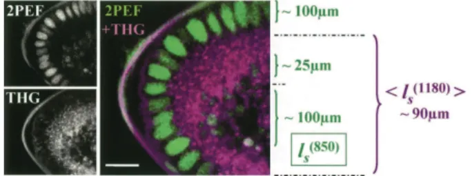
We characterized early Drosophila embryo prop-erties (Fig. 1) by using a custom-built combined 2PEF– THG microscope incorporating a femtosecond titanium:sapphire oscillator (Coherent, Inc.), an optical parametric oscillator (APE), galvanometer mirrors (GSI Lumonics), water-immersion objectives (Olym-pus), and photon-counting photomultiplier modules (Electron Tubes). 2PEF was epidetected when green f luorescent protein- (GFP-) expressing embryos were excited at 820 –920 nm. Alternatively, THG was detected in the transmitted direction when embryos were excited at 1180 nm. In either case, the signal was selected by use of appropriate f ilters (Chroma).
Combined 2PEF–THG imaging of embryos with nuclear GFP labeling illustrates the complementarity of these imaging modalities (Fig. 1). At the onset of gastrulation (the universal primary morphogenetic process in animal development), Drosophila embryos comprise a single cell layer surrounding a hetero-geneous medium (yolk), which is totally opaque in transmitted light microscopy. As expected, GFP-labeled nuclei produce a strong 2PEF signal when they are excited at 820 nm. THG is enhanced for
2882 OPTICS LETTERS / Vol. 29, No. 24 / December 15, 2004
Fig. 1. Optical properties of early Drosophila embryos investigated by 2PEF– THG microscopy. 2PEF imaging (820-nm excitation) reveals GFP-labeled nuclei and en-dogenous yolk f luorescence. Lipid droplets are a major source of contrast in THG images (1180-nm excitation).
ls, scattering mean free path (see text); scale bar, 15 mm.
micrometer-sized optical heterogeneities.6 When
em-bryos are illuminated at 1180 nm, lipid droplets that are present around the nuclei and at the periphery of the central yolk region produce a readily detectable THG signal (5 10 3 106 detected photons兾s in our
imaging conditions). These droplets have typical sizes of the order of 0.5 mm,7which contribute to THG
signal enhancement.6
The key parameter for nonlinear microscopy of tis-sues is the scattering mean free path of excitation light
ls共ex兲, because it defines the increase in laser power
that is necessary to compensate for the scattering of excitation light with depth.8 When absorption is
ne-glected and with the assumption of constant collection efficiency, the detected signal for an nth-order pro-cess locally scales as exp关2z兾ls共ex兲兴nfor a given incident
power, where z is the imaging depth. We estimated
ls共850兲 (850-nm excitation) at different depths within
embryos at the onset of gastrulation and found that three regions with distinct optical properties could be defined: (i) the peripheral region (nuclei) for which
ls共850兲⬃ 100 mm, (ii) a highly scattering region at the
yolk surface [ls共850兲 ⬃ 25 mm], and (iii) the internal
yolk [ls共850兲⬃ 100 mm]. As is evident from THG
im-ages, the highly scattering nature of the yolk’s sur-face is related to the density of lipid droplets in this region, which strongly hampers inner tissue imaging by conventional techniques. We estimated an average scattering length 具ls共1180兲典 for THG imaging over the
entire yolk region (see Fig. 1). We found that 具ls共1180兲典 ⬃
90 mm, indicating that THG microscopy performs well deep within embryos owing to the reduced scattering of excitation wavelengths in the 1100– 1300-nm range. Rapid THG imaging of embryos with ⬃80-MHz pulse trains requires instantaneous intensities in the TW兾cm2regime (90-mW average power with a 0.8-N.A.
objective). Although THG microscopy is believed to be noninvasive because harmonic generation processes do not involve energy deposition in the sample, the possibility of laser-induced perturbation with such intensities cannot be excluded without experimental evidence. As a matter of fact, we observed bubble formation in isolated cells with slightly higher pulse energy in the same wavelength range (data not shown). To establish the validity of THG microscopy for measuring the dynamics of developmental pro-cesses we monitored the process of cellularization, a critical and temperature-sensitive dynamic event
of embryonic cells. Cellularization involves oocyte plasma membrane folding between nuclei, resulting in the formation of individual cells. The rate and the completion of cellularization front invagination (CFI) are sensitive indicators of the integrity of cytoskeleton dynamics.9 We found that CFI can be
followed in vivo by THG microscopy because it induces changes in the distribution of lipid droplets about the nuclei. We compared the CFI rate in control cytoskeleton-labeled GFP (sGMCA) embryos by using 2PEF microscopy and in unlabeled wild-type embryos by THG microscopy. As illustrated in Fig. 2, we found similar dynamics and phase durations for the two imaging modalities. These data also corroborate transmitted-light measurements (not shown). We conclude that for these imaging conditions (1180 nm, 80 MHz, 0.9 nJ, 0.8 N.A., 120-mm兾ms scan speed, 20 s between successive images), sustained THG imaging does not induce significant perturbation. Moreover, we observed a normal survival rate (85 6 3%) af-ter long-af-term imaging, which further confirms the validity of THG microscopy for measuring embryo developmental dynamics.
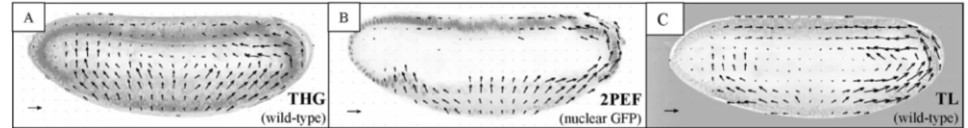
To provide a micrometer-scale quantitative descrip-tion of morphogenetic movements, we estimated in-stantaneous velocity fields from THG image sequences of developing embryos by use of PIV,10 a technique
commonly used in hydrodynamics. PIV relies on cor-relation calculations and can extract velocimetric data with subpixel resolution even when moving structures are not clearly defined in the images, in contrast to standard tracking methods. THG microscopy is sen-sitive to micrometer-sized optical inhomogeneities and provides rich structural information throughout entire unstained embryos. As a consequence, THG image sequences are ideally suited for correlation analy-sis (Fig. 3). PIV analyanaly-sis was performed with the MatPIV software package11 with 64 3 64 pixel
inter-rogation windows.10 As shown in Fig. 3, velocimetric
analysis of THG data provides information about tissue
Fig. 2. Cellularization dynamics were followed at 19±C
in control sGMCA embryos by 2PEF microscopy (920-nm excitation) and in wild-type embryos by THG microscopy (1180-nm excitation). Image sequences were recorded, and space –time (YT) projections of the signal in the white rectangle areas were extracted. In the YT representation, slopes ref lect the rate of CFI; see text. Similar speeds (indicated in micrometers per minute) are found in both ex-periments, which show that sustained THG imaging does not perturb the dynamics of this sensitive, temperature-dependent process.
December 15, 2004 / Vol. 29, No. 24 / OPTICS LETTERS 2883
Fig. 3. A, Velocimetric THG microscopy provides in vivo a micrometer-scale description of morphogenetic movements through entire unstained wild-type embryos. B, Velocimetric analysis of 2PEF images of a nuclei-labeled embryo. C, Velocimetric analysis of transmitted-light (TL) images of an unstained embryo does not provide three-dimensional sectioning. Scale arrow, 5 mm兾min. One image was recorded every 20 s, with 0.6-mm pixel size.
Fig. 4. THG microscopy provides micrometer-scale quantif ication of disrupted morphogenetic movements in unstained mutant embryos. The mean velocity in the area defined by a black rectangle, as determined by veloci-metric THG in a wild-type embryo, is shown as a function of time. The same measurements in a Dorsal embryo with disrupted movements and in a wild-type embryo from transmitted-light (TL) microscopy are also shown. Veloci-ties are projected in the vertical direction, positive up.
movements similar to that obtained from GFP lines without the need to stain structures selectively. It has the additional benefit of providing deeper imaging in unstained areas. Despite the highly scattering nature of early Drosophila embryos, clear velocimetric THG whole-embryo images could be obtained down to the equator within rapid acquisition times (2.3 s; Fig. 3A), providing information that is not visible in 2PEF and transmitted-light images. Since morphogenetic movements are symmetric relative to the equatorial plane (antero– posterior, dorso–ventral axis), they all occur within the accessible volume of view. As is apparent from Fig. 3, velocimetric THG imaging pro-vides a simultaneous description of tissue and internal structure dynamics and reveals their coupling.
We expect that a major application of this tech-nique will be micrometer-scale characterization of morphogenetic movements in mutant embryos, for which GFP constructs can be diff icult to obtain. We illustrate this point by comparing developmental dy-namics of unstained wild-type embryos and unstained embryos from dorsal mutant mothers (so-called dorsal embryos).2 Dorsal embryos lack dorso – ventral
polar-ization and exhibit disrupted movements. Figure 4 illustrates that velocimetric THG imaging clearly extracts distinct velocity fields from internal regions of wild-type and dorsal embryos during ventral furrow invagination and that this information is not accessible to conventional imaging techniques.
Our experiments establish the combination of THG microscopy and PIV as a functional imaging
technique for the micrometer-scale characterization of morphogenetic movements in live unstained embryos. We showed that this approach is applicable to early
Drosophila embryos, a major model in developmental
genetics that is difficult to observe with conventional imaging techniques. Our combined 2PEF –THG data provide insight about optical properties of embryos. Importantly, we showed that sustained THG imag-ing does not introduce significant perturbation to sensitive dynamic processes under practical imaging conditions. Thus, biologically relevant quantitative descriptions of morphogenetic movements can be obtained in both unstained wild-type and mutant em-bryos. This versatile approach should find a broad range of applications in developmental biology.
We thank Catherine Schaffner, Marcel Bierry, Jean-Marc Sintes, and Xavier Solinas for technical assis-tance and Eric Brouzés and Charlotte Py for fruitful discussions. We are grateful to Daniel P. Kiehart and Ruth A. Montague for the gift of the sGMCA transgenic line. This work was supported by the Délégation Générale pour l’Armement. E. Beaurepaire’s e-mail address is [email protected]. References
1. L. Solnica-Krezel and S. Eaton, Development 130, 4229 (2003).
2. M. Bate and A. Martinez-Arias, The Development of
Drosophila melanogaster (Cold Spring Harbor
Labora-tory Press, New York, 1993).
3. Y. Barad, H. Eisenberg, M. Horowitz, and Y. Silberberg, Appl. Phys. Lett. 70, 922 (1997).
4. D. Yelin and Y. Silberberg, Opt. Express 5, 196 (1999), http://www.opticsexpress.org.
5. S.-W. Chu, S.-Y. Chen, T.-H. Tsai, T.-M. Liu, C.-Y. Lin, H.-J. Tsai, and C.-K. Sun, Opt. Express 11, 3093 (2003), http://www.opticsexpress.org.
6. J.-X. Cheng and X. S. Xie, J. Opt. Soc. Am. B 19, 1604 (2002).
7. M. A. Welte, S. P. Gross, M. Postner, S. M. Block, and E. F. Wieschaus, Cell 92, 547 (1998).
8. M. Oheim, E. Beaurepaire, E. Chaigneau, J. Mertz, and S. Charpak, J. Neurosci. Methods 112, 205 (2001). 9. A. Royou, C. Field, J. C. Sisson, W. Sullivan, and R.
Karess, Mol. Biol. Cell 15, 838 (2004).
10. M. Raffel, C. Willert, and J. Kompenhans, Particle
Image Velocimetry: A Practical Guide
(Springer-Verlag, Berlin, 1998).
11. J. K. Sveen, “An introduction to MatPIV v. 1.6.1,” eprint series (Department of Mathematics, University of Oslo, Oslo, Norway, 2004).