Publisher’s version / Version de l'éditeur:
Vous avez des questions? Nous pouvons vous aider. Pour communiquer directement avec un auteur, consultez la première page de la revue dans laquelle son article a été publié afin de trouver ses coordonnées. Si vous n’arrivez pas à les repérer, communiquez avec nous à PublicationsArchive-ArchivesPublications@nrc-cnrc.gc.ca.
Questions? Contact the NRC Publications Archive team at
PublicationsArchive-ArchivesPublications@nrc-cnrc.gc.ca. If you wish to email the authors directly, please see the first page of the publication for their contact information.
https://publications-cnrc.canada.ca/fra/droits
L’accès à ce site Web et l’utilisation de son contenu sont assujettis aux conditions présentées dans le site LISEZ CES CONDITIONS ATTENTIVEMENT AVANT D’UTILISER CE SITE WEB.
2D Materials, 7, 1, pp. 015002-1-015002-19, 2019-10-04
READ THESE TERMS AND CONDITIONS CAREFULLY BEFORE USING THIS WEBSITE.
https://nrc-publications.canada.ca/eng/copyright
NRC Publications Archive Record / Notice des Archives des publications du CNRC :
https://nrc-publications.canada.ca/eng/view/object/?id=bdad48cd-5ae9-45ad-8114-9921008bf191 https://publications-cnrc.canada.ca/fra/voir/objet/?id=bdad48cd-5ae9-45ad-8114-9921008bf191
NRC Publications Archive
Archives des publications du CNRC
This publication could be one of several versions: author’s original, accepted manuscript or the publisher’s version. / La version de cette publication peut être l’une des suivantes : la version prépublication de l’auteur, la version acceptée du manuscrit ou la version de l’éditeur.
For the publisher’s version, please access the DOI link below./ Pour consulter la version de l’éditeur, utilisez le lien DOI ci-dessous.
https://doi.org/10.1088/2053-1583/ab4554
Access and use of this website and the material on it are subject to the Terms and Conditions set forth at
CVD grown nitrogen doped graphene is an exceptional visible-light driven photocatalyst for surface catalytic reactions
Alam, Kazi M.; Kumar, Pawan; Manuel, Ajay P.; Vahidzadeh, Ehsan;
Goswami, Ankur; Zeng, Sheng; Wu, Wenjie; Mahdi, Najia; Cui, Kai; Kobryn, Alexander E.; Gusarov, Sergey; Song, Yenan; Shankar, Karthik
CVD Grown Nitrogen Doped Graphene is an Exceptional
Visible-Light Driven Photocatalyst for Surface Catalytic
Reactions
Kazi M. Alam,1 Pawan Kumar,1 Ajay Manuel,1 Ehsan Vaidzadeh,1 Ankur Goswami,1 Sheng Zeng,1 Wenjie Wu,3,4 Najia Mahdi,1 Kai Cui,2 Alexander E. Kobryn,2 Sergey Gusarov,2 Yenan Song3,4* and Karthik Shankar1,4
1
Department of Electrical & Computer Engineering, University of Alberta, 9211-116 St., Edmonton, AB T6G 1H9, Canada
2
Nanotechnology Research Centre, National Research Council Canada, 11421 Saskatchewan Drive, Edmonton, AB T6G 2M9, Canada
3
Engineering Research Center for Nanophotonics & Advanced Instrument, School of Physics and Electronic Science, East China Normal University, Shanghai, 200062 China
4
Joint Institute of Advanced Science and Technology, East China Normal University, Shanghai, 200062 China
Abstract
The photocatalytic potential of large area CVD grown nitrogen doped graphene (NGr) has been explored though the surface catalytic chemical transformation of 4-nitrobenzene thiol into p,p-dimercaptoazobenzene. Decoration of NGr with Ag nanocubes with rounded edges to form NGr/Ag nanohybrids resulted in a slight increase in the work-function and a decrease in the n-type character of NGr due to ground state transfer of negative charge from NGr to Ag. The Ag nanocubes exhibited a localized surface plasmon resonance (LSPR) at ~ 425 nm. When the NGr/Ag nanohybrids were illuminated with visible light of wavelength close to the LSPR peak, KPFM indicated a dramatic change in surface potential of −225 mV and Raman spectra detected electron accumulation in NGr, which are
attributed to a high local field enhancement-mediated hot electron injection into NGr and the formation of long-lived charge separated states. Pristine nitrogen doped graphene and its coupled system with plasmonic Ag nanoparticles showed superior photocatalytic performance compared to bare plasmonic Ag catalyst. While standalone Ag NPs were unable to complete the transformation of 4-NBT into DMAB even at a laser power of 10 mW, NGr/Ag nanohybrids completed this transformation at a laser power of 1 mW, pointing to the high photoreduction strength of NGr/Ag. Density functional theory (DFT) based computational modeling was used to examine the electronic structure of graphene doped with graphitic, pyridinic and pyrrolic nitrogen dopant atoms. DFT results indicated a stronger localization of charge at the dopant sites and a pronounced difference in the projected density of states (PDOS) for carbon atoms in proximity to, and distant from, the nitrogen dopant sites.
Keywords:
Nitrogen doped graphene, surface plasmon resonance, plasmonic energy transfer, hot electron injection, surface catalytic reactions, plexciton.1. Introduction
Graphene, a single atom-thick two-dimensional (2D) sheet of sp2 hybridized carbon atoms, has been extensively studied experimentally and theoretically, due to its distinct electronic properties governed by the relativistic quantum mechanical Dirac equation 1-2. Graphene has long range -conjugation and can be considered as the basic building block of all other carbon based allotropes that are comprised of sp2 hybridized carbon atoms. Since its discovery, it has been explored by many research groups around the globe, in order to advance the fundamental science of 2D materials and unveil technological potential 3. However pristine graphene is a zero-band gap semiconductor possessing semimetallic character, which renders it ineffective as a stand-alone photocatalyst. Incorporation of an impurity heteroatom through compositional doping is an effective route to alter the electronic properties of graphene. Such behavior
modification is desirable for a plethora of applications; for instance, chemical doping increases the on/off current ratio of a graphene-based transistor 4. A nitrogen atom has one extra electron than a carbon atom, and analogous atomic size. Therefore N-doping of graphene is a viable way to tune the electronic properties of graphene without compromising its high mobility 4-9. Both experimental and theoretical works showed an upward shifting of the Fermi level relative to the Dirac point in N-doped graphene (NGr), giving rise to a finite bandgap in this n-type semiconductor 5-7, 10. In addition to the creation of a bandgap, nitrogen doping modifies the charge distribution over the carbon atoms and increases chemical reactivity through creation of catalytically active sites, thus making it suitable as a catalyst 6-7.
NGr has been demonstrated to have potential applications in many fields of current technological interest, particularly those related to energy research. In non-energy related fields, NGr has been used in field-effect transistors 11-14, electrochemical biosensors and medical devices 15-16, nonvolatile memories
17
, and light emitting diodes 18. Insofar as energy-related applications are concerned, NGr has been used in lithium ion batteries 19-20, as an electrocatalyst in fuel-cells 21-22, and in supercapacitors 21, 23. However, research papers on the potential of NGr in solar energy harvesting devices, such as photocatalysts, are surprisingly scarce 7-8, 24-25. Li Jia et. al. reported CdS/NGr composite photocatalyst for photoelectrochemical water splitting 25, where NGr plays the role of a co-catalyst, anchoring CdS nanocrystals on its 2-D honeycomb nanosheet and enhances photogenerated electron-hole separation under visible light illumination 25. Here in this communication, we report the photocatalytic performance of stand-alone NGr and its composite with lab-grown Ag nanoparticles in the surface chemical transformation of 4-nitrobenzenethiol (4-NBT) into p,p-dimercaptoazobenzene (DMAB) under visible light. To the best of our knowledge this is the first report on the photocatalytic performance of pristine two-dimensional NGr and Ag/NGr hybrid.
In a semiconductor-noble metal nanocomposite, the bound electron hole pair (exciton) in the semiconductor and the surface plasmon in the noble metal can interact with each other to form a plexciton provided the illumination wavelength is such that both species are photo-excited. In this study, the photocatalytic activity of both excitonic (NGr) and plexcitonic (Ag/NGr) systems have been studied and compared with plasmonic pristine silver nanoparticles 26. Photoconversion of 4-NBT to DMAB is a reduction reaction that has been observed on plasmonic metal surfaces such as Ag, Au and Cu 27, in hybrid systems comprising noble plasmonic particles such as Ag, and semiconductor photocatalysts such as TiO2 28, MoS2 29, and graphene oxide 30. Strong exciton-plasmon coupling has been
experimentally observed in Ag nanowire and graphene composite system 26. The exciton plasmon co-driven photochemical transformation under visible wavelength laser illumination is a consequence of coupling between generated surface plasmons at the metal nanoparticle/semiconductor interfaces with the photogenerated excitons in the semiconductor. Ag nanowire−graphene composite system showed a huge accumulation of hot electrons as well as increased plasmon to electron conversion efficiency caused by strong plasmon-exciton coupling 26. Recently plexcitonics has become an active field of research since it holds great promise for solar energy harvesting applications 31. We have chosen Ag nanoparticles due to their low intrinsic loss that gives rise to a narrow localized surface plasmon resonance (LSPR) with large electromagnetic field enhancement 32. Both the Ag/NGr and pristine NGr systems showed superior performances in the photocatalytic reactions through plexcitonic and excitonic effects respectively, compared to the pristine Ag nanoparticles that utilizes the plasmonic effect alone. Although solution based techniques have been used to form graphene flakes, NGr and their heterojunctions 33-38, the highest quality graphene is still primarily obtained by chemical vapor deposition (CVD) on catalytic metal substrates. The extended delocalization of π-electrons in large area
CVD-grown NGr has enabled electron mobilities of 1000 cm2V-1s-1 to 9000 cm2V-1s-1 to be measured in such NGr films 39-40. Herein we used CVD-grown NGr to form hybrids with Ag nanocubes.
2. Experimental Details
2.1 Synthesis of large area nitrogen doped graphene
Nitrogen doped graphene was grown on copper foil (Alfa Aesar, purity of 99.8%, and thickness of 25 μm). A low pressure chemical vapor deposition (LPCVD) process was used with the pressure under 1 Pa in the furnace at the initial stage. 10 sccm H2 (purity of 99.999%) was introduced into the furnace tube
with the pressure of 12 Pa, and the system was heated to 1000 °C in 1.5 h. Then the temperature was held for 100 min to anneal the copper substrate. Acetonitrile was used as the carbon source and nitrogen source. The acetonitrile was evaporated from a stainless steel tank using a pump, and the pressure of acetonitrile was controlled using a pressure meter. The growth temperature and hydrogen/acetonitrile pressure in LPCVD were 1000 °C and 100 Pa, respectively. The growth time was 1 h, and the furnace was cooled to room temperature with non-stop gas flow.
2.2 Transfer of NGr films
A protective PMMA (dissolved in anisole with a concentration 8%) layer was spin-coated on one side of the as grown graphene films on copper. After heating at 90 °C for 5 min on a hot plate, the other side of the copper was exposed to O3 plasma for 60 s to remove the unnecessary graphene, and the sandwich
structure was obtained. After that, the 1M FeCl3 was used to etch the Cu foils, resulting in a
PMMA/graphene film floating on the surface of the solution. HCl and deionized water were used to wash the PMMA/graphene film for several times, and then transferred to ITO glass. After air drying, the PMMA film was removed by acetone, and then the substrate with graphene was rinsed with isopropyl alcohol and finally blow dried with N2 gas.
2.3 Synthesis of silver nanoparticles
Silver nanocubes were synthesized by a polyol synthesis process reported earlier.41 Silver nitrate (AgNO3) was the Ag precursor in this synthesis process. Na2S and poly(vinyl pyrrolidone) (PVP) were
used as the reducing and capping agents respectively. Briefly ethylene glycol (EG) was first heated up to 160 °C followed by addition of AgNO3, PVP and Na2S into the reaction medium. After a few minutes,
the color of the solution turned to ruddy-brown which indicated the formation of Ag nanocubes. The size of the nanocubes was controlled by changing the duration of the reaction. The reaction time used in this study was seven minutes.
2.4 Physicochemical characterization
Field emission scanning electron microscopy (FESEM) was employed to characterize the surface morphological features of Ag nanoparticle coated N-doped graphene. A Zeiss Sigma FESEM with an accelerating voltage of 5 kV was used for this purpose. The fine structural and elemental analysis of lab-grown Ag nanoparticles were performed using a JEOL 2200 FS transmission electron microscope (TEM) equipped with a field emission gun. The accelerating voltage was 200 kV. Scanning TEM (STEM) mode with a nominal probe size of 1 nm was employed for the EDX mapping. An ultra-dilute solution of Ag nanoparticles was cast onto lacey carbon-coated copper TEM grids followed by drying under a solar simulator. Gatan micrograph was used to process the HRTEM (high resolution TEM) files (.dm3) and obtain lattice spacings. In order to reveal the crystalline properties of the plasmonic Ag nanoparticles, we used a Bruker D8 advance X-ray diffractometer (XRD) equipped with a 2-D detector (VANTEC-500). The X-ray radiation source was CuK, IS ( = 1.5406 Å) operating at 50 W at room
temperature. The vibrational properties of pristine N-doped graphene and Ag coated N-doped graphene were studied using a Raman spectrometer (Nd:YAG laser Raman Microscope, Nicolet Omega XR). The excitation wavelength of the Raman laser was 532 nm; the incident power was varied. Diffuse
reflectance spectra (DRS) for pristine N-doped graphene and Ag coated N-doped graphene, and absorption spectra (transmission mode) of pristine Ag nanoparticles in aqueous solution were collected using a Perkin Elmer Lambda-1050 UV–vis−NIR spectrophotometer equipped with an integrating sphere accessory.
The surface chemical attributes and elemental binding energies of N-doped graphene (NGr) and Ag decorated NGr were determined using X-ray photoelectron spectroscopy (XPS) acquired on a XPS (Axis-Ultra, Kratos Analytical) instrument equipped with a monochromatic Al-Kα source (15 kV, 50 W) and photon energy of 1486.7 eV under ultrahigh vacuum (∼10−8 Torr). The binding energy of adventitious carbon’s C1s peak (BE ≈ 284.8 eV) was used as a standard to assign the binding energies (BE) of other elements. The obtained raw data (.vms files) were deconvoluted into various peak components using CasaXPS and the extracted data was plotted using Origin 2018. Ultraviolet photoemission spectroscopy (UPS) was performed to acquire work function and valence band spectra of materials using a 21.21 eV He lamp source. The surface topographical features of the transferred NGr from Cu surface to glass was acquired with an atomic force microscope (Bruker Dimension Edge) operating in air using tapping mode. A silicon AFM probe (TAP300-G-50) with resonant frequency 300 kHz and force constant 40 N/m was used in this characterization. KPFM (Kelvin probe force microscopy) was used to measure the surface potential of the samples under investigation. A Dimension Fast Scan Atomic Force Microscope (Bruker Nanoscience Division, Santa Barbara, CA, USA) was employed for this purpose. A diode laser (425 nm) was used to perform KPFM measurements under illumination, in addition to measurements in the dark. Conducting copper tape was used to ground the samples with the AFM chuck prior to data collection. A SCM-PIT cantilever with 4.4 N/m stiffness was used in this experiment. The relevant parameters associated with this experiment are cantilever lift height of 75 nm, operating at 2 kHz lock-in bandwidth while maintaining a scan speed of 1 Hz. Tip bias
was set to zero as a reference. Prior to each repetitive data collection step under light and in dark, a minimum of 5 minutes wait-time was used to ensure carrier transport equilibrium. The platinum tip work function was calibrated by measuring the contact potential difference of HOPG and the tip.
2.5 Measurement of surface photocatalytic transformation of 4-NBT to DMAB
A dilute aqueous solution of lab-grown Ag nanoparticles was spin-cast onto N-doped graphene surface and bare glass substrates followed by 30 minutes of baking at 100 °C on a hot plate. Prior to the Raman surface catalytic experiments, an ultra-dilute solution of 4-NBT (5 X 10⁻⁵
M) in methanol was cast onto the N-graphene/Ag, bare glass/Ag and bare N-graphene samples followed by vacuum drying at 60 °C. The excitation wavelength of the Raman laser was 532 nm with variable power from 0.1 mW to 10 mW. The other experimentally relevant parameters were fluorescence correction factor of 4, aperture size of 50 m pinhole, 2 m spot size, a 10X objective, and 2 cm-1/CCD pixel element 900 lines/mm spectral
dispersion grating. The Raman spectra of DMAB was digitized from previously reported work 42.
Raman spectra were accumulated for 5 X 20 s exposure time in air at room temperature.
3. Optical Simulations
Optical properties of the nanocube structures were investigated using Lumerical FDTD (finite difference time domain) simulation software. The smoothed and rounded nanocube structures were simulated with reference to the experimental SEM and TEM images obtained and spanned edge lengths of 45 nm with a radius of curvature of 17 nm. The simulated nitrogen-doped graphene substrate was of thickness 30 nm. The simulations were performed for various combinations of dimer nanocube structures where the nanocubes were placed at a distance of 34 nm from each other or were in physical contact with each, both in the presence and the absence of the nitrogen-doped graphene substrate. The simulations were exclusively performed in vacuum. Scattering and absorption cross sections, and electric field intensity profiles at the wavelength of 532 were captured using near-field and far-field profile and frequency
monitors. Optical constants for nitrogen-doped graphene are obtained from the work of Shen et al. 43, who calculated the refractive index of nitrogen-doped graphene thin films using spectroscopic ellipsometry. A light source of bandwidth range 200-800 nm was incident upon the structures at a normal angle from above. Lumerical’s in-built refractive index monitor was utilized to confirm that the structures were appropriately configured and modeled throughout the course of the simulation. PML absorbing boundary conditions were utilized for the simulation, allowing for the absorption of light waves (both propagating and evanescent) with minimal reflections. The relevant results pertaining to the electric field intensity profiles were obtained at the wavelength of 532 nm as viewed along different planes, such as xy, xz, and yz planes.
4. Results and Discussion
4.1 Physicochemical properties
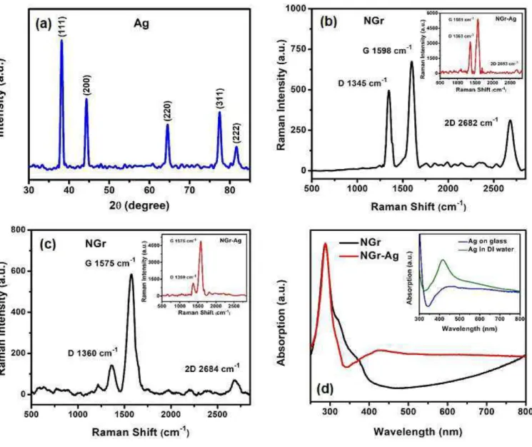
The field emission scanning electron microscopic (FESEM) image of pristine N-doped graphene and with synthesized Ag nanoparticles on its surface are shown in Fig. 1 (a)-(c). The low magnification image shows some white patches (Fig. 1a) and a closer view (Fig. 1b) reveals the wrinkles of two-dimensional nanosheets, which is a general topological feature for graphene and N-doped graphene 15, 19. The monolayer of Ag nanoparticles (Fig. 1c) were dried on a hot plate at 100 °C before casting the 4-NBT solution on it. This dense distribution of Ag nanoparticles on N-doped graphene substrate is expected to enhance local electromagnetic field desired for SERS (surface enhanced Raman spectroscopy) experiments 44. Fig. 1d shows the transmission electron microscopic image of these Ag nanoparticles on a TEM copper grid. The size and shape of these lab-synthesized particles were determined from these images. They have an intermediate structural shape between a cube and sphere, in other words cubes with rounded corners. In order to determine the fine structural features, we collected high resolution TEM (HRTEM). Figs. 1(e)-(f) show the HRTEM images of Ag nanoparticles.
The obtained lattice spacing, d has a value of 0.24 nm which is associated with (111) crystal plane of Ag and is in good agreement with X-ray diffraction peak shown in Fig. 2a 45-46. All the XRD peaks in Fig. 2a are indexed to face-centered-cubic silver 45-46. We also opted for the elemental analysis of home-grown Ag nanoparticles. Fig 1 (g)-(i) show the presence of Ag with negligible amount of oxygen in the EDX mappings collected in STEM mode. Thus, the lab-synthesized Ag nanoparticles have high crystallinity and compositional features desirable for harnessing plasmonic properties in solar energy harvesting applications. 200 nm 200 nm
Ag
200 nmO
(c)
(b)
(e)
(g)
(h)
(d)
(i)
(b)
(a)
(f)
200 nm 500 nm 200 nmFig. 1. (a)-(b) FESEM image of N-doped graphene on glass at low and high magnification respectively (c) FESEM
image of a monolayer of Ag nanoparticles on N-doped graphene (d) TEM image of Ag nanoparticles (e) HRTEM image of Ag nanoparticle (f) Selected magnified region of Fig. (e) for identifying crystal plane (111) (g) Bright field STEM image of a lone Ag nanoparticle and (h-i) STEM elemental mapping for (h) Silver and (i) Oxygen.
Raman spectroscopy is a convenient and powerful technique to determine crystallite size, number of layers, doping and disorder in graphene and other carbon-based materials 4, 7. The Raman spectral features (532 nm excitation) of N-doped graphene and composite systems with Ag nanoparticles are presented in Fig. 2 (b-c). The D, G and 2D are the important and prominent bands for analyzing graphene and graphene-based derivatives. The first order Raman scattering process that gives rise to doubly degenerate E2g phonons at the Brillouin zone, constitutes G band, whereas the D and 2D bands
are the results of a zone-boundary phonon induced second order, double-resonance process 7, 47. The external doping breaks the translational symmetry of graphene and the D band represents doping induced defects in graphene-based materials 48. In our pristine N-doped graphene samples, we found two distinct regions, one with a high intensity ratio of the D and G bands ID/IG, and the other one with low
ID/IG. This nonuniformity in the peak intensity ratio ID/IG, is a consequence of inhomogeneous doping of
nitrogen in graphene and has been observed in many earlier reports 47. This peak intensity ratio is proportional to defects and is inversely proportional to crystallite size 48. Therefore ID/IG is also
proportional to N-doping level as reported earlier 49. Fig. 2b represents high ID/IG, corresponding to high
doping, high defects and smaller crystallite size and Fig. 2c depicts low doping, low defects and larger crystallite size as it has lower ID/IG. N-doping induced reduction in crystallite size is consistent with
previous works 49. The intensity ratio IG/I2D is a measure of the thickness of graphene or the number of
atomic layers 50. The higher value of IG/I2D in Fig. 2c (pristine NGr) compared to that in Fig. 2b supports
the crystallite size comparison as well. However, we found supressed 2D bands for both kinds of regions in N-doped graphene/Ag hybrid (insets of Fig 2b and 2c), which indicates that two zone-boundary
phonons mediated intervalley double resonance scattering around the K-point of the Brillouin zone 47 is largely inhibited in the composite system. The reduction in peak intensity ratio I2D/IG is an indication of
higher electron concentration 51-52. This is expected, since the Ag surface plasmon decay generated hot electron injection into graphene, can potentially increase electron concentration, resulting in reduction of I2D/IG. Plasmon induced charge carrier enhancement can occur through other possible mechanisms as
well. This is discussed later in this article. The absorption spectra of pristine doped graphene and N-doped graphene/Ag composites on glass are presented in Fig 2d (main). The inset shows the absorption spectra of pristine Ag nanoparticles in aqueous solution and on glass. The plasmonic peaks for the N-doped graphene/Ag composites and pristine Ag nanoparticles on glass are relatively broad compared to the absorption peak of pristine Ag nanoparticle in aqueous solution, possibly due to an enhanced aggregation of Ag particles on glass and graphene/glass substrate 53. The main absorption peak of Ag nanoparticles is sensitive to the size and shape of the Ag nanoparticles. The position of this peak is at ~ 425 nm, for the synthesized Ag nanoparticles, is consistent with previously reported data 41, 53. The enhanced absorption in the NGr/Ag hybrid is a general feature of exciton-plasmon coupled systems 32.
Fig. 2. (a) X-Ray diffractograms of pristine Ag nanoparticles (b-c) Raman spectra of pristine N-doped graphene for
high and low defect area respectively; insets show corresponding spectra for the N-doped graphene/Ag composites (d) DRS absorption spectra of pristine N-doped graphene and N-doped graphene/Ag composite; inset shows pristine Ag on glass and in aqueous solution.
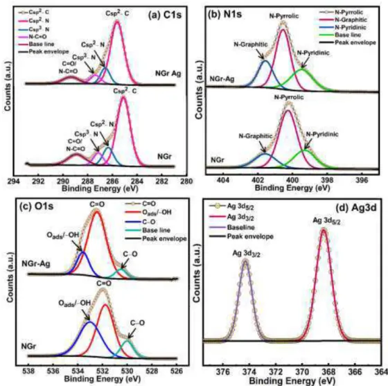
X-ray photoelectron spectroscopy (XPS) was used to determine surface and sub-surface (up to ~10 nm) chemical composition of NGr and NGr-Ag. The elemental survey scan (Fig. 3) using XPS of NGr displayed all the low energy and deep core level features corresponding to C1s, N1s, O1s, OKLL peaks. The N atom doping level was found to be 3.17 at%. For NGr-Ag, all the relevant peaks of NGr were observed along with additional peaks of Ag3d, Ag4s and Ag4p confirming the presence of Ag
nanoparticles on the surface of N-doped graphene. High resolution core-level XPS spectra of NGr in C1s region exhibited an intense peak at BE≈285.11 eV with shoulder peak in the high BE region (Fig. 4a). The C1s peak can be deconvoluted into four well-separated chemically shifted peak components centered at BE ≈ 285.11, 286.34, 287.15 and 288.86 eV. The peaks at BE ≈ 285.11 and 286.34 eV originated due to sp2 hybridized carbon that compose the graphene skeleton (Csp2-C) and sp2 carbons bonded with graphitic, pyridinic and pyrrolic nitrogens (Csp2-N) while peaks at 287.15 and 288.86 eV were assigned to Csp3-N and C=O/N-C=O species of carbon 54-57.
Fig. 3. XPS elemental survey scan of compact NGr (black) and Ag/NGr (red).
The high resolution N1s spectrum of NGr can be deconvoluted into three peak components located at 399.29, 400.25 and 401.56 eV, corresponding to pyridinic, pyrrolic and graphitic nitrogen atoms present in N doped graphene, respectively (Fig. 4b) 58-60. The deconvoluted spectra of NGr in O1s region showed three peak components at 530.00, 531.76 and 533.98 eV, attributed to C-O, C=O and surface adsorbed oxygens (-OH/Oads) respectively (Fig. 4c) 61-62. After drop casting Ag nanoparticles on the NGr
(NGr-Ag), the peak position for all elements was almost identical to pristine NGr, demonstrating that the chemical structure of NGr is preserved after the decoration of AgNPs (Fig. 4a-4c). Further, the presence of two well resolved XPS peaks at 368.33 and 374.28 eV in Ag3d region of NGr-Ag confirms the presence of Ag (0) nanoparticles (Fig. 4d) 63-64.
Fig. 4. HR-XPS core level spectra of compact NGr and NGr-Ag in (a) C1s region, (b) N1s region, (c) O1s region and
HR-XPS of NGr-Ag in (d) Ag3d region.
Defect-free pristine graphene is metallic in nature with a theoretical zero-bandgap. However, the introduction of N atoms into a graphenic framework followed by bonding with adjacent sp2 hybridized carbons modifies the charge distribution over neighboring carbon atoms. Additionally, the orbital
overlap of sp2 hybridized graphitic nitrogen bonded to three sp2 carbons releases two extra electrons into the π conjugated aromatic system which increases the electron density on the surface of NGr, which in turn breaks the symmetry of the graphene sub-lattice and results in the shifting of the Fermi level above the Dirac point and a band gap is created between the valence and conduction band 5, 7, 10, 65.
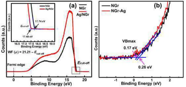
Fig. 5 (a) Ultraviolet pS work function spectra of NGr and NGr-Ag. Inset shows cut-off energy (Ecut-off) of the
secondary electron and (b) UPS valence band spectra showing the position of valence band edge (VBmax) below the Fermi level.
The electronic band structures of NGr and NGr-Ag materials were determined from the ultraviolet photoelectron spectra (UPS) (Figs. 5a and 5d). The expression WF (ϕ) = 21.21−Ecut-off was used to
estimate the value of WF where 21.21 eV is the energy of the incident, He I laser light, and Ecut-off is the
cut-off energy of secondary electrons. The extrapolation of the linear region of the graph to the vertical and horizontal axes gave the value of cut-off energy of secondary electrons Ecut-off at the point of
intersection. The Ecut-off values for compact NGr and NGr-Ag were calculated to be 17.74 and 17.49 eV,
and corresponded to work function (ϕ) values of 3.47 and 3.72 eV respectively (Fig. 5a and inset). The slight increase in the WF energy after addition of Ag nanoparticles demonstrate downshifting of Fermi level which indicates transfer of negative charge from N doped graphene sheets to Ag. Further, the
positions of the valence band maxima obtained from the intersection point of the extrapolated linear region of the graph for NGr and NGr-Ag were found to be 0.26 and 0.17 eV, respectively (Fig. 5b). Keeping in mind that the Fermi level lies close to the conduction band edge for a n-type semiconductor, the obtained VB maxima can be regarded as bandgap values. The obtained bandgap value for NGr was consistent with previous literature values reported for approximately similar N doping (2.9 at%) 49. Further, a smaller energetic separation between the VB maximum and the Fermi level after Ag decoration in NGr-Ag indicated a reduced n-type character and thus complied with WF values demonstrating a downshift of Fermi level due to electron transfer from N graphene to Ag.
(a) Surface topographic AFM images of pristine N-doped graphene; inset shows the line scan for height corresponding to the main image (b)-(d) KPFM surface potential distribution in the dark and under
(b)
(d)
(c)
(d)
illumination condition (LED 425 nm) for pristine Ag particles, pristine N-doped graphene and N-doped graphene/Ag composite respectively.
Fig. 6a shows the AFM height image of N-doped graphene nanosheets on glass after being transferred from copper. A thick layer of NGr nanosheets can be visible in the image. The transfer process of CVD-grown NGr generally induces wrinkles on the graphene surface (Fig. 1). There exist some thick patches of nanosheets of NGr in the FESEM images as mentioned earlier in this section. The inset of Fig. 6a shows the corresponding line cross-section of the height image of this thick wrinkled area. The line scan shows an average height of ~ 34 nm of NGr nanosheets. Thick layers are not very useful for electronic device applications such as transistor channels, but for photocatalysis a large thickness is not detrimental for this high mobility material. Surface potential, which is defined as the contact potential difference between KPFM tip and the material surface has been measured in dark and under illumination. It is a powerful technique to elucidate the nature of charge carrier dynamics 66-68. The charge carrier photogeneration in the photocatalyst can be imaged by this microscopic method. We have obtained the comparative photoresponse of all three systems, namely plasmonic, excitonic and plexcitonic, comprising bare Ag, bare NGr and NGr/Ag hybrid respectively. Figs. 6b-d show the surface potential distribution of pristine Ag nanoparticles, pristine NGr and NGr/Ag hybrid in the dark and under light illumination (425 nm) obtained using KPFM. Illumination produces photogenerated charge carriers whose redistribution causes the shifting of work functions for the systems under study, thus changing the surface potentials. Different light induced carrier enhancement mechanisms in bare Ag, bare NGr, and NGr/Ag composite occur, such as plasmon induced electric field amplification followed by accumulation of charge carriers, optical transition leading to formation of excitons and coupling of plasmon-exciton respectively. Therefore, the surface potential difference between dark and illumination conditions is a measure of the photoresponse of the surface. From Fig. 6 (b-d), it is evident that NGr/Ag hybrid system shows the highest photoresponse, resulting in a large negative shift of surface potential,
compared to the other two systems, pristine Ag and pristine NGr. This is reminiscent of the significant reduction of Raman peak intensity ratio I2D/IG for the hybrid system, as a result of huge charge carrier
accumulation discussed earlier. A correlation between plasmon induced local field enhancement and surface potential change has also been reported in GaN and Ag nanoparticle hybrid, where a concomitant reduction in surface potential was observed with optical field enhancement 69.
The wavelength corresponding to the illumination used during photo-KPFM measurements (450 nm) was deliberately chosen to be close to the LSPR peak of the rounded Ag nanocubes (425 nm, see Fig. 2d and inset). The bare Ag particles exhibit a mere −100 mV shift in surface potential upon illumination. Bare NGr shows a small shift in surface potential of ~ −40 mV upon illumination. However, NGr decorated with Ag nanocubes shows a large shift of −225 mV upon illumination, which is indicative of long-lived charge separation in the NGr/Ag hybrid. Considering the ground state charge transfer in the NGr/Ag system indicated by UPS data (Fig. 5) and the proximity of the illumination wavelength to the LSPR of Ag nanocubes, the most likely mechanism for extended charge separation is efficient injection of hot electrons from Ag into NGr following the decay of the Ag surface plasmons. We surmise that ultrafast chemical interface damping of the Ag surface plasmon may be responsible for the clear accumulation of electrons in NGr and long-lived charge separation. This is because the sequential mechanism consisting Landau damping of the plasmon followed by hot electron injection is known to exhibit much poorer charge separation and higher losses due to much faster competing thermalization processes involving electron-electron scattering and hot electron-phonon collisions 31.
4.2 Surface photocatalytic transformation of 4-NBT to DMAB
In this work, we have investigated a comparative performance analysis of surface catalytic reactions for converting 4-NBT to DMAB using three systems, pristine Ag nanoparticles, pristine N-doped graphene
and N-doped graphene/Ag composite. These systems represent three distinct mechanisms of photocatalysis with different charge carrier dynamics, such as plasmonic, excitonic and plexcitonic (coupled plasmon and exciton) respectively. The photoconversion or dimerization of 4-NBT to DMAB is a reduction reaction that involves four electrons (Fig. 7) 27, 70. Surface plasmon decay generated hot electrons in plasmonic metals are injected into 4-NBT with high kinetic energy, and are responsible for performing this reduction 27, 30. A large kinetic energy is required to overcome the potential barrier and complete the reduction reaction. It is worth mentioning that surface plasmon driven oxidation reaction involves a completely different mechanism. It can be performed either by a hot hole generated by plasmon decay or by the plasmonic hot electron driven transformation of singlet oxygen into triplet oxygen, where triplet oxygen plays the role of the catalyst 71-72. The excitonic reduction of 4-NBT to DMAB has been observed on Cu2O surfaces 42. The photogenerated electrons and holes in the
conduction and valence bands respectively under light illumination, perform the catalytic reduction and oxidation for any excitonic photocatalyst, constrained by the energy requirements of electrons and holes with respect to reduction and oxidation potentials of the reactions. Apart from pure plasmonic and pure excitonic photocatalysis, plexcitonic or plasmon-exciton co-driven photocatalysis has the immense potential to drive these surface catalytic reactions as the exciton-plasmon interaction increases the plasmon to electron conversion efficiency, as well as prolongs the hot electron lifetime 26, 28-29, 73.
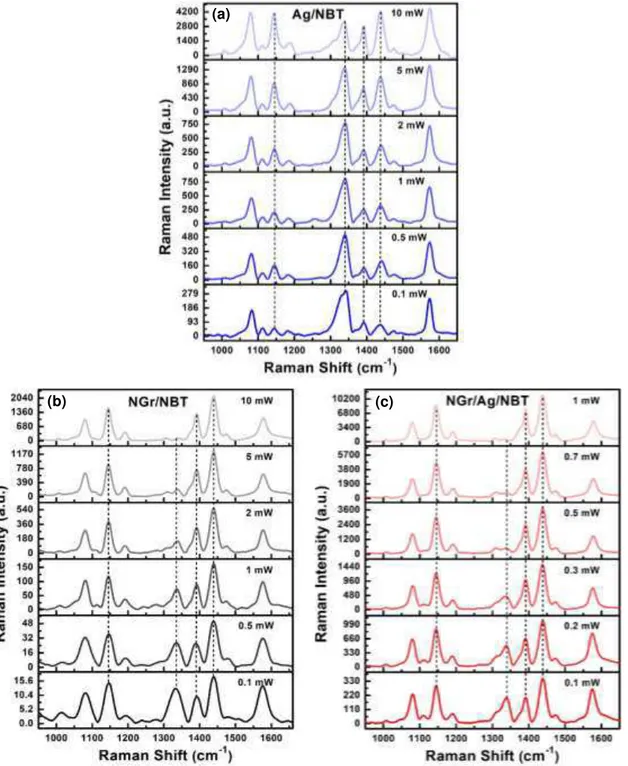
Fig. 7 shows the Raman spectra of 4-NBT and DMAB. The Raman peaks at 1101, 1332 and 1576 cm-1 of 4-NBT are assigned to S-C stretching vibration, vS(NO2) vibration and C = C stretching
vibration of benzyl ring respectively 27, 74. DMAB Raman peaks are identified at 1142, 1392 and 1439 cm-1. The band at 1142 cm-1 is attributed to C-N stretching vibration, and the peaks at 1392 and 1439 cm-1 arise due to ag16 and ag17 vibrational modes respectively, related to N = N stretching 27, 75. The
The surface catalytic reduction of 4-NBT to DMAB is highest for the NGr/Ag hybrid system as evidenced by the complete disappearance of the vS(NO2) vibration of 4-NBT at 1332 cm-1, followed by
pristine NGr and pristine Ag respectively. Fig. 8 shows the laser power dependent gradual evolution of DMAB peaks for these three systems. In all the three systems gradual decrease of main 4-NBT peaks (1332 cm-1) and a simultaneous gradual increase of the DMAB peaks mentioned earlier, occurs with increasing laser power. From Fig. 8 it evident that, the surface plasmons in Ag nanoparticle alone cannot completely transform 4-NBT into DMAB even at laser power as high as 10 mW. Notice that in this experiment we used 10X objective for all three systems, which gives much lower power compared to 50X or 100X. Pristine graphene completed this chemical transformation at 10 mW power, while NGr/Ag hybrid catalyst required only 1 mW. These results demonstrate that pristine N-doped graphene alone can drive the surface catalytic reduction reaction without any plasmonic co-catalyst. The Raman spectral intensity at higher laser power of 4-NBT/DMAB on NGr surface is also comparable to that of pristine Ag supported system, but indeed much weaker than the hybrid system NGr/Ag. This implies that NGr has potential for SERS (surface enhance Raman spectroscopy) application for system where it does not have any reactivity with adsorbed molecule, in other words where it is expected to behave noninvasively. Unlike graphene-Ag nanowire system reported earlier 26, we have observed the DMAB peak at 1142 cm-1 in both the excitonic N-doped graphene and plexcitonic NGr/Ag systems. Metal-semiconductor hybrids have been thoroughly explored in the recent past as these hybrid systems combine two systems possessing complementary optical properties. Fundamental optical excitations in metal are surface plasmons that are collective oscillations of conduction band electrons, while in semiconductor these are excitons or bound electron-hole pairs which are the product of transitions between conduction and valence band energy levels 32. The plasmonic metals have the potential to enhance optical field through concentrating electromagnetic energy and the semiconductors can generate
and support long-live excitations that are capable of harvesting solar energy 32. NGr/Ag composite system, promotes plasmon−exciton coupling that induces a significantly high amplification of local electromagnetic field, accumulation of high-energy hot electrons following plasmon dephasing, in addition to having high absorption cross-sections, thus demonstrates the superior photocatalytic prowess. The different possible plasmon-exciton coupling mechanisms have been discussed later in this communication.
Fig. 7 Left panel: Comparative performance for the Raman surface catalytic reduction of 4-NBT (4-nitrobenzenethiol)
to DMAB (p, p-dimercaptoazobenzene); from top, Raman spectrum of 4-NBT (green), 4-NBT on NGr/Ag hybrid (red), 4-NBT on pristine NGr (black), 4-NBT on pristine Ag nanoparticles (blue) and DMAB (orange). Laser wavelength was 532 nm at 1 mW power. Right panel: Chemical structure of 4-NBT and DMAB; Electron source for the 4-NBT dimerization in three different prototypical systems.
Fig. 8 Surface catalytic conversion of 4-NBT to DMAB monitored by Raman spectroscopy (excitation 532 nm); Laser
power dependent spectral evolution of reactant (4-NBT ) and product (DMAB) for different systems - (a) Ag nanoparticles (plasmonic) (b) N-doped graphene (excitonic) and (c) N-doped graphene/Ag hybrid (plexcitonic or plasmon-exciton interaction-induced).
4.3 Interaction with incident optical field
(a)
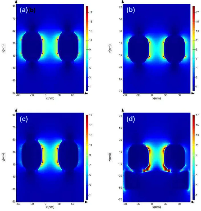
FDTD simulations were carried out to model the plasmonic and plexcitonic systems of the Ag nanoparticles in the absence and presence of NGr substrate respectively. The relevant physical features of Ag nanoparticles were obtained from SEM and TEM data. Different arrangements of Ag nanoparticles with and without NGr substrate have been studied as mentioned earlier. Fig. 9 and Fig. S2 (supplementary information) show the electric field distribution for two Ag nanoparticles in the absence and presence of NGr substrate for two arrangements, separated by 34 nm and in physical contact. The k vector and field polarization direction were along z and x directions respectively. In both systems E-field intensity is highly amplified between the Ag particles and between NGr and Ag. Similar local E-field enhancement can be observed in Fig. S1 and Fig. S3 also, where these two systems are exactly same as Fig 9 and Fig. S2, except the E-filed polarization direction, which is y. The maximum field is concentrated between NGr and Ag particles (in the vicinity of the interface) for all four systems (Fig. 9d, S1d, S2d and S3d). This SPR induced local field enhancement has the immense potential to enhance electron concentration in NGr through various mechanisms that has been discussed later. The reduction of E-field intensity in the regions where Ag nanoparticles are in direct contact with NGr (Fig. 9d, S1d, S2d and S3d) implies plasmon induced energy transfer from Ag into NGr. Fig. (S1-S4) show simulated absorption and scattering results for these systems with E-field polarized in y direction only. The absorption peaks of isolated Ag nanoparticles without and with NGr substrate show a blue shift (Fig. S1 and S2) compared to the experimentally obtained data (Fig. 2d). However, the when two Ag nanoparticles are in direct physical contact these peaks (Fig. S3 and S4) are redshifted. The drop-cast Ag nanoparticles on NGr have both systems present as evidenced from SEM (Fig. 1c). LSPR peaks are broadened on the NGr substrate relative to the isolated nanoparticles, similar to the experimental data. In all the plots (Fig S1-S4) scattering is dominated by absorption, implying faster plasmon dephasing 76.
Fig. 9. Electric field intensity distribution of two 34 nm apart Ag nanoparticles (cubes with rounded corners) at an
incident wavelength of 532 nm; (a) in the xy plane without any substrate, (b) in the xz plane without any substrate, (c) in the xy plane with N-doped graphene substrate, and (d) in the xz plane with N-doped graphene substrate. The electric field component was x-polarized.
5. DFT Modeling of N-doped graphene
(b)
(c)
(d)
5.1 Computation and modeling details
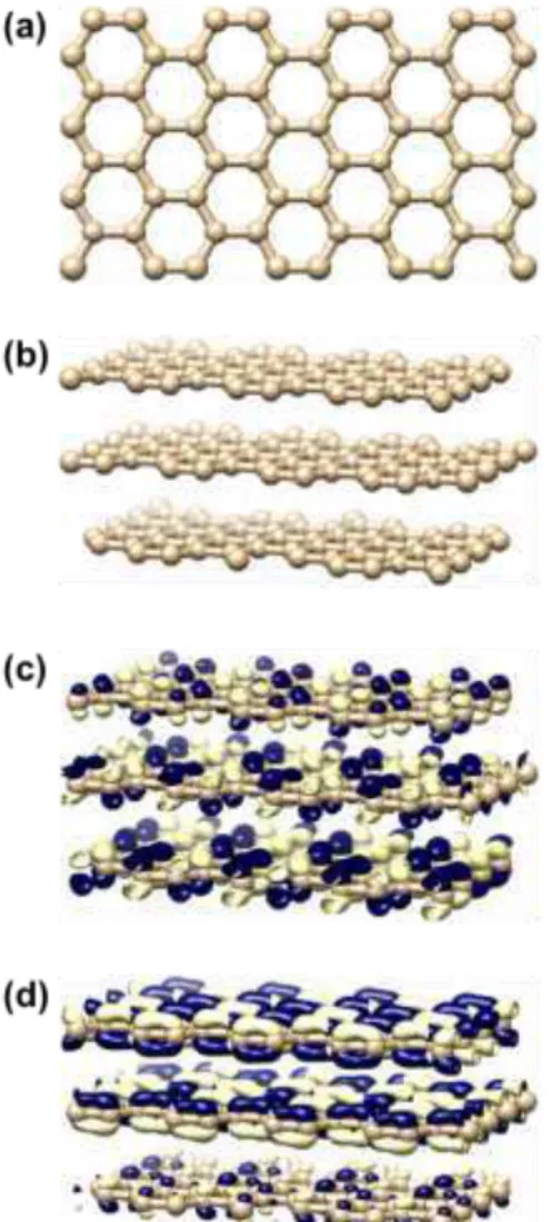
The electronic properties of three different types of nitrogen doped graphene, namely graphitic, pyridinic and pyrrolic8 were calculated and compared with pristine undoped graphene within the framework of density functional theory77 as implemented in the software package OpenMX 3.8 [A.K.]. Spin polarization was considered in all the calculations using Perdew-Burke-Ernzerhof (PBE) exchange correlation functional with the general gradient approximation (GGA)78. Numerical pseudoatomic orbitals centered on atomic sites were used for the basis set. Periodical system with the supercell consisting of 192 carbon atoms were used for pristine graphene. Three single layers of graphene were vertically stacked in z-direction. The N-doped graphene systems were also constructed in a similar way with the dopant N atoms residing in each three layers. Three systems were studied containing only one type of doping and the fourth one comprised of all three types of dopants, such as, graphitic N, pyridinic N and pyrrolic N (Fig. 10). Fig. 10 (e) shows these three types of N-atoms as N1, N2 and N3 for graphitic, pyridinic and pyrrolic respectively. The ratio of N and C atoms were kept low, in order to be consistent with the XPS results, discussed earlier. Gaussian broadening method was used to study the projected density of states (PDOS) for the dopant N atoms, neighboring and the remote C atoms from the dopant atoms.
5.2 Results and analysis
Molecular orbital distribution and the projected density of states (PDOS) of the optimized structures were plotted and analyzed in order to compare the electronic properties of the nitrogen doped and the pristine undoped graphene. Highest occupied molecular orbital (HOMO) and lowest unoccupied molecular orbital (LUMO) have been shown in Fig. 10 and Fig. S13 (supplementary section). HOMO and LUMO are delocalized in pristine graphene, whereas N-doping introduces significant spatial
localization of these molecular orbitals, indicative of high chemical reactivity79. Pyrrolic and pyridinic systems have stronger localization compared to graphitic systems. These localization is a consequence of introduction of an unpaired electron into graphene upon N-doping79.
Fig. 10. Optimized structures and calculated spatial distributions of molecular orbitals for pristine graphene and
N-doped graphene. Pristine graphene; (a) top surface, (b) perspective view, (c) HOMO and (d) LUMO. N-N-doped graphene doped with graphitic, pyridinic and pyrrolic N; (e) top surface, (f) perspective view, (g) HOMO and (h) LUMO.
PDOS have been plotted separately for sp2 and pz like states, of individual atomic sites in pristine and N-doped graphene. These plots have been demonstrated in Fig. 11 and Fig. S14 (supplementary section). For the N-doped systems PDOS at the dopant N atoms, neighboring and far-located C atoms have been selected. These results indicate that high number of states near the Fermi level (set at 0 eV) can be created at the dopant sites and the neighboring C atoms compared to the remote C atom in the N-doped graphene as well as pristine graphene. This implies the chemical reactivity enhancement upon nitrogen doping. Fig. 11 and Fig. S14 show that all three types of doping can enhance chemical reactivity of pristine graphene. Pyrrolic N and the neighboring C sites have highest reactivity, followed by pyridinic and graphitic systems respectively. Therefore a correlation exist between molecular orbital localization and accumulation of states near the Fermi level.
Fig. 11. Projected density of states (PDOS) for selected sites of pristine graphene and N-doped graphene surface.
Pristine graphene; (a) at a surface C atom. N-doped graphene doped with graphitic, pyridinic and pyrrolic N; (b) at a graphitic N site, (c) at a neighboring C atom of a graphitic N, (d) at a pyridinic N site, (e) at a neighboring C atom of a pyridinic N, (f) at a pyrrolic N site, (g) at a neighboring C atom of a pyrrolic N, (h) at a remote C atom.
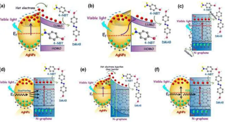
6. Mechanisms of Photoreduction
Now, let us revisit the scenarios of photocatalytic reduction reactions for three different systems, namely plasmonic, excitonic and plexcitonic. When light falls on the Ag nanoparticles, it excites collective oscillations of conduction electrons, called plasmon. This surface plasmons are analogous to quantum harmonic oscillators, creating alternating regions of higher and lower electron densities 80. Some of the stored light quanta stored in plasmons are reemitted as light but part of it decays into electrons and holes. When incident photon energy is in resonance with the restoring energy of the oscillating valence electrons on the metal surface (Ag nanoparticles), localized surface plasmon resonance (LSPR) is excited 81. This localized surface plasmon resonance are coherent oscillations of electrons in space and energy. They have the potential to drive photocatalytic reactions through different mechanisms. The dephasing of this coherent oscillation can occur by different mechanisms, such as, elastic re-radiation of photons and nonradiative Landau damping that gives rise to the high energetic hot electrons and holes 81. These hot electrons can perform the photocatalytic reduction reaction of 4-NBT to DMAB 27. The ability of hot electrons generated by Landau damping to perform useful work is severely constrained by their short lifetime, since hot carriers in metals experience electron-electron scattering and electron-phonon collisions over femtosecond to picosecond timescales that cause their thermal equilibration 31. There is another route of direct plasmonic photocatalysis for plasmonic metal nanoparticles when they are in contact with adsorbate molecules. In this mechanism, excited plasmonic states can directly inject electrons into adsorbate acceptor states by a process called chemical interface damping 81. Therefore the latter process does not require nonradiative Landau damping of plasmons to perform photocatalysis. Fig. 10a and 10b show these two routes of photocatalytic reduction, which in our study, are primarily relevant to the case where bare Ag nanoparticles function as a stand-alone photocatalyst for the
transformation of 4-NBT into DMAB. In the case of the excitonic reduction reaction, excitons are generated in bare NGr upon illumination of light, when incident photon energy is larger than the band gap of NGr. According to the UPS result, discussed earlier in this report, bandgap of NGr is very low, thus a large number of electron-hole pair (exciton) generation is likely in this material. An efficient charge separation enables the catalytic reduction of 4-NBT. Excitonic photoreduction mechanism in NGr is shown in Fig. 10c. Plasmon-exciton interaction can take place through different routes, scattering or trapping of light in semiconductor, energetic hot electron injection from plasmonic metal into the adjacent semiconductor and plasmon-induced resonant energy transfer (PIRET) 76, 82-83. In this project, the plexcitonic system, comprising the NGr/Ag hybrid, is expected to involve these different mechanisms of charge carrier dynamics as well, even though the relative strengths might be different. Scattering or trapping of light in the semiconductor can occur after the illumination of light as a result of plasmonic metal nanoparticle induced light scattering and subsequent penetration into the adjacent semiconductor 76, 82. This process increases the effective optical absorption cross-section and effective optical path length in the semiconductor, in addition to local amplification of the electromagnetic field
83
. The plasmonic energy transfer from metal to semiconductor has been schematically drawn in Fig. 10d. The plasmon decay (Landau damping) generated hot electrons in Ag nanoparticles can be directly injected into NGr conduction band. There are two possible paths available relevant to this process, the more energetic electrons, having energies greater than the Schottky barrier that forms between Ag and NGr, will be directly injected into the conduction band of NGr, while comparatively lower energetic electrons can tunnel through the barrier to reach NGr 80, 84. N-doped graphene, being a n-type semiconductor, would be expected to undergo a downward band-bending at the metal semiconductor Schottky junction, which facilitates the tunneling of hot electrons. Fig. 10e shows the two possible ways of hot electron injection from Ag nanoparticles into NGr. However, theoretical calculation showed that
efficient injection of hot electrons from plasmonic metals to adjacent semiconductors require very small nanocrystals 85. There are other possible routes to increase the number of charge carriers in semiconductors coupled with plasmonic metal nanoparticles. The surface plasmon mediated local electromagnetic field can generate electron-hole pair in semiconductors through a radiative process 83. An alternate resonant energy transfer mechanism has been proposed to induce electron-hole pair generation through a nonradiative localized surface plasmon dipole relaxation 83. This process is called plasmon-induced resonant energy transfer (PIRET) 82-83. The incident photon energy is stored in plasmon’s polarization prior to its decay or dephasing into hot electrons. This energy can be transferred to the semiconductor through the near-field 76. PIRET process is dipole-dipole or higher order coupling mechanism in the near field, that directly excites electron-hole pairs in semiconductor through a nonradiative localized surface plasmon relaxation 83. Fig. 10f schematically shows the PIRET mechanism for surface photocatalytic reactions in plasmon-exciton coupled systems. Under light the electron concentration is significantly enhanced in the NGr/Ag hybrid system, as was evidenced from Raman spectroscopy and KPFM, discussed earlier in this report. 4-NBT molecules can adsorb onto Ag NPs through the thiol linkage. However, they can also
Thus far, we have discussed how plasmonic metal nanoparticles coupled with semiconductor can enhance charge carriers in semiconductor and consequently enhance photocatalytic performance through three different mechanisms, far-field scattering or trapping of light in semiconductor, hot electron generation and injection into semiconductor and near-field dipole-dipole interaction mediated resonant energy transfer. For a small band gap semiconductor such as N-doped graphene, it is expected that all three mechanisms will play some role. Even though all three mechanisms are simultaneously contributing in plasmonic energy transfer, it is reasonable to expect that scattering and near-field PIRET mechanisms are the dominant ones as there is some overlap between LSPR absorption band with that of
NGr (Fig. 2d), in other words, plasmon energy is larger than the NGr band gap. However, from Fig. 2d it is obvious that LSPR band is away from the prominent NGr absorption band located between 250-400 nm, which implies the absence of dominant contribution from PIRET and scattering and a significant contribution of hot electron injection mechanism in this hybrid composite system 82. Theoretical simulation results (Fig. S1-S4, supplementary information) show larger absorption compared to scattering, indicative of a faster plasmon dephasing, characteristics of small plasmonic nanoparticles 76. Theoretical calculations showed that smaller plasmonic nanoparticles have higher number of high energetic hot electrons compared to larger nanoparticles 85. The size reduction of metal nanoparticles increases the probability of generation of more hot electrons than dephasing the plasmons into scattering or near-field processes 76. Thus our results show hot electron injection could be significant in small band gap semiconductors, as opposed to generally accepted fact that it is predominantly operative in large band gap semiconductors only.
Fig. 12. Photocatalytic reduction mechanisms for various systems. (a) Hot electron injection into 4-NBT following
plasmon at Ag-4NBT interface (c) Light-induced electron-hole pair (exciton) generation in semiconductor (NGr) (d) plasmon induced energy transfer from metal to semiconductor by light scattering or trapping (e) hot electron injection into Ngr from Ag and (f) nonradiative resonant energy transfer (PIRET). Figure 10 needs to be modified.
7. Conclusion
The surface-catalyzed single-electron reduction of 4-NBMT to DMAB was used to probe the photocatalytic activity of NGr, Ag and Ngr/Ag substrates. Both the bare NGr-based excitonic photocatalyst and the plexcitonic NGr/Ag hybrid photocatalysts showed much stronger activities for the transformation of 4-NBT to DMAB in comparison to bare Ag-based plasmonic photocatalyst. The NGr/Ag hybrid system has great potential in photocatalysis as evidenced by its highest performance. Exciton-plasmon interaction leads to a synergistic enhancement of surface photocatalytic activity in the NGr/Ag plexcitonic photocatalyst. FDTD simulation results showed a sizeable electric field enhancement in the vicinity of NGr-Ag interface. Raman spectroscopic results indicated a higher charge concentration in NGr/Ag system under illumination of light. This phenomenon was in good agreement with the observations from KPFM, that showed large surface potential reduction under light compared to in dark condition. The enhanced charge carrier concentration in NGr/Ag plexcitonic system holds immense potential for other photocatalytic applications.
Acknowledgements
This work was supported by the National Research Council Canada (NRC). The authors would also like to acknowledge NSERC, AITF, CMC Microsystems, Future Energy Systems, ECNU-UofA JIAST and CFI for direct and indirect support. The University of Alberta nanofab personnel Shiau-Yin Wu is acknowledged for her help in collecting AFM data. Prof. Thomas Thundat is kindly acknowledged for
allowing the use of the KPFM facility. Yenan Song would like to thank Fund of Research Innovation, East China Normal University.
References
1. Novoselov, K. S.; Geim, A. K.; Morozov, S. V.; Jiang, D.; Katsnelson, M. I.; Grigorieva, I. V.; Dubonos, S. V.; Firsov, A. A., Two-dimensional gas of massless Dirac fermions in graphene. Nature 2005, 438 (7065), 197-200.
2. Matthew J. Allen, V. C. T., and Richard B. Kaner, Honeycomb Carbon- A Review of Graphene. Chemical Reviews 2010, 110 (1), 132-145.
3. Ferrari, A. C.; Bonaccorso, F.; Fal'ko, V.; Novoselov, K. S.; Roche, S.; Boggild, P.; Borini, S.; Koppens, F. H.; Palermo, V.; Pugno, N.; Garrido, J. A.; Sordan, R.; Bianco, A.; Ballerini, L.; Prato, M.; Lidorikis, E.; Kivioja, J.; Marinelli, C.; Ryhanen, T.; Morpurgo, A.; Coleman, J. N.; Nicolosi, V.; Colombo, L.; Fert, A.; Garcia-Hernandez, M.; Bachtold, A.; Schneider, G. F.; Guinea, F.; Dekker, C.; Barbone, M.; Sun, Z.; Galiotis, C.; Grigorenko, A. N.; Konstantatos, G.; Kis, A.; Katsnelson, M.; Vandersypen, L.; Loiseau, A.; Morandi, V.; Neumaier, D.; Treossi, E.; Pellegrini, V.; Polini, M.; Tredicucci, A.; Williams, G. M.; Hong, B. H.; Ahn, J. H.; Kim, J. M.; Zirath, H.; van Wees, B. J.; van der Zant, H.; Occhipinti, L.; Di Matteo, A.; Kinloch, I. A.; Seyller, T.; Quesnel, E.; Feng, X.; Teo, K.; Rupesinghe, N.; Hakonen, P.; Neil, S. R.; Tannock, Q.; Lofwander, T.; Kinaret, J., Science and technology roadmap for graphene, related two-dimensional crystals, and hybrid systems. Nanoscale 2015, 7 (11), 4598-810.
4. Liu, H.; Liu, Y.; Zhu, D., Chemical doping of graphene. J. Mater. Chem. 2011, 21 (10), 3335-3345. 5. Xinran Wang, X. L., Li Zhang, Youngki Yoon, Peter K. Weber, Hailiang Wang, Jing Guo, Hongjie Dai, N-doping of graphene through electrothermal reactions with ammonia. Science 2009, 324, 768. 6. Laref, A.; Ahmed, A.; Bin-Omran, S.; Luo, S. J., First-principle analysis of the electronic and optical properties of boron and nitrogen doped carbon mono-layer graphenes. Carbon 2015, 81, 179-192.
7. Wang, H.; Maiyalagan, T.; Wang, X., Review on Recent Progress in Nitrogen-Doped Graphene: Synthesis, Characterization, and Its Potential Applications. ACS Catalysis 2012, 2 (5), 781-794.
8. Xu, H.; Ma, L.; Jin, Z., Nitrogen-doped graphene: Synthesis, characterizations and energy applications. Journal of Energy Chemistry 2018, 27 (1), 146-160.
9. Lin, C. K., Theoretical study of nitrogen-doped graphene nanoflakes: Stability and spectroscopy depending on dopant types and flake sizes. J Comput Chem 2018, 39 (20), 1387-1397.
10. Lherbier, A.; Blase, X.; Niquet, Y. M.; Triozon, F.; Roche, S., Charge transport in chemically doped 2D graphene. Phys Rev Lett 2008, 101 (3), 036808.
11. Oh Seok Kwon, S. J. P., Jin-Yong Hong, A-Reum Han, Jun Seop Lee, James S. Lee, Joon Hak Oh, and Jyongsik Jang, Flexible FET-Type VEGF Aptasensor Based on Nitrogen-Doped Graphene Converted from Conducting Polyme. ACS Nano 2012, 6 (2), 1486-1493.
12. Chang, D. W.; Lee, E. K.; Park, E. Y.; Yu, H.; Choi, H. J.; Jeon, I. Y.; Sohn, G. J.; Shin, D.; Park, N.; Oh, J. H.; Dai, L.; Baek, J. B., Nitrogen-doped graphene nanoplatelets from simple solution edge-functionalization for n-type field-effect transistors. J Am Chem Soc 2013, 135 (24), 8981-8.
13. Qian, W.; Cui, X.; Hao, R.; Hou, Y.; Zhang, Z., Facile preparation of nitrogen-doped few-layer graphene via supercritical reaction. ACS Appl Mater Interfaces 2011, 3 (7), 2259-64.
14. Lherbier, A.; Botello-Mendez, A. R.; Charlier, J. C., Electronic and transport properties of unbalanced sublattice N-doping in graphene. Nano Lett 2013, 13 (4), 1446-50.
15. Ying Wang, Y. S., Dean W. Matson, Jinghong Li, and Yuehe Lin, Nitrogen-Doped Graphene and Its Application in Electrochemical Biosensing. ACS Nano 2010, 4 (4), 1790-1798.
16. Fan, H.; Li, Y.; Wu, D.; Ma, H.; Mao, K.; Fan, D.; Du, B.; Li, H.; Wei, Q., Electrochemical bisphenol A sensor based on N-doped graphene sheets. Anal Chim Acta 2012, 711, 24-28.
17. Sohyeon Seo, Y. Y., Junghyun Lee, Younghun Park, and Hyoyoung Lee, Nitrogen-Doped Partially Reduced Graphene Oxide Rewritable Nonvolatile Memory. ACS Nano 2013, 7 (4), 3607–3615.
18. Jin Ok Hwang, J. S. P., Dong Sung Choi, Ju Young Kim, Sun Hwa Lee, Kyung Eun Lee, Yong-Hyun Kim, Myoung Hoon Song, Seunghyup Yoo, and Sang Ouk Kim, Workfunction-Tunable, N-Doped Reduced Graphene Transparent Electrodes for High-Performance Polymer Light-Emitting Diodes. ACS Nano 2012, 6 (1), 159-167.
19. Wang, H.; Zhang, C.; Liu, Z.; Wang, L.; Han, P.; Xu, H.; Zhang, K.; Dong, S.; Yao, J.; Cui, G., Nitrogen-doped graphene nanosheets with excellent lithium storage properties. Journal of Materials Chemistry 2011, 21 (14).
20. Lu, H.; Chen, R.; Hu, Y.; Wang, X.; Wang, Y.; Ma, L.; Zhu, G.; Chen, T.; Tie, Z.; Jin, Z.; Liu, J., Bottom-up synthesis of nitrogen-doped porous carbon scaffolds for lithium and sodium storage. Nanoscale 2017, 9 (5), 1972-1977.
21. Yang Zhao, C. H., Yue Hu, Huhu Cheng, Gaoquan Shi, and Liangti Qu, A Versatile, Ultralight, Nitrogen‐Doped Graphene Framework. Angew. Chem. Int. Ed. 2012, 51, 11371 –11375.
22. Liangti Qu, Y. L., Jong-Beom Baek, and Liming Dai, Nitrogen-Doped Graphene as Efficient Metal-Free Electrocatalyst for Oxygen Reduction in Fuel Cells. ACS Nano 2010, 4 (3), 1321–1326.
23. Jeong, H. M.; Lee, J. W.; Shin, W. H.; Choi, Y. J.; Shin, H. J.; Kang, J. K.; Choi, J. W., Nitrogen-doped graphene for high-performance ultracapacitors and the importance of nitrogen-Nitrogen-doped sites at basal planes. Nano Lett 2011, 11 (6), 2472-7.
24. Deng, Y.; Tang, L.; Feng, C.; Zeng, G.; Wang, J.; Lu, Y.; Liu, Y.; Yu, J.; Chen, S.; Zhou, Y.,
Construction of Plasmonic Ag and Nitrogen-Doped Graphene Quantum Dots Codecorated Ultrathin Graphitic Carbon Nitride Nanosheet Composites with Enhanced Photocatalytic Activity: Full-Spectrum Response Ability and Mechanism Insight. ACS Appl Mater Interfaces 2017, 9 (49), 42816-42828. 25. Jia, L.; Wang, D.-H.; Huang, Y.-X.; Xu, A.-W.; Yu, H.-Q., Highly Durable N-Doped Graphene/CdS Nanocomposites with Enhanced Photocatalytic Hydrogen Evolution from Water under Visible Light Irradiation. The Journal of Physical Chemistry C 2011, 115 (23), 11466-11473.
26. Ding, Q.; Shi, Y.; Chen, M.; Li, H.; Yang, X.; Qu, Y.; Liang, W.; Sun, M., Ultrafast Dynamics of Plasmon-Exciton Interaction of Ag Nanowire- Graphene Hybrids for Surface Catalytic Reactions. Sci Rep 2016, 6, 32724.
27. Dong, B.; Fang, Y.; Chen, X.; Xu, H.; Sun, M., Substrate-, wavelength-, and time-dependent plasmon-assisted surface catalysis reaction of 4-nitrobenzenethiol dimerizing to
p,p'-dimercaptoazobenzene on Au, Ag, and Cu films. Langmuir 2011, 27 (17), 10677-82.
28. Ding, Q.; Li, R.; Chen, M.; Sun, M., Ag nanoparticles-TiO2 film hybrid for plasmon-exciton co-driven surface catalytic reactions. Applied Materials Today 2017, 9, 251-258.
29. Yang, X.; Yu, H.; Guo, X.; Ding, Q.; Pullerits, T.; Wang, R.; Zhang, G.; Liang, W.; Sun, M., Plasmon-exciton coupling of monolayer MoS 2 -Ag nanoparticles hybrids for surface catalytic reaction. Materials Today Energy 2017, 5, 72-78.
30. Wu, H.-Y.; Lai, Y.-H.; Hsieh, M.-S.; Lin, S.-D.; Li, Y.-C.; Lin, T.-W., Highly Intensified Surface Enhanced Raman Scattering through the Formation ofp,p′-Dimercaptoazobenzene on Ag
Nanoparticles/Graphene Oxide Nanocomposites. Advanced Materials Interfaces 2014, 1 (8). 31. Manuel, A. P.; Kirkey, A.; Mahdi, N.; Shankar, K., Plexcitonics – fundamental principles and optoelectronic applications. Journal of Materials Chemistry C 2019, 7 (7), 1821-1853.
32. Acher a , M., Excito −Plas o I teractio s i Metal−Se ico ductor Na ostructures. The Journal of Physical Chemistry Letters 2010, 1 (19), 2837-2843.
33. Kumar, P.; Boukherroub, R.; Shankar, K., Sunlight-driven water-splitting using two-dimensional carbon based semiconductors. J. Mater. Chem. A 2018, 6 (27), 12876-12931.
34. Zeng, B.; Xia, Q.; Zeng, W., Fabrication and characterization of novel graphene/iodine doped CdS nanoplates and their photocatalytic performances. J. Mater. Sci.: Mater. Electron. 2019, 30 (12), 11619-11626.
35. Zeng, B.; Liu, W.; Zeng, W.; Jin, C., A general method for the synthesis of graphene-metal sulphide nanosheets. Journal of Nanoparticle Research 2018, 20 (3), 55.
36. Singh, D. K.; Iyer, P. K.; Giri, P. K., Improved chemical synthesis of graphene using a safer solvothermal route. Int. J. Nanosci. 2011, 10 (01n02), 39-42.
37. Rajender, G.; Kumar, J.; Giri, P. K., Interfacial charge transfer in oxygen deficient TiO2-graphene quantum dot hybrid and its influence on the enhanced visible light photocatalysis. Appl. Catal. B-Environ. 2018, 224, 960-972.
38. Giglio, C. S.; Osazuwa, O.; Kontopoulou, M.; Docoslis, A., Achieving high yield of graphene nanoplatelets in poloxamer-assisted ultrasonication of graphite in water. J. Colloid Interface Sci. 2019, 539, 107-117.
39. Liu, B.; Yang, C. M.; Liu, Z. W.; Lai, C. S., N-Doped Graphene with Low Intrinsic Defect Densities via a Solid Source Doping Technique. Nanomaterials 2017, 7 (10).
40. Liu, Q. B.; Yu, C.; He, Z. Z.; Gu, G. D.; Wang, J. J.; Zhou, C. J.; Guo, J. C.; Gao, X. D.; Feng, Z. H., Chemical vapor deposition graphene of high mobility by gradient growth method on an 4H-SiC (0001) substrate. Appl. Surf. Sci. 2018, 454, 68-73.
41. Siekkinen, A. R.; McLellan, J. M.; Chen, J.; Xia, Y., Rapid synthesis of small silver nanocubes by mediating polyol reduction with a trace amount of sodium sulfide or sodium hydrosulfide. Chem Phys Lett 2006, 432 (4-6), 491-496.
42. You, T.; Jiang, L.; Yin, P.; Shang, Y.; Zhang, D.; Guo, L.; Yang, S., Direct observation ofp,p ′-dimercaptoazobenzene produced fromp-aminothiophenol andp-nitrothiophenol on Cu2O
nanoparticles by surface-enhanced Raman spectroscopy. Journal of Raman Spectroscopy 2014, 45 (1), 7-14.
43. Shen, C. C.; Tseng, C. C.; Lin, C. T.; Li, L. J.; Liu, H. L., Optical properties of nitrogen-doped graphene thin films probed by spectroscopic ellipsometry. Thin Solid Films 2014, 571, 675-679.
44. Mohammadi, A.; Nicholls, D. L.; Docoslis, A., Improving the Surface-Enhanced Raman Scattering Performance of Silver Nanodendritic Substrates with Sprayed-On Graphene-Based Coatings. Sensors 2018, 18 (10), 3404.