Publisher’s version / Version de l'éditeur:
Journal of Alzheimer's Disease, 57, 4, pp. 1265-1279, 2017-04-19
READ THESE TERMS AND CONDITIONS CAREFULLY BEFORE USING THIS WEBSITE. https://nrc-publications.canada.ca/eng/copyright
Vous avez des questions? Nous pouvons vous aider. Pour communiquer directement avec un auteur, consultez la première page de la revue dans laquelle son article a été publié afin de trouver ses coordonnées. Si vous n’arrivez pas à les repérer, communiquez avec nous à [email protected].
Questions? Contact the NRC Publications Archive team at
[email protected]. If you wish to email the authors directly, please see the first page of the publication for their contact information.
NRC Publications Archive
Archives des publications du CNRC
This publication could be one of several versions: author’s original, accepted manuscript or the publisher’s version. / La version de cette publication peut être l’une des suivantes : la version prépublication de l’auteur, la version acceptée du manuscrit ou la version de l’éditeur.
For the publisher’s version, please access the DOI link below./ Pour consulter la version de l’éditeur, utilisez le lien DOI ci-dessous.
https://doi.org/10.3233/JAD-160133
Access and use of this website and the material on it are subject to the Terms and Conditions set forth at
Apolipoprotein E isoforms differentially regulate Alzheimer's disease
and amyloid-β-induced inflammatory response in vivo and in vitro
Dorey, Evan; Bamji-Mirza, Michelle; Najem, Dema; Li, Yan; Liu, Hong;
Callaghan, Debbie; Walker, Douglas; Lue, Lih-Fen; Stanimirovic, Danica;
Zhang, Wandong
https://publications-cnrc.canada.ca/fra/droits
L’accès à ce site Web et l’utilisation de son contenu sont assujettis aux conditions présentées dans le site LISEZ CES CONDITIONS ATTENTIVEMENT AVANT D’UTILISER CE SITE WEB.
NRC Publications Record / Notice d'Archives des publications de CNRC:
https://nrc-publications.canada.ca/eng/view/object/?id=78f57561-0310-4b77-861f-b9a22981b766 https://publications-cnrc.canada.ca/fra/voir/objet/?id=78f57561-0310-4b77-861f-b9a22981b766AUTHOR COPY
IOS Press
Apolipoprotein E Isoforms Differentially
Regulate Alzheimer’s Disease
and Amyloid--Induced Inflammatory
Response in vivo and in vitro
Evan Dorey
a,b,1, Michelle Bamji-Mirza
a,b,1, Dema Najem
a,b, Yan Li
a,b, Hong Liu
b,
Debbie Callaghan
b, Douglas Walker
c, Lih-Fen Lue
c, Danica Stanimirovic
a,band Wandong Zhang
a,b,∗aFaculty of Medicine, University of Ottawa, Ottawa, Canada
bHuman Health Therapeutics, National Research Council Canada, Ottawa, Canada cBanner Sun Health Research Institute, Sun City, AZ, USA
Accepted 14 February 2017
Abstract. Neuroinflammation plays a critical role in neuronal dysfunction and death of Alzheimer’s disease (AD). ApoE4
is a major risk factor of AD, while ApoE2 is neuroprotective. Little is known about the roles of ApoE isoforms in the neuroinflammation seen in AD. Their roles and mechanisms in A-induced/neuroinflammation were investigated in this study using in vivo and in vitro models. Rat astrocytes were treated with lipid-poor recombinant hApoE and/or A42.
Mouse astrocyte lines-expressing lipidated hApoE were treated with A42and/or vitamin D receptor (VDR) agonist,
1␣,25-dihydroxyvitamin D3. Cells and media were harvested for cytokine ELISA, RNA isolated for qRT-PCR, and nuclear protein
for transcription factor (TF) arrays and EMSA. hApoE-transgenic and AD mice were mated to generate hApoE2/AD and hApoE4/AD mice. Mice were euthanized at 6 months of age. Brain tissues were collected for cytokine ELISA array, A ELISA, immunoblotting, and immunohistochemistry. hApoE4/AD mice had significantly higher levels of inflammatory cytokines than hApoE2/AD mice. Lipidated hApoE4 significantly promoted inflammatory gene expression induced by A42 but not recombinant hApoE4 in astrocytes as compared to controls. Lipidated hApoE3 provided a certain degree
of protection against A42-induced inflammatory response but not recombinant hApoE3 as compared to controls. Both
lipidated and recombinant hApoE2 provided protection against A42-induced inflammatory response compared to controls.
TF array revealed that ApoE2 strongly activated VDR in A42-treated astrocytes. Application of 1␣,25-dihydroxyvitamin D3
completely inhibited A-induced inflammatory gene expression in hApoE4-expressing astrocytes. The results suggest that ApoE4 promotes, but ApoE2 inhibits, AD/A-induced neuroinflammation via VDR signaling. Targeting VDR signaling or active form of VD3 may relieve AD neuroinflammation or/and neurodegeneration.
Keywords: Alzheimer’s disease, amyloid- peptides, ApoE isoform proteins, neuroinflammation, vitamin D receptor signaling
1These authors contributed equally to this work.
∗Correspondence to: Dr. Wandong Zhang, National Research
Council Canada, 1200 Montreal Road, Building M-54, Ottawa, Ontario, K1A 0R6, Canada. Tel.: +1 613 993 5988; Fax: +1 613 941 4475; E-mails: [email protected]; [email protected]
INTRODUCTION
Neuroinflammation is one of the main patholog-ical characteristics of Alzheimer’s disease (AD), which leads to synaptic dysfunction and neurode-generation. A number of factors are involved in initiating and propagating the cascade of the inflam-matory response and signaling, such as amyloid-
AUTHOR COPY
(A) peptides, hyperphosphorylated tau proteins, reactive oxygen species, metal ions, and variants of the genes involved in inflammatory response. Among these, A peptides can form oligomers, aggregates, and plaques, which are toxic to neu-ronal cells and pro-inflammatory. Astrocytes exert influence over a range of CNS activities, including microglial-mediated neuroinflammatory responses. They respond to soluble chemical signals released from tissue during injury and disease by mobiliz-ing to lesion sties, clearmobiliz-ing toxic molecules, and releasing chemical signals of their own [1]. A pep-tides are associated with the activation of microglia and astrocytes, which surround amyloid plaques and mediate the release of pro-inflammatory medi-ators [2]. Microglial-mediated neuroinflammation in AD brain remains an area of intense investigation, the mechanisms underlying regulation of aber-rant microglial responses by astrocytes are largely unstudied [1].
Apolipoprotein E (ApoE) is a well-known regula-tor of cholesterol homeostasis and plays major roles in the modulation of the innate immune response [1]. A number of epidemiological, molecular, and clinical studies have demonstrated that the poly-morphism of the gene encoding ApoE is associated with the risk of late-onset AD. There are three ApoE alleles, 2, 3, and 4, which encode ApoE2 (cys112, cys158), ApoE3 (cys112, arg158), and ApoE4 (arg112, arg158) proteins, respectively. The 2 allele is associated with neuroprotection for AD [3, 4], while the 4 allele is a very strong risk fac-tor for AD. The 3 allele is the most frequent allele in the population and is considered to be neutral. An 4 homozygote has ∼12 times the risk of contract-ing AD, compared to an 3 homozygote [5]. While ApoE4’s status as an AD risk factor is very well estab-lished, the underlying mechanisms of the risk are still largely unclear. Similarly, the mechanism of the neuroprotective effect of ApoE2 on AD also remains largely unknown. ApoE isoform-related differences have been reported at a variety of stages in A pro-cessing, deposition, and degradation. In both mice and AD patients, ApoE4 is associated with higher levels of A peptides and more advanced amyloid plaques in the brain, relative to ApoE3, with ApoE2 showing lower levels than the other isoforms [6–8]. Jiang et al. reported that ApoE isoforms also differed in their ability to facilitate neprilysin-mediated degra-dation of A1-42 within Apoe–/– murine microglia,
with ApoE4 being least effective compared to ApoE2 or ApoE3 [9].
Little is known about the roles of ApoE, and its isoforms, in AD neuroinflammation. ApoE defi-ciency resulted in impaired clearance of apoptotic cells, and a systemic proinflammatory condition in mice [10, 11]. Similar reduction of ApoE in humans may contribute to a range of chronic dis-eases, including osteoporosis, atherosclerosis, and dementia. Studies have shown that ApoE4 allele had significantly greater systemic and brain eleva-tions of pro-inflammatory cytokines compared with their ApoE3 counterparts in vivo [12], and increased levels of inflammation relative to other isoforms in cultured macrophages [13–15]. Ophir et al. treated ApoE3 and ApoE4 transgenic mice with lipopolysac-charide (LPS) and observed that ApoE3 can regulate LPS-induced astrogliosis but not ApoE4 [16]. Vitek et al. [17] reported that mice expressing one human 3 allele (3/0) had lower inflammatory response upon LPS challenge than 4/4 mice [17]. These studies suggest that ApoE does play a role in the inflammatory response and that ApoE4 may be pro-inflammatory while apoE2 is anti-pro-inflammatory.
A number of studies have indicated that neu-roinflammation is an important mechanism leading to synaptic dysfunction and neurodegeneration. We hypothesize that the various isoforms of ApoE protein may differentially regulate AD or A-induced neu-roinflammation. In this study, we show that ApoE4 promotes AD neuroinflammation while ApoE2 has a protective effect against AD neuroinflammation, acting via the vitamin D receptor (VDR) signaling pathway.
METHODS
Chemical reagents
Dulbecco’s modified Eagle’s medium (DMEM), Advanced DMEM, TRIzol, geneticin, sodium pyruvate, 0.25% trypsin/EDTA, and antibi-otic/antimycotic were purchased from Life Technologies Inc. (Burlington, ON). Fetal bovine serum (FBS) was purchased from Hyclone (Logan, UT, USA). Dimethyl sulfoxide (DMSO), rosigli-tazone and 1␣, 25-Dihydroxyvitamin D3 were
purchased from Sigma (Oakville, ON). Human recombinant ApoE isoforms were purchased from Leinco (St. Louis, MO, USA). Antibodies for NFκB, pGSK, pcJun73, and VDR were purchased from Santa Cruz Biotechnologies (Santa Cruz, CA, USA). A1-42and a scrambled control, featuring the same
AUTHOR COPY
amino acids in a randomized order, were purchased from r-Peptide (Bogart, GA, USA).
Generation of ApoE2/AD and ApoE4/AD mice Human ApoE2 (stock #004632) and ApoE4 (stock #004631) transgenic mice were purchased from Jackson Laboratory and mated with the APPsw/PS1dE9 (AD) mice (stock #005864) (Jack-son Laboratory). Importantly, the human ApoE2 and ApoE4 isoforms in the transgenic mice are under the direction of the human glial fibrillary acidic protein and do not express endogenous mouse apoE (http://jaxmice.jax.org). The double transgenic ApoE2/AD and ApoE4/AD mice were generated and confirmed by genotyping PCR as described [18]. The double transgenic mice express an endogenous mouse apoE allele and a respective human ApoE allele (either ApoE2 or ApoE4). Also, there have been reports of gender difference in the levels of A in the plasma and brains of APP/PS1 mice [19]. However, we did not observe differences in the levels of A between male and female mice at 6 months of age in the animals we generated and thus there was a mixture of genders in the groups. The use of animals in this study was approved by the Animal Care Committee of the National Research Council Canada in Ottawa. The mice were maintained on a 12-h light/dark cycle in a temperature- and humidity-controlled environ-ment with free access to food and water. Both male and female mice were used in the study.
Cell culture
The neonatal rat astrocytes (NRA) generated from the cortex of 4–8 day old Sprague-Dawley rats and immortalized using SV40 large T antigen were grown in DMEM with 10% FBS and 1% antibiotic/
antimycotic and used in previous studies as described [20, 21]. Immortalized mouse astrocytes express-ing human ApoE isoforms were provided by Dr. D. Holtzman from Washington University (St. Louis, MO, USA). These cells were generated and immortalized as described [22], and were grown in advanced DMEM containing 10% FBS and 200 g/mL geneticin.
Aβ, ApoE, and VDR agonist treatments
A1-42or scrambled A1-42treatment of the
astro-cyte cell lines was performed at a concentration of 5 M. A peptide and the scrambled control pep-tide were brought up in 0.25% acetic acid, which served as the vehicle control. Human recombinant ApoE was brought up in 20 mM sodium phosphate +0.5 M DTT. 1␣, 25-Dihydroxyvitamin D3was
dis-solved in 99% DMSO and used at a concentration of 0.1 M.
RNA isolation and RT-qPCR
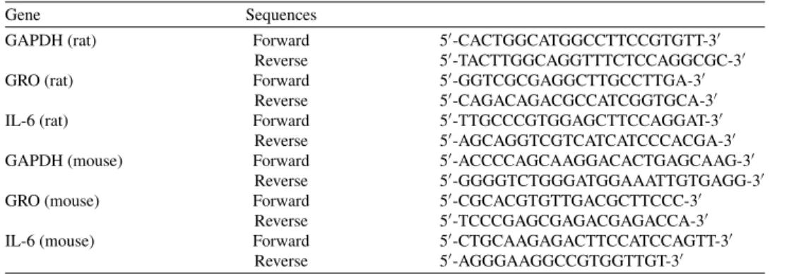
Total RNA was isolated from treated cells using TRIzol reagent (Life Technologies) accord-ing to manufacturer’s instructions. RNA samples were removed of DNA contaminates using Ambion DNA-free kits (Life Technologies), and RNA qual-ity and integrqual-ity were confirmed using Experion Automated Electrophoresis Station and Experion RNA StdSens analysis kit following manufacturer’s instructions (Bio-Rad). RNA was reverse-transcribed into cDNA using iScript kits (BioRad, Berkeley, CA, USA) according to manufacturer’s instructions. qPCR primers were generated using Primer-Blast and ordered from IDT (Coralville, IA, USA) (Table 1). The PCR efficiency was assessed by performing stan-dard curves using pooled cDNA material and by
Table 1 qPCR primer sequences
Gene Sequences
GAPDH (rat) Forward 5′-CACTGGCATGGCCTTCCGTGTT-3′
Reverse 5′-TACTTGGCAGGTTTCTCCAGGCGC-3′
GRO (rat) Forward 5′-GGTCGCGAGGCTTGCCTTGA-3′
Reverse 5′-CAGACAGACGCCATCGGTGCA-3′
IL-6 (rat) Forward 5′-TTGCCCGTGGAGCTTCCAGGAT-3′
Reverse 5′-AGCAGGTCGTCATCATCCCACGA-3′
GAPDH (mouse) Forward 5′-ACCCCAGCAAGGACACTGAGCAAG-3′
Reverse 5′-GGGGTCTGGGATGGAAATTGTGAGG-3′
GRO (mouse) Forward 5′-CGCACGTGTTGACGCTTCCC-3′
Reverse 5′-TCCCGAGCGAGACGAGACCA-3′
IL-6 (mouse) Forward 5′-CTGCAAGAGACTTCCATCCAGTT-3′
AUTHOR COPY
plotting the log of the starting quantity of the template against the Cq values to determine the equation of the linear regression line. The BioRad SsoFast EvaGreen qPCR reaction mix was used on a CFX96 Real-time PCR detection system (BioRad) with the following conditions: 98◦C for 2 min, then 39 cycles of 98◦C
for 2 s and 55◦C for 5 s (for IL-6 reactions, 60◦
for 5 s was used). CFX Manager software (BioRad) was used to calculate the levels of gene expres-sion. The gene expression levels were normalized to GAPDH.
Enzyme-linked immunosorbent assay (ELISA) kits
Commercial kits for A1-40and A1-42were
pur-chased from Life Technologies (Burlington, ON). The levels of total A peptides were measured in the mouse brain tissues using the kits following the manufacturer’s instructions as described previously [18, 23]. Each sample was measured in duplicate and compared to linear standard curves. Total protein con-centrations were determined using BCA protein assay (BioRad, Hercules, CA, USA). The concentrations of A1-40and A1-42in brain samples were calculated
and expressed as pg/mg protein. Commercial kits for IL-6 ELISA were purchased from R&D Biosystems (Minneapolis, MN, USA) and from Life Technolo-gies (Burlington, ON) for TNF-␣. For the TNF-␣ assay, conditioned media from treated cells was used, while for the IL-6 ELISA, whole cell protein of the treated cells was harvested using RIPA buffer (1% NP40, 0.5% Deoxycholate, 0.1% SDS, 1X PBS). For both assays, protein levels were normalized to total protein, as measured by BCA protein assay.
Cytokine ELISA array
The mouse cytokine ELISA plate array was purchased from Signosis (Santa Clara, CA). The AD/ApoE2 and AD/ApoE4 mice (6 mice/genotype) were euthanized at 6 months of age and their brains were harvested and homogenized. Three sets of pooled homogenates were created per genotypic group (pool 2 brain homogenates x3). Applied each pooled sample to one ELISA plate (therefore, 3 plates per genotypic group), and the analysis was performed as per manufacturer’s instructions. The cytokine values were corrected to the blank and the average level of each cytokine was plotted for the two groups.
Isolation of nuclear extract and protein/DNA array
NRA cells were treated with a combination of A or scrambled control and recombinant ApoE isoforms. Nuclear material was isolated using a kit purchased from Panomics Inc. (Santa Clara, CA, USA). Protein concentration of each sample extract was then determined by BCA protein assay. The Protein/DNA Combo array was purchased from
Table 2
TFs changing in ApoE2 + A treatment relative to the ApoE3 + A treatment, as determined by Protein/DNA arrays
TF Fold Change VDR/DR-3 68.7 RXR/DR-1 32.9 SIE 24.6 SMAD-3/4 16.9 Stat-1 12.8 ERE 2.1 NF-E1/YY1 2.1 CP1/CTF/CBTF –2.1 PU.1 –2.1 TFE-3L –2.3 PPAR –2.5 E12/E47 –3.5 AFP-1 –4.9 TEF-1/AP-5 –8.6 LH2/Lim-1 –10.3 PAX-6 –20.5 PAX-5 –20.8 TIF-1 –23.1 CP-1 –23.2 TTF-1 –31.7 IL-6-RE-BP –42.1 CREB-2 –44.7 Stat-3(1) –50.4 AIC/CBF –61.5 OCT –82.001 HOX4C –87.589 p53 –98 X2 BP –111.08 GATA-1 –150.8 NFkB –159.08 c-Fos BP –159.3 Tat –171.4 CP-1B –175.4 COUP-TF –195.3 Mfh-1 –199.16 PTF-1 –216.16 NF-1/L –238.362 TFE3 –262.843 msx-1/2/3 –375.7 SIF-2 –412.47 XBP-1 X2 BP –522.9 PUR –713.5 MAZ –1006.25
TFs displaying at least a 2-fold change between ApoE2 + A and ApoE3 + A treatments are listed.
AUTHOR COPY
Table 3TFs found to change in the ApoE2 + A treatment, relative to the ApoE3 + A treatment, and which have links to AD/inflammation
TF Relative to ApoE3 + A AD Inflammation
Vitamin D receptor (VDR) 68.7 Y [37] Y [49]
Retinioid X receptor (RXR) 32.9 Y [50] Y [51, 52] Mothers against decapentaplegic homolog (Smad)-3 16.9 Y [53] N Estrogen receptor element (ERE) 2.15 Y [54–57] Y [58]
Yin Yang 1 (YY1) 2.11 Y [59] N
Peroxisome proliferator-activated receptor (PPAR) 0.402 Y [60–63] Y [64, 65] Interleukin-6 response-element-binding-protein (IL-6-RE-BP) 0.023 N Y [66] Signal transducer and activator of transcription (STAT)-3 0.019 Y [67, 68] Y ApoA-I gene (AIC) promoter C region 0.016 Y [69] N
p53 0.010 Y [70, 71] Y [72, 73]
NFκB 6.29 × 10⁻³ Y [16, 74] Y [75]
X-box binding protein 1 (XBP-1) 1.96 × 10⁻³ Y [76] N
PUR 1.4 × 10⁻³ Y [77] N
Myc-associated zinc finger protein (MAZ) 1.22 × 10⁻⁴ Y [78, 79] Y [80]
Panomics Inc. (Santa Clara, CA, USA). Hybridiza-tion, probe binding and detection were performed according to manufacturer’s instructions. Blots were analyzed using UN-SCAN-IT gel software (Silk Sci-entific, Inc, Orem, UT, USA). Three steps were used to identify the TFs of interest. The initial analysis identified the TFs that showed at least a 2-fold change in intensity between ApoE2 + A and ApoE3 + A treatments. The second processing step identified TFs that also did not change between the ApoE2 + A scrambled and ApoE3 + A scrambled treatments. The last screening step identified those TFs that also changed between the A relative to scrambled treat-ments. This yielded 7 TFs that were upregulated and 36 TFs that were downregulated with ApoE2 + A treatment as compared to ApoE3 + A treatment (Table 2). This list was narrowed even further by list-ing only those TFs that had a reported link to either AD and/or inflammation (Table 3).
Electrophoretic mobility shift assay (EMSA) The 3’ end of the oligonucleotide DNA probes (Table 4) was labelled using the DNA 3’End Biotiny-lation Kit (ThermoFisher Scientific, Waltham, MA, USA), following manufacturers’ instructions. Nuclear extracts were then used for the binding reactions. Thermo Scientific LightShift
Chemilumi-nescent EMSA kits were obtained by ThermoFisher Scientific Inc. (Waltham, MA, USA) and were per-formed following manufacturer’s instructions. To generate the supershift reactions, 2 g of anti-body was added to one of the reaction mixes. To detect the bands on the cross-linked membranes, Chemiluminescent Nucleic Acid Detection Mod-ule (ThermoFisher Scientific, Waltham, MA, USA) detection kits were used. Bands were then visualized on X-ray film at a variety of exposure times. Immunoblotting
Total protein in the brain homogenates was resolved on 12% SDS-PAGE and transferred to PVDF membrane. The blots were probed with pGSK3 (ser9), pcJun (Ser73), and -actin antibod-ies (1:1000 dilution), appropriate HRP-conjugated secondary antibodies (1:5000), and the images were quantified by densitometry and presented relative to -actin.
Statistical analysis
Statistical analysis for all experiments was done using GraphPad Prism from GraphPad Software (La Jolla, CA, USA). For comparisons between multiple treatments, One-Way ANOVA was used, with post-hoc analysis using the Bonferroni method.
Table 4
DNA probe sequences for EMSA reactions Target Sequences
NFκB Sense 5′-TTTCGCGGGGACTTTCCCGCGC-3′
Anti-sense 5′-TTTGCGCGGGAAAGTCCCCGCG-3′
VDR Sense 5′-AGCTTCAGGTCAAGGAGGTCAGAGAGC-3′
AUTHOR COPY
For comparisons between single treatments, Stu-dent’s t-test was used.
RESULTS
An association exists between ApoE4, AD, and neuroinflammation
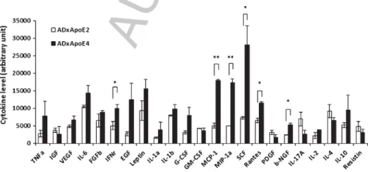
Our previous study had shown that pro-inflammatory cytokines were significantly increased in AD brains relative to age-matched non-demented (ND) controls [24]. Of note, 40–70% of the AD patients carry the ApoE4 allele whereas most ND control individuals carry the ApoE2 and/or ApoE3 alleles. These findings highlight a complex relationship between ApoE4, AD, and inflammation. This APOE isoform-modulated A-induced neuroinflammation is a very important factor in AD [21]. To determine the contribu-tions of ApoE2 and ApoE4 to AD phenotypes, particularly in inflammation, we analyzed inflam-matory phenotypes from ApoE2 and ApoE4 alleles both in the in vivo and in vitro models. First, the brains of ApoE2/APPsw/PS1dE9 (ApoE2/AD) and ApoE4/APPsw/PS1dE9 (ApoE4/AD) mice were assessed for inflammatory markers. ApoE2/AD and ApoE4/AD mice (males and females) were gener-ated and sacrificed at 6 months of age. Cytokine ELISA arrays were performed on brain tissue lysates from the mice and revealed that the levels of IFN␥, MCP-1, MIP-1␣, SCF, and RANTES were signifi-cantly higher in the brains of ApoE4/AD relative to ApoE2/AD mice (Fig. 1, two-tailed t-test,∗p< 0.05, ∗∗p< 0.01). Based on the amyloid-hypothesis,
that increased A is observed in AD patients, it was next assessed whether A levels were altered in ApoE4/AD compared to ApoE2/AD mouse brains. A1-40 and A1-42 ELISAs revealed that
homogenized brains of ApoE4/AD mice exhibited increased levels of A1-42 relative to ApoE2/AD
mice (Fig. 2A, left panel, two-tailed t-test,∗p< 0.05),
but the levels of A1-40 were not significantly
altered from ApoE4/AD versus ApoE2/AD express-ing mice (Fig. 2A, middle panel) resultexpress-ing in a slight but insignificant increase in the A42/A40
ratio for AD/ApoE4-containing mice (Fig. 2A, right panel). A peptides can activate the p38MAPK [20], NFκB [21], and JNK-AP1 [24] signaling pathways and modulate GSK3 activity for tau pathology. Thus, we evaluated the levels of phosphorylated c-Jun Ser73 and phosphorylated GSK3 ser9 in the brains of ApoE4/AD relative to ApoE2/AD by west-ern blotting. The data show that AD/ApoE4 mice displayed decreased levels of pGSK3 (Fig. 2B, two-tailed t-test,∗∗p< 0.01), and increased levels of
p-cJun/ser73 (Fig. 2C, two-tailed t-test, ∗p< 0.05),
relative to AD/ApoE2 mouse brain. The results show that ApoE4/AD mice exhibit higher levels of inflammatory cytokines and the JNK-AP1 signaling pathway (that activates inflammatory response) than ApoE2/AD mouse brains. ApoE4/AD mouse brains also display decreased GSK3 phosphorylation at ser9 (indicating an increased activity of the enzyme for tau phosphorylation) and increased levels of neu-rotoxic A1-42 peptides. To gain an understanding
of the underlying mechanisms of ApoE isoforms and A-induced inflammation we used in vitro models.
Fig. 1. Cytokine levels in ApoE2/AD and ApoE4/AD mouse brains. ApoE2/APPsw/PS1dE9 and ApoE4/APPsw/PS1dE9 mice (6 mice/group) were sacrificed at 6 months of age, and the brains were harvested and homogenized. The brain homogenates were applied to the mouse cytokine ELISA plate array, and the average level of the cytokines were plotted. Two-tailed t-test,∗∗p< 0.01,∗p< 0.05.
AUTHOR COPY
Fig. 2. Levels of A1-40and A1-42peptides, pGSK, and pc-Jun73 in the brains of ApoE2/AD and ApoE4/AD mice. ApoE2/APPsw/PS1dE9
and ApoE4/APPsw/PS1dE9 mice (6 mice/group) were sacrificed at 6 months of age, and the brains were harvested. A) Brain homogenates were applied to A1-42and A1-40ELISA kits, and the levels of A1-42and A1-40(pg/ml) were normalized to total brain protein, averaged
and plotted (left and middle panels). The A42/40peptide ratio was also calculated and plotted (right panel). B) Total protein in the brain
homogenate was solubilized by sample loading buffer, resolved on SDS-PAGE and transferred to PVDF membrane. pGSK and actin were detected by western blotting (left panel), and quantified by densitometry and presented relative to actin (right panel). C) To save on antibody, brain homogenates 1&2, 3&4, and 5&6 were pooled from each group (AD/E2 and AD/E4) to create new homogenates 1, 2, and 3, respectively. The brain protein was solubilized by sample loading buffer, resolved on SDS-PAGE and transferred to PVDF membrane. pcJun73 and actin were detected by western blotting (left panel), and quantified by densitometry and presented relative to actin (right panel). Two-tailed t-test,
∗∗p< 0.01,∗p< 0.05).
ApoE isoforms modify Aβ-induced inflammatory response in vitro
Neonatal rat astrocytes (NRA) were used to study the relationship between ApoE isoforms and A1-42-induced inflammation in vitro. To show that
A activated inflammation in NRAs, the cells were treated with A1-42 and the expression levels of
pro-inflammatory cytokines were detected by RT-qPCR. NRA cells were treated with 5 M A1-42,
scrambled A42-1peptide or vehicle for 6 h, and the
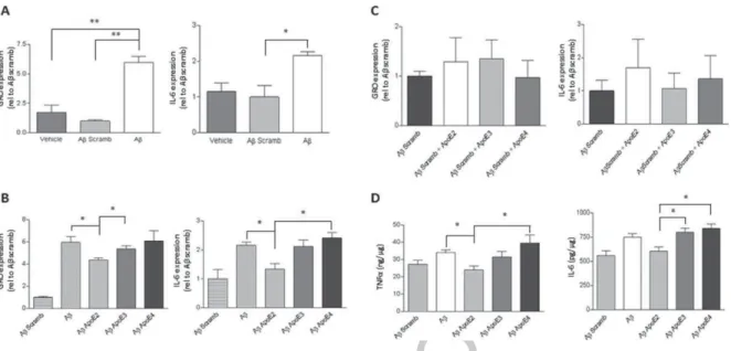
levels of GRO and IL-6 were measured by RT-qPCR. Expression of GRO was significantly increased upon A challenge, relative to both vehicle and scrambled A controls (Fig. 3A, left panel, One-way ANOVA, Bonferoni post-hoc test, ∗∗p< 0.01). Expression of
IL-6 was also significantly increased in the presence of A relative to scrambled A (Fig. 3A, right panel,
One-way ANOVA, Bonferoni post-hoc test,
∗p< 0.05). Thus, A
1-42 elicited an inflammatory
response in NRA cells as evidenced by the increase in expression levels of pro-inflammatory cytokines GRO and IL-6, among others (data not shown), and as such the levels of these two cytokines were used as markers of inflammation in this study. Next, it was examined whether various isoforms of recombinant ApoE alter the A-induced inflammatory response. NRA cells were treated with one of the three isoforms of recombinant human ApoE (E2, E3, or E4) (lipid-poor form of ApoE) at a concentration of 3 M for 24 h, and then 5 M A1-42was added for
6 h, and inflammation was assessed by measuring the expression levels of the pro-inflammatory cytokines GRO and IL-6. There was a trend with ApoE2 + A treatment exhibiting reduced expression of GRO relative to ApoE3 + A, ApoE4 + A or
AUTHOR COPY
Fig. 3. The effects of A1-42peptides and ApoE isoforms on inflammatory gene expression in NRA cells. A) NRA cells were treated
with 5 M A1-42, scrambled A or vehicle for 6 h. Expression of GRO (left panel) and IL-6 (right panel) were determined by RT-qPCR,
normalized to GAPDH and plotted (One-way ANOVA, Bonferroni post-hoc,∗∗p< 0.01,∗p< 0.05, n = 3). B) NRA cells were treated with
ApoE isoforms at 3 M for 24 h, and then with A1-42at 5 M for 6 h. Expression of GRO (left panel) and IL-6 (right panel) were
determined by RT-qPCR, normalized to GAPDH and plotted (n = 3) (One-way ANOVA, Bonferroni post-hoc, *p ˙< 0.05). C) Cells were treated as described in B, but with scrambled A instead of A1-42. Expression of GRO (left panel) and IL-6 (right panel) were determined
by RT-qPCR, normalized to GAPDH and plotted. D) Cells treated as described in B. Conditioned media was applied to TNF-␣ ELISA and the TNF-␣ levels were normalized to total cellular protein, and presented as ng/g protein (n = 3) (left panel). Whole cell lysate was applied IL-6 ELISA and the levels of IL-6 were normalized to total cellular protein, and presented as pg/g protein (right panel) (One-way ANOVA, Bonferroni post-hoc, *p < 0.05, N = 3).
to A alone (Fig. 3B, left panel). There was no difference in GRO expression between ApoE3 + A or ApoE4 + A and A treatment alone. ApoE2 + A treatment also exhibited a trend of reduced expression of IL-6 relative to A alone or ApoE4 + A treatments (Fig. 3B, right panel). In the absence of A1-42, there was no difference in GRO or IL-6
expression between scrambled A and the ApoE isoform treatments (Fig. 3C). To determine if A and ApoE isoforms treatments also affected protein levels of inflammatory markers, media and cell protein were harvested from treated NRA cells, and TNF-␣ and IL-6 were measured, respectively, by ELISA. TNF-␣ levels in media collected from cells treated with ApoE2 + A showed a trend to be reduced as compared to the ApoE4 + A treated cells or to A treatment alone (Fig. 3D, left panel). IL-6 levels in whole cell lysate exhibited a trend of reduction in the ApoE2 + A treated cells relative to ApoE3 or ApoE4 + A treated NRA cells (Fig. 3D, right panel). These data show that ApoE isoforms can alter the A-induced inflammatory response in NRA cells. The presence of recombinant ApoE2 seems to reduce the A-induced inflammatory gene
expression while the presence of ApoE3 or ApoE4 does not. The changes in inflammatory gene expres-sion in response to different ApoE isoforms were not exclusively an RNA effect, but did ultimately result in changes in protein levels of these cytokines.
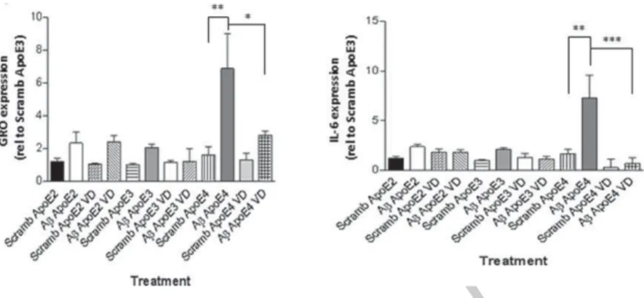
Immortalized mouse astrocytes, with native murine apoE knocked-out and human ApoE iso-forms knocked-in, were also used as an in vitro model to test whether lipidated ApoE isoforms mod-ify A-induced inflammation. The mouse astrocytes expressing hApoE isoforms were challenged with A1-42under the same conditions used for NRA cells.
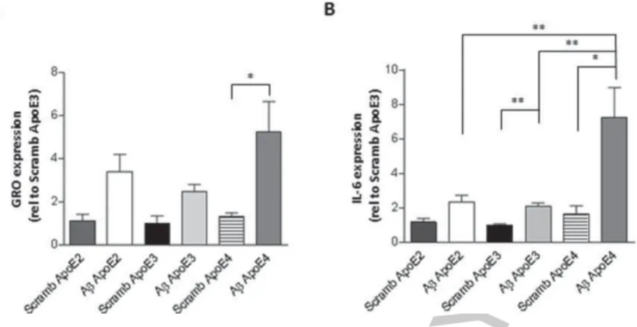
Each of the three cell lines showed a trend of increase in GRO and IL-6 expression upon A1-42treatment
(Fig. 4). In ApoE4-expressing cells, the increase in GRO expression upon A1-42treatment was
signif-icant relative to scrambled A (Fig. 4A, one-way ANOVA, Bonferoni post-hoc test ∗p< 0.05). IL-6
expression was significantly increased in ApoE4-expressing cells upon A1-42treatment relative to the
scrambled control (Fig. 4B, one-way ANOVA, Bon-feroni post-hoc test ∗p< 0.05). ApoE4-expressing
cells displayed significantly higher IL-6 expression after A challenge, compared to ApoE3 + A or
AUTHOR COPY
Fig. 4. Inflammatory gene expression in mouse apoE knock-out, human ApoE knock-in, astrocytes upon challenge with A1-42. Mouse
astrocytes expressing either hApoE2, E3, or E4 were treated with 5 M A1-42for 6 h. Expression of GRO (A) and IL-6 (B) were determined
by RT-qPCR, normalized to GAPDH and plotted (one-way ANOVA & Bonferroni post-hoc,∗p< 0.05,∗∗p< 0.01).
ApoE2 + A (Fig. 4B, One-way ANOVA, Bon-feroni post-hoc test, ∗∗p< 0.01). A similar trend
was observed with GRO expression (Fig. 4A). IL-6 expression upon A1-42 treatment in the
ApoE2-expressing mouse astrocytes was not significantly increased relative to the scrambled control (Fig. 4B). Together the data from the NRA and mouse astrocytes suggest that ApoE2 can reduce A-induced inflam-matory response, while ApoE3 and mainly ApoE4 can exacerbate the inflammatory response.
Signaling pathways are differentially activated by ApoE isoforms and Aβ combination treatments
To determine if the differential signaling path-ways are activated from A + ApoE2 versus A + ApoE3 treatments, nuclear materials from treated NRA cells were run on Protein/DNA Combo TF arrays. Densitometry analysis of the blots revealed the levels of activation of each TF from each treat-ment (Fig. 5). The TF arrays identified 7 TFs that
were upregulated, and 36 TFs that were downregu-lated in cells treated with ApoE2 + A as compared to the cells treated with ApoE3 + A (Table 2) (refer to Materials and Methods section for selection crite-ria). Table 3 lists those TFs that were also reported to be associated with AD and/or inflammation; this includes 5 TFs (VDR/DR-3, RXR/DR-1, SMAD-3/4, ERE and NF-E1/YYI) that were upregulated and 9 TFs (PPAR, IL-6-RE-BP, STAT-3, AIC, p53, NFκB, XBP-1, PUR, and MAZ) that were downregulated in cells treated with ApoE2 + A as compared to the cells treated with ApoE3 + A. EMSA validations were performed for NFκB (which was 6.29 × 10⁻³ in the ApoE2 + A treatment relative to ApoE3 + A (Table 3)) and VDR (which was increased by ∼ 70x in ApoE2 + A treatment relative to ApoE3 + A (Table 3). The NFκB EMSA confirmed that the signalling pathway was activated by A1-42 as
compared to A scrambled, and that the ApoE2 + A treatment resulted in significantly reduced DNA binding than A alone or ApoE3 + A treatment
Fig. 5. Protein/DNA arrays to identify TFs activated in rat astrocytes treated with ApoE isoforms and A42. Protein/DNA array blots from
cells treated with ApoE2 + A (left panel) or ApoE3 + A (right panel) are shown. Densitometry analysis of the arrays yielded 7 spots that were upregulated in ApoE2 + A treatment (at-least 2-fold change in intensity compared to ApoE3 + A) (Table 2), and thirty-six spots yielded the inverse relationship (downregulation in ApoE2 + A as compared to ApoE3 + A) (Table 2). VDR and MAZ were highlighted.
AUTHOR COPY
Fig. 6. Effect of VDR agonist 1␣, 25-Dihydroxyvitamin D3on the expression of inflammatory genes in mouse apoE knock-out, human
ApoE knock-in astrocytes upon challenge with A1-42. Mouse astrocytes expressing ApoE isoforms were treated with 0.1 M 1␣,
25-dihydroxyvitamin D3. Expression of GRO (A) and IL-6 (B) were measured by RT-qPCR, normalized to GAPDH and presented (One-way
ANOVA, Bonferroni post-hoc,∗∗∗p< 0.001,∗∗p< 0.01,∗p< 0.05, n = 3).
(data not shown). The VDR EMSA results also agreed with the array observations. A treatment showed significantly lower VDR binding than the scrambled control; while there was not a significant difference between ApoE2 + A and ApoE3 + A treatments, there was a clear trend that agreed with the array results (data not shown). These data show that the TF hits identified from the arrays highlight differential cellular pathways in the presence of ApoE isoforms and A, and are potential candidates of interest in AD inflammation.
The effects of signaling modulation on Aβ-induced inflammatory response
To test if VDR signaling affects the expression of inflammatory genes in astrocytes, a VDR agonist 1␣, 25-Dihydroxyvitamin D3(0.1 M) was applied for
24 h to mouse astrocytes expressing human ApoE isoforms. Activation of VDR caused a significant reduction of the A-induced upregulation of GRO and IL-6 observed in the ApoE4-expressing cell line (Fig. 6, One-way ANOVA, Bonferoni post-hoc test, ∗∗∗p< 0.001, ∗∗p< 0.01, ∗p< 0.05). There was
no significant difference in either marker upon 1␣, 25-Dihydroxyvitamin D3 treatment of ApoE2- or
ApoE3-expressing lines in the presence of A or scrambled A (Fig. 6). The expression of VDR in ApoE4/AD or ApoE2/AD mouse brains was not detected by immunoblot analysis (data not shown). BioGPS expression profile for mouse VDR shows that VDR expression is low in the brain and the anti-bodies we used may not be sensitive enough to detect the signal. The results suggest that inhibiting VDR signaling may be one of the main mechanisms of
ApoE4-mediated promotion of A-induced inflam-matory response. Enhancement of VDR signaling by agonist can suppress ApoE4-promoted inflammatory response induced by A1-42in astrocytes.
DISCUSSION
A number of studies have shown that ApoE isoforms have differential effects on A aggre-gation, degradation and clearance and may affect synaptic function [8, 25–30]. However, little is known whether ApoE isoforms differentially regulate AD/A-induced neuroinflammation and what mech-anisms might be involved. Our study is the first to show that ApoE4 promotes AD/A-induced inflam-mation while ApoE2 inhibits it. The roles of ApoE isoforms in AD neuroinflammation may be one of the mechanisms by which ApoE2 offers protection but ApoE4 promotes AD pathology. The presence of ApoE4 in AD mice increased brain inflamma-tory response compared to the ApoE2/AD mice. We also noticed that the level of A1-42 was higher in
ApoE4/AD than in ApoE2/AD mice. Higher level of A1-42may induce stronger inflammatory response,
but this cannot explain the observations that A1-42
evoked significantly stronger inflammatory response in cultured ApoE4-expressing astrocytes than that in ApoE2- or ApoE3-expressing astrocytes. Fur-thermore, recombinant ApoE2 significantly inhibited A1-42-induced expression of inflammatory genes in
cultured rat astrocytes as compared to recombinant ApoE3 or ApoE4. The combination of in vitro and in vivodata supports the notion that ApoE isoforms play differential roles in AD/A-induced inflamma-tory response.
AUTHOR COPY
One intriguing question is why A1-42 induced
significantly stronger inflammatory response in ApoE4-expressing mouse astrocytes as compared to A-treated ApoE3-or ApoE2-expressing cells, while recombinant ApoE4 + A treatment did not promote a stronger inflammatory response compared to A alone or recombinant ApoE3 + A treatment in NRA cells. It has been shown that hApoE expression in immortalized murine astrocytes results in lipidated ApoE particles [22]. This suggests that lipidation state may affect ApoE4’s role [22]. Thus, lipidated ApoE4 may promote stronger inflammatory response than ApoE2- or ApoE3-expressing cells treated with A, whereas lipid-poor ApoE4 in NRA cells does not exacerbate the A-induced inflammation rel-ative to incubation with recombinant (lipid-poor) ApoE3. It should be noted from our in vitro and in vivoresults that both lipid-poor and lipidated ApoE2 provided protection against A1-42-induced
inflam-matory gene expression. In rat astrocytes, lipid-poor ApoE2 inhibited A-induced inflammatory gene expression as compared to astrocytes treated with A alone or with A + recombinant ApoE3 or ApoE4. Although inflammatory gene expression induced by A1-42was higher than that induced by scrambled A
in ApoE2-expressing cells, there is no significant sta-tistical difference between them, and furthermore, the expression of inflammatory genes induced by A1-42
was significantly lower in ApoE2-expressing cells than that in ApoE4-expressing cells. More impor-tantly, ApoE2/AD mice had significantly lower levels of inflammatory cytokines than those in ApoE4/AD mice. This further endorses the notion that both lipid-poor and lipidated forms of ApoE2 offers pro-tection against A-induced inflammatory response. This may also explain why ApoE2 provides a greater degree of protection than ApoE3. Lipidated ApoE produced in mouse astrocytes is similar in structure to that seen in human brain, though not identical [22]. Delipidation of ApoE, as seen in ABCA1-knockout mice, promoted the formation of A plaques in AD mice [31–33], while ABCA1 upregulation shows the opposite effect, decreasing A levels [34]. The differ-ence in ApoE4’s pro-inflammatory activity between the two cell model systems may depend on ApoE’s lipidation state, and suggests that further study into the role of lipidation in the inflammation seen in AD is warranted. Since both lipid-poor and lipi-dated ApoE proteins are present in human cells and tissues, the observed effects of lipid-poor and lip-idated ApoE proteins could both be present in the brain.
One of the objectives of this study was to iden-tify signaling pathways of importance in ApoE/A inflammatory response. While some of the identified pathways are quite well-known (such as JNK-AP1 and NFκB pathways), many have not been substan-tially studied in the AD context. Vitamin D (VD) is a steroid hormone, primarily acting through VDR. VDR is a 427-amino acid protein belonging to the nuclear factor family of transcription factors. Sev-eral studies have found that polymorphisms in VDR gene are associated with risk of AD [35–37], further implicating this pathway in AD pathology. VDR is located in cytoplasm, and upon activation by active form of VD3, VDR forms a heterodimer with RXR,
translocates into nucleus, and binds to the vitamin D response elements (VDRE) on DNA to regu-late target gene expression [38]. Interestingly, both VDR and RXR were found to be highly activated in ApoE2 + A treated rat astrocytes on the Pro-tein/DNA array, relative to ApoE3 + A (Table 3). Studies have shown that VD is an important regulator of immune and inflammatory response. Zhang et al. reported that VD inhibits production of inflammatory cytokines by monocytes/macrophages and inhibits inflammatory response [39, 40]. VDR signaling can attenuate Toll-like receptor mediated inflammation [41]. In vitro studies have shown that VD protects neurons from A-induced cytotoxicity and apoptosis [42], and VD/VDR signaling suppress APP produc-tion in cells [43]. Our study shows that active form of VD3 almost completely inhibited inflammatory
gene expression in ApoE4-expressing cells follow-ing A treatment in comparison to the cells treated with A1-42. This suggests that VDR may be one
of the major signaling pathways regulating AD/A-induced inflammatory response. Interestingly, VD3
did not affect A-induced expression of inflamma-tory genes in ApoE3- or ApoE2-expressing cells. Since ApoE4-expressing cell line showed much higher inflammatory response to A than the other cell lines, it is possible that VDR’s effect was only observable in these highly inflammatory conditions. On the other hand, VDR might have already been acti-vated in the presence of lipidated ApoE2 or ApoE3 with A1-42since inflammatory gene expression had
been inhibited in the absence of active form of VD3.
This is also supported by our observation that both VDR and RXR were highly activated in rat astro-cytes pre-treated with lipid-poor recombinant ApoE2 for 24 h followed by treatment with A1-42. It is
known that many VDR targets have neuroprotec-tive and anti-inflammatory effects, including within
AUTHOR COPY
astrocytes [44]. Thus, active form of VD3 may be
a preventative and/or therapeutic agent to relieve AD/A-induced inflammatory response for reduced neuronal dysfunction and neurodegeneration, espe-cially in ApoE4-carrier AD patients. VDR activation could be a potential AD therapeutic approach as a part of a wider attempt to affect a number of inflammatory pathways.
A clinical study [45] found that treatment of AD patients with memantine plus VD improved cognition performance. Annweiler and colleagues conducted a meta-analysis for 9 published studies and found that AD patients had lower levels of serum 25-hydroxylvitamin D3than matched controls
[46], but the ApoE genotypes of the AD patients were not investigated in the analyses. Huebbe et al. found that ApoE4 carriers were associated with higher 25-hydorxylvitamin D3 levels than ApoE2
and ApoE3 carriers in animal models and in nor-mal human subjects [47]. There is no report in the literature of whether ApoE4/AD patients display lower or higher 25-hydorxylvitamin D3 levels than
age-matched non-demented controls or ApoE3/AD patients. 25-hydroxylvitamin D3is not an active form
of VD and does not activate VDR signaling unless it is converted to 1,25-hydroxylvitamin D3. It is likely
that ApoE4 transgenic mice or ApoE4/AD patients may be less efficient in synthesizing active 1,25-dihydroxylvitamin D3from 25-hydroxylvitamin D3,
which remains to be investigated.
In summary, our studies have shown that ApoE isoforms play differential roles in regulating AD/A-induced inflammatory response. ApoE4 seems to promote A-induced inflammatory response while ApoE2 protects against it. In vivo experiment shows that ApoE4/AD mice had significantly higher lev-els of inflammatory cytokines than ApoE2/AD mice. In vitro studies reveal that lipidation states of ApoE isoforms may affect their roles in A-induced inflammatory response. Lipidated ApoE4 enhanced A42-induced inflammatory response in astrocytes
but not the recombinant lipid-poor ApoE4. Inter-estingly, lipidated ApoE3 provided a certain degree of protection while lipid-poor ApoE3 did not. Both lipidated and recombinant ApoE2 offered protec-tion against A42-induced inflammatory response
in astrocytes. VDR signaling was strongly acti-vated in astrocytes pre-treated with recombinant ApoE2. Application of a VDR agonist, 1␣, 25-dihydroxylvitamin D3, almost completely inhibited
A42-induced inflammatory gene expression in
ApoE4-expressing cells. This suggests that VDR may
be a major signaling pathway by which ApoE iso-forms (lipidated and lipid-poor iso-forms) play their differential roles in regulating ADs/A-induced neu-roinflammation. Active form of VD3 or targeting
VDR signaling may relieve the neuroinflammation and neurodegeneration seen in AD.
ACKNOWLEDGMENTS
This study was supported by research grants, CIHR#109606, CIHR#106886, and #TAD 125698 to W. Zhang (uOttawa); and was conducted at the National Research Council Canada. The authors would like to thank Dr. D. Holtzman (Washington University at St. Louis) for providing the immor-talized mouse ApoE astrocyte cells for the study. The authors would like to thank the staff at the NRC-Ottawa Animal Facility for maintaining the transgenic mouse colonies, mating the mice and col-lecting blood samples.
Authors’ disclosures available online (http://j-alz. com/manuscript-disclosures/16-0133r1).
REFERENCES
[1] Cudaback E, Yang Y, Montine TJ, Keene CD (2015) APOE genotype-dependent modulation of astrocyte chemokine CCL3 production. Glia 63, 51-65.
[2] Kitazawa M, Yamasaki TR, LaFerla FM (2004) Microglia as a potential bridge between the amyloid beta-peptide and tau. Ann N Y Acad Sci 1035, 85-103.
[3] Corder EH, Saunders AM, Risch NJ, Strittmatter WJ, Schmechel DE, Gaskell PC Jr, Rimmler JB, Locke PA, Con-neally PM, Schmader KE, Small GW, Roses AD, Haines JL, Pericak-Vance MA (1994) Protective effect of apolipopro-tein E type 2 allele for late onset Alzheimer disease. Nat
Genet 7, 180-184.
[4] Genin E, Hannequin D, Wallon D, Sleegers K, Hiltunen M, Combarros O, Bullido MJ, Engelborghs S, De Deyn P, Berr C, Pasquier F, Dubois B, Tognoni G, Fievet N, Brouw-ers N, Bettens K, Arosio B, Coto E, Del Zompo M, Mateo I, Epelbaum J, Frank-Garcia A, Helisalmi S, Porcellini E, Pilotto A, Forti P, Ferri R, Scarpini E, Siciliano G, Sol-frizzi V, Sorbi S, Spalletta G, Valdivieso F, Vepsalainen S, Alvarez V, Bosco P, Mancuso M, Panza F, Nacmias B, Bossu P, Hanon O, Piccardi P, Annoni G, Seripa D, Galim-berti D, Licastro F, Soininen H, Dartigues JF, Kamboh MI, Van Broeckhoven C, Lambert JC, Amouyel P, Campion D (2011) APOE and Alzheimer disease: A major gene with semi-dominant inheritance. Mol Psychiatry 16, 903-907. [5] Roses AD (1996) Apolipoprotein E alleles as risk factors in
Alzheimer’s disease. Annu Rev Med 47, 387-400. [6] Bien-Ly N, Gillespie AK, Walker D, Yoon SY, Huang Y
(2012) Reducing human apolipoprotein E levels attenu-ates age-dependent Abeta accumulation in mutant human amyloid precursor protein transgenic mice. J Neurosci 32, 4803-4811.
AUTHOR COPY
[7] Bales KR, Liu F, Wu S, Lin S, Koger D, DeLong C, HansenJC, Sullivan PM, Paul SM (2009) Human APOE isoform-dependent effects on brain beta-amyloid levels in PDAPP transgenic mice. J Neurosci 29, 6771-6779.
[8] Castellano JM, Kim J, Stewart FR, Jiang H, DeMattos RB, Patterson BW, Fagan AM, Morris JC, Mawuenyega KG, Cruchaga C, Goate AM, Bales KR, Paul SM, Bateman RJ, Holtzman DM (2011) Human apoE isoforms differentially regulate brain amyloid-beta peptide clearance. Sci Transl
Med 3, 89ra57.
[9] Jiang Q, Lee CY, Mandrekar S, Wilkinson B, Cramer P, Zelcer N, Mann K, Lamb B, Willson TM, Collins JL, Richardson JC, Smith JD, Comery TA, Riddell D, Holtzman DM, Tontonoz P, Landreth GE (2008) ApoE pro-motes the proteolytic degradation of Abeta. Neuron 58, 681-693.
[10] Grainger DJ, Reckless J, McKilligin E (2004) Apolipopro-tein E modulates clearance of apoptotic bodies in vitro and in vivo, resulting in a systemic proinflammatory state in apolipoprotein E-deficient mice. J Immunol 173, 6366-6375.
[11] LaDu MJ, Shah JA, Reardon CA, Getz GS, Bu G, Hu J, Guo L, Van Eldik LJ (2001) Apolipoprotein E and apolipoprotein E receptors modulate A beta-induced glial neuroinflammatory responses. Neurochem Int 39, 427-434. [12] Lynch JR, Tang W, Wang H, Vitek MP, Bennett ER, Sullivan PM, Warner DS, Laskowitz DT (2003) APOE genotype and an ApoE-mimetic peptide modify the systemic and central nervous system inflammatory response. J Biol Chem 278, 48529-48533.
[13] Jofre-Monseny L, Loboda A, Wagner AE, Huebbe P, Boesch-Saadatmandi C, Jozkowicz A, Minihane AM, Dulak J, Rimbach G (2007) Effects of apoE genotype on macrophage inflammation and heme oxygenase-1 expres-sion. Biochem Biophys Res Commun 357, 319-324. [14] Maezawa I, Zaja-Milatovic S, Milatovic D, Stephen C,
Sokal I, Maeda N, Montine TJ, Montine KS (2006) Apolipoprotein E isoform-dependent dendritic recovery of hippocampal neurons following activation of innate immu-nity. J Neuroinflammation 3, 21.
[15] Colton CA, Needham LK, Brown C, Cook D, Rasheed K, Burke JR, Strittmatter WJ, Schmechel DE, Vitek MP (2004) APOE genotype-specific differences in human and mouse macrophage nitric oxide production. J Neuroimmunol 147, 62-67.
[16] Ophir G, Amariglio N, Jacob-Hirsch J, Elkon R, Rechavi G, Michaelson DM (2005) Apolipoprotein E4 enhances brain inflammation by modulation of the NF-kappaB signaling cascade. Neurobiol Dis 20, 709-718.
[17] Vitek MP, Brown CM, Colton CA (2009) APOE genotype-specific differences in the innate immune response.
Neurobiol Aging 30, 1350-1360.
[18] Zeng Y, Callaghan D, Xiong H, Yang Z, Huang P, Zhang W (2012) Abcg2 deficiency augments oxidative stress and cognitive deficits in Tg-SwDI transgenic mice. J Neurochem
122, 456-469.
[19] Ordonez-Gutierrez L, Anton M, Wandosell F (2015) Periph-eral amyloid levels present gender differences associated with aging in AbetaPP/PS1 mice. J Alzheimers Dis 44, 1063-1068.
[20] Giovannini MG, Scali C, Prosperi C, Bellucci A, Vannucchi MG, Rosi S, Pepeu G, Casamenti F (2002) Beta-amyloid-induced inflammation and cholinergic hypofunction in the rat brain in vivo: Involvement of the p38MAPK pathway.
Neurobiol Dis 11, 257-274.
[21] Garberg P, Ball M, Borg N, Cecchelli R, Fenart L, Hurst RD, Lindmark T, Mabondzo A, Nilsson JE, Raub TJ, Sta-nimirovic D, Terasaki T, Oberg JO, Osterberg T (2005) In
vitromodels for the blood-brain barrier. Toxicol In Vitro 19, 299-334.
[22] Morikawa M, Fryer JD, Sullivan PM, Christopher EA, Wahrle SE, DeMattos RB, O’Dell MA, Fagan AM, Lashuel HA, Walz T, Asai K, Holtzman DM (2005) Production and characterization of astrocyte-derived human apolipoprotein E isoforms from immortalized astrocytes and their interac-tions with amyloid-beta. Neurobiol Dis 19, 66-76. [23] Xiong H, Callaghan D, Wodzinska J, Xu J, Premyslova
M, Liu QY, Connelly J, Zhang W (2011) Biochemical and behavioral characterization of the double transgenic mouse model (APPswe/PS1dE9) of Alzheimer’s disease. Neurosci
Bull 27, 221-232.
[24] Vukic V, Callaghan D, Walker D, Lue LF, Liu QY, Couraud PO, Romero IA, Weksler B, Stanimirovic DB, Zhang W (2009) Expression of inflammatory genes induced by beta-amyloid peptides in human brain endothelial cells and in Alzheimer’s brain is mediated by the JNK-AP1 signaling pathway. Neurobiol Dis 34, 95-106.
[25] Bu G (2009) Apolipoprotein E and its receptors in Alzheimer’s disease: Pathways, pathogenesis and therapy.
Nat Rev Neurosci 10, 333-344.
[26] Chalmers K, Wilcock GK, Love S (2003) APOE epsilon 4 influences the pathological phenotype of Alzheimer’s disease by favouring cerebrovascular over parenchymal accumulation of A beta protein. Neuropathol Appl
Neuro-biol 29, 231-238.
[27] Deane R, Sagare A, Hamm K, Parisi M, Lane S, Finn MB, Holtzman DM, Zlokovic BV (2008) apoE isoform-specific disruption of amyloid beta peptide clearance from mouse brain. J Clin Invest 118, 4002-4013.
[28] Korwek KM, Trotter JH, Ladu MJ, Sullivan PM, Weeber EJ (2009) ApoE isoform-dependent changes in hippocampal synaptic function. Mol Neurodegener 4, 21.
[29] Dorey E, Chang N, Liu QY, Yang Z, Zhang W (2014) Apolipoprotein E, amyloid-beta, and neuroinflammation in Alzheimer’s disease. Neurosci Bull 30, 317-330. [30] Nwabuisi-Heath E, Rebeck GW, Ladu MJ, Yu C (2014)
ApoE4 delays dendritic spine formation during neuron development and accelerates loss of mature spines in vitro.
ASN Neuro 6, e00134.
[31] Wahrle SE, Jiang H, Parsadanian M, Hartman RE, Bales KR, Paul SM, Holtzman DM (2005) Deletion of Abca1 increases Abeta deposition in the PDAPP transgenic mouse model of Alzheimer disease. J Biol Chem 280, 43236-43242. [32] Hirsch-Reinshagen V, Maia LF, Burgess BL, Blain JF, Naus
KE, McIsaac SA, Parkinson PF, Chan JY, Tansley GH, Hayden MR, Poirier J, Van Nostrand W, Wellington CL (2005) The absence of ABCA1 decreases soluble ApoE levels but does not diminish amyloid deposition in two murine models of Alzheimer disease. J Biol Chem 280, 43243-43256.
[33] Koldamova R, Staufenbiel M, Lefterov I (2005) Lack of ABCA1 considerably decreases brain ApoE level and increases amyloid deposition in APP23 mice. J Biol Chem
280, 43224-43235.
[34] Riddell DR, Zhou H, Atchison K, Warwick HK, Atkinson PJ, Jefferson J, Xu L, Aschmies S, Kirksey Y, Hu Y, Wagner E, Parratt A, Xu J, Li Z, Zaleska MM, Jacobsen JS, Pangalos MN, Reinhart PH (2008) Impact of apolipoprotein E (ApoE) polymorphism on brain ApoE levels. J Neurosci 28, 11445-11453.
AUTHOR COPY
[35] Gezen-Ak D, Dursun E, Ertan T, Hanagasi H, Gurvit H,Emre M, Eker E, Ozturk M, Engin F, Yilmazer S (2007) Association between vitamin D receptor gene polymor-phism and Alzheimer’s disease. Tohoku J Exp Med 212, 275-282.
[36] Gezen-Ak D, Dursun E, Bilgic B, Hanagasi H, Ertan T, Gurvit H, Emre M, Eker E, Ulutin T, Uysal O, Yilmazer S (2012) Vitamin D receptor gene haplotype is associated with late-onset Alzheimer’s disease. Tohoku J Exp Med 228, 189-196.
[37] Lehmann DJ, Refsum H, Warden DR, Medway C, Wilcock GK, Smith AD (2011) The vitamin D receptor gene is asso-ciated with Alzheimer’s disease. Neurosci Lett 504, 79-82. [38] Haussler MR, Haussler CA, Bartik L, Whitfield GK, Hsieh JC, Slater S, Jurutka PW (2008) Vitamin D recep-tor: Molecular signaling and actions of nutritional ligands in disease prevention. Nutr Rev 66, S98-112.
[39] Zhang Y, Leung DY, Richers BN, Liu Y, Remigio LK, Riches DW, Goleva E (2012) Vitamin D inhibits mono-cyte/macrophage proinflammatory cytokine production by targeting MAPK phosphatase-1. J Immunol 188, 2127-2135.
[40] Zhang Y, Leung DY, Goleva E (2013) Vitamin D enhances glucocorticoid action in human monocytes: Involvement of granulocyte-macrophage colony-stimulating factor and mediator complex subunit 14. J Biol Chem 288, 14544-14553.
[41] Chen Y, Liu W, Sun T, Huang Y, Wang Y, Deb DK, Yoon D, Kong J, Thadhani R, Li YC (2013) 1,25-Dihydroxyvitamin D promotes negative feedback regulation of TLR signal-ing via targetsignal-ing microRNA-155-SOCS1 in macrophages.
J Immunol 190, 3687-3695.
[42] Dursun E, Gezen-Ak D, Yilmazer S (2011) A novel perspective for Alzheimer’s disease: Vitamin D receptor suppression by beta and preventing the amyloid-beta induced alterations by vitamin D in cortical neurons.
J Alzheimers Dis 23, 207-219.
[43] Wang L, Hara K, Van Baaren JM, Price JC, Beecham GW, Gallins PJ, Whitehead PL, Wang G, Lu C, Slifer MA, Zuch-ner S, Martin ER, Mash D, Haines JL, Pericak-Vance MA, Gilbert JR (2012) Vitamin D receptor and Alzheimer’s dis-ease: A genetic and functional study. Neurobiol Aging 33, 1844 e1841-1849.
[44] Garcion E, Sindji L, Montero-Menei C, Andre C, Bra-chet P, Darcy F (1998) Expression of inducible nitric oxide synthase during rat brain inflammation: Regulation by 1,25-dihydroxyvitamin D3. Glia 22, 282-294.
[45] Annweiler C, Herrmann FR, Fantino B, Brugg B, Beauchet O (2012) Effectiveness of the combination of memantine plus vitamin D on cognition in patients with Alzheimer disease: A pre-post pilot study. Cogn Behav Neurol 25, 121-127.
[46] Annweiler C, Llewellyn DJ, Beauchet O (2013) Low serum vitamin D concentrations in Alzheimer’s disease: A sys-tematic review and meta-analysis. J Alzheimers Dis 33, 659-674.
[47] Huebbe P, Nebel A, Siegert S, Moehring J, Boesch-Saadatmandi C, Most E, Pallauf J, Egert S, Muller MJ, Schreiber S, Nothlings U, Rimbach G (2011) APOE epsilon4 is associated with higher vitamin D levels in targeted replacement mice and humans. FASEB J 25, 3262-3270.
[48] McKee AC, Kosik KS, Kowall NW (1991) Neuritic pathol-ogy and dementia in Alzheimer’s disease. Ann Neurol 30, 156-165.
[49] Fernandes de Abreu DA, Eyles D, Feron F (2009) Vitamin D, a neuro-immunomodulator: Implications for neurodegenerative and autoimmune diseases.
Psychoneu-roendocrinology 34(Suppl 1), S265-S277.
[50] Tai LM, Koster KP, Luo J, Lee SH, Wang YT, Collins NC, Ben Aissa M, Thatcher GR, LaDu MJ (2014) Amyloid-beta pathology and APOE genotype modulate retinoid X receptor agonist activity in vivo. J Biol Chem 289, 30538-30555.
[51] Roszer T, Menendez-Gutierrez MP, Cedenilla M, Ricote M (2013) Retinoid X receptors in macrophage biology. Trends
Endocrinol Metab 24, 460-468.
[52] Zhuang J, Zhang H, Zhou R, Chen L, Chen J, Shen X (2013) Regulation of prostaglandin F2alpha against beta amyloid clearance and its inflammation induction through LXR/RXR heterodimer antagonism in microglia.
Prostaglandins Other Lipid Mediat 106, 45-52.
[53] Tichauer JE, von Bernhardi R (2012) Transforming growth factor-beta stimulates beta amyloid uptake by microglia through Smad3-dependent mechanisms. J Neurosci Res 90, 1970-1980.
[54] Morinaga A, Ono K, Takasaki J, Ikeda T, Hirohata M, Yamada M (2011) Effects of sex hormones on Alzheimer’s disease-associated beta-amyloid oligomer formation in
vitro. Exp Neurol 228, 298-302.
[55] Pike CJ, Carroll JC, Rosario ER, Barron AM (2009) Protec-tive actions of sex steroid hormones in Alzheimer’s disease.
Front Neuroendocrinol 30, 239-258.
[56] Long J, He P, Shen Y, Li R (2012) New evidence of mito-chondria dysfunction in the female Alzheimer’s disease brain: Deficiency of estrogen receptor-beta. J Alzheimers
Dis 30, 545-558.
[57] Razmara A, Duckles SP, Krause DN, Procaccio V (2007) Estrogen suppresses brain mitochondrial oxidative stress in female and male rats. Brain Res 1176, 71-81.
[58] Brown CM, Choi E, Xu Q, Vitek MP, Colton CA (2008) The APOE4 genotype alters the response of microglia and macrophages to 17beta-estradiol. Neurobiol Aging 29, 1783-1794.
[59] Nowak K, Lange-Dohna C, Zeitschel U, Gunther A, Luscher B, Robitzki A, Perez-Polo R, Rossner S (2006) The tran-scription factor Yin Yang 1 is an activator of BACE1 expression. J Neurochem 96, 1696-1707.
[60] Combs CK, Johnson DE, Karlo JC, Cannady SB, Lan-dreth GE (2000) Inflammatory mechanisms in Alzheimer’s disease: Inhibition of beta-amyloid-stimulated proinflam-matory responses and neurotoxicity by PPARgamma agonists. J Neurosci 20, 558-567.
[61] Heneka MT, Reyes-Irisarri E, Hull M, Kummer MP (2011) Impact and Therapeutic Potential of PPARs in Alzheimer’s Disease. Curr Neuropharmacol 9, 643-650.
[62] Mandrekar-Colucci S, Karlo JC, Landreth GE (2012) Mechanisms underlying the rapid peroxisome proliferator-activated receptor-gamma-mediated amyloid clearance and reversal of cognitive deficits in a murine model of Alzheimer’s disease. J Neurosci 32, 10117-10128. [63] Wang R, Li JJ, Diao S, Kwak YD, Liu L, Zhi L, Bueler H,
Bhat NR, Williams RW, Park EA, Liao FF (2013) Metabolic stress modulates Alzheimer’s beta-secretase gene transcrip-tion via SIRT1-PPARgamma-PGC-1 in neurons. Cell Metab
17, 685-694.
[64] Xu J, Chavis JA, Racke MK, Drew PD (2006) Peroxisome proliferator-activated receptor-alpha and retinoid X recep-tor agonists inhibit inflammarecep-tory responses of astrocytes.
AUTHOR COPY
[65] Heneka MT, Sastre M, Dumitrescu-Ozimek L, Hanke A,Dewachter I, Kuiperi C, O’Banion K, Klockgether T, Van Leuven F, Landreth GE (2005) Acute treatment with the PPARgamma agonist pioglitazone and ibuprofen reduces glial inflammation and Abeta1-42 levels in APPV717I trans-genic mice. Brain 128, 1442-1453.
[66] Brechner T, Hocke G, Goel A, Fey GH (1991) Interleukin 6 response factor binds co-operatively at two adjacent sites in the promoter upstream region of the rat alpha 2-macroglobulin gene. Mol Biol Med 8, 267-285.
[67] Wan J, Fu AK, Ip FC, Ng HK, Hugon J, Page G, Wang JH, Lai KO, Wu Z, Ip NY (2010) Tyk2/STAT3 signaling medi-ates beta-amyloid-induced neuronal cell death: Implications in Alzheimer’s disease. J Neurosci 30, 6873-6881. [68] Chiba T, Yamada M, Sasabe J, Terashita K, Shimoda M,
Matsuoka M, Aiso S (2009) Amyloid-beta causes memory impairment by disturbing the JAK2/STAT3 axis in hip-pocampal neurons. Mol Psychiatry 14, 206-222.
[69] Lefterov I, Fitz NF, Cronican AA, Fogg A, Lefterov P, Kodali R, Wetzel R, Koldamova R (2010) Apolipoprotein A-I deficiency increases cerebral amyloid angiopathy and cognitive deficits in APP/PS1DeltaE9 mice. J Biol Chem
285, 36945-36957.
[70] Behrens MI, Lendon C, Roe CM (2009) A common biolog-ical mechanism in cancer and Alzheimer’s disease? Curr
Alzheimer Res 6, 196-204.
[71] Hooper C, Meimaridou E, Tavassoli M, Melino G, Love-stone S, Killick R (2007) p53 is upregulated in Alzheimer’s disease and induces tau phosphorylation in HEK293a cells.
Neurosci Lett 418, 34-37.
[72] Kim H, Lee SH, Lee MN, Oh GT, Choi KC, Choi EY (2013) p53 regulates the transcription of the anti-inflammatory molecule developmental endothelial locus-1 (Del-1).
Onco-target 4, 1976-1985.
[73] Schauer IG, Zhang J, Xing Z, Guo X, Mercado-Uribe I, Sood AK, Huang P, Liu J (2013) Interleukin-1beta promotes ovarian tumorigenesis through a p53/NF-kappaB-mediated inflammatory response in stromal fibroblasts. Neoplasia 15, 409-420.
[74] Bales KR, Du Y, Holtzman D, Cordell B, Paul SM. (2000) Neuroinflammation and Alzheimer’s disease: Critical roles for cytokine/Abeta-induced glial activation, NF-kappaB, and apolipoprotein E. Neurobiol Aging 21, 427-432; dis-cussion 451-423.
[75] Gilmore TD (2006) Introduction to NF-kappaB: Players, pathways, perspectives. Oncogene 25, 6680-6684. [76] Casas-Tinto S, Zhang Y, Sanchez-Garcia J,
Gomez-Velazquez M, Rincon-Limas DE, Fernandez-Funez P (2011) The ER stress factor XBP1s prevents amyloid-beta neurotoxicity. Hum Mol Genet 20, 2144-2160.
[77] Darbinian N, Cui J, Basile A, Del Valle L, Otte J, Miklossy J, Sawaya BE, Amini S, Khalili K, Gordon J (2008) Neg-ative regulation of AbetaPP gene expression by pur-alpha.
J Alzheimers Dis 15, 71-82.
[78] Gomez Ravetti M, Rosso OA, Berretta R, Moscato P (2010) Uncovering molecular biomarkers that correlate cognitive decline with the changes of hippocampus’ gene expression profiles in Alzheimer’s disease. PLoS One 5, e10153. [79] Jordan-Sciutto KL, Dragich JM, Caltagarone J, Hall DJ,
Bowser R (2000) Fetal Alz-50 clone 1 (FAC1) pro-tein interacts with the Myc-associated zinc finger propro-tein (ZF87/MAZ) and alters its transcriptional activity.
Biochem-istry 39, 3206-3215.
[80] Ray A, Shakya A, Kumar D, Benson MD, Ray BK (2006) Inflammation-responsive transcription factor SAF-1 activ-ity is linked to the development of amyloid A amyloidosis.
J Immunol 177, 2601-2609.
View publication stats View publication stats