Applying a Microfluidic 'Deformability Cytometry' to
Measure Stiffness of Malaria-infected Red Blood Cells
at Body and Febrile Temperatures
by
Sha Huang
MASsACHUSETTS INS E OF TECHNOLOGY[JUN
17
2011
LIBRARIES
Submitted to the Department of Electrical Engineering and Computer
Science in partial fulfillment of the requirements for the degree of
Masters of Science in Electrical Engineering and Computer Science
at the
MASSACHUSETTS INSTITUTE OF TECHNOLOGY
ARMRMS
June 2011
© Massachusetts Institute of Technology 2011. All rights reserved.
Signature of Author:
Department of Electrical Engineering and Computer Science
May 20, 2011
Certified by:
__Jongyoon Han
Associate Professor of Electrical Engineering and Computer Science
Thesis Supervisor
Accepted by:
Leslie A. Kolodziejski
,'-Professor of Electrical Engineering and Computer Science
Chairman, Committee for Graduate Students
Applying a Microfluidic 'Deformability Cytometry' to
Measure Stiffness of Malaria-infected Red Blood Cells
at Body and Febrile Temperatures
by
Sha Huang
Submitted to the Department of Electrical Engineering and Computer
Science on May 20. 2011, in partial fulfillment of the
requirements for the degree of
Masters of Science in Electrical Engineering and Computer Science
Abstract
Red blood cells (RBCs) undergo repeated deformation as they traverse blood vessel, capillaries and splenic cords; RBC deformability is therefore crucial in maintaining normal blood circulation. During falciparum malaria, parasite proteins interact with the spectrin network of host RBCs, moderately stiffening the ring stage infected cells (rings). The subtle modification in the deformability of rings is however believed to be significant enough to trigger their retention by human spleen. In addition, recent studies demonstrated considerable stiffening of parasitized RBCs at febrile temperature, highlighting the temperature-dependent physiological consequences in microcirculation.
A quantitative characterization of the dynamic process of RBC deformation at
physiologically relevant temperatures is therefore highly desirable. In this work, a microfluidic device with bottleneck arrays is developed to mimic RBCs' travelling through narrow in-vivo constrictions such as splenic cordal meshwork and blood capillaries. For the first time, we report the dynamic mechanical responses of rings in a large population of co-cultured uninfected cells at both body and febrile temperatures. Experiments revealed that the deformability cytometer can differentiate parasitized RBCs from normal RBCs most efficiently at febrile temperature, suggesting a potential role of fever in facilitating splenic clearance. Similar dynamic deformability measurements were also conducted on RBCs with anti-malarial drug treatment; the drug effect on the deformability of both normal and parasitized cells is assessed.
Thesis Supervisor: Jongyoon Han
Title: Associate Professor of Electrical Engineering and Computer Science
and Biological Engineering
Acknowledgements
It has been almost two years since I first embarked on my journey as a graduate student in MIT. Life has been even more enriching and exciting than I had envisaged. The ample collaboration opportunities amongst dissimilar research groups and the state of the art lab facilities have offered me the maximum research freedom one could possibly have.
First of all, I would like to express my utmost gratitude to my research advisor Professor Jongyoon Han, who introduced me into this amazing field of BioMEMS. I would like to thank Prof. Han for his guidance, support and encouragement throughout. His enthusiasm for science and his insights in conducting scientific research have influenced me greatly and redefined my perspective towards research. In addition, I am also deeply indebted to Prof. Han for the enormous research freedom he endowed me. He encouraged me to explore areas of my own interests and he has always been accepting to my new ideas, even though most of the time they did not work out well. I could not have wished for a better advisor.
I would also like to thank all the Han group members who have been so
supportive and helpful. It has been a very comfortable and conductive environment to work in. I greatly appreciate Dr. Pan Mao and Dr. Hansen Bow, both offered me a lot of hands-on trainings and supervisions and helped me identify possible research directions. Dr. Pan Mao's expertise in device design and fabrication helped me enormously when I first entered Microsystems Technology Laboratories (MTL). Under his coach, I learnt that thoughtfulness and patience are the two essential qualities for device fabrication and for research in general. Dr. Hansen Bow was the person who initiated this malaria project when he was a graduate student in Han group. He built the foundation for this project and gave me a lot of guidance and advice during the initial phase of my research work. The weekly project discussions with Hansen have been really educational and rewarding. The other members in the group are also great people to work with and to spend time with including Lih-Feng Cheow, Aniruddh Sarker, Lidan Wu, Leon Li, Rhokyun Kwak, Dr. Chia-Hung Chen, Dr. Hiong Yap Gan, Dr. Sung Jae Kim, and Dr. Yong-Ak Song. During my hard times at MIT, they offered me the warmest companionship.
Prof. Subra Suresh and his nanomechanics laboratory have also offered me a lot of support without which this project could not have been accomplished. As current director of national science foundation (NSF), Prof. Suresh still meet us whenever it is possible and offered us a lot of invaluable advice on the general direction of the project. His research group, currently led by Dr. Ming Dao, has been a pleasure to work with. Special thanks to Dr. Monica Diez-Silva whose expertise in Malaria research has been a great asset for this project.
During my summer internship at the National University of Singapore, the constructive interactions I had with Prof. Chwee Teck Lim, Dr. Ali Asgar Bhagat and graduate student Hanwei Hou have been also very valuable.
Last but not least, I would like to thank my dearest parents and my husband Yuan Fang. I greatly appreciate their support and understanding. I would like to dedicate this thesis to them to express my love and gratitude.
Contents
1. Introduction ...
7
Red Blood Cell (RBC) Deformability...7
Plasmodium falciparum Malaria...12
2. Existing Tools for RBC Deformability Measurement...16
RBC deformability measurement by Optical Tweezers...16
RBC deformability measurement by Micropipette Aspiration...18
RBC deformability measurement by ektacytometry...19
RBC deformability measurement in MicroFluidic devices...19
3. Materials and Methods...23
Device Fabrication...23
Parasite Culture...
25
Solution Preparation...25
Experim ent Protocol...25
4. Effect of Febrile Temperature on the Deformability...28
of Malaria-infected Erythrocytes
Temperature-dependent iRBC deformability ...
28
Temperature-dependent uRBC deformability ...
32
Temperature-dependent hRBC deformability ...
33
Febrile condition enhances the separation resolution ...
34
between iRBCs and uRBCs
Spleen as a mechanical filter...36
5. Effect of Anti-malarial Drugs on the Deformability...38
of Malaria-infected Erythrocytes
Artemisinin and its derivatives...39
6. Effect of the Ring-infected Erythrocyte Surface...44
Antigen (RESA) on the Deformability of
Malaria-infected Erythrocytes
7. Discussions...47
Body temperature corresponds to the maximum...47
hRBC deformability
Fever in the pathogenesis of malaria...49
Antim alarial drug effect...
50
Reduced uRBC deformability may exacerbate malaria anemia...53
8. Conclusion and Future Plans...55
Chapter 1
Introduction
Human blood consists of approximately 55% plasma and 45% blood cells, of which 99% are red blood cells (RBCs). One important function of RBC is to take up oxygen in the lungs or gills and deliver it to all body tissues. During the 120-day life-span of an average RBC, it circulates through the body for approximately 500,000 times and undergoes repeated severe deformations while squeezing through blood capillaries and splenic cords. RBC deformability is therefore crucial in maintaining normal blood circulation. Decreased RBC deformability is both a cause of and biomarker for potentially severe diseases such as diabetes, sickle cell anemia and malaria. Crossing splenic interendothelial slits is probably the most stringent challenge on RBC deformability because the slit size is estimated to be only 1pm. Spleen is therefore perceived as a mechanical filter which facilitates the clearance of RBCs with reduced deformability.
Red Blood Cell (RBC) Deformability
Red blood cells are also known as erythrocytes. Mature human erythrocytes are non-nucleated discoid with average diameter and thickness of 7gm and 2pm respectively. The biconcave shape evolves from the multilobulated reticulocyte during 48 hours of maturation first in the bone marrow and then in blood circulation' (Figure 1). As the only structural component of a mature RBC, the plasma membrane enclosing a large amount of hemoglobin accounts for all of its diverse antigenic, transport and mechanical characteristics. Essentially, RBC deformability comprises two major components: membrane stiffness and cytosol (hemoglobin) fluidity.
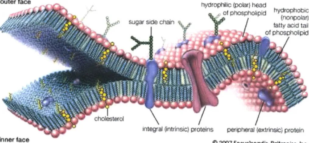
Red blood cell membrane is currently viewed as a composite structure in which a membrane envelope, composed of cholesterol and phospholipids, is anchored to an elastic network of skeletal proteins via transmembrane proteins embedded in the lipid bilayer' (Figure 2 and Figure 3). The principal skeletal proteins that form the spectrin network are a- and
P-
spectrin, actin, protein 4.1 R, adducing, dematin, tropomyosin and tropomodulin. Spectrin tetramer, the major structural component of the skeletal network, is formed by the lateral interaction between a solitary helix of the a-chain from 1 dimer (at the N-terminus) and 2 helices of the 0- chain from the other (at the C-terminus) to create a stable triple-helical repeat2. The shift in dimer-tetramer ratio could be induced byshear-stress3, oxidative stress4 or temperature5
and the shift in dimer-tetramer ratio is
believed to have a significant impact on the mechanical stability of RBCs4. On the other
hand, the lipid bilayer composes both cholesterol and phospholipids (Figure 4). While the cholesterol is believed to be equally disposed between the two leaflets, the four phospholipids are asymmetrically distributed. Aminophospholipids including phosphatidylserine (PS) and phosphatidylethanolamine are usually confined to the inner leaflet, whereas cholinephospholipids, phosphatidylocholine (PC) and sphingomvelin are predominately located in the outer leaflet. Several enzyme activities are involved in the regulation of this membrane lipid asymmetry. "Flippase" moves membrane lipids from outer to inner leaflet, and "floppases" does the opposite. Because macrophages recognize and phagocytose RBCs which expose PS at their outer surface, the localization of PS to the inner leaflet is essential for cells to survive their frequent encounter with macrophages in the reticuloendothelial system. In fact, loss of lipid asymmetry leading to
PS exposure to the macrophages is believed to play a major role in premature destruction
of thalassemic and sickle cells"6.
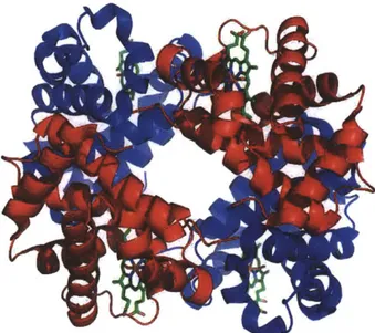
Hemoglobin (Hgb) is the iron-containing oxygen-transport protein that is involved in oxygen transportation. Abnormal Hgb concentration or altered Hgb structure can lead to anemia and other genetic disease. In adult humans, Hgb protein typically contains 4 subunit proteins non-convalently bound. Each subunit consists of a protein chain associated with a non-protein heme group (Figure 5). The heme group consists of an iron ion held in a heterocyclic ring, known as a porphyrin. Under homeostasis, heme is controlled by its insertion into the "heme pockets" of hemoglobins. However, under oxidative stress, some hemoglobin may release their heme prosthetic groups which are
highly cytotoxic. It is believed that the iron ion in the protoporphyrin IX ring undergoes
Fenton chemistry to catalyze in an unfettered manner and produces free radicals. This deleterious effect could indeed play an important role in the pathogenesis malaria 7.
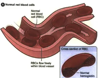
ROCS Now ftly m
within blood vessel
W w W
Figure 1: Normal Red Blood Cells in Blood Flow. Adapted from National Heart Lung and Blood Institute Diseases and Conditions Index 8
Ankyrin 4.1R
Dernatin
-'Acti
S SlasctTropomyosin - protohitament
C
~~~elf
association Tooouisite Tropomodulin
MmtbY O Ind dembft 02MW by Meabt.
br-Figure 2: A schematic model of the red-cell membrane.
This figure was published in: Young NS, Gerson SL, High KA, eds. Clinical hematology. Mohandas
Membrane
Aprin
Glycophorin
Figure 3: Red Cell Spectrin Network. Adapted from Sigma Aldrich
http://www.sigmaaldrich.com/life-science/metabolomics/enzyme-explorer/learning-center/structural-proteins/spectrin.html0
hydrophiic (polar} head
Sof phospholipId hydrophobic(nonpolar)
fatty acid tal of phosphoipid
s
/cholesterol/
inteal (intrinsic) protns peipherat (extrinsic) protein
Inner face @ 2007 Encyclopadia Britannica, Inc. Figure 4: Schematic picture of the lipid bilayer. Adapted from Techno-Science http://conquerordany.blogspot.com/2011/04/cell-structure-and-function.html" outer face
Figure 5: Structure of human hemoglobin. The protein's a and
P
subunits are in red and blue, and the iron-containing heme groups in green. Adapted from PDB IGZX Proteopedia Hemoglobin2Plasmodium falciparum Malaria
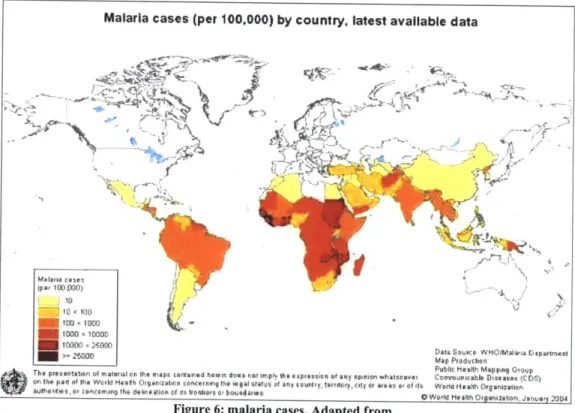
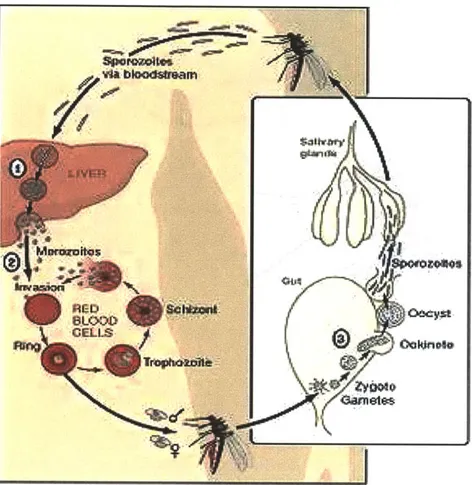
Malaria is the most deadly parasitic disease which affects 200 million people worldwide and accounts for one million deaths annually3 (Figure 6). The most virulent
malarial parasite Plasmodium falciparum can lead to severe complications and has the highest mortality rate4. Cyclic febrile attack is a characteristic clinical feature of P.
faciparum malaria. The intermittent fever paroxysm corresponds to the release of
merozoites (free parasites) following schizont rupture. During intra-erythrocytic development, the invasion of merozoites to other red blood cells (RBCs) reinitiates a 48 hour asexual reproduction cycle5. The infected cells (iRBCs) then undergo notable
morphological and rheological changes from the ring-form (rings) to trophozoite and finally schizont (Figure 7). While rings are only moderately less deformable than uninfected cells (uRBCs), trophozoites and schizonts can be 10 to 50 times stiffer6.
Malaria cases (per 100,000) by country, latest available data
fr10D]
1j
11000 1000 < Dt oreW O~laDaatnn
> Mp Produclierr
Publit Ke slh Mapping Group The presetntation of m atofial con the rinaps coritiand hervim does n ot imply the e aptessie n of any apnien whatsoaver Cornenncabip Dialog assCDS) mn the panl of the Worid Hetith Otraal conceirnng the legal Stattis Lif ar'j courtly, tovrfaci Irty Of afoast o of -is Worid Moadh Organizatioan
P0h7t s
, 2cotn iflgi the delif eioff tit 110"0110 0 bo u 0nd aits 0 W&Mi He Aftin Otgomi23110o , J#,nuse 2004
Figure 6: malaria cases. Adapted from
Figure 7: Plasmodium falciparum malaria parasite life cycle. Adapted from http://www.leidenuniv.nl/en/researcharchive/index.php3-c=217.htm' 8
In the pathophysiology of P. faciparum malaria, the alteration in the dynamic deformability of iRBCs is critical for their clearance in the spleen. RBCs undergo repeated deformations to traverse blood capillaries (3-5um) and splenic cordal meshwork. The narrow splenic inter-endothelial slits (-lum) pose the most stringent mechanical challenge on RBC deformability'9. In vivo, only rings can be seen in the peripheral blood
whereas stiffer iRBCs such as trophozoites and schizonts are typically sequestered during microcirculation. In fact, a substantial proportion of rings would also be retained by human spleen as illustrated by recent studies on isolated-perfused human spleens20. The
study suggests that even a subtle deformability shift in the rings could trigger their clearance in the spleen.
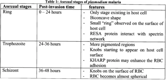
Table 1: Asexual stages of plasmodium malaria
Asexual stages Post-invasion time features
Ring 0 - 24 hours - Only stage existing in host cell
- Biconcave shape
- Small "ring" observed on the surface of host cell
- RESA protein interact with spectrin network
Trophozoite 24-36 hours - More pigmented regions
- Knobs starting to appear on host cell
surface
- KHARP protein may enhance the RBC adhesion
Schizont 36-48 hours - Knobs on the surface of RBC
- RBC becomes almost spherical
To better understand the dynamic deformation of RBCs in vivo, the nature of the RBC deformability should be carefully characterized2 1
. Previously, the deformability of individual RBCs has been measured by a number of ways, including micropipette aspiration , atomic force microscopy , and optical tweezers" . Many of these measurements apply quasi-static loads to attain notable deformation. The deformability of the cells is hence characterized by the shear and bending moduli of the cell membrane. However, when RBCs circulate in the blood capillaries and splenic cordal meshwork, their ability to deform is also a time-dependent response which conventional static measurements fail to correlate directly. The best and most direct way to study cells' dynamic deformation under viscous fluid flow is to develop a microfluidic "deformability cytometer" which simulates in vivo flow condition.
Another important aspect in simulating in vivo RBC deformation is the effect of temperature. Almost every malarial patient displays intermittent fever, which pathophysiologically corresponds to the onset of schizont rupture. Yet, how the febrile conditions stimulate changes in the mechanical properties of both normal and parasitized cells is still unclear. Recent studies shed light on the vital role of temperature in malaria pathophysiology by demonstrating significant stiffening of rings at febrile conditions21.
Fever could then be perceived as part of the body's protective mechanism which facilitates more efficient parasite clearance in human spleen by stiffening the infected cells. The attachment of parasite-derived protein, Ring-infected Erythrocyte Surface Antigen (RESA), is believed to be responsible for the stiffening of infected cells at febrile conditions . Compared to the iRBCs, the temperature effect on co-cultured uninfected
RBCs has often been disregarded, although the main cause of malarial anemia is the massive loss of uninfected RBCs in patient'9. An important question posed is whether
this loss of uninfected RBCs has any biomechanical reason (e.g. stiffening of uninfected RBCs), and if so, how we can understand the mechanism of such mechanical changes in both iRBCs and uninfected RBCs.
Clinical studies revealed that patients treated with anti-malarial drug exhibit a more rapid decline in parasitemia. However the accelerated parasite clearance is delayed or even obscured in splenectomized patients2 5. Therefore, active splenic retention is
believed to be the underlying mechanism facilitating rapid parasite clearance after anti-malarial treatment'9. As spleen could mechanically filter stiffer cells from
microcirculation; the more efficient splenic clearance after drug treatment seems to suggest that some of the tested anti-malarial drugs may be able to modify the mechanical properties of healthy or parasitized cells. However, there is a lack of experimental proof to confirm the exact role of anti-malarial drug on RBC deformability. In this project, we would focus on two most popular anti-malarial drugs that have relatively fewer drug resistance reported: Malarone (Proguanil) and Artemisinin with its derivatives. Two questions are to be addressed in this thesis: 1) whether these two anti-malarial drugs have similar mechanical impact on ring stage infected RBCs; 2) whether the drugs have any adverse effect on healthy (uninfected) RBCs.
To answer these questions, our group previously introduced a microfabricated 'deformability cytometer' which measures dynamic mechanical responses of many
(-1000) RBCs in a population2 6
. Distinct from conventional tools for single cell analysis, our microfluidic device is able to process approximately 10 cells per second. The high throughput enables us to measure a considerable proportion of cells from population, permitting high sensitivity and low sampling error. In this thesis, we employ this tool to investigate the subtle RBC deformability changes in response to temperature changes and drug treatments, both for infected and normal (uninfected) RBCs. Enhanced deformability difference between healthy and infected RBCs at febrile temperature, or after drug treatment (but not in an additive manner) are shown from our experiments. This observation suggests that temperature or drug may act on similar biological pathway to stiffen the iRBCs in order to facilitate splenic clearance. Also from this study, we demonstrated that this simple, high-throughput and inexpensive device could also be potentially used as a malaria diagnostic tool in rural Africa where advanced imaging tools
Chapter 2
Existing Tools for RBC Deformability
Measurement
As the red blood cell deformability plays a key role in blood circulation under both physiological and pathophysiological conditions, searching for the right tool to characterize RBC deformability has been an active research field over a few decades. Several measurement techniques were developed which measure red cell "deformability" either statically or dynamically. In this chapter, more commonly used measurement tools are outlined and discussed. Some of these tools are able to produce single cell analysis with low throughput whereas others are bulk measurements with high throughput but fail to reflect individual trait.
RBC Deformability Measurement by Optical Tweezers (OT)
Optical tweezers (OT) generate force via a highly focused laser beam which could trap micron-sized dielectric particles and manipulate sub-nanometer displacements. The dielectric particle that is trapped by the laser beam experiences an attractive force towards the laser beam focus and the restoring force is linear with respect to the displacement between the particle and the focus. In this way, the force acting on the particle can be modeled as a simple spring which follows Hooke's law:
F= -kU
where the constant k is the trap stiffness depending on the OT design and the particle size. size. Experimentally, k is usually in the order of 50pN/pm, which corresponds to a resolution of 0.5pN in force measurement.
Though the OT technique has been used to characterize micron particles for almost 20 years, its application to measure cell deformability was fairly recent. In the paper published by Mills et a24
in 2007, OT was used to measure single RBC force-displacement response, from which membrane shear modulus was derived based on computational modelings2 7
. The schematic set up for a typical OT experiment is sketched in Figure 8.
Figure 8A: picture of optical tweezers stretching a red cell. Adapted from http://www.bioeng.nus.edu.sg/nanolab/galleryresearch.htm 21; B: schematic diagrapm of optical tweezers. Adapted from Wikipedia: http://en.wikipedia.org/wiki/Opticaltweezers#Physics of optical-tweezers29
One distinct advantage of optical tweezers measuring RBC membrane stiffness is its high precision and specificity. By stretching the beads that are strategically attached to cell surface, optical tweezers can stretch RBCs in one or more directions0. Several limitations are also associated to this method. For example, typically the maximum optical force is limited to several hundred pico-Newton, which could be insufficient to induce large deformation in the cells commonly encountered in vivo. Moreover, care needs to be taken to avoid overheating the cells by laser light during operation.
RBC Deformability Measurement by Micropipette Aspiration (MP)
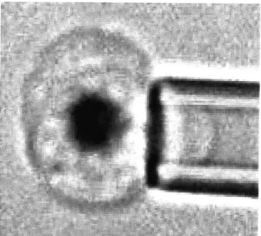
Micropipette aspiration is one of the most classic methods that measure RBC membrane stiffness. During a typical MP experiment, RBCs are usually diluted by 1000 times from whole blood and suspended in a saline solution with comparable osmolarity.A small micropipette with inner diameter between 1 to 3 pm is carefully inserted in the
RBC suspended solution and the target RBC is aspired into the mouth of the pipette under a known suction pressure AP.
Compared to OT, MP is a more versatile method for measuring the mechanical properties of living cells as the suction pressure could ranges from 0. 1pN/pm2 to almost atmospheric. That means this technique can probe soft, fluid-like cells, such as RBCs, as well as very rigid cells. On the other hand, the high flexibility of red cells and their biconcave shape often cause "buckling" problem while performing MP experiment. That means, even at a low suction pressure, it is very easy for the entire cell to be sucked into the micropipette. Additionally, as a tool aimed to measure RBC membrane property, a considerable amount of hemoglobin would also be sucked into the pipette, depending on the size of pipette. Though many complex models try to accommodate these variations in pipette inner diameter, it remains a question whether measured membrane stiffness can be consistent with pipettes of different sizes. Figure 9 illustrates a micropipette aspirating a malaria infected red blood cell.
- s---.
Figure 9: picture of micropipette aspiration of a malaria infected red blood cell. Adapted from http://www.bioeng.nus.edu.sg/nanolab/galleryresearch.html2 8
RBC Deformability Measurement by Ektacytometry (LORCA)
Ektacytometer is another method to measure cell deformation in Couette flow and was first developed by Bessis and Mohandas in 1975 3. The basic idea is to suspend cells in the narrow gap between two concentric cylinders and apply various shears on the cells
by rotating the outer cylinder at different speed. As a laser beam is passed through the
cell solution, the cells scatter the light to form a diffraction pattern which is circular at low shear force and becomes ellipsoidal at higher shear. The ratio of the major and minor axis of the ellipsoid indicates how deformable the cells are under a given shear force, and the deformability index (DI) was properly defined as follows:
A - B
DI =
A + B
where A is the major axis of the ellipsoid and B is the minor axis of the ellipsoid.
Distinct from MP and OT measurement, Laser-assisted Optical Rotational Cell Analyzer (LORCA) ektacytometry measures the average deformability of cell populations. Therefore, as a bulk measurement tool, ektacytometry has a much higher throughput and is capable of producing population-wide trait but it fails to reflect the deformability of individual cells in a given population. This becomes an important drawback when the target cells to be studied form a minority population in a given sample, such as in malaria culture.
RBC Deformability Measurement by MicroFluidic devices (MF)
Most of the above mentioned measurement methods may be a good indicator of how flexible a red blood cell is, but it is still difficult to correlate the calculated DI or shear modulus directly with the in vivo blood flow. Hence, several microfuidic devices were designed to mimic the RBC deformation in small capillaries.
The first realistic in vitro microfluidic realization of in vivo RBC deformation was presented by Brody et al in 1995 3 (Figure 10). Though Brody et al. did not attempt to measure malaria infected red blood cells, the pillar array structure proposed in that work is the basis of the current malaria-deformability cytometry used in this project. Comparing the single capillary system by Shelby et al. Brody's pillar array structure has several distinct advantages: 1) the pillar array structure minimizes cell-cell interaction, achieving single cell accuracy measurement; 2) clogging is less likely a problem as there are several channels in parallel and the constriction is fairly short; 3) the pillar array
structure could be a better depiction of in vivo RBC deformation in microcirculation as the cells have to undergo "repetitive" "severe" deformations while each deformation time is not necessarily long.
Figure 10: Video frames of healthy RBCs passing through a 4pm constrictions. Picture adapted from Brody et al. Biophy J 1995"
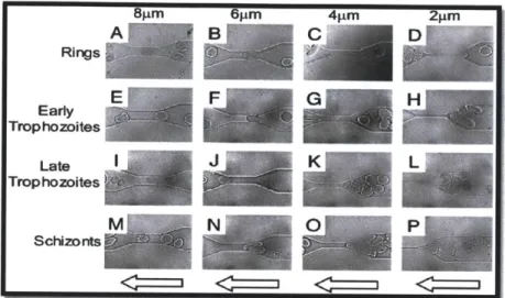
In 2003, Shelby et al developed a microfluidic model for single-cell capillary obstruction34. Red blood cells at different stages of malaria infection were passed through the microchannel of 2, 4, 6, and 8pm size. It was found that while Ring-stage infected erythrocytes were able to pass through all constricted channels, cells with later infection stage exhibited decreased ability to squeeze through the microchannels and Schizont stage infected erythrocytes were blocked even at 6 pm channel. At 8 pm channel, the channel size is already comparable to average RBC size and little deformation is needed to pass through the channel. (Figure 11). Though this experiment did not derive cell deformability in a very rigorous manner, it demonstrated several in vivo concepts such as "pitting" and "capillary blockage". However, this technique would probably suffer from serious clogging issue and no quantitative data on cell deformability can be obtained directly. Cell-cell interaction may play a significant role and it is difficult to interpret single cell deformability based on this channel design.
6~±m 4iim Rings Early Trophozoites Late Trophozoites Schizonts
Figure 11: Video images of four stages of malaria-infected RBCs (early ring stage, early trophozoite, late trophozoite, and schizont) passing through microchannel with different size (2, 4, 6, and 8
sm).
Figure adapted from Shelby et al PNAS 2003 3
Based on the similar design principles first proposed by Brody et al, Bow et al. optimized the pillar array structure for malaria diagnostic and related deformability studies. The "microfluidic cytometry" could detect the minute deformability shift from healthy to Ring stage malaria infected RBCs26
. This optimized device is used for this project and the Figure 12 depicts how single cells could pass through repeated 3 Prn constrictions
Figure 12: Video frames of healthy RBCs passing through a 3sam constrictions.
Compare the aforementioned measurement tools which are commonly used to probe RBC deformability, each has its own advantages and limitations in terms of throughput, precision, specificity, and ease of operation. However, distinct from many other material stability tests, the need for RBC deformation study is built on its physiological importance. Therefore, when performing these deformation tests, it is important to recreate a physiologically relevant environment, such as shear rate, pressure gradient etc. At very low shear rate, the pseudo-static measurement could overlook the inherent viscoelasticity of cell membrane and leads to misrepresentation. On the other extreme when very high shear rate is applied, the red blood cells could be permanently damaged. As RBCs pass through the splenic slit in vivo, shear rate is estimated to be -10 sec' and this number could be important potentially in interpreting the difference in experimental results by different measurement tools3 5. The table below summarizes
several key features of above mentioned measurement tools.
Table 2: Key features of different tools measuring RBC deformability
Tool Dynamic Single cell / Throughput Shear Rate
/Static Bulk (sec-)
OT Static Single cell <1 cell / min 7-50
(Mills et al24
)
MP Static/dynami Single cell <1 cell / min <1
(Nash et a122) C
LORCA Static Bulk ~ 1000 cell/min 50036 (Bessis et al 3 2 )
MF Dynamic Single cell ~ 100 cell/min 10-100
Chapter 3
Materials and Methods
Device Fabrication
Layout program CleWin3.0 was used to design our microfluidic device, which consists of a 500 x 500ptm2
inlet reservoir, a 500 x 500 pLm2
outlet reservoir, and parallel capillary channels with triangular pillar arrays. Three different pore-size of 2.5, 3.0 and
4.0 pm were designed for the capillary channels to test optimum deformation condition for the experiment (Figure 13). A silicon mold of the device was made using standard silicon processing techniques. The photolithography step was done using a 5X reduction step-and-repeat projection stepper (Nikon NSR2005i9, Nikon Prevision) and reactive-ion etching (RIE) techniques were used to give the mold a final depth of 4.2pjm. Final device was then casted from the silicon mold using polydimethylsiloxane (PDMS) and was sealed by a glass slide using oxygen plasma.
a. Silicon Mold Fabrication
Silicon molds were fabricated at the Microsystems Technology Laboratories (MTL) with the help of the staff members. The table below describes the process flow to fabricate these glass devices.
Starti g material: 6" p test wafers, GREEN process in TRL/ICL
Step Action Machine Parameters
Cleaning
1_1 Piranha cleaning Acid-hood 5 min; hydrogen peroxide:sulfuric acid = 1:3
Pattern
Alignment Marks
2_1 Resist coating Coater 6 ICL, ipm thickness, "T1HMDS", 4600rpm
2 2 Exposure Stepper ICL, 170ms
2 3 Development Coater 6 ICL, "PUDDLE2" Etch
Alignment Marks
3_1 Reactive Ion Etch AME5000 ICL,
"ISABELLA LTO" :descum:20s, oxide
"pan Si etch" etch rate: 4-4.5nm/s. etch
35s
3 2 Resist removal Asher 3 min
Pattern
Microchannels
4_1 Resist coating Coater 6 ICL, 1p m thickness, "T1IHMDS",
4600rpm
4 2 Exposure Stepper ICL, 170ms
4 3 Development Coater 6 ICL, "PUDDLE2"
Etch
Microchannels
5_1 Reactive Ion Etch STS2 TRL
"JBETCH" etch rate 2-2.5pm/min. Etch
2 mi
5 2 Resist removal Asher 3 min
Measurement
6_1 Measure final depth P1O Measure 4 corners + centre point and take average.
b. PDMS-Glass Device Fabrication (Soft Lithography)
Polydimethylsiloxane (PDMS) is prepared by mixing 10:1 w/w base to curing
agent and degassing under vacuum for at least 1 hour. The degassed mixture is poured
onto the silicon master and postbake at 65'C over overnight. Cured PDMS is peeled from the master following punching the 1.5mm holes at the reservoirs. Tubings will be connected to the reservoir during subsequent experiment.
PDMS-glass bonding is made by plasma treatment of both PDMS and glass slide for 1 minute. A strong bond forms instantaneously. Annealing on a hotplate at 95'C
for >1 hour significantly increase the bond strength. Detailed steps are outlined in the table below:
1) Pre-clean the PDMS with tapes and preclean the glass slide with ethanol followed by water rinsing.
2) Place the PDMS and glass in the vacuum chamber and turn on the pump.
3) Make sure the plasma is at the "off' position. Make sure the valve is fully closed.
4) Wait 60 second.
5) Turn the plasma on to HIGH. Fine tune the needle value counterclockwise until the a bright purple color is observed.
6) Wait for 60 second.
7) Turn off the plasma and vacuum pump. Release the valve slowly. 8) Remove the PDMS and glass from the chamber and press them together.
Parasite Culture
P. falciparum 3D7A parasites (from Malaria Research and Reference Reagent
Source, American Type Culture Collection) were cultured in leukocyte-free human RBCs (Research Blood Components, Brighton MA) in RPMI 1640 complete medium as described3 7. Parasites cultures were synchronized by sorbitol lysis3 8 two hours after
merozoite invasion.
Solution Preparation
1x Phosphate buffered saline (PBS) was mixed with 1% w/v Bovine Serum Albumin (BSA) (Sigma-Aldrich, St. Louis, MO) as a stock solution and was freshly made on every experimental day. For experiments tracking fluid flow velocity, 200 nm FluoSpheres europium luminescent microspheres (Molecular Probes, Eugene, OR) were used at a final concentration of 5.0 x 10⁻⁴ percent solids. For experiments involving only healthy RBCs, fresh whole blood (Research Blood Components, Brighton, MA) was used at 100 times dilution (i.e. 1Il whole blood with 99 pl stock solution). For experiments involving parasitized cells, Iml of cultured cells were spun down at 1,000rpm for 5 minute; 1 pl of the pellet was then aliquot to 200 pl stock solution.
1 pl of 50 pg/ml Cell Tracker Orange (Invitrogen, Carlsbad, CA), which stains the membrane of the cell, was added to aforementioned 100 pl healthy RBC solution for better imaging, 20 minutes before the experiment. To ensure no adverse effect on the cell deformability was induced by the dye, a control experiment of same RBC solution without staining was performed. No statistically significant difference in deformability was found between the sample with and without staining.
10 l of 1 x 10⁻⁶ M thiazole orange dye (Invitrogen, Carlsbad, CA), which stains
the RNA of the cell, was added to the aforementioned 200 pl iRBC solution 20 minutes before the experiment. The infected cells would appear bright under the green fluorescence filter set (488nm excitation) whereas the uninfected cells were seen as dark shadows.
Experimental Protocol
To accurately control the ambient temperature, the microscope surface was replaced by a heating chamber (Olympus), which was preheated to a desired temperature range (i.e. 30-400C) for 30 minutes before the beginning of every experiment. Meanwhile, the PBS-BSA stock solution was injected into the device to coat the PDMS walls to
prevent adhesion. This filling step need not be done inside the heating chamber, but the PBS-BSA filled device needed to be placed into the heating chamber at least 5 minutes before loading 10 pL of diluted blood sample in order to guarantee the temperature stability. During temperature calibration phase, a thermocouple thermometer was used to probe the exact temperature inside the heating chamber. When the temperature needed to be adjusted to a different value, at least 5 minutes of waiting time was required to ensure a new stable ambient temperature.
To apply a constant pressure gradient across the device, the pressure difference between inlet and outlet reservoir was generated hydrostatically by fixing the difference between inlet and outlet water column height. To ensure the pressure difference is constant throughout the experiment, a large volume (60ml) syringe was selected to be connected to the inlet reservoir, so that the water column height would not vary
significantly within several hours.
A
Heating chamberOutlet
Inleth
- .A..X.A.Tubing
PDMS dev lce \Glass slide Tbn
101pm 10 pm
FHold F
3 pim ]"m
3 pmn
Figure 13: Experimental schematics. A heating chamber (Olympus) was mounted to the inverted microscope stage. Four heaters for the inner water bath, microscope stage, chamber top, and lens were designed to accurately control the ambient temperature inside the chamber. The PDMS-glass bonded device consists of the inlet and outlet reservoirs and main channels with triangular pillar arrays. It was placed inside the heating chamber with only the inlet reservoir connecting to an external syringe via a 20cm-tubing. The reservoirs were 500x500 Pm2 squares with 20
sm-interspacing cylindrical pillar arrays; these pillar arrays could pre-filter white blood cells from whole blood, allowing only RBCs to pass through the main channels. Each of the parallel channels was 10 pillars wide and 200 pillars long. Along the flow direction, the inter-pillar spacing was 10 pm. This spacing would allow deformed cells to recover and ready for subsequent deformations. Perpendicular to the flow direction, the pore size varied from 2.5 to 4 pm for different channels. Cells would experience different level of deformation when passing through these pores.
To capture the movement of RBCs inside the microchannels, a CCD camera (Hamamatsu Photonics, C4742-80-12AG, Japan) was connected to the inverted fluorescent microscope (Olympus IX71, Center Valley, PA). Images were automatically acquired by IPLab (Scanalytics, Rockville, MD) at 100ms time interval and the post-imaging analysis was done using ImageJ (NIH, http://rsb.info.nih.gov/ij/). The velocity of individual RBCs was defined as the distance the cells moved divide by the time in seconds.
The device constitutes a series of equally spaced triangular pillar arrays with pore size ranging from 2.5 to 4um (Figure 13 of Chapter 1 illustrates the case of 3um pore
size). Compared to the diameter of an average RBC (~8um), the considerably smaller pore sizes are designed to impose similar mechanical constrains on the cells as if they are passing through blood capillaries and splenic meshwork. Driven by constant pressure gradient in the sub-Pascal-per-micrometer range, RBCs are able to deform substantially at each constriction and traverse along the channel26. The dynamic deformability of
RBCs is then characterized by their velocity: the ability to deform repeatedly in order to pass through multiple constrictions in series. Stiffer cells with longer entrance time at each constriction would assume lower velocity and vice versa.
Chapter 4
Effect of Febrile Temperature on the
Deformability of Malaria-infected Erythrocytes
In this chapter, we study the temperature dependent deformability changes of iRBCs and uninfected RBCs (uRBCs). One important note: Once the blood is extracted from the body and cultured / maintained ex vivo for an extended period of time, RBCs' chemical and mechanical properties shift significantly over time (storage lesion)39'4 0
Therefore, as a proper control for the iRBCs (which underwent extended parasite culture processes), 'uRBCs' in this thesis designates uninfected RBCs contained in the malaria culture. On the other hand, healthy RBCs (hRBCs) in this thesis mean freshly drawn (from less than a day old sample in vacutainer) RBCs.
Figure 14 presents microscopic screenshots illustrating both iRBCs and co-cultured uRBCs moving in the microchannel at different temperature conditions. The uninfected cells appear as dark shadows indicated by blue arrows, and the infected cells with thiazole orange (TO) staining appear as bright dots indicated by white arrows. The velocity of individual cells can then be derived by recording the time period for each cell passing through 10 constrictions in series (i.e. equivalent to 200pjm distance travelled). In this work, cell velocity is used to characterize the dynamic deformability of individual RBCs. Larger cell velocity value corresponds to larger RBC deformability). The temperature-dependent modifications on the dynamic deformability of both uRBCs and iRBCs are investigated. The clinical relevance of these results is discussed on the basis that human spleen mechanically filters stiffer RBCs19.
Temperature-dependent iRBC deformability
Figure 15A demonstrates the temperature-dependent modification on iRBC deformability. The velocity of infected cells exhibited a significant raise from 300C to 370C followed by
a notable drop at 400C. The peak at 370C marked the optimum temperature for maximum
iRBC deformability.
To investigate the deformability increase from 300C to 370C, several factors were
buffer solution viscosity as well as possible confounding effects caused by the modification of cytoskeletal structure and membrane proteins. It was noted that the PBS buffer viscosity decreases by 33% from 19 C to 37 C and the blood viscosity decreases
by 2% for every degree C temperature increment (i.e. -31% decrease from 19 to 37'C).
At a given pressure gradient, elevated temperature would therefore increase the bulk fluid flow speed, leading to a projected increase in cell velocity measurement. For better comparison, normalized cell deformability was measured by performing the experiment at equalized bulk fluid velocity over all temperatures. Assuming the combined viscosity shift in iRBC and PBS buffer solution is linear, we computed the pressure gradient to be applied at each temperature for constant fluid flow. This calibration was also experimentally verified by measuring fluid velocity via 200nm non-deformable polystyrene beads (Figure 15B). With constant beads speed of 226 ptm/s, the normalized cell deformability (Figure 15C) inside 4pm-channel was found to be fairly constant from
300C to 37'C. This result is consistent with similar measurements using other techniques,
such as micropipette aspiration and optical tweezers24. Compare Figure 15A and C, we
concluded that observed -50% increase in cell deformability from room to body temperature was predominantly caused by temperature related viscosity change in both iRBCs and PBS buffer solution. Other factors such as the cytoskeletal structural modification, membrane protein alternation played a minor role.
On the other hand, the significant drop in iRBC velocity between 37 C and 40 C was preserved at constant local fluid velocity (Figure 15C). This remarkable stiffening effect at febrile condition agrees with recent deformability measurement by optical tweezers 24. The attachment of parasite-derived protein, Ring-infected Erythrocyte
Surface Antigen (RESA), is believed to be responsible for the notable drop in cell deformability at febrile temperatures24. While RESA would be necessary for the
parasitized cells to survive heat-induced damages, the protein related stiffening also facilitates more efficient splenic clearance. Detailed discussion on RESA effect is presented in a separate section.
Os
1s
2s infected cel
<-> : distance moved by a uninfected cell over one second time period
: distance moved by an Infected cell over one second time period
Figure 14: Experimental images of iRBCs (white arrows) and uRBCs (blue arrows) in 3pm channels. Driven by a constant pressure gradient of 0.36 Pa/sm, the cell motion was tracked at three different temperatures: 30 oC, 37 oC, and 40 oC. While a uninfected cell appeared as a dark shadow, the GFP-transfected parasite inside an infected cell would be seen as a small fluorescent dot. The red and black arrows indicated the distance moved by iRBCs and uRBCs respectively. With one second time interval, the lengths of the arrows revealed the velocity of cells. The images on the top right corner illustrated how a cell would pass through the pores.
uW T
50-~CED
00 30 3500 30 0 0 20 -10P > 200 CO 00 100 80020
4
150
EI
0 30 0 3C 3CFigre 5 ACostat PesureGraieT elvlc semperature pltfrinetdcll0asn
3 00- 80
0-0
though 3pim channels. B 200nm microspheres were used to track fluid velocity at different temperature and by adjusting the pressure gradient, equalized fluid velocity is achieved. C Constant
Fluid Velocity Cell velocity vs. temperature plot for infected cells passing through 4 pim channels.
Data was obtained at a constant local fluid velocity of 230 pim/s as calibrated by beads. Translated to pressure gradients, the gradient applied was 0.36 Pa/sm at 30 *C, 0.312 Pa/sm at 37 "C, and 0.288 Pa/sm at 40 "C
Temperature-dependent uRBC deformability
Whereas iRBC deformability is commonly accepted as an important factor in the pathogenesis of p. falciparum malaria, the temperature-dependent modification on co-cultured uRBC deformability has often been overlooked. With the understanding that the human spleen is capable of sequestering stiffer cell during microcirculation , deformability of uRBCs, as well as the deformability separation between iRBCs and co-cultured uRBCs, are indeed important in order to better understand the mechanism of splenic retention.
Figure 16 demonstrates the temperature-dependent modification on uRBC deformability. Similar to that of iRBCs, the uRBC velocity increased significantly from
30*C to 37"C. From 37 "C to 40 "C, instead of a 40% drop displayed by iRBCs, the
decrease in uRBC deformability was subtle but still statistically significant (p <0.01). Following the same viscosity argument as presented earlier; normalized uRBC velocity was also measured at equalized bulk fluid velocity inside 4pm-channel. The result was similar to that of normalized iRBC deformability: no significant change in the normalized uRBC deformability was observed from 30"C to 37'C, and the velocity drop from 37"C to 404C was preserved.
The result again confirms the major role of viscosity in influencing the RBC deformability from 30'C to 370
C. Compared to the 50% drop in the average deformability
of iRBCs from body to febrile temperatures, the temperature-dependent effect on uRBC deformability is subtle but still statistically meaningful (P<0.01). This result indirectly confirmed that the significant drop in iRBC velocity at febrile condition is mainly due the effect of the parasite-derived protein RESA, and is not something inherent in non-parasitized cells. This result is also consistent with similar measurement using other techniques. 100 120 80
A
100.B
S80-E60A
60-400 0 40 Z 20- 0 ,20 - 2 30 37 4b 030 0C 37 0C 40 OC Temperature (0)0Figure 16 A Constant Pressure Gradient: Cell velocity vs. temperature plot for uninfected cells passing though 3pm channels. B Constant Fluid Velocity Cell velocity vs. temperature plot for uninfected cells passing through 4 pm channels.
Temperature-dependent healthy RBC deformability
The dynamic deformability of healthy RBCs (hRBCs) from room temperature (25
C) to febrile temperature (400
C) was measured, in order to be compared with the result for uRBCs (Figure 17). Similarly, under constant pressure-driven scheme, the hRBCs became more deformable from room to body temperature, and the deformability peaked at 370C. The measured temperature-dependent RBC deformability was independent of the thermal history of the sample, which was confirmed by changing the order in which measurements are made at different temperature values. In addition, temperature-induced deformability changes were reversible under the conditions tested (from 25 to 40 "C), meaning that returning a sample to its original temperature restored the deformability value measured at that temperature.
120-100* 0 80-20- 0 40-66 2 27.5 30.0 32.5 35.0 37.5 Temperature (0C) 40.0 "U 4-pim channel Ave fluid flow =226 pm/s
100-
80- 60-
40-
20-0.36OPa/pm 0.312Palpm 0.288Pa/pm
0
30 3mr7 40
TemfPerature
(
0C)
Figure 17 A Constant Pressure Gradient: Cell velocity vs. temperature plot for healthy cells passing though 3pm channels. B Constant Fluid Velocity Cell velocity vs. temperature plot for healthy cells passing through 4 pm channels. Data was obtained at a constant local fluid velocity of 230 pm/s as calibrated by beads. Translated to pressure gradients, the gradient applied was 0.36 Pa/pm at 30 "C, 0.312 Pa/sm at 37 *C, and 0.288 Pa/pm at 40 *C
Febrile condition enhances the separation resolution between iRBCs
and uRBCs
The deformability separation resolution between normal and parasitized cells is previously noted as the key parameter for efficient splenic clearance of infected RBCs. The temperature-dependent cell deformabilities for both iRBCs and co-cultured uRBCs were simultaneously measured at 30'C, 37*C, and 40'C (Figure 18). The deformability separation resolution Rs between normal and infected cells was analyzed using the formula below, where X1, X2 and o1, o-2 denote the mean and standard deviation of
normal and infected cell mobilities. A higher Rs value implies better separation.
SX2
- X1
2(o1 +
q2)While at all tested temperatures the infected RBCs displayed statistically significant stiffening compared to uninfected cells (p < 0.001), the deformability
separation resolution between uRBCs and iRBCs enhanced substantially with increasing temperature (Table 3). At 30'C, the separation resolution was only 0.28; the value increased to 0.46 at body temperature, and to 0.94 at febrile condition. Furthermore, at 40 'C, the average iRBC velocity was 3.02a (i: standard deviation of uRBC velocity distribution) away from the average uRBC velocity. This result implied a more sensitive and specific deformability differentiation between normal and parasitized RBCs at febrile condition The result is consistent with similar measurement using other techniques2,32 and reconciles with our hypothesis that febrile condition facilitates more efficient splenic retention. The velocity difference between normal and parasitized cells can be explained
by the RESA effect.
Table 3: temperature dependent resolution
Temperature ("C) Resolution (R,)
30 0.28
37 0.46
120 30 0C 100- 80- 60- 40- 20-01 Uninfected RBCs Infected RBCs 120 37 0C 100- 80-60 40* 20-Uninfected RBCs Infected RBCs 120 40 0C 100- 80- 6040
20
- 0-Uninfected RBCs Infected RBCsFigure 18 Cell velocity of both infected and co-cultured uninfected cells passing though 3sm channels at different temperatures. Approximately 600 RBCs were tracked at a pressure gradient of
0.36 Pa/
sm.
The velocity of uninfected RBCs was found to be statistically higher than parasitized cells at all temperatures, and the cell velocity difference between uRBCs and iRBCs is maximised at febrile condition (40 *C).Spleen as a mechanical filter
Spleen is believed to work as a mechnical filter which removes stiffer cells from circulation. The splenic retention model has been hypothesized by Buffet in his recent review paper 22. To quantitative illustrate splenic clearance, Figure 18 is replotted into Figure 19 at body (370C) and febrile (40C) temperatures. A hypothetical 'filtration
threshold' value can be assumed such that all the RBCs with velocity below that value will be retained (filtered) at spleen. This threshold cannot be too high, because it will lead to the loss of too many too many normal RBCs. If this splenic filtration threshold is set too low, then many of iRBCs will pass and survive the splenic clearance mechanism. Presumably, the splenic filtration threshold in vivo will be determined (by evolution) based on this tradeoff.
Our result shows that splenic mechanical filtration process can be more 'specific' at fever temperature, due to the greater separation between iRBCs and uRBCs. For example, if the 'filtration threshold' is set to be 34ptm/s, which is 2a away from the average uRBC velocity measured at 370C. At 370C, only 12 out of 25 iRBCs traverse
below the threshold velocity, indicating the efficiency of splenic filtration of iRBC at
370C is only 48%; however at 40C, 24 out of 25 iRBCs has a velocity value lower than 34um/s, suggesting 96% of iRBCs will be cleared by spleen at febrile condition.
The significant improvement in splenic filtration efficiency (from 48% to 96%) suggest a potentially important role of fever in the pathophysiology offalciparum malaria, and in splenic clearance in general. On the other hand, as we compare the splenic retention of uninfected RBCs at body and febrile temperatures, we found that while only
1.5% of uRBCs would be removed from blood stream at 37 0C, 9% of uRBCs are below
the threshold velocity at 400C. The exact mechanism why febrile condition would mildly
reduce uRBC deformability is still unclear, but the significantly increased amount of uRBC removal might be the related to the pathology of malarial anemia.
uninfected RBCs 37 OC EM52 Infected RBCs 3 10-:0 0 _ 0 10 20 30 40 50 60 70 80 91
RBC Velocity (gm/s) RBC Velocity (tim/s)
100-
80-10 20 30 40 50 60 70 80
RBC Velocity (gm/s)
Figure 19: A Cell velocity histogram with normal fit for both infected (red curve) and co-cultured uninfected (black curve) RBCs at 37 "C. For the uninfected RBCs, their mean velocity and standard deviation were (52.02, 9.41). For the infected RBCs, their mean velocity and standard deviation were (33.04, 11.61). B Cell velocity histogram with normal fit for both infected (red curve) and co-cultured uninfected (black curve) RBCs at 40 *C. For the uninfected RBCs, their mean velocity and standard deviation were (44.82, 8.19). For the infected RBCs, their mean velocity and standard deviation were (21.29, 5.87). The normal fit graphs at 37 "C and 40 "C were overlaid in C. 10 I 'I I / I I I I /
Chapter 5
Effect of Anti-malaria Drugs on the
Deformability of Malaria-infected Erythrocytes
As one of the most deadly infectious disease, malaria is responsible for over 200 million clinical cases in 200941. Though a number of anti-malarial drugs were widely used clinically, antimalarial drug resistance has increasingly emerged to be a major public health problem which hinders the control of malaria. Figure 20 illustrates the dire situation of chloroquine resistance in malaria endemic areas. While the urgent need for a new malaria drug is clearly justified, it is also important to understand how these malaria drugs are working. In fact, the exact drug mechanism of artemisinin - one of the most commonly used malaria drug currently - is still an active area of research. In this report, several existing malaria drugs including artemisinin and malarone are studied to examine their effect on the deformability of both healthy and malaria-infected red blood cells. It is hoped that by studying these existing drug, we could gain a better understanding of the mechanisms of drug action in vivo.
Figure 20: Chloroquine resistant in Malaria-endemic countries in the Eastern Hemisphere. Adapted from http://wwwne.cdc.gov/travel/yellowbook/2010/chapter-2/malaria.htm
Artemisinin and its derivatives
Figure 21: Chemical structure of artemisinin and its derivatives. Adapted from Wikipedia: http://upload.wikimedia.org/wikipedia/commons/a/a5/Artemisininskeletal.svg 2 9
Artemisinin, also known as qinghaosu, was extracted from a traditional Chinese herbal Qinghao for the treatment of fever 2000 years ago . Only in the late 1960s, Chinese scientists discovered that it is also a natural candidate for malaria treatment: it could clear malaria parasite from the body faster than any other drug known. The peak plasma concentration is reached within 1-2 hours and the half-life of the drug is only 2-3 hours. The rapid absorption as well as fast parasite elimination makes Artemisinin an efficient curative drug for the treatment of Plasmodium malaria.
Multidrug resistance has always been a serious problem in the treatment of malaria. As a relatively new anti-malaria drug, artemisinin and its derivatives made a remarkable impact on the reduction of P. falciparum malaria. However in recent years, an increasing number of clinical cases report treatment failures with artemisinin monotherapy.
a) Current Hypotheses on the Mechanisms of Artemisinin Drug Action
Though the exact mechanism of artemisinin drug action is still unclear, clinical studies have shown two important characteristics of artemisinin derivatives: 1) The endo-peroxide bridge is believed to be mainly responsible for the drug action. Deoxyartemisinin, an artemisinin analog lacking endoperoxide bridge, is clinically