Publisher’s version / Version de l'éditeur:
Seed Dormancy: Methods and Protocols, pp. 113-134, 2011-10-02
READ THESE TERMS AND CONDITIONS CAREFULLY BEFORE USING THIS WEBSITE. https://nrc-publications.canada.ca/eng/copyright
Vous avez des questions? Nous pouvons vous aider. Pour communiquer directement avec un auteur, consultez la première page de la revue dans laquelle son article a été publié afin de trouver ses coordonnées. Si vous n’arrivez pas à les repérer, communiquez avec nous à PublicationsArchive-ArchivesPublications@nrc-cnrc.gc.ca.
Questions? Contact the NRC Publications Archive team at
PublicationsArchive-ArchivesPublications@nrc-cnrc.gc.ca. If you wish to email the authors directly, please see the first page of the publication for their contact information.
NRC Publications Archive
Archives des publications du CNRC
This publication could be one of several versions: author’s original, accepted manuscript or the publisher’s version. / La version de cette publication peut être l’une des suivantes : la version prépublication de l’auteur, la version acceptée du manuscrit ou la version de l’éditeur.
For the publisher’s version, please access the DOI link below./ Pour consulter la version de l’éditeur, utilisez le lien DOI ci-dessous.
https://doi.org/10.1007/978-1-61779-231-1_8
Access and use of this website and the material on it are subject to the Terms and Conditions set forth at
In vitro assay for ABA 8'-hydroxylase: implications for improved assays for cytochrome P450 enzymes
Krochko, Joan E.; Cutler, Adrian J.
https://publications-cnrc.canada.ca/fra/droits
L’accès à ce site Web et l’utilisation de son contenu sont assujettis aux conditions présentées dans le site LISEZ CES CONDITIONS ATTENTIVEMENT AVANT D’UTILISER CE SITE WEB.
NRC Publications Record / Notice d'Archives des publications de CNRC:
https://nrc-publications.canada.ca/eng/view/object/?id=fac896bf-c28c-4104-8b1e-365818d74faf https://publications-cnrc.canada.ca/fra/voir/objet/?id=fac896bf-c28c-4104-8b1e-365818d74faf
In Vitro
Assay for ABA 8'-Hydroxylase: Implications for Improved
Assays for Cytochrome P450 Enzymes
Joan E. Krochko and Adrian J. Cutler
Plant Biotechnology Institute, National Research Council of Canada Saskatoon, Saskatchewan, Canada
Abstract
In vitro assays for cytochrome P450 enzymes developed from plant-derived microsomal extracts have not been used extensively for the characterization and quantification of enzyme activities in plant tissues. We describe here an in vitro assay for ABA 8'-hydroxylase that was developed using microsomes extracted from (+)-ABA induced corn suspension cultures. This assay may be useful for further characterization and monitoring of ABA 8'-hydroxylase activities in germinating seeds, seedlings and other tissues. Additionally, the optimization protocols provided here may be adapted towards improving in vitro enzyme assays for other cytochrome P450 enzymes expressed in plants.
Key Words: Cytochrome P450; in vitro assay; microsomes; (+)-ABA 8'-hydroxylase; abscisic acid; phaseic acid; germination; dormancy.
1. Introduction
Cytochrome P450 enzymes are responsible for a wide array of biological oxidations in plants, including the production of secondary metabolites, primary structural compounds and several plant hormones (e.g. auxins, gibberellins, brassinosteroids, abscisic acid) (1-3). In particular, a large number of distinct cytochrome P450 enzymes are involved in upstream pathways for the synthesis of phytohormone precursors, late conversions of these to more active forms, and subsequent catabolism of phytohormones to less active (or inactive) metabolites
(1-3).
Four members of the CYP707A subfamily of cytochrome P450 enzymes in Arabidopsis (i.e. CYP707A1, CYP707A2, CYP707A3, CYP707A4) are responsible for the oxidative inactivation of abscisic acid (ABA) to its biologically less active metabolite, 8'-hydroxyABA (4-7). This latter compound cyclizes spontaneously to the stable and (almost) inactive metabolite, phaseic acid (PA), and as a consequence the effective levels of the active hormone are dramatically reduced by these tandem reactions. The CYP707A subfamily genes are expressed at high levels in imbibing and germinating seeds, young seedlings, leaves/stomata, and in response to stress or leaf/stem submergence (6-10). Most importantly, the enzyme activity associated with this subfamily (e.g. CYP707A2) has been implicated in the catabolism/inactivation of ABA in seeds to promote seed germination and overcome dormancy (11-16).
In many cases it has been difficult to measure cytochrome P450 activities directly in extracts from plant tissues. Characteristically, these enzymes are in low abundance and enzyme activity measurements are inferred by indirect means from gene expression measurements based on northern blots, PCR-based assays, EST frequencies or microarray data. A robust in vitro assay developed from plant microsomal extracts can provide direct quantitative and qualitative data for a particular enzyme activity across tissues and developmental stages. We present here a detailed
protocol for the development and optimization of the in vitro assay for ABA 8'-hydroxylase (4) and suggest where it might be applied to future studies of germination and dormancy, and as a model for studies of cytochrome P450 enzymes in general. With recent improvements in the sensitivity of detection methods for various metabolites it may now be possible to analyze and measure the in vivo activities of highly expressed cytochrome P450s through tissue-based in vitro assays on a more routine basis (17-20).
2. Materials
2.1. Corn Suspension Culture and Tobacco Seedlings
1. Black Mexican Sweet (BMS) corn suspension cultures (Zea mays L. cv. Black Mexican Sweet) are initiated and maintained as described previously (21). A culture was grown continuously at the NRC-Plant Biotechnology Institute under sterile conditions for more than 20 years. Although these cells are no longer available from NRC-PBI, verified cultures (callus or suspension) are currently available from the German Collection of Microorganisms and Cell Cultures (DSMZ; http://www.dsmz.de/plant_cell_lines/; cat. PC-1116).
2. Thiamine stock solution: 0.5 mg/mL. Use tissue culture quality water (see Note 1), and store at 4°C protected from light.
3. 2,4-D stock solution: 0.5 mg/mL. The 2,4-D (Sigma-Aldrich) is first dissolved in ethanol, then water is added to make the correct volume. Use tissue culture quality water (see Note 1), and store at 4°C protected from light.
4. Culture medium for the BMS corn suspension cultures (SPECIAL2-MS Medium; SP2-MS). This is prepared from single strength (1X; 4.4 g/L) Murashige and Skoog (MS) Basal
Medium (22) (Sigma-Aldrich; M5519). The MS basal medium contains macronutrients, micronutrients and vitamins, and is supplemented with 0.5 mg/L thiamine (1 mL of 0.5 mg/mL thiamine stock per L of medium), 2 mg/L 2,4-D (4 mL of 0.5 mg/mL 2,4-D stock per L of medium), 150 mg/L L-asparagine and 20g/L sucrose, with pH adjusted to 5.8.
5. Aliquots of the SP2-MS medium are placed into 250-mL Erlenmeyer flasks (50 mL in each). These are stoppered with cotton ball plugs (rolled cotton batting with a cheesecloth outer layer), and autoclaved for 30 min.
6. Vacuum apparatus: Büchner funnel (with rubber bung adapter) set on a large Büchner flask (side-arm flask) (Fisher Scientific) with low vacuum applied through a water aspirator. If necessary, the flask is securely clamped to prevent it from falling over.
7. Abscisic acid stock solution for induction of corn suspension cultures: 25 mg of (+)-abscisic acid (Sichuan Lomon Corporation, Sichuan, China; see Note 2) is dissolved in 1.25 mL of 100% ethanol to make a 76 mM stock solution (F.W. 264.3 g) (see Note 3). The stock solution is tightly sealed, protected from light, and stored at -20°C. Amber vials with a lined cap are useful for storing ABA and ABA metabolite stock solutions.
8. Ethanol (100%), HPLC grade.
9. Whatman No. 1 filter paper, sized to fit the Büchner funnel. 10. Aluminum foil.
11. Spatula (Spoonula Lab Spoon; Fisher Scientific).
12. Tobacco: Nicotiana tabacum L cv. Xanthi NN (Lehle Seeds, Round Rock, TX). Tobacco seedlings are grown at 22°C, 16/8 h light/dark cycle. Cotyledons and expanding leaves are harvested from 9 and 15 day-old seedlings by shearing the seedlings at the surface of the soil
(roots are discarded). This tissue is frozen, stored at -80°C, extracted and analyzed for ABA 8'-hydroxylase activity as described for corn suspension cells.
2.2. Microsomal Fraction Preparation
1. A mortar and pestle (8.8 cm O.D. mortar) provide the best grinding action with these volumes of tissue.
2. 200 mM stocks of potassium phosphate monobasic (KH2PO4) and potassium phosphate
dibasic (K2HPO4). Do not adjust the pH. To make a 200 mM potassium phosphate buffer (pH
7.6), mix together prescribed volumes of the 200 mM monobasic and dibasic potassium phosphate solutions, with stirring and monitoring using a pH meter, until pH 7.6 is reached (requires volumes in the ratio of ~ 15:85, monobasic to dibasic). Store at 4°C.
3. Extraction buffer: 0.1% bovine serum albumin (BSA), 0.33 M sucrose, 40 mM ascorbate, 100 mM K-phosphate buffer pH 7.6(from stock above) (see Note 4). Make fresh each time and chill on ice.
4. Miracloth (EMD Chemicals, Gibbstown, NJ).
5. Weighing boat: hexagonal polystyrene weighing dish (bottom I.D. 2.5 cm, top I.D. 3.6 cm) (Fisher Scientific).
6. Corex centrifuge tubes (15 mL; now manufactured by Kimble) (see Note 5). Adaptors are required for rotors accommodating 50-mL tubes.
7. High-speed refrigerated preparative centrifuge: Sorvall RC-5B with SS-34 fixed-angle rotor (8 x 50 mL) and adaptors, or an equivalent centrifuge and rotor.
8. Polyethylene transfer pipettes (various sizes): large opening (and non-breakable) for re-suspending pellets and transferring supernatants (Fisher Scientific).
9. Ultracentrifuge: Beckman L8-M with TY-65 fixed-angle rotor and Beckman polyallomer Quick-Seal tubes (16 x 76 mm; capacity 12.5 mL) (Beckman Coulter, Brea, CA). Use tubes recommended by the manufacturer for that rotor. The Quick-Seal tubes may require a sealing device. Proper operation of an ultracentrifuge requires strict adherence to the manufacturer’s instructions and these should be reviewed and consulted at all stages.
10. Resuspension buffer (same as assay buffer, see below): 100 mM K-phosphate pH 7.6 or 100 mM NaOH-HEPES pH 7.6.
2.3. In vitro ABA 8'-Hydroxylase Assay
1. Screw-capped 1.5-mL conical microfuge tubes, with O-ring. 2. 1 N HCl (diluted with water from concentrate).
3. Bio-Rad Protein assay (Bio-Rad) (23). 4. Glass test tubes (13 x 100 mm).
5. Protein standards (bovine serum albumin, BSA; Sigma-Aldrich). These are prepared from a 20 mg/mL stock, and stored in 1-mL aliquots at -20°C. This concentrated solution is thawed, mixed thoroughly, diluted to 2 mg/mL with resuspension buffer, then further diluted with measured aliquots of resuspension buffer in 1.5-mL microfuge tubes to give a set of eight BSA standard solutions (1 mL each) at concentrations ranging from 100 to 1400 µg/mL (see
Note 6).
6. Spectrophotometer: visible range required. Use disposable polystyrene cuvettes (Bio-Rad). 7. Assay buffer (same as resuspension buffer): 100 mM K-phosphate pH 7.6 or HEPES-NaOH,
pH 7.6. Use either the K-phosphate buffer for both resuspension of the microsomal pellet and the in vitro assay, or use the HEPES-NaOH buffer for the resuspension and assay, but do not
include both buffers in the same experiment. We used the K-phosphate buffer almost exclusively.
8. NADPH (Sigma-Aldrich). Make a fresh quantity of 10 mM NADPH in resuspension buffer each time. Keep on ice. Use the highest quality of NADPH that is available.
9. Radioactive substrate for the in vitro assay: [3H]-(+)-abscisic acid. This was prepared for us by others and is reasonably stable under proper storage conditions (Specific Activity: from 30 to 300 Ci/mole, depending on the lot). The [3H]-(+)-ABA is dissolved in 100% ethanol,
measured aliquots are distributed to several dozen pre-weighed vials, solvent is evaporated from each vial, and these aliquots of [3H]-(+)-ABA are stored at -20°C (sealed and protected from light) for long-term storage. When required, stocks are made by dissolving the [3 H]-(+)-ABA residue in each vial in 100% ethanol to a final concentration of 20 mM (up to 6.86 µCi/µL); these solutions are stored at -20°C (tightly sealed and protected from light). One µL of the 20 mM [3H]-(+)-ABA stock and 4 µL of 20 mM non-radioactive (+)-ABA (see below) are added to each assay reaction tube (200 µM final concentration) to initiate the enzyme reaction. Anyone wishing to pursue this detection method should obtain the radiolabeled material by contract from an institute having the appropriate radiation license and chemistry expertise (see Note 7). Observe all stipulated precautions when handling radioactive
material, and dispose of all waste properly.
10. Non-radioactive substrate for the in vitro assay: (+)-Abscisic acid is dissolved in 50 mM sodium carbonate at 20 mM. Store at -20°C (seal tightly and protect from light). For non-radioactive assays 5 µL of the 20 mM stock is added to each assay reaction tube for a final concentration of 200 µM (see Note 2).
11. Thermomixer (Eppendorf) (see Note 8).
2.4. Detection and Quantification of ABA 8'-Hydroxylase Activity
1. Glass screw-cap round-bottomed test tubes with lined caps (Fisher Scientific; 13x100 mm). 2. Solvents: high quality (or HPLC grade) methanol, ethanol, ethyl acetate, toluene, acetic acid,
acetonitrile.
3. Nitrogen tank with valve, and evaporating apparatus. To increase the evaporation of the ethyl acetate the tubes are placed in a 24 place heating block (use very low heat) and a manifold, with appropriately placed blunt needles, is positioned above the tubes to gently dispense the nitrogen gas. Use in a fume-hood.
4. Waters Oasis HLB extraction cartridges (Waters, Milford, MA).
5. Hamilton Gastight syringes with blunt needle tip, 50 µL and 10 µL (Fisher Scientific). 6. (+)-Abscisic acid standard (non-radioactive) for thin layer chromatography (TLC). Dissolve
(+)-abscisic acid at 3mg/mL in 100% ethanol; use 6 µL for each spot required on the TLC plate. Store solution at -20°C (seal tightly and protect from light). The other ABA-related standards are prepared similarly.
7. ABA and ABA-metabolites used as standards for TLC and HPLC detection were purchased commercially (Sigma-Aldrich) or synthesized in-house (NRC-Plant Biotechnology Institute). Some ABA metabolites can be purchased from the NRC-Plant Biotechnology Institute (http://www.nrc-cnrc.gc.ca/eng/facilities/pbi/plant-hormone.html).
8. Thin Layer Chromatography (TLC) plates: silica gel aluminum-backed plates with a fluorescence indicator, 20 x 20 cm (EMD Chemicals).
9. TLC solvent system: toluene:ethyl acetate:acetic acid (25:15:2, v/v).
10. Ethanol
11. Chromatography tanks, with lids.
12. Whatman chromatography paper (Grade 3). 13. Hand-held UV light and eye protection.
14. EN3HANCE spray surface autoradiography enhancer (Perkin Elmer, Waltham, MA). 15. Plastic food wrap
16. X-ray film: Kodak X-OMAT XRP-1 or BioMax XAR film (8x10 inches; Perkin Elmer) 17. Kodak Biomax Film Cassette, without screen (Perkin Elmer).
18. HPLC System (High Performance Liquid Chromatography). 19. Supelcosil HISEP reverse-phase column (150 x 4.6 mm).
20. HPLC solvent system: 75:25 (v/v) mixture of 1% acetic acid (in water) and acetonitrile. 21. Sample vials and capping tool, for HPLC analyses.
3. Methods
Cytochrome P450 enzymes are typically associated with the endoplasmic reticulum (ER) so successful development of an authentic in vitro assay requires preparation of a microsomal fraction enriched in ER by centrifugation. However, successful assays are contingent upon identification of a tissue that displays, or can be induced to display, high expression of the enzyme activity in question. Isolation of the microsomal fraction must be done in such a manner as to preserve the activity, and in vitro assay conditions must support optimal activity. Our studies of the ABA 8'-hydroxylase enzyme led us to develop an in vitro assay, using both radioactive and non-radioactive substrates, which could be used for studies of enzymatic
parameters and classification of agonists and inhibitory substrate analogs (5). As well, such studies could be used for quantifying kinetic parameters in vitro and improving an understanding of metabolic flux in vivo.
Using these techniques, an in vitro assay for ABA 8'-hydroxylase activity was developed for (+)-ABA induced Black Mexican Sweet corn suspension cultures, and later for germinating seedlings of tobacco to document natural plant levels of this enzyme activity. In germinating tobacco seedlings the enzyme activity was estimated at approximately 1000 times lower than in membranes isolated from the (+)-ABA induced corn suspension cell cultures. The amount of ABA 8'-hydroxylase activity measured in microsomes from tobacco seedlings is similar to the values reported previously for this enzyme from embryonic axes of chickpea (24).
3.1. BMS Corn Suspension Cultures and Induction of ABA 8'-Hydroxylase
1. BMS corn suspension cultures are grown in liquid SP2-MS Medium (MS Basal Medium supplemented with 0.5 mg/L thiamine, 2mg/L 2,4-D, 150 mg/L L-asparagine and 20g/L sucrose), at room temperature with gentle shaking (100 rpm) and diffuse fluorescent light (day-length in the lab may be variable) (21).These are sub-cultured twice a week (Mondays and Fridays), with transfer of half of the culture (approximately 50 mL) into 50 mL of fresh media in a 250-mL flask. To maintain sterility, flasks are stoppered with tight-fitting cotton ball plugs made from rolled cotton batting and a layer of cheesecloth. Additional long-term cultures are maintained on solid SP2-MS media in Petri plates, sealed with parafilm, and incubated at room temperature under low light (in a plastic box); these require sub-culturing once a month.
2. (+)-ABA 8'-hydroxylase activity is induced in fresh BMS cultures (24 h after sub-culturing, see Note 9) with addition of approximately 200 µM (+)-ABA (250 µL of 76 mM stock). The cultures are incubated in the dark for 16 h with gentle shaking (100 rpm).
3. After 16 h of incubation with (+)-ABA, cells are harvested by gentle vacuum filtration to remove excess media. Cultures are slowly poured onto 2 layers of dampened Whatman No. 1 filter paper in a vacuum apparatus. The collected cells are immediately weighed in 2.5 g aliquots in small polystyrene weighing boats, wrapped securely in aluminum foil (includes the weighing boat) and dropped into liquid nitrogen for rapid freezing. These pre-measured aliquots of cells (2.5 g) are stored at –80°C until required. Sterile conditions are not required during harvest.
3.2. Extraction of Microsomes for In Vitro Assay
1. Wear laboratory quality gloves for all subsequent steps, in all sections of all protocols.
2. The preparative centrifuge (Sorvall RC-5B), tube adaptors and rotor (SS-34 fixed-angle rotor) are pre-chilled to 4°C.
3. Frozen tissue (2.5 g) is ground rapidly to a powder with added liquid nitrogen in a pre-chilled mortar with pestle (mortar and pestle chilled to -20°C) (see Note 10).
4. Eight volumes (20 mL) of extraction buffer at 4°C are added as the last of the liquid nitrogen is vaporized, and gently mixed with the frozen ground tissue using the pestle (see Note 11). The mortar is kept on ice during this step to keep the tissue extract as cold as possible. 5. The tissue homogenate is filtered through two layers of Miracloth into a chilled 50-mL beaker
on ice, then divided between two 15-mL Corex centrifuge tubes (pre-chilled on ice). Tubes in
adaptors occupying opposite positions in the rotor are adjusted to identical weights (see Note
5).
6. The samples are centrifuged at 13,100xg (12,000 rpm) for 15 min at 4°C in a Sorvall RC-5B preparative centrifuge using a SS-34 rotor with adaptors (see Note 5). After centrifugation, the supernatants are removed with a pipette and collected in cold beakers on ice (identical treatments are pooled). The pelleted cellular debris is discarded (see Note 12)
7. The collected supernatants are re-centrifuged at 200,000xg (55,000 rpm) under vacuum at 4°C for 60 min in sealed and balanced ultracentrifuge tubes in a high-speed ultracentrifuge. We used a Beckman L8-M ultracentrifuge with a fixed-angle TY65 rotor and polyallomer Quick-Seal tubes. Operation of an ultracentrifuge requires strict adherence to the
manufacturer’s instructions and these should be reviewed and consulted at all steps. Follow the manufacturer’s recommendations for choice of rotor and tube for your ultracentrifuge. Always accurately weigh oppositely-placed tubes (and adjust weights if necessary) to
properly balance the rotor, and practice sealing tubes before using your protein sample. After centrifugation the tubes are opened, the supernatant is discarded and the pelleted membrane fraction is retained.
8. Each microsomal pellet is resuspended (on ice) with 0.625 mL of cold (4°C) resuspension buffer (1.25 mL /original 2.5 g tissue), by gentle aspiration with a plastic disposable pipette. The enzyme solution is stored on ice until required (see Note 13).
9. Protein concentration of the resuspended pellet is measured using the BioRad protein assay (BioRad) with BSA standards from 100 µg/mL to 1400 µg/mL (see Note 14). One hundred µL of each solution (sample or protein standard) is pipetted into a clean glass test-tube. Add 5.0 mL of the diluted dye reagent (see manufacturer’s instructions) to each tube and vortex to
mix thoroughly. Allow the assay to develop for 30 min. Measure the absorbance at 595 nm against a blank consisting of 100 µL of the resuspension buffer and 5.0 mL of dye reagent, incubated similarly to the protein-containing samples. The microsomal sample(s) might have to be diluted to get an accurate measure of protein concentration.
10. Alternative methods for preparing microsomes, involving Ca2+ or polyethylene glycol (PEG) precipitation of membranes and low speed centrifugation, also have produced satisfactory results (25, 26), and may be used when some equipment (i.e. ultracentrifuge) is not available.
3.3. In Vitro Assay for ABA 8'-Hydroxylase
1. A sensitive, but poorly quantitative, measure of enzyme activity was developed first using radiolabelled [3H]-(+)ABA as the precursor substrate. This first in vitro assay utilized a radioactive substrate, [3H]-(+)ABA, followed by ethyl acetate extraction, separation of metabolites by thin layer chromatography (TLC), and fluorography to visualize the metabolites (Fig. 1A).
2. When using radioactive materials follow all guidelines for using such materials to prevent contamination of lab spaces and equipment. The enzyme assays with radioactive substrates are conducted in screw-capped conical 1.5-mL microfuge tubes, fitted with O-rings, to
prevent any leakage and /or aspiration of the radioactive material into the centrifuge rotor and casing.
3. The hydroxylation reaction is initiated by the addition of 5 µL of substrate (1µL of [3H]- (+)-ABA and 4 µL of non-radioactive (+)-(+)-ABA from 20 mM stocks) to a 1.5-mL microfuge tube containing an aliquot of microsomal suspension (0.3 to 0.6 mg protein), diluted with assay
buffer to 245 µL, and 250 µL of NADPH (from 10 mM stock; freshly prepared in assay buffer). The total reaction volume after the substrate is added is 500 µL.
4. Mix gently, but do not vortex the assay mixture.
5. The NADPH concentration for optimal activity is 5 mM.
6. When developing an in vitro assay for any enzyme it is necessary to include several controls (for example, inactivated protein preparation (boiled) vs. active preparation, plus and minus cofactor (NADPH)) in order to ensure that the activity detected (i.e. product accumulated) is genuinely due to the expected enzyme reaction (Fig. 1A).
7. The reaction is incubated for 3 h at 30°C with gentle shaking in a temperature-controlled incubator (Thermomixer) (see Note 15). The reaction is stopped by the addition of 0.2 volumes of 1 N HCl to each tube and centrifuged for 5 min to remove precipitated protein (high speed at room temperature in a microcentrifuge).
8. Ethyl acetate extraction: This entire extraction must be done in a fume-hood. The assay samples are transferred to individual glass screw-top round-bottomed test tubes (13 x 100 mm) with lined caps to prevent solvent spillage and allow for vigorous vortexing. ABA and ABA-metabolites are isolated from the acidified and clarified assay mixture by repeated extraction (3x) with 3 mL of ethyl acetate each time and vortexing for 30 sec (see Note 16). After vortexing, the ethyl acetate/aqueous mixture is allowed to separate into two phases by standing, or low speed centrifugation in a bench-top swing bucket centrifuge. The upper phase (ethyl acetate phase), now containing ABA and its metabolites, is collected into a clean glass test tube using a glass Pasteur pipette. After successive extractions of the aqueous phase, the collected ethyl acetate fractions are evaporated under a stream of nitrogen gas with
gentle warming in the fume-hood. The dried residue (containing ABA and ABA metabolites) is re-dissolved in 100 µL of methanol for sample analysis by TLC (see Note 17).
9. TLC separation: Extracts of in vitro assays using [3H]-(+)ABA as substrate are separated on TLC plates (with a fluorescence indicator). Note that all chromatography steps should be conducted in a fume-hood.
10. The TLC plate is prepared by lightly drawing a pencil line (using a soft pencil) evenly about 2.5 cm above the bottom edge. This line is placed such that samples spotted along this line will be entirely above the initial solvent front when the plate is placed within the
chromatography tank. Pencil dots are placed equidistant along this line (1.5 cm apart) to show the positions where the individual samples will be applied.
11. A measured aliquot of the methanol extract for each of the samples (20 to 40 µL) is carefully spotted in small increments at one of the pencil dots on the TLC plate (with drying between each application to concentrate the material) using a 50-µL Hamilton Gastight glass syringe. Additional samples are applied successively to the plate and allowed to dry completely before chromatography in a sealed chromatography tank in a fume-hood.
12. All chromatography steps are conducted in a fume-hood. Initially the plate is run for only 4 to 5 min with a small amount of ethanol in a chromatography tank, such that the initial solvent level in the tank is below the sample positions on the TLC plate. This short pre-run vertically concentrates the samples at a new origin.
13. After drying, the plate is run in the TLC chromatography solvent (toluene:ethyl acetate:acetic acid; 25:15:2, v/v). This tank is set-up prior to spotting the plate to ensure an equilibrated and saturated atmosphere. A piece of chromatography paper placed along the back wall of the tank acts as a wick to improve equilibration (see Note 18).
14. The chromatography run is allowed to proceed until the solvent front is at a pre-determined point, ~ 2.5 cm from the upper edge of the plate (this takes approximately 1 h). The TLC plate is removed and dried in the fume-hood. The plate is run a second time in fresh solvent, in a second tank, equilibrated as before. Abscisic acid has an Rf of approximately 0.50 at the
completion of the chromatography runs..
15. After air-drying the plate is examined using a hand-held UV light. The fluorescing standards and samples are marked (circled with a pencil) on the TLC plate. Eye protection (i.e. UVEX glasses) must be worn while using the UV light.
16. The TLC plate is sprayed with the EN3HANCE autoradiography enhancer in a fume-hood, dried for 30 min in a fume-hood, and then wrapped securely in a plastic food wrap (e.g. Saran Wrap). The wrapped TLC plate is placed onto film in a dark-room. All manipulations with the film occur in a light tight darkroom with a red (film safe) light to allow viewing. It is very important to align and record the position of the TLC plate against the X-ray film to confirm the relative positions of the samples, substrate and metabolites, for later. We mark the upper right side edge of the X-ray film by a diagonal cut, and tape the wrapped TLC plate to the film, marking that position with a permanent marker. The film is placed in a film cassette, and this is further wrapped in light tight material (foil, or a photography bag) and placed in a -80°C freezer for 24 h. The film is developed using an automatic film developer or using standard X-ray film development solutions available from Kodak.
17. After the film is developed, comparisons are made between the marked positions of the metabolites and standards on the TLC plate (previously viewed under UV light) and the positions of the radioactive compounds visualized on the film. The identification of substrate and products is made by comparisons to the migration patterns of authentic standards applied
in additional lanes. Identifications can be confirmed by co-migration of the radioactive metabolites from the in vitro assays with separately spotted quantities of unlabelled
standards. In co-migration comparisons the radioactive experimental sample is spotted onto the TLC plate first, and after drying, the unlabelled standard for comparison also is spotted on the same place on the TLC plate.
18. The radioactive assay was the most sensitive assay and permitted the initial optimization of the in vitro assay (Fig. 1). Thereafter, it was most convenient and efficient to switch to a non-radioactive assay with detection of the products by HPLC. This permits the quantitative measurement of enzyme activities and broader assessments of activity-related parameters within a single experiment (Fig. 1B). The non-radioactive assay is identical to the assay with the radioactive substrate, except that the substrate concentration is increased to 200 µM (+)-ABA.
19. An additional modification to the protocol involves the switch from ethyl acetate extraction to the Oasis HLB column purification of metabolites. The sample assays are diluted to 1 mL after acidification and clarification. The Oasis HLB columns are secured in a holder, with test tubes below to catch the waste. A vacuum manifold is attached and the vacuum set to 5 inches Hg. The columns are conditioned with 1 mL of methanol, equilibrated with 1 mL of water, and then 1 mL of sample is added. The column is washed with 1 mL of 5% methanol in water (v/v), the tubes with waste are removed and replaced with clean tubes to catch the sample. The vacuum is reattached and the sample eluted with 1 mL of methanol. Samples are evaporated to dryness with a Vacufuge.
20. Non-radioactive assay products purified by either ethyl acetate extraction or with Oasis HLB columns are re-dissolved in methanol and analyzed by HPLC detection. ABA and its
metabolites are separated on a Supelcosil HISEP reverse-phase column (150 x 4.6 mm) by isocratic elution with a 75:25 (v/v) mixture of 1% acetic acid (in water) and acetonitrile and UV detection at 262 nm. ABA and PA are identified by retention times, and quantitative measurements of the amounts of these compounds are calculated from peak areas by
comparison with known quantities of (-)-PA and (+)-ABA (see Note 19). A calibration curve representing different amounts of PA or ABA is developed. The initial products of the catabolism of ABA, 8'-hydroxyABA and phaseic acid, are summed and expressed as nmoles product per mg proteinover the duration of the reaction (usually 3 h) (see Note 20).
3.4. Modifications and Optimization of ABA 8′-Hydroxylase In Vitro Assay
The in vitro assay conditions, although reasonably simple and straightforward in this assay, were extensively optimized for substrate and cofactor concentrations, buffer, pH and
temperature, extraction conditions etc. to ensure maximum and reproducible activity from the experimental material. All of the initial conditions and protocols used to develop this assay were focused, as a first step, on generating some/any measurable amount of detectable enzyme activity (detectable product/phaseic acid formation). The assay was then optimized in a more systematic way to develop it as a useful and reproducibly quantitative assay (see Note 21).
1. The supernatant of the initial centrifugation contained the majority of the enzyme activity (as expected) and this crude fraction could have been used directly to monitor enzyme activity if less sensitivity had been required. This fraction was quite dilute for the enzyme activity and still contained many other soluble proteins and cell factors/metabolites from the initial
extract, and thus, the enzyme may not have retained its activity as long if this fraction had been used.
2. The most important factor in developing a high activity (+)-ABA 8'-hydroxylase in vitro assay is the identification of a tissue expressing large amounts of in vivo activity. When this in vitro ABA 8'-hydroxylase assay was first developed (4) very few cytochrome P450s from plants had been functionally characterized; currently, there are approximately 50-60 plant
cytochrome P450 enzymes with known activities. It should now be possible to develop in vitro assays for some of these enzymes to monitor substrate specificities and quantitative expression from tissues with high activities, or tissues transformed to over-express the enzyme, coupled with very sensitive product detection methods (20, 27).
3. The appearance of enzyme activity induced by (+)-ABA in corn suspension cells is transient
(4, 28), and this should be considered in developing any such assays involving induction protocols. A time-course of enzyme activity after induction should be considered and well-documented.
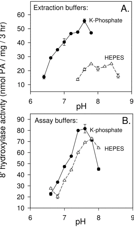
4. The choice of buffering agent and pH for the extraction buffer is critical in preserving activity. Much greater activity was always found with phosphate buffers rather than HEPES-based extraction buffers (Fig. 2). The efficiencies of these extraction buffers were compared through combinations of phosphate and HEPES at varying ratios (with or without EDTA, and/or EGTA) (Krochko, unpublished data). The results suggest that HEPES is not inhibitory as an extraction buffer, but rather that phosphate exerts a concentration-dependent positive effect on activity that could be supplemented, but not entirely replaced by EDTA.
Additionally, the pH and buffer choice for the assay buffer are important as well, and this should be determined empirically and confirmed once some initial enzyme activity has been
established (Fig. 2). For the ABA 8'-hydroxylase enzyme from corn, both the K-phosphate and HEPES buffers are equally appropriate for the assay/resuspension buffer. The optimum temperature of the reaction is 32-38 °C.
5. Neither EDTA nor an osmotic agent, i.e. sucrose, is required within the enzyme assay to maintain activity.
6. Losses of enzyme activity when the purified microsomal enzyme preparation is kept on ice over the course of a day are relatively minor (~20% over 6 h). In fact, induced enzyme activity is lost faster in the intact incubating (+)-ABA induced suspension cultures over the course of a day than in the enzyme extracts (see Fig. 3A). Nonetheless, delays or
interruptions in the extraction and purification of the microsomal enzyme preparation once the protocol is initiated could result in very substantial losses of enzyme activity. Any such delays should be rigorously avoided.
7. Enzyme activities can be compared to mRNA accumulations/concentrations across the time-course of induction by northern blots (Fig. 3B). A tight correlation can be used to deduce a causal relationship between the enzyme activity and the gene/probe used for hybridization on the northern blot.
8. The choice of cofactor and concentration also are important. NADPH at high concentrations (5 mM) is required to maximize ABA 8'-hydroxylase activity in the assay. NADH does not adequately substitute for NADPH in supporting the enzyme activity; however, NADH does support ABA 8'-hydroxylase activity at sub-optimal NADPH concentrations (Fig. 4) (4). This synergism could be exploited to reduce the costs of each reaction by reducing the amounts of the very expensive NADPH required for each reaction. Alternatively, the
NADPH could be replaced with an enzymatic NADPH regenerating system which can be considerably cheaper, but which we did not try (available from BD Biosciences).
3.5. In Vitro Assay for ABA 8'-Hydroxylase from Tobacco Leaves
We were interested in developing a prototype in vitro assay for ABA 8'-hydroxylase using differentiated tissues from intact plants as a quantitative comparison to the corn suspension cells. We were able to demonstrate in vitro ABA 8'-hydroxylase activity in microsomes isolated from young seedlings of tobacco (Nicotiana tabacum cv. Xanthi) (Krochko, unpublished data). The standard in vitro assay with HPLC detection (described previously) is not sensitive enough in this case to detect products of the enzyme activity; however, the standard in vitro assay using [3H](+)ABA as the substrate (see Fig. 1A) and very long exposure to Xray film (4.5 weeks at -80°C) of the TLC-separated ABA metabolites from the reaction provides clear evidence of phaseic acid production (see Fig. 5). It is estimated that the differences in enzyme activity between (+)-ABA induced corn suspension cultures and the young tobacco leaves are in the order of a 1,000 fold difference (based on the intensity of the phaseic acid signals, the exposure time taken to develop a visible band for phaseic acid on the X-ray film, and with the assumption that the extraction and assay conditions optimized for corn microsomes are also optimal for the tobacco microsomal preparations).
The enzyme activity we recorded using young tobacco leaf tissue is of a similar magnitude to the values reported previously for embryonic axes of chickpea (24). Using the detection
methods described in this chapter it is not possible to routinely sample this enzyme activity in microsomes from young tobacco leaves (or other such tissues). However, newer detection methods, i.e. LC MS/MS, that are more sensitive than using radiolabeled compounds now make
it feasible to use in vitro assays more frequently to directly measure cytochrome P450 enzyme activities. This also permits more accurate comparisons with gene expression estimates based on northern blots, microarray, EST frequencies, and real time RT-PCR. In fact, in vitro assays for direct measurements of cytochrome P450 activities in plant tissues have been used more frequently in recent years (20).
4. Notes
1. All solutions and media described in this chapter are made with distilled deionized water (other high quality water can be substituted).
2. Chemically synthesized preparations of abscisic acid are often available as racemic mixtures of the (+) and (-)-abscisic acid stereoisomers. It is important to recognize that the natural form of ABA is S(+)-2-cis,4-trans-abscisic acid, and other formulations such as R(-)-abscisic acid or (+/-)-abscisic acid will have weaker or diluted responses, and thus pose difficulties in correctly assigning the enzyme activity to the natural form of abscisic acid.
3. The response of a plant tissue or enzyme extract to any solvent should be checked in order to confirm that there is no effect from the solvent alone. For some purposes ABA can be dissolved in ethanol, methanol or DMSO (dimethyl sulfoxide).
4. Other compounds are not required in the extraction buffer to maintain enzyme activity (e.g. catalase, protease inhibitors, chelating agents). For some plant tissues it might be advisable to add protease inhibitors to this extraction solution.
5. When this protocol was published (4), we used 15-mL Corex tubes at 15,000 rpm (20,000xg) for 10 min in a SS-34 rotor (with adaptors) for the first centrifugation. Current information
on the 15-mL Corex tubes (now made by Kimble) specifies that the maximum RCF for these is 13,100xg, and therefore the speed indicated in the Methods section in this chapter has been changed to 12,000 rpm (13,100xg) for 15 min. There are a number of plastic tubes with enhanced strength now available for high-speed preparative centrifugation, and further information on these can be obtained from various manufacturers.
6. The sequential dilutions of BSA described in making the protein standards for the protein assay are designed to maximize accuracy and reproducibility of these concentrations. 7. Abscisic acid is radiolabeled at 6 positions (3', 5', 5', 7’, 7’, 7’) at high specific activities. It is
prepared using methods involving the exchange of hydrogen atoms on the abscisic acid molecule with radioactive hydrogen atoms from radiolabeled water (29). This exchange reaction is encouraged under basic conditions, but requires appropriately licensed facilities and chemistry expertise. The product of the enzymatic reaction, phaseic acid, has the same labeling pattern, although it can be reduced through exchange reactions during purification and work-up of the sample. Stored samples of radiolabeled ABA are examined regularly by TLC to determine purity, and quantities of radiolabeled (+)-abscisic acid are re-purified by TLC if required (the purified [3H]-(+)-abscisic acid is eluted from scrapings of the TLC plate with ethanol).
8. The programmable Thermomixer (Eppendorf) is invaluable for these assays. This apparatus maintains the desired temperature (hotter or cooler than ambient), while gently shaking the assay tubes.
9. Large amounts of culture could be grown and induced for enzyme expression at one time. Large harvests allow for scale-up and reproducible comparisons of several treatments and/or
chemicals at a time, with the advantage that base activity for the enzyme is constant across all samples in that tissue lot.
10. Care is taken to prevent any warming of the tissue during this process. Ample liquid nitrogen is always present.
11. The chilled buffer will frequently freeze slightly when added to the ground tissue in the mortar. This slightly frozen slurry is stirred gently with the pestle until it is thawed and then immediately strained through Miracloth.
12. It is very important to proceed rapidly to the isolation of the microsomes. All solutions, including the extraction buffer and enzyme solution, and all tubes are pre-chilled and kept chilled at all times.
13. The enzyme activity of the microsomal preparation is reasonably stable when stored on ice over the course of the day.
14. Other protein measurement assays can be used. This is dependent upon the additives (detergents, reducing agents) required in the extraction and re-suspension buffers and their compatibilities with various protein assays (see Bio-Rad).
15. The standard assay period is three h but this can sometimes be shortened to accommodate more samples. In either case, the appropriate reaction duration is determined empirically to cover the linear stage of the enzymatic reaction. Enzyme activity in our standard assays is based on the conversion or appearance of the phaseic acid (from abscisic acid) over a 3-h period (see Figs. 1- 3).
16. Acidification of the assay mixture improves the extraction of ABA and its metabolites into ethyl acetate during the purification prior to TLC or HPLC.
17. The first product of the reaction, 8'-hydroxyABA, is not stable under these conditions and quickly cyclizes to phaseic acid. Any 8'-hydroxyABA detected in the assays (TLC or HPLC detection) is added to the phaseic acid to estimate/quantify the reaction rate through total product produced.
18. The chromatography runs are much improved using a wick (Whatman filter paper) along the back of the tank and allowing the sealed tank to equilibrate before running the TLC plate. These modifications prevent ‘smiling’ of the bands.
19. Two assay extraction and two detection methods are provided for the benefit of the reader. The Oasis cartridges improve time efficiencies, allowing one to increase the number of samples handled and the size of the individual experiments. Likewise, HPLC detection is more efficient (though not as sensitive as the radioactive method) and permits quantitative comparisons between treatments.
20. We did not have a calibration curve for 8'-hydroxyABA, because under these conditions this compound is transient, but we made the assumption that the response factor for
8'-hydroxyABA is the same as phaseic acid (the stable derivative).
21. It should be noted that in vitro assays from tissue-derived microsomal extracts cannot easily confirm or identify the specific gene/protein product (cytochrome P450 enzyme) responsible for the activity; rather these assays only show that the enzyme activity is expressed in that tissue. The enzyme activity in question can be attributed to any one of the many P450 enzymes accumulating in that tissue. Confirmation of a specific enzyme activity for an individual cytochrome P450 gene may require heterologous expression in yeast, bacteria or insect cultures and/or manipulated over- and under-expression in a host plant system.
Acknowledgements
The authors would like to thank Drs. Garth Abrams and Patricia Rose for their invaluable help in development of the corn ABA 8'-hydroxylase in vitro assay, Sandra Gillett for help with the tobacco in vitro assay, and Ning Zhou for the northern blot analysis. This is National Research Council of Canada publication No. 50161.
References
1. Kim GT, Tsukaya H (2002) Regulation of the biosynthesis of plant hormones by cytochrome P450s. J Plant Res 115:169-77
2. Ehlting J, Sauveplane V, Olry A, Ginglinger JF, Provart NJ, Werck-Reichhart D (2008) An extensive (co-)expression analysis tool for the cytochrome P450 superfamily in Arabidopsis thaliana. BMC Plant Biol 8:47
3. Yinghong P, Michael TP, Hudson ME, Kay SA, Chory J, Schuler MA (2009) Cytochrome P450 monooxygenases as reporters for circadian-regulated pathways. Plant Physiol 150:858-878
4. Krochko JE, Abrams GD, Loewen MK, Abrams SR, Cutler AJ (1998) (+)-Abscisic acid 8'-hydroxylase is a cytochrome P450 monooxygenase. Plant Physiol 118:849-860
5. Cutler AJ, Rose PA, Squires TM, Loewen MK, Shaw AC, Quail JW, Krochko JE, Abrams SR (2000) Inhibitors of abscisic acid 8'-hydroxylase. Biochemistry 39:13614-13624
6. Kushiro T, Okamoto M, Nakabayashi K, Yamagishi K, Kitamura S, Asami T, Hirai N, Koshiba T, Kamiya Y, Nambara E (2004) The Arabidopsis cytochrome P450 CYP707A encodes ABA 8'-hydroxylases: key enzymes in ABA metabolism. EMBO J 23:1647-1656
7. Saito S, Hirai N, Matsumoto C, Ohigashi H, Ohta D, Sakata K, Mizutani M (2004) Arabidopsis CYP707As encode (+)-abscisic acid 8'-hydroxylase, a key enzyme in the oxidative catabolism of abscisic acid. Plant Physiol 134:1439-1449
8. Umezawa T, Okamoto M, Kushiro T, Nambara E, Oono Y, Seki M, Kobayashi M, Koshiba T, Kamiya Y, Shinozaki K (2006) CYP707A3, a major ABA 8'-hydroxylase involved in
dehydration and rehydration response in Arabidopsis thaliana. Plant J 46:171-182
9.Yang SH, Choi D (2006) Characterization of genes encoding ABA 8'-hydroxylase in ethylene-induced stem growth of deepwater rice (Oryza sativa L.). Biochem Biophys Res Commun 350:685-690
10. Okamoto M, Tanaka Y, Abrams SR, Kamiya Y, Seki M, Nambara E (2009) High humidity induces abscisic acid 8'-hydroxylase in stomata and vasculature to regulate local and systemic abscisic acid responses in Arabidopsis. Plant Physiol 149:825-834
11. Millar AA, Jacobsen JV, Ross JJ, Helliwell CA, Poole AT, Scofield G, Reid JB, Gubler F (2006) Seed dormancy and ABA metabolism in Arabidopsis and barley: the role of ABA 8'-hydroxylase. Plant J 45:942-954
12. Okamoto M, Kuwahara A, Seo M, Kushiro T, Asami T, Hirai N, Kamiya Y, Koshiba T, Nambara E (2006) CYP707A1 and CYP707A2, which encode abscisic acid
8'-hydroxylases, are indispensable for proper control of seed dormancy and germination in Arabidopsis. Plant Physiol 141:97-107
13. Chiang GCK, Barua D, Kramer EM, Amasino RM, Donohue K (2009) Major flowering time gene, Flowering Locus C, regulates seed germination in Arabidopsis thaliana. Proc Natl Acad Sci USA 106:11661-11666
14. Liu Y, Shi L, Ye N, Liu R, Jia W, Zhang J (2009) Nitric oxide-induced rapid decrease of abscisic acid concentration is required in breaking seed dormancy in Arabidopsis. New Phytol 183:1030-1042
15. Liu Y, Zhang J (2009) Rapid accumulation of NO regulates ABA catabolism and seed dormancy during imbibitions in Arabidopsis. Plant Signal Behav 4:905-907
16. Matakiadis T, Alboresi A, Jikumaru Y, Tatematsu K, Pichon O, Renou JP, Kamiya Y, Nambara E, Truong HN (2009) The Arabidopsis abscisic acid catabolic gene CYP707A2 plays a key role in nitrate control of seed dormancy. Plant Physiol 149:949-960
17. Werck-Reichhart D, Hehn A, Didierjean L (2000) Cytochrome P450 for engineering herbicide tolerance. Trends Plant Sci 5:116-123.
18. Schuhegger R, Nafisi M, Mansourova M, Petersen BL, Olsen CE, Svatos A, Halkier BA, Glawischnig E (2006) CYP71B15 (PAD3) catalyzes the final step in camalexin biosynthesis. Plant Physiol 141:1248-1254
19. Kandel S, Sauveplane V, Compagnon V, Franke R, Millet Y, Schreiber L, Werck-Reichhart D, Pinot F (2007) Characterization of a methyl jasmonate and wounding responsive
cytochrome P450 of Arabidopsis thaliana catalyzing dicarboxylic fatty acid formation in vitro. FEBS J 274:5116-5127
20. Böttcher C, Westphal L, Schmotz C, Prade E, Scheel D, Glawischnig E (2009) The multifunctional enzyme CYP71B15 (PHYTOALEXIN DEFICIENT3) converts cysteine-indole-3-acetonitrile to camalexin in the cysteine-indole-3-acetonitrile metabolic network of Arabidopsis thaliana. Plant Cell 21:1830-1845
21. Ludwig SR, Somers DA, Peterson WL, Pohlmann BF, Zarovitz MA, Gengenbach BG, Messing J (1985) High-frequency callus formation from maize protoplasts. Theor Appl Genet 71:344-350
22. Murashige T, Skoog F (1962) A revised medium for rapid growth and bioassays with tobacco tissue cultures. Physiol Plant 15:473-497
23. Bradford MM (1976) A rapid and sensitive method for quantification of microgram
quantities of protein utilizing the principle of protein-dye-binding. Anal Biochem 72:248-254 24. Babiano MJ (1995) Metabolism of [2-14C]abscisic acid by a cell-free system from embryonic
axes of Cicer arietinum L. seeds. J Plant Physiol 145:374-376
25. van der Hoeven TA (1981) Isolation of hepatic microsomes by polyethylene glycol 6000 fractionation of the post mitochondrial fraction. Anal Biochem 115:398-402
26. Hamilton RL, Moorehouse A, Lear SR, Wong JS, Erickson SK (1999) A rapid calcium precipitation method of recovering large amounts of highly pure hepatocyte rough endoplasmic reticulum. J Lipid Res 40:1140-1147
27. Alden PG, Plumb RS, Jones MD, Rainville PD, Shave D (2009) A rapid ultra-performance liquid chromatography/tandem mass spectrophotometric methodology for the in vitro analysis of Pooled and Cocktail cytochrome P450 assays. Rapid Commun Mass Spectrom 24:147-154
28. Cutler AJ, Squires TM, Loewen MK, Balsevich JJ (1997) Induction of (+)-abscisic acid 8'-hydroxylase by (+)-abscisic acid in cultured maize cells. J Exp Bot 48:1787-1795
29. Bonnafous JC, Fonzes L, Mousseron-Canet M (1971) Synthese d’acide abscisique radioactive. II. Marquage au tritium. Bull Soc Chim Fr 1971 :4552-4554
Fig. 1. (+)-ABA 8'-hydroxylase activity in microsomes extracted from BMS corn suspension cultures. (A) After tissue homogenization, the initial 20,000xg supernatant and subsequent 200,000xg supernatant and microsomal pellet were assayed for in vitro ABA 8'-hydroxylase activity with (+)-[3H]ABA as a substrate. The pellet was re-suspended in 0.1 volume of the original homogenate, and therefore proteins were concentrated 10-fold over the first supernatant or the original homogenate. Identification of substrate and products on TLC plates was based on co-migration with non-radioactive authentic standards (identified as fluorescent spots under UV light) that had been run in separate lanes, or superimposed on the assay extracts for verification of co-migration. Standards (stds) used were: a, trans-ABA; b, ABA; c, (±)-ABA trans-1',4'-diol; d, phaseic acid (PA); e, (±)-ABA cis-1',4'-diol; f, 8'-hydroxy-ABA; g, 7'-hydroxy-ABA; and h, dihydrophaseic acid (DPA). The filled arrowheads indicate the ABA metabolites identified in these assays. (B) Output from an HPLC separation using a reverse-phase column with UV detection at 262 nm. (Reproduced from 4).
Fig. 2. Effect of buffers and pH on in vitro assayable ABA 8'-hydroxylase activity. (A)
Comparison of the efficacy of 100 mM HEPES-NaOH and 100 mM potassium phosphate as the initial extraction buffers over a range of pH values. (B) Comparison of 100 mM HEPES-NaOH and 100 mM potassium phosphate as assay buffers over a range of pH values. The initial extraction was performed with 100 mM potassium phosphate buffer, pH 7.6. After ultracentrifugation the microsomal pellets and the cofactor were resuspended for the assay in the buffers indicated at each data point on the graph. (Reproduced from 4).
Fig. 3. Time course of the induction of ABA 8'-hydroxylase activity following addition of (+)-ABA to BMS corn suspension cultures. Tissue was harvested at the times indicated after the addition of (+)-ABA to the culture medium. (A) Phaseic acid (PA) and 8'-hydroxy-ABA were assayed by HPLC and summed to represent the total ABA 8'-hydroxylase activity (expressed as total PA produced/mg protein/3h). (B) Messenger RNA accumulation for ABA
8'-hydroxylase (CYP707A subfamily) was monitored through northern blots on total RNA using a probe to a corn CYP707A gene (Adapted from 4).
Fig. 4. The relative efficiencies of NADPH, NADH, and various combinations of these cofactors in supporting (+)-ABA 8'-hydroxylase activity. Concentrations: NADPH, 5, 1, 0.5 mM; NADH, 5 mM; and NADPH, 5, 1, 0.5 mM in combination with NADH at 5 mM (Adapted from 4).
Fig. 5. Development of an in vitro assay for ABA 8'-hydroxylase from young tobacco leaves. Microsome preparations and the in vitro assay with [3H]-(+)-ABA were conducted as described previously for corn tissues. Tobacco leaves were collected from 9 and 15 day-old seedlings. The TLC plate was exposed to the film for 4.5 weeks at -80°C for detection of phaseic acid by fluorography. H, tissue homogenate; S1, first supernatant; P1, first pellet; S2, second supernatant; M, microsomal pellet; PA, phaseic acid; ABA, abscisic acid. (Krochko et al., unpublished.)
Figure 1.
32 32