Split-rib reconstruction of the frontal sinus: two
cases and literature review
M B SOYKA1, M GUGGENHEIM2, A ARNOUX3, D HOLZMANN1
Departments of1Otolaryngology Head and Neck Surgery and2Plastic and Reconstructive Surgery, University Hospital Zurich, and3Department of Otolaryngology Head and Neck Surgery, Kantonsspital Aarau, Switzerland
Abstract
Background: Large defects of the anterior wall of the frontal sinus require closure using either autologous or foreign material. In cases of osteomyelitis, the reconstruction must be resistant to bacterial infection. Split-rib osteoplasty can be used in different sites.
Methods: Two patients with malignant sinonasal tumours underwent repeated treatment, and subsequently developed osteomyelitis of the frontal bone. After adequate therapy, a large defect of the anterior wall persisted. Reconstruction was performed using the split-rib method. The literature on this topic was reviewed.
Results: Both patients’ treatment were successful. No complications occurred. A PubMed search on the topic of rib reconstruction of the frontal sinus and skull was performed; 18 publications matched the inclusion criteria. From these sources, we noted that 182 reconstructions yielded good results with few complications.
Conclusion: Large defects of the anterior wall of the frontal sinus can be closed successfully using autologous split-rib grafting. Aesthetic outcome is good and donor site morbidity is minimal.
Key words: Frontal Sinus; Osteomyelitis; Ribs; Reconstructive Surgical Procedures
Introduction
Bony defects of the anterior wall of the frontal sinus may occur due to frontal mucoceles, pyocoeles, malformations and tumours, and following trauma and infection. Many different techniques exist for closing such defects. Autologous tissue graft reconstruction using split calvaria is very much favoured in the literature, but fixation with osteo-synthetic material is usually required.1–3Sometimes, a bony transplant is resorbed over time, depending on its origin.4
Other, alloplastic graft materials can also be used, together with various types of cement. The use of such materials avoids the risk of donor site morbidity; however, such allo-grafts may be susceptible to rejection and infection.5
In the cases presented below, we were faced with the problem of preceding infection, and therefore needed to avoid using foreign material implants or osteosynthesis. In addition, it was essential to seal the nasal cavity with vascu-larised autografts, in order to avoid potential ascending bac-terial contamination of the frontal implant.
As is known from cranioplasty, rib reconstruction consti-tutes an alternative approach for treating larger defects of the skull.6,7In this article, we describe the surgical procedure for rib reconstruction in the frontal sinus region; we also discuss the available literature on this topic.
Patients and methods
Patient one
In 2007, a 47-year-old, Caucasian man was diagnosed with sinonasal undifferentiated carcinoma, by endonasal biopsy.
The initial tumour-node-metastasis (TNM) stage was cT4 cN0 cM0, according to the Union for International Cancer Control (UICC) classification (Figure 1). The tumour was placing pressure on both optic nerves, causing the patient’s vision to deteriorate.
Therefore, we performed emergency tumour debulking via transnasal transcribriform resection, with optic nerve decompression. This was followed by transfrontal subcranial resection of the adjacent dura. The defect was reconstructed with a pericranial flap, and the anterior wall of the frontal sinus was restored using a split calvarium graft fixed with titanium mini-plates.
The peritumoural area received adjuvant chemoradiation, comprising cisplatin (total dose 65 mg) and a total radiation dose of 60 Gy (intensity modulated radiotherapy).
The patient’s vision returned to almost normal values (uncorrected vision of 70 and 100 per cent), and his initial double vision resolved.
One and a half years after initial diagnosis, the patient developed intermittent eyelid swelling on both sides, with subsequent formation of a right upper lid abscess.
Postradiogenic osteitis of the frontal bone was found to be the underlying pathology, involving infection with multiple bacteria (Pseudomonas aeruginosa, Staphylococcus aureus, coagulase-negative staphylococcal species and group B streptococci), which required treatment with long term intravenous antibiotics.
The foreign osteosynthetic material was removed, all necrotic bone was debrided, and the frontal sinus was oblit-erated with abdominal fat.
Accepted for publication 17 May 2011 First published online 6 October 2011
The Journal of Laryngology & Otology (2011),125, 1301–1308.
CLINICAL RECORD
© JLO (1984) Limited, 2011 doi:10.1017/S0022215111002611
The infection resolved; however, a large hole was left in the anterior wall of the frontal sinus, together with a bony defect in the right orbit (Figures 2 and 3b). As a conse-quence, the patient’s forehead had a large dent which was not cosmetically desirable (Figure 3b).
Patient two
A 43-year-old, Caucasian woman was treated by combined transnasal and transfrontal resection for a cT4 cN0 cM0,
malignant solitary fibrous tumour of the frontal sinus and nasal cavity. The anterior wall of the frontal sinus was fixed by titanium mini-plating, a bony defect was restored with a Palacos (Heraeus Kulzer GmbH, Hanau, Germany) cement plasty, and the skull base was reconstructed using titanium mesh.
The operation was followed by intensity modulated radio-therapy with a cumulative dose of 64 Gy.
The patient subsequently developed greenish rhinorrhoea. This was discovered to be due to P aeruginosa infection, requiring antibiotic treatment with aminoglycosides and ceftazidim.
However, the infection advanced and frontal osteomyelitis developed. Repeated craniotomy was necessary. All foreign material was removed and the osteomyelitic bone debrided. The skull base was reconstructed using a pedicled periosteal flap, and the anterior frontal sinus wall was again restored using Palacos cement plasty.
Antibiotics were continued for one month post-operatively. However, this treatment was subsequently discontinued because of bilateral vestibular dysfunction, and substituted with piperacillin with tazobactam.
Unfortunately, the patient again developed infection, this time with a cutaneous fistula. The Palacos cement plasty was again removed and wide local debridement performed. A free radial forearm flap was used to cover the external defect.
This flap failed, and the procedure was repeated from the contralateral side.
Finally, the patient was treated with Rimactane® and ciprofloxacin, and the infection resolved.
Again, the patient was left with a large dent in her fore-head due to the bony defect.
Materials and methods
The informed consent of both patients was obtained for pub-lication of the contents of this article.
The same reconstructive surgical procedures were under-taken for both patients.
We used a two-team approach, working with our colleagues from the plastic and reconstructive surgery department.
The left side of the thorax was incised between the eight and ninth rib, and a thorough subperiosteal dissection per-formed. We then harvested either the bony parts of both ribs, in patient one, or only one rib, in patient two, without any injury to the pleura. The periosteum was incised only ventrally, and was closed after removal to promote rib regrowth. The skin was closed in two layers, and two suction drains inserted.
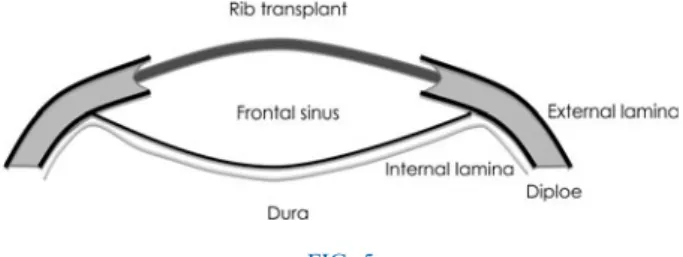
The approximately 15 cm long transplants were then split in half using a hand-driven chisel and thinned using flat-nosed pliers, in order to render them more flexible (Figure 4). After coronal incision and preparation of the scalp down to the supraorbital rim, the supraorbital nerves were identified and spared. The edges of the bony defect were debrided, and a groove was drilled into the diploe, using a 2 mm drill, just between the external and internal tabulae (Figures 5 and 6). The rib transplants were tailored in length to fit the gap. The rib transplants were then placed into the laterally drilled groove, bent and under tension, to form the new contour of the forehead (Figures 7 and 8).
In patient one, a small piece of calvarial bone was placed onto the orbital roof, in order to reconstruct a smaller defect in this area.
FIG. 1
Axial, T1-weighted, gadolinium-enhanced magnetic resonance imaging scan of patient one prior to surgery, showing a sinonasal
undifferentiated carcinoma at the level of the optic nerves.
FIG. 2
Three-dimensional computed tomography reconstruction of patient one, showing the pre-reconstruction defect in the frontal sinus.
Finally, all edges were levelled using a sharp 5 mm drill, the scalp was put back in place, and the incision was closed using a bilayer technique, after placement of two non-suction drains.
We also conducted a literature review on split-rib recon-struction of the cranium. The PubMed database was searched using the following key words: ‘split rib’, ‘frontal sinus reconstruction’, ‘frontal sinus + rib’, ‘frontal sinus osteo-plasty’, ‘bone grafting frontal sinus’, ‘frontal sinus osteo-myelitis’, ‘sinus + rib grafts’, ‘cranial vault reconstruction’, ‘frontal cranioplasty’, ‘rib cranioplasty’ and ‘frontal rib grafts’. We assessed only material published between 1950 and 2010, concurrent with the modern antibiotic era. We considered articles if abstracts were available electronically, or if the article title indicated that its content may be relevant. Selection was limited to English language literature.
We included in the review only those publications describ-ing the use of a split rib technique to close a cranial defect, preferably in the frontal sinus region.
All 18 publications selected were reviewed as full text articles, in order to record the total number of patients, defect site, reconstruction material used, aesthetic results, complications and graft osteointegration.
Results
Patients’ post-operative course
In our two patients, post-operative X-rays of the thorax showed no signs of pneumothorax. The post-operative course was uneventful in both cases. Minimal pain in the left hemithorax was treated with non-steroidal anti-inflammatory drugs. Patient one was discharged from hospital after 10 days of prophylactic FIG. 3
antibiotic treatment, as recommended by colleagues in the infectious diseases department. Patient two left hospital after six days and needed no further antibiotic treatment. Sutures and clips were removed on day 10.
Follow up was performed on a monthly basis, using mag-netic resonance imaging (MRI), computed tomography (CT) and positron emission tomography (PET) CT. The final follow-up CT scan of patient two, performed eight months
after reconstruction, is shown in Figure 9, and clearly demon-strates signs of osteointegration of the rib graft.
At the time of writing (40 and 36 months after initial diag-nosis), there were no signs of tumour recurrence in either patient. Despite this, follow up was ongoing.
Literature review
Almost 1000 articles were identified by our PubMed search, although overlapping results were encountered. Thirty-four publications were selected and retrieved as full text articles, of which only 18 met the inclusion criteria (see Table I).
Articles were excluded for the following reasons: non-human subjects (one paper), reconstruction in the wrong anatomical area (one paper), insufficient data (six papers), inappropriate initial selection (i.e. paper did not refer to split-rib reconstruction; six papers), and results overlapping with those of a previously identified study (two papers). FIG. 4
Intra-operative photograph showing flattening of the split-rib graft, using pliers, for patient one.
FIG. 5
Diagram of the reconstructed site.
FIG. 6
Early post-reconstruction, axial computed tomography scan of the reconstruction site in patient one. F= Foot
FIG. 7
Intra-operative photograph showing reconstruction in patient one.
FIG. 8
Early post-reconstruction, three-dimensional computed tomography reconstruction for patient one. A= anterior
The accepted 18 publications described a total of 250 patients undergoing skull reconstruction. In 241 cases, split ribs were the only reconstructive material used (aside from fixation wires). In nine cases, other materials were used in combination with rib transplants (alloplastic graft material in six cases, soft tissue in one and calvarial bone in two). Fifty-nine cases involved frontal area reconstruction and 12 involved parietal area reconstruction; in 181 cases, the site of reconstruction was not clearly defined, but was stated to be in the skull area.
The overall results were reported to be very good. Six (2.5 per cent) cases of bony resorption were reported, and 10 (4 per cent) patients underwent revision surgery due to unsatisfactory results. One early displacement needed immediate revision, one haematoma required evacuation, and three local infections were controlled with systemic anti-biotics. No transplant losses were encountered in patients with infections, and in only one case was local debridement needed to control infection.9There were three cases of major complications, all of which involved haemato- or pneu-mothorax requiring drainage. In two studies, with a total of five patients, post-operative complications were not evalu-ated. Osteointegration was discussed and confirmed in only four studies, involving a total of 129 patients. Three studies (with a total of 24 patients) did not report aesthetic outcomes, and two studies (with a total of five patients) did not clearly assess complications.
Discussion
There is not much information in the otolaryngological lit-erature on reconstructions of the anterior wall of the frontal sinus. In our literature search, only two of the finally reviewed articles were from otolaryngological journals, and these involved a total of only five patients. Most descriptions were in case reports or small series introducing new materials and techniques in this field. Because frontal sinus defects are often found in close proximity to other cranial defects, our neurosurgical colleagues and collaborating plastic and recon-structive surgeons seem to be more familiar with this problem, and have found several ways of coping with even larger defects of the cranium. However, reconstructions of bony defects in the frontal area are aesthetically more chal-lenging than those located parietally or occipitally.
Current skull reconstruction techniques include the use of alloplastic and autologous materials.23However, certain situ-ations require different approaches to achieve successful FIG. 9
Eight months post-reconstruction, axial computed tomography scan of patient two, showing signs of osteointegration. F= Foot
TABLE I
PREVIOUS STUDIES OF SPLIT-RIB CRANIAL RECONSTRUCTION∗
Study Pts (n) Site Material Results Complication(s) Osteoint? Longacre & Destefano8 118 Cranium & face Rib Good 1 infection Yes
1 haematoma
Leivy & Tovi9 7 Cranium Rib NA 1 infection Yes 1 pneumothorax Körlof et al.10 46 23 frontal Rib 50% good 3 haematomas NA
11 parietal 25% slightly uneven 1 haematothorax 12 other 20% definitely uneven
5% marked depression
Marchac & Cophignon11 12 Cranium Rib Some irregularities 1 displacement NA Shaw & Thering12 7 Cranium Rib Good None
Steinhäuser & Hardt13 5 Cranium Rib NA 2 resorptions NA Munro & Guyuron7 12 Frontal Rib NA 1 pneumothorax NA
Cababbe et al.14 8 Frontal 6 rib Good 1 infection NA 2 rib+ alloplast
Stueber et al.15 1 Parietal Rib+ soft tissue Good None Yes Forte & de Souza16 1 Frontal Rib Good NA NA
Argenta & Dingman17 1 Cranium Rib Good None NA Chicarilli & Ariyan18 4 Cranium Rib+ silicone Good NA NA
Edwards & Ousterhout3 2 Frontal Rib+ calvaria Good None NA Pochon & Klöti19 6 Cranium Rib All irregular 3 resorptions NA
Stal et al.20 2 Frontal Rib All irregular 1 resorption NA Viterbo et al.21 2 Frontal & skull Rib Good None NA
Taggard & Menezes22 13 Mostly frontal (8/13) Rib 1 revision None NA Beekmans et al.6 3 Cranium Rib Good None Yes ∗Identified by literature review. Pts= patients; osteoint = osteointegration; NA = not specified
closure of cranial defects. Live bone seems to be more resist-ant to infection.24 The advantages of autologous calvarial reconstruction, over the use of alloplastics, have been reported previously.5 Baumeister and colleagues described an algorithm for the treatment of neurosurgical bone flap losses due to infection.4After treatment of a potential soft tissue defect, they recommended alloplastic material as the first choice, followed by autologous grafts and allogenic bone. However, they emphasised the need to confirm that any previous infection is completely healed by the time of intervention, when alloplastic material is used. In complicated infectious situations, autologous bone is preferred.
Thus, expert opinion currently favours the use of split-cal-varial bone grafts. However, in specific situations (e.g. when one needs to cover a large area in a repeatedly infected site), alternative approaches and techniques are often required. One such alternative, with proven good results, is split-rib cranioplasty.
Reconstruction of the skull in the area of the frontal sinus is prone to infection, due to ascending bacteria from the nasal cavity. We, and others, believe that closure of the frontal recess (including meticulous removal of sinus mucosa) is an important step in preventing such infection, as it isolates the frontal sinus from the nasal cavity.25In addition, perfused tissue (e.g. a pedicled pericranial flap) needs to be brought in to cover badly vascularised tissue, especially in irradiated areas.26
In cases of osteitis and osteomyelitis (anywhere in the body) in which antibiotic therapy has failed, thorough debri-dement of the affected bone is essential, and removal of any foreign material in proximity to the focus of infection is also required. Therefore, in cases of repeated infection, we avoid using alloplastic implants during subsequent reconstruction. (In unproblematic cases, we use split-calvarium grafting to cover frontal defects, but this reconstruction usually needs to be kept in place by mini-plate osteosynthesis or wires, especially if large areas are involved.)
In all the reviewed cases of split-rib reconstruction of the frontal sinus, there was not a single case of graft loss due to infection. With split-rib grafting, it is possible to close even large defects in the skull area, using a‘living’ graft, where split-calvarium grafting could not provide sufficient material.27Split-rib grafts can be kept in place without any use of fixation, simply by drilling a rim into the diploe of the calvarium and placing the graft under slight tension to follow the natural curvature of the skull.11Thus, the use of split-rib grafting is favoured in previously infected regions, as foreign material is always more prone to bacterial coloni-sation. The tension on the transplant promotes healing by axial compression, as seen in other osteosynthetic pro-cedures.28 There is excellent integration of the split-rib grafts, as shown in an experimental study by Longacre and Destefano.29
Both our patients required regular radiological follow up, using MRI, CT and PET-CT imaging, to monitor outcomes following treatment of their malignant tumours. The use of bony grafts without osteosynthetic materials has the additional advantage of good radiolucency, producing no magnetic interference on MRI, as well as no artefacts on electroencephalography (in patients with seizures).12
Another indisputable advantage of rib grafts is the ability of the rib to regrow, when the periosteum is closed over the site of the harvested rib. In their large series, Körlof et al. reported almost 100 per cent regrowth of the harvested rib.10 One of the main disadvantages of the split-rib technique is donor site morbidity. Although post-operative pain is usually not severe, there is the potential risk of injury to the pleura and subsequent pneumo-haematothorax, requiring pleural drainage.30 This complication has been reported in as many as 30 per cent of patients following rib removal.31 However, other studies of facial and cranial reconstruction using rib grafts have reported an incidence of 1–2.4 per cent.30,32 In our study, we observed this complication in only three cases (approximately 1 per cent), of a total study group of 252 (i.e. our own two patients plus 250 literature
FIG. 10
review cases undergoing cranial reconstruction using split-rib grafting). Clinically, if a pleural tear is suspected intra-operatively, this can be tested by filling the surgical wound with water and applying positive pressure ventilation; any pleural tear should cause bubbles to rise. An X-ray of the thorax, taken during expiration, should confirm the findings. Another reported disadvantage of split-rib grafting is an inferior cosmetic result, in terms of an uneven surface. In our limited series, results were good for the large defects that needed to be covered (Figure 10). A minor degree of unevenness, such as seen in our first patient (Figure 3b) can be easily addressed by injection of autologous abdomi-nal fat. Of the studies identified in our literature, the best aes-thetic outcome was reported by Körlof et al.: 75 per cent of reconstructions achieved good or slightly uneven results, with only 5 per cent retaining an obvious depression of the reconstructed area.10 Of those identified articles which reported their aesthetic outcomes, good results were reported for 182/214 reconstructions (85 per cent). However, in our opinion these descriptions were often not sufficiently detailed, so this rate may overestimate the incidence of a favourable cosmetic outcome.
• Multimodal treatment of sinonasal malignancies may result in osteomyelitis and cosmetic deformity • In such cases, frontal sinus reconstruction can be a
challenge
• In the two presented cases, split-rib reconstruction had good cosmetic results, adequate
osteointegration and minimal donor site morbidity • Split-rib reconstruction should be considered
when avoidance of foreign materials is required
It was not the aim of this study to compare different recon-structive materials. The advantages and disadvantages of the various graft materials are discussed elsewhere, as are their indications in cranial reconstructive surgery.23,33–36 The choice of reconstructive materials for the two patients presented is discussed above. In both patients, alloplastic materials were too prone to infection, and had already failed previously. For similar reasons, we chose not to use preformed or manufactured titanium implants for the final reconstruction.37 Split calvarium had either already been used or would have failed to fill the large gap. Furthermore, split-calvarium grafting would have required wire or mini-plate fixation, leading to implantation of foreign material which could cause untreatable infection. (The same applies to iliac bone grafting, although other tech-niques have been described.)38
The overall results of the split-rib method are good and morbidity is low; however, extensive experience in the frontal sinus region is lacking. Further research is needed into the outcomes of split-rib reconstruction in this region.
Conclusion
Split-rib osteoplasty of the frontal sinus is a valid method for closing even quite large defects. It has the advantage of not relying on foreign material or osteosynthesis, therefore mini-mising the risk of infection. Furthermore, it causes no dis-tracting imaging artefacts, and functional and aesthetic morbidity at the graft donor site is minimal.
Acknowledgement
We thank Dr Walter Künzi, Department of Plastic and Reconstructive Surgery, for his support with manuscript preparation.
References
1 Grundmann T, Kehrl W. Reconstruction of the frontal sinus with a calvarium split galea periosteum transplant after inflammatory complications [in German]. HNO 2004;52:57–62
2 Artico M, Ferrante L, Pastore F, Ramundo EO, Cantarelli D, Scopelliti D. Bone autografting of the calvaria and craniofacial skeleton: historical background, surgical results in a series of 15 patients, and review of the literature. Surg Neurol 2003;60: 71–9
3 Edwards M, Ousterhout D. Autogeneic skull bone grafts to reconstruct large or complex skull defects in children and ado-lescents. Neurosurgery 1987;20:273–80
4 Baumeister S, Peek A, Friedman A, Levin L, Marcus J. Management of postneurosurgical bone flap loss caused by infection. Plast Reconstr Surg 2008;122:195–208e
5 Sahoo N, Roy I, Desai A, Gupta V. Comparative evaluation of autogenous calvarial bone graft and alloplastic materials for sec-ondary reconstruction of cranial defects. J Craniofac Surg 2010; 21:79–82
6 Beekmans S, Don Griot J, Mulder J. Split rib cranioplasty for aplasia cutis congenita and traumatic skull defects: more than 30 years of follow-up. J Craniofac Surg 2007;18:594–7 7 Munro I, Guyuron B. Split-rib cranioplasty. Ann Plast Surg
1981;7:341–6
8 Longacre J, Destefano G. Further observations of the behavior of autogenous split-rib grafts in reconstruction of extensive defects of the cranium and face. Plast Reconstr Surg 1957;20:281–96 9 Leivy D, Tovi D. Autogenous bone cranioplasty.
Neurochirurgia (Stuttg) 1970;13:82–6
10 Körlof B, Nylén B, Rietz K. Bone grafting of skull defects. A report on 55 cases. Plast Reconstr Surg 1973;52:378–83 11 Marchac D, Cophignon J. Technique for embedding split ribs in
a cranioplasty. Plast Reconstr Surg 1975;55:237–9
12 Shaw R, Thering H. Reconstruction of cranial defects. Clin Plast Surg 1975;2:539–49
13 Steinhäuser E, Hardt N. Secondary reconstruction of cranial defects. J Maxillofac Surg 1977;5:192–8
14 Cabbabe E, Shively R, Malik P. Cranioplasty for traumatic deformities of the frontoorbital area. Ann Plast Surg 1984;13: 175–84
15 Stueber K, Salcman M, Spence R. The combined use of the latis-simus dorsi musculocutaneous free flap and split-rib grafts for cranial vault reconstruction. Ann Plast Surg 1985;15:155–60 16 Forte V, de Souza F. Autogenous rib grafts in facial surgery.
J Otolaryngol 1985;14:201–2
17 Argenta L, Dingman R. Total reconstruction of aplasia cutis congenita involving scalp, skull, and dura. Plast Reconstr Surg 1986;77:650–3
18 Chicarilli Z, Ariyan S. Cranioplasty with a silicone prosthesis and split rib grafts. Head Neck Surg 1986;8:355–62
19 Pochon J, Klöti J. Cranioplasty for acquired skull defects in chil-dren– a comparison between autologous material and methyl-methacrylate 1974–1990. Eur J Pediatr Surg 1991;1:199–201 20 Stal S, Netscher D, Shenaq S, Spira M. Reconstruction of
calvar-ial defects. South Med J 1992;85:812–19
21 Viterbo F, Palhares A, Modenese E. Cranioplasty: the autograft option. J Craniofac Surg 1995;6:80–3
22 Taggard D, Menezes A. Successful use of rib grafts for cranio-plasty in children. Pediatr Neurosurg 2001;34:149–55 23 Goiato M, Anchieta R, Pita M, dos Santos D. Reconstruction of
skull defects: currently available materials. J Craniofac Surg 2009;20:1512–18
24 Hancock D. The fate of replaced bone flaps. J Neurosurg 1963; 20:983–4
25 Manson P, Crawley W, Hoopes J. Frontal cranioplasty: risk factors and choice of cranial vault reconstructive material. Plast Reconstr Surg 1986;77:888–904
26 Snyderman C, Janecka I, Sekhar L, Sen C, Eibling D. Anterior cranial base reconstruction: role of galeal and pericranial flaps. Laryngoscope 1990;100:607–14
27 Weber R, Kearns D, Smith R. Split calvarium cranioplasty. Arch Otolaryngol Head Neck Surg 1987;113:84–9
28 Bagby G. Compression bone-plating: historical considerations. J Bone Joint Surg Am 1977;59:625–31
29 Longacre J, Destefano G. Experimental observations of the repair of extensive defects of the skull with split-rib grafts. Plast Reconstr Surg Transplant Bull 1958;21:372–88 30 Tessier P, Kawamoto H, Matthews D, Posnick J, Raulo Y, Tulasne
JF et al. Taking long rib grafts for facial reconstruction– tools and techniques: III. A 2900-case experience in maxillofacial and cra-niofacial surgery. Plast Reconstr Surg 2005;116(Suppl. 5): 38S–46S, 92–4S
31 Whitaker L, Munro I, Salyer K, Jackson I, Ortiz-Monasterio F, Marchac D. Combined report of problems and complications in 793 craniofacial operations. Plast Reconstr Surg 1979;64: 198–203
32 James D, Irvine G. Autogenous rib grafts in maxillofacial surgery. J Maxillofac Surg 1983;11:201–3
33 Gosain A, Committee PSEFD. Biomaterials for reconstruction of the cranial vault. Plast Reconstr Surg 2005;116:663–6 34 Friedman C, Costantino P, Synderman C, Chow L, Takagi S.
Reconstruction of the frontal sinus and frontofacial skeleton with hydroxyapatite cement. Arch Facial Plast Surg 2000;2: 124–9
35 Mann W, el-Khatieb A. Cranioplasty with Palacos R®in recon-struction of frontal sinus defects. J Laryngol Otol 1988;102: 824–7
36 Kuttenberger J, Hardt N. Long-term results following recon-struction of craniofacial defects with titanium micro-mesh systems. J Craniomaxillofac Surg 2001;29:75–81
37 Lethaus B, Poort Ter Laak M, Laeven P, Beerens M, Koper D, Poukens J et al. A treatment algorithm for patients with large skull bone defects and first results. J Craniomaxillofac Surg 2010;39:435–40
38 Grocott J. Experiences in cranial bone grafting. Br J Plast Surg 1952;5:51–9
Address for correspondence: Dr Michael B Soyka,
Department of Otolaryngology Head and Neck Surgery, University Hospital Zurich,
Frauenklinikstrasse 24, 8091 Zurich, Switzerland Fax:+41 44 255 45 56
E-mail: [email protected]
Dr M B Soyka takes responsibility for the integrity of the content of the paper