M.Eugenia Heres-Pulido1, Irma DuenÄas-GarcõÂa1,
Laura CastanÄeda-Partida1, Antonio SaÂnchez-GarcõÂa1,
Martha Contreras-Sousa1, AÂngel DuraÂn-DõÂaz1 and
Ulrich Graf2,3
1Laboratorio de GeneÂtica ToxicoloÂgica, FES Iztacala, Universidad Nacional
AutoÂnoma de MeÂxico, 54090 Tlalnepantla, Estado de MeÂxico, MeÂxico and
2Institute of Animal Sciences, Section Physiology and Animal Husbandry,
Swiss Federal Institute of Technology, Schorenstrasse 16, CH-8603 Schwerzenbach, Switzerland
Tamoxifen (TAM) is an anti-oestrogen used for treatment and prevention of human breast cancer, but it is also related to human endometrial and uterine cancer. The wing spot test in Drosophila melanogaster was employed to determine the genotoxic effects of TAM and 4-nitroquino-line-1-oxide (4-NQO), a carcinogen that produces adducts similar to TAM±DNA adducts detected in rodent liver and human liver microsomes. As Drosophila spp. have no oestrogen receptor, no effects can result in binding of TAM to a receptor. Chronic treatments with TAM citrate were performed with 3-day-old larvae of the standard (ST) and high bioactivation (HB) crosses of the wing spot test at concentrations of 0.66, 1.66 and 3.33 mM. In addi-tion, the carcinogen 4-NQO was administered at 2.5 and 5.0 mM. Somatic spots on normal wings from marker-heterozygous ¯ies and on serrate wings from balancer-heterozygous ¯ies were scored to determine mutation and recombination events in somatic cells for each compound. The results showed genotoxic effects of TAM at 1.66 and 3.33 mM in the ST cross only and without a clear dose± response effect. This suggests a weak genotoxicity of this anti-oestrogen. The negative results obtained with TAM in the HB cross may indicate ef®cient detoxi®cation of the compound by the increased xenobiotic metabolism present in this cross. As reported before, 4-NQO showed genotoxic effects in the ST cross with a clear dose±response effect. For the ®rst time, we report enhanced effects of this com-pound in the HB cross. It is concluded that the geno-toxicity of TAM in the Drosophila wing spot test is different from that of 4-NQO.
Introduction
Breast cancer is the most common cancer in women, but is also present in 1% of men. Tamoxifen citrate salt (TAM), a triphenylethylene type I non-steroid anti-oestrogen [(Z)-1-(p-dimethylaminoethoxyphenyl)-1,2-diphenyl-1-butene, Figure 1], is approved for humans that have or are at risk of getting breast cancer. TAM is a selective oestrogen response modi®er (SERM) that exerts antagonistic effects in breast cancer cells and agonistic effects in other tissues, such as endometrium and bone (White, 1999). It is proposed that this non-steroid and its reactive intermediates increase the risk of endometrial and
uterine cancer in breast cancer patients receiving TAM therapy (IARC, 1996). The mechanism for this increase is unclear, although two plausible hypotheses have been proposed: (i) TAM has agonistic effects on oestrogen receptors (ERs) which induces the development of endometrium or uterine tumour cells; (ii) cancer is caused by TAM±DNA adducts that lead to mutations in those cells (see Poirier and Schild, 2003). The two principal routes of TAM metabolism are a-C-hydroxylation of the ethyl group that produces a-hydroxytamoxifen (a-OHT), de®ned as group II adducts (Randerath et al., 1994), and xenobiotic metabolism by cytochrome P450 2D6 (CYP2D6), which yields detectable levels of 4-OHT in human liver microsomes (Dehal and Kupfer, 1997; White, 1999), described as group I adducts (Randerath et al., 1994). The concentrations of TAM correlate with the in vitro production of 4-OHT (Crewe et al., 2002). Low TAM and 4-OHT levels signi®cantly increased CYP3A4 expression (Desai et al., 2002). It has been demonstrated that CYP3A4 is the only cytochrome responsible for yielding a-OHT (Boocock et al., 2002) in correlation with protein and DNA adduct formation (Notley et al., 2002). CYP2B prevents the production of TAM±DNA adducts (Stiborova et al., 2002). In a human lymphoblastoid cell line (MCL-5) with elevated CYP1A1 and transfected human cDNAs (CYP1A2, CYP2A6, CYP2E1 and CYP3A4), exposure to TAM increased micronuclei and caused aneuploidy and structural abnormalities (Styles et al., 1997). It is clear that cytochrome P450 enzymes play an important role in the genotoxic effects of TAM.
The a position of a-OHT joins covalently with the exocyclic amino group of deoxyguanosine in DNA (N2G alkylation) or in
lesser proportion with the amino group of deoxyadenine (N6A
alkylation) leading in vitro to >90% of total TAM±DNA adducts, which are identical to those detected in the liver of rats treated with TAM (Osborne et al., 1997). The variability in the amounts of adducts could represent differences in oxidative metabolism of TAM and/or cellular sulphotransferases that convert a-OHT into an activated form that produces adducts (Shibutani et al., 1998) and/or differences in nucleotide excision repair (Shibutani et al., 2000). In mammalian cells, being an anti-oestrogen, TAM binds to steroid receptors and shows antagonistic or partial agonistic action. Drosophila melano-gaster does not have a nuclear ER. It has a nuclear steroid receptor, a heterodimer that includes the ecdysone receptor (ECR) and the ultraspiracle (USP) receptor, which is homo-logous to the retinoid X receptor (RXR). Thackray et al. (2000) demonstrated that in D.melanogaster TAM exerts its antag-onism in imaginal eye cells only in an oestrogen-responsive reporter system that uses transgenic ¯ies carrying an ER. They proved that some co-regulators and basal transcription factors in Drosophila are homologous to many mammalian signal proteins. Without the ER reporter system, TAM cannot exert any effects related to oestrogenic activity in Drosophila assays.
3To whom correspondence should be addressed. Tel: +41 1 655 74 40; Fax: +41 1 655 72 01; Email: [email protected]
Genotoxicity of tamoxifen citrate and 4-nitroquinoline-1-oxide in the
wing spot test of Drosophila melanogaster
1The carcinogen 4-nitroquinoline-1-oxide (4-NQO) (Figure 1) is a pluripotent carcinogen and a strong mutagen in standard short-term assays. This mutagen produces DNA single-strand breaks and alkali-labile sites by reactive oxygen species (ROS) in mammalian cells. Xenobiotic metabolism induces alkali-stable bulky DNA lesions in purines, via oxidative damage (N2G > C8G > N6A alkylation), two of them similar to bulky
adducts detected with TAM in rat liver cells (Mirzayans et al., 1999). 4-NQO has been shown to be genotoxic in the standard (ST) cross of the wing somatic and mutation recombination test (SMART) in D.melanogaster (Graf et al., 1989; Hayatsu et al., 1992; Negishi et al., 1994; Batiste-Alentorn et al., 1995; Kaya et al., 2002), but it has not been assayed in the high bioactivation (HB) cross of this in vivo test.
Our experiments were performed to determine the muta-genic and recombinamuta-genic activity of TAM and 4-NQO (as a positive control) in somatic cells of D.melanogaster, assuming that the constitutive or regulated xenobiotic metabolism of both compounds generates or eliminates genotoxic effects. We used larvae from the ST and HB crosses of the wing spot test (Graf et al., 1984, 1998). It is based on the loss of somatic heterozygosity of two recessive wing cell markers by mutation and recombination. The ST cross is characterized by regulated CYP450 genes, whereas the HB cross shows a high constitutive production of these enzymes (FroÈlich and WuÈrgler, 1989; Graf and van Schaik, 1992). Drosophila melanogaster has been shown to have cytochrome P450 enzymes similar to those found in the S9 fraction of mammalian liver (Clark, 1982; HaÈllstroÈm et al., 1984). Furthermore, Danielson et al. (1997, 1998) have proven that the CYP6 family of D.melanogaster shows strong regional homologies with the CYP3 family of vertebrates which, along with the CYP2 family, are responsible for drug metabolism in vertebrates. Finally, Saner et al. (1996) and Dunkov et al. (1997) functionally characterized the CYP6A2 gene of D.melanogaster responsible for xenobiotic metabolism in HB strains. Possible genotoxicity of TAM in this in vivo assay would indicate DNA damage unrelated to ER. The results could help to clear up the controversies about its carcinogenicity in TAM users. They may also contribute to the determination of endometrial or uterine tumour risk for humans, as has been shown previously with the determination
of genotoxicity of tricyclic antidepressants in Drosophila (van Schaik and Graf, 1991, 1993). For this group of compounds a positive correlation between genotoxicity in Drosophila and increased risk for breast cancer was found in human epidemiological studies (Sharpe et al., 2002).
Materials and methods
Chemicals
Tamoxifen citrate salt (TAM) (CAS no. 54965-21, 99% purity), 4-nitroquinoline-1-oxide (4-NQO) (CAS no. 56-57-5, 97% purity) and N-nitrosodimethylamine (NDMA) (CAS no. 62-75-9, 99% purity) were purchased from Sigma (St Louis, MO). TAM and 4-NQO were dissolved in a 1:1 mixture of 3% Tween-80 (CAS no. 9005-65-6) from Sigma and 3% ethanol (Merck, Darmstadt, Germany). NDMA was dissolved in distilled water. The chemical structures of TAM, 4-NQO and NDMA are shown in Figure 1. Strains and crosses
The wing spot test uses two markers located on the left arm of chromosome 3: (i) multiple wing hairs (mwh, 3±0.3), a recessive mutation which produces multiple trichomes per cell instead of the normally unique trichome; (ii) ¯are-3 (¯r3, 3±38.8), a recessive mutation which produces malformed wing hairs that
have the shape of a ¯are. All three mutant alleles of ¯are are recessive zygotic lethals. However, homozygous cells in the imaginal discs are viable and lead to mutant wing cells. The ¯r3allele is kept over a balancer chromosome carrying
multiple inversions [In(3LR)TM3] and a dominant marker which is homozygous lethal (BdS, Beaded-Serrate, serrate wings). FroÈlich and
WuÈrgler (1989) constructed a strain (ORR) with increased cytochrome P450-dependent xenobiotic metabolism which facilitates the detection of promuta-gens. In particular, the level of CYP6A2 is increased in this strain (Saner et al., 1996). For the ST cross, females of the strain ¯r3/In(3LR)TM3, BdSwere mated
with mwh males. For the HB cross, females of the strain ORR [ORR; ¯r3/
In(3LR)TM3, BdS] and mwh males were crossed.
Collection of larvae and larval feeding
From the two crosses, eggs were collected for 8 h in bottles containing a thick layer of fermenting live baker's yeast supplemented with sucrose at 25°C, 60± 80% humidity and in the dark. Three days later, the larvae (72 6 4 h) were washed out of the bottles with tap water through a ®ne-meshed stainless steel strainer (Graf et al., 1998). Chronic feeding (until pupation, ~48 h) was performed by adding equal batches of larvae to vials with 0.5 g Drosophila Instant Medium (Carolina Biological Supply, Burlington, NC) plus 2 ml solution of the test compounds or controls (Graf et al., 1984; Graf and van Schaik, 1992). TAM was administered at three concentrations (0.66, 1.66 and 3.33 mM). As a positive control for DNA adducts, 4-NQO was tested at 2.5 and 5.0 mM. NDMA (1.0 and 2.0 mM) is a monofunctional alkylating promutagen that depends on CYP enzymes to exert genotoxicity (RodrõÂguez-Arnaiz et al., 1996). It is used routinely as a positive control to document the high bioactivation characteristics of the HB cross (FroÈlich and WuÈrgler, 1989; Graf and van Schaik, 1992). For all three test compounds, the appropriate negative controls were run in parallel. All compounds were tested in three independent experiments and three replicates for each treatment.
Phenotypes obtained in the wing spot test
Approximately 10±12 days after treatment, the emerging adult ¯ies were collected from the feeding vials and stored in 70% ethanol. The ST and HB crosses produce two types of progeny which differ phenotypically based on the BdSmarker: (i) MH, marker-heterozygous ¯ies (mwh+/+ ¯r3or ORR; mwh+/+
¯r3): wild-type wings; (ii) BH, balancer-heterozygous ¯ies (mwh+/TM3, BdSor
ORR; mwh+/TM3, BdS): serrate wings. Wings of both phenotypes and both
sexes from the two crosses were mounted on slides using Faure's solution (30 g gum arabic, 20 ml glycerol, 50 g chloral hydrate, 50 ml water).
Wing analysis
The dorsal and ventral surfaces of the wings were scored under a microscope at 4003 magni®cation for the occurrence of small single spots, large single spots and twin spots (Graf et al., 1984). Single spots are due to mutational or recombinational events, while twin spots are produced by somatic recombination only. The sizes of the mwh clones are given as mean size class, whereby class 1 represents the smallest clone size possible, i.e. class 1 corresponds to 1 cell, class 2 to 2 cells, class 3 to 3±4 cells, class 4 to 5±8 cells, etc. Hence, the size class represents the number of cell division cycles that occurred between the time of induction of the clone in the larval imaginal disk cells and the beginning of differentiation of the wing (Graf et al., 1984). In order to evaluate the contribution of mitotic recombination events to total genotoxicity, the frequencies of mwh clones per ¯y were scored in both phenotypes (MH and BH). In the BH genotype, recombination is suppressed Fig. 1. Chemical structures of (A) tamoxifen citrate salt (TAM),
(B) 4-nitroquinoline-1-oxide (4-NQO) and (C) N-nitrosodimethylamine (NDMA).
due to the inversion-heterozygosity; no twin spots are observed. A comparison of the frequencies of mwh clones obtained in the MH and BH phenotypes gives a measure of the relative recombinagenic activity of a compound (RodrõÂguez-Arnaiz et al., 1996; Spano et al., 2001).
Data evaluation and statistical analysis
For the statistical assessment of genotoxicity, the frequencies of each type of spot per ¯y were compared pairwise with the corresponding solvent or water control. To resolve inconclusive and weak results obtained with the standard computer program SMART (Frei and WuÈrgler, 1988), we used the special SMART software (Frei and WuÈrgler, 1995) based on the non-parametric U-test of Mann, Whitney and Wilcoxon (a = b = 0.05, one-sided).
Results and discussion
The results obtained in the three independent experiments with the wing SMART with TAM, 4-NQO and NDMA are given in Table I. Since no statistical differences were found between the results of individual experiments, the data obtained for both phenotypes of the two crosses were pooled. The wings of a total of 1820 ¯ies were analysed in this study. The numbers of mwh clones and their mean size class as well as the mwh clone formation frequencies per 105cells per cell division (Frei and
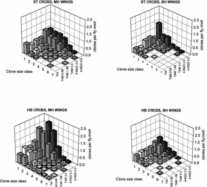
WuÈrgler, 1995) are also shown in Table I. The size distribu-tions of the mwh clones recorded after chronic feeding with TAM and 4-NQO are plotted in Figure 2.
TAM
In the ST cross, TAM gave statistically signi®cant positive results for the frequencies of total spots per ¯y at the two higher concentrations in the MH phenotype and at the highest concentration in the BH phenotype (Table I). The size distributions of the mwh clones show that in the MH wings, the frequencies of large single spots are signi®cantly increased, while in the BH wings this is the case for the small single spots (see also Figure 2). It is a well-known phenomenon that the sizes of the mwh clones in BH wings are always smaller than those in MH wings, which may be due to clones with induced segmental aneuploidy that show reduced proliferation capacity (Frei et al., 1992; Frei and WuÈrgler, 1996). This is best demonstrated here by the values of the mean mwh clone size class (Table I). This observation is in agreement with Styles et al. (1997), who reported that TAM is an aneugenic compound.
Because D.melanogaster has no ER, DNA damage cannot be produced by any pathway related to this receptor. The lipophilicity characteristics of TAM and its metabolites indicate that they can diffuse inside the cell and reach the nuclear chromatin (El-Kattan et al., 2001). The absence of a nuclear ER then suggests that the weak genotoxicity of TAM in the ST cross is the result of DNA adducts. However, the genotoxic effects observed in the MH wings show no clear concentration response and only a very weak one in the BH wings. For this reason, it is not possible to determine in this cross the contribution of recombinational events to total genotoxicity in a quantitative way. From the nearly identical frequencies of mwh clones at the highest concentration in the two phenotypes it can be concluded that TAM most probably has mainly mutagenic activity. This conclusion is further supported by the fact that no positive results were obtained for the induction of twin spots (Table I), which are exclusively due to mitotic recombination.
In contrast to these positive results in the ST cross, TAM was clearly negative in both phenotypes of the HB cross (Table I and Figure 2). We expected that a-hydroxylation of the ethyl group of TAM produced by cytochrome P450 enzymes would
lead to more spots in the HB cross. The negative results obtained in this cross may be explained by an ef®cient elimination of reactive intermediates by the constitutively enhanced xenobiotic metabolism in this cross. Similar effects were demonstrated in mouse liver cells (Moorthy et al., 1997). Thus, it would seem plausible that group II adducts of TAM (a-OHT) cause the weak mutagenicity observed in the ST cross.
The data given in Table I show that a signi®cantly lower frequency of small single spots was observed with 0.66 mM TAM in the MH wings of the ST cross compared with the corresponding negative control frequency. In a similar fashion, signi®cantly reduced frequencies of large single spots were observed with 1.66 and 3.33 mM TAM in the MH wings of the HB cross. This might indicate a possible anti-genotoxic activity of TAM against spontaneous genetic events, but it may also be due to chance variation. It might be possible that other effects of TAM, such as inhibition of oxidative stress by low doses (Bhimani et al., 1993) or by regulation of xenobiotic metabol-ism (Moorthy et al., 1997), could cause this decrease. Further studies with well-known genotoxic agents in combined treat-ments are needed to demonstrate true anti-genotoxic properties of small doses of TAM.
4-NQO
As shown in previous studies with the wing spot test, chronic treatments with 4-NQO gave statistically signi®cant results in the ST cross for all three types of spots, with a clear dose± response effect (Graf et al., 1989; Hayatsu et al., 1992; Negishi et al., 1994; Batiste-Alentorn et al., 1995; Kaya et al., 2002). In particular, the frequencies of twin spots were also signi®cantly increased, which indicates recombinagenic activity. Positive results were also obtained with the HB cross for all three types of spots (Table I and Figure 2). However, with this cross the genotoxic effects were considerably higher than with the ST cross, which is here reported for the ®rst time. The frequencies of total spots per ¯y in MH wings induced by 2.5 and 5.0 mM 4-NQO are ~1.7-fold higher in the HB cross than in the ST cross. This increase in genotoxicity is due to the high constitutive levels of cytochrome P450 enzymes in the HB cross (Saner et al., 1996). In addition, it is quite evident that 4-NQO is far more genotoxic than TAM (Figure 2). Furthermore, linear regression analysis of the effects observed in the two phenotypes of both crosses (data not shown) indicates that 4-NQO has a high recombinagenic activity in the HB cross (~73% recombination). Theoretically, 4-NQO gener-ates ROS that can directly produce DNA single-strand breaks and alkali-labile sites in a similar manner to ionizing radiation. Xenobiotic metabolism of 4-NQO produces the metabolite acetoxyaminoquinoline, which leads to three purine adducts, like UV light lesions that are repaired by excision repair (Mirzayans et al., 1999). Direct damage by oxidative stress would mainly produce mutation events in both crosses. DNA adducts produced by xenobiotic metabolism could induce repair activities that may increase recombinational events. Thus, our results indicate that 4-NQO is a mutagenic but also recombinagenic compound that is dependent on metabolism by cytochrome P450 enzymes.
NDMA
The positive control NDMA produced total spot frequencies ~3-fold higher in the HB cross than in the ST cross (Table I). It gave more small single spots than large single spots, but also signi®cant frequencies of twin spots as shown previously by
Table I. Summar y of result s obtained in the D rosophi la w ing spot test Cros s b Type c Co nc. (mM ) No. of ¯ies Spo ts per ¯y (no. of spot s) statistical diag nosis a Mean mw h clon e size class Clone fo rmation per 10 5cells per cell div ision d Sma ll single spot s (1 ±2 cells) (m =2 ) Large sin gle spots (>2 cells) (m =5 ) Twin spots (m =5 ) Total spots (m =2 ) mwh clone s Obser ved Co ntrol cor rected Tamoxi fen cit rate (Twe en±etha nol) ST MH 0.0 56 0.89 (50) 0.11 (6) 0.05 (3) 1.05 (59) 1.05 (59) 1.56 2.2 0.66 50 0.78 (39)+ f 0.18 (9)± 0.02 (1)± 0.98 (49) ± 0.98 (49) 1.78 2.0 ±0.2 1.66 56 1.00 (56)± 0.32 (18)+ 0.04 (2)± 1.36 (76) + 1.30 (73) 2.00 2.6 0.5 3.33 73 0.89 (65)± 0.22 (16)+ 0.04 (3)± 1.15 (84) + 1.12 (82) 1.93 2.3 0.2 BH e 0.0 37 0.43 (16) 0.03 (1) 0.46 (17) 0.46 (17) 1.35 0.9 0.66 60 0.33 (20)± 0.02 (1)± 0.35 (21) ± 0.35 (21) 1.38 0.7 ±0.2 1.66 22 0.68 (15)+ 0.00 (0)± 0.68 (15) ± 0.68 (15) 1.13 1.4 0.4 3.33 42 0.83 (35)+ 0.12 (5)± 0.95 (40) + 0.95 (40) 1.52 1.9 1.0 HB MH 0.0 60 1.23 (74) 0.42 (25) 0.12 (7) 1.77 (106) 1.76 (106) 2.25 3.6 0.66 56 1.73 (97)+ 0.43 (24)± 0.16 (9)± 2.32 (130) ± 2.16 (121) 2.09 4.4 0.8 1.66 55 1.38 (76)± 0.31 (14)+ f 0.15 (8)± 1.84 (101) ± 1.72 (95) 2.08 3.5 0.1 3.33 54 1.56 (84)± 0.30 (34)+ f 0.15 (8)± 2.00 (108) ± 1.85 (100) 1.95 3.8 0.2 BH e 0.0 55 0.71 (39) 0.09 (5) 0.80 (44) 0.80 (44) 1.70 1.6 0.66 54 0.74 (40)± 0.06 (3)± 0.80 (43) ± 0.80 (43) 1.44 1.6 0.0 1.66 42 0.69 (29)± 0.10 (4)± 0.79 (33) ± 0.79 (33) 1.67 1.6 0.1 3.33 57 0.61 (35)± 0.09 (5)± 0.70 (38) ± 0.67 (38) 1.37 1.3 ±0.2 4-Nitr oquino line-1-oxide (Tw een±ethanol) ST MH 0.0 56 0.89 (50) 0.11 (6) 0.05 (3) 1.05 (59) 1.05 (59) 1.56 2.1 2.5 70 1.71 (120)+ 1.59 (111)+ 0.50 (35)+ 3.80 (266) + 3.57 (250) 2.69 7.3 5.1 5.0 68 1.76 (120)+ 2.00 (136)+ 0.84 (57)+ 4.60 (313) + 4.11 (280) 2.90 8.5 6.3 BH e 0.0 37 0.43 (16) 0.03 (1) 0.46 (17) 0.45 (17) 1.35 0.9 2.5 18 0.83 (15)+ 0.39 (7)+ 1.22 (22) + 1.22 (22) 2.18 2.5 1.5 5.0 28 1.96 (55)+ 1.14 (32)+ 3.11 (86) + 3.07 (86) 2.26 6.3 5.3 HB MH 0.0 60 1.23 (74) 0.42 (25) 0.12 (7) 1.77 (106) 1.76 (106) 2.25 3.6 2.5 52 3.48 (181)+ 1.67 (87)+ 1.25 (65)+ 6.40 (333) + 5.84 (304) 2.39 12.0 8.3 5.0 48 3.44 (165)+ 2.48 (119)+ 1.69 (81)+ 7.60 (365) + 7.20 (346) 2.70 14.8 11.1 BH e 0.0 55 0.71 (39) 0.09 (5) 0.80 (44) 0.80 (44) 1.70 1.6 2.5 55 1.44 (79)+ 0.40 (22)+ 1.84 (101) + 1.83 (101) 1.97 3.7 2.1 5.0 41 1.59 (65)+ 0.68 (28)+ 2.27 (93) + 2.26 (93) 2.08 4.6 3.0 N -n itrosod imethylamine (wat er) ST MH 0.0 73 0.48 (35) 0.10 (7) 0.03 (2) 0.60 (44) 0.60 (44) 1.84 1.2 1.0 54 6.72 (363)+ 2.70 (146)+ 0.59 (32)+ 10.00 (541) + 9.57 (517) 2.11 19.6 18.4 2.0 11 9.73 (107)+ 1.73 (19)+ 0.55 (6)+ 12.00 (132) + 11.0 0 (121) 1.72 22.5 21.3 BH e 0.0 55 0.33 (18) 0.05 (3) 0.38 (21) 0.38 (21) 2.05 0.8 1.0 32 4.47 (143)+ 1.00 (32)+ 5.47 (175) + 5.46 (175) 1.81 11.2 10.4 2.0 17 8.12 (138)+ 1.06 (18)+ 9.18 (156) + 9.00 (156) 1.57 18.4 17.7 HB MH 0.0 76 0.63 (48) 0.30 (23) 0.16 (12) 1.09 (83) 1.07 (82) 2.57 2.2 1.0 10 16.7 0 (167)+ 8.30 (83)+ 4.80 (48)+ 29.80 (298) + 27.7 0 (277) 2.24 56.8 54.6 2.0 4 23.7 5 (95)+ 12.75 (51)+ 2.25 (9)+ 38.75 (155) + 34.0 0 (136) 2.05 69.7 67.5 BH e 0.0 67 0.66 (44) 0.04 (3) 0.70 (47) 0.68 (47) 1.39 1.4 1.0 4 33.5 0 (134)+ 4.25 (17)+ 37.75 (151) + 36.7 5 (151) 1.63 75.4 73.9 aStatistica ldiag noses w ith Mann ±Whitn ey±Wilcoxon U -test acco rding to Fre iand Wu Èrgler (1988 ,1995). m ,mini mal risk mu ltiplicat ion factor for the assessm ent of nega tive results. One sid ed bin omial test s, signi®c ance levels a and b: positive res ults, + (a < 0.05); negat ive res ults, ± (b < 0.05 ). bST, standa rd cross; H B ,high bioa ctivation cross. cMH, marke r-heterozygous wing s; BH, ba lancer-heteroz ygous wing s. dClone frequencies per ¯y divided by the number of cells ex amined per ¯y (48 800) estim ate formatio n frequenc ies per cell and per cell div ision in chroni c ex posure experiments (Frei an d W uÈrg ler, 1995 ). eOnly mwh single spots can be observed in mwh /TM3 heterozygot es as the balan cer chromosome TM3 does not carry a ¯r mutation. fSigni®can tly lower than control.
Kawai (1998) and RodrõÂguez-Arnaiz et al. (1996). These frequencies were considerably higher than those observed with 4-NQO. As expected, the genotoxicity of NDMA was higher in the HB cross than in the ST cross. However, the compound was also de®nitively more toxic in the HB cross so that only very few ¯ies could be analysed. Regression analysis for the two phenotypes of the ST cross (data not shown) leads to a value of 36% recombinagenicity, which is lower than the 67% value observed previously by RodrõÂguez-Arnaiz et al. (1996). It is most probable that differences in concentrations used and sample size (Frei and WuÈrgler, 1995) are responsible for this discrepancy.
General conclusions
Our results indicate that TAM is a weak mutagen in the wing spot test of Drosophila. It is suggested that xenobiotic metabolism (Moorthy et al., 1997) in the HB cross with high levels of cytochromes P450 decreases group II DNA adducts in such a way that this compound and its metabolites were not genotoxic in this cross. The weak genotoxicity of TAM was thus con®rmed in this eukaryotic in vivo assay. In order to
interpret these results, we must also consider that Drosophila has been shown to have some co-regulators and basal transcription factors homologous to human metabolism of TAM (Thackray et al., 2000). Furthermore, there are strong homologies between the CYP6 family of insects and the CYP3 family of vertebrates (Danielson, 1997, 1998), and CYP3A4 yields a-OHT (Boocock et al., 2002). The difference in the regulated and high constitutive synthesis of CYP450 enzymes in the ST and HB crosses, respectively, could result in a high ability to metabolize TAM and thus avoid DNA damage in the HB cross but not in the ST cross. These quantitative enzymatic differences between the two crosses which produce different genotoxicity results resemble other data obtained with human and rat tissues or organs exposed to TAM (Poirier and Schild, 2003). In humans, 4-OHT metabolism is by a thermostable sulphotransferase. This polymorphic enzyme has a wild-type allele SULT1A1*1 and an altered allele SULT1A1*2, with ~40% activity. Poirier and Schild (2003) speculated that TAM detoxi®cation accomplished by SULT1A1 reduces the con-centration of drug available for genotoxic activation. Therefore, the less ef®cient SULT1A1*2 could increase
Fig. 2. Distribution of the mwh clone sizes after feeding of larvae of the ST and the HB cross (MH and BH wings) with different concentrations (mM) of TAM and 4-NQO (TW, Tween±ethanol 3%). The clone sizes are 1, 2, 3±4, 5±8, 9±16, 17±32 and >32 cells.
genotoxic activation. Phillips (see Poirier and Schild, 2003) suggested that the presence of another sulphotransferase SULT2A1, an enzyme found only in rat hepatocytes exposed to TAM and a-OHT, is related to TAM±DNA adduct detection. On the other hand, the absence of SULT2A1 in other cells and tissues of rat and human (lymphocytes and endometrium, respectively) is related to a lack of TAM±DNA adducts. This evidence supports the idea that enzymatic differences may be responsible for the presence or absence of TAM±DNA adducts. We conclude that our study in Drosophila supports the hypothesis that the magnitude of genotoxicity of TAM may be related to quantitative enzymatic differences. Furthermore, our results indicate that the genotoxicity of TAM differs from that of 4-NQO because the latter possesses signi®cant recombinagenic activity and shows a high bioactivation effect.
Acknowledgement
We thank Dr H.Frei for his contributions to this study and his help with the statistical evaluation of the data.
References
Batiste-Alentorn,M., Xamena,N., Creus,A. and Marcos,R. (1995) Genotoxic evaluation of ten carcinogens in the Drosophila wing spot test. Experientia, 51, 73±76.
Bhimani,R.S., Troll,W., Grunberger,D. and Frenkel,K. (1993) Inhibition of oxidative stress in HeLa cells by chemopreventive agents. Cancer Res., 53, 4528±4533.
Boocock,D.J., Brown,K., Gibbs,A.H., Sanchez.E., Turteltaub,K.W. and White,I.N.H. (2002) Identi®cation of human CYP forms involved in the activation of tamoxifen and irreversible binding to DNA. Carcinogenesis, 23, 1897±1901.
Clark,A.M. (1982) The use of larval stages of Drosophila in screening for some naturally occurring mutagens. Mutat. Res., 92, 89±97.
Crewe,H.K., Notley,L.M., Wunsch,R.M., Lennard,M.S. and Gillam,E.M.J. (2002) Metabolism of tamoxifen by recombinant human cytochrome P450 enzymes: formation of the 4-hydroxy, 4¢-hydroxy and N-desmethyl metabolites and isomerization of trans-4-hydroxytamoxifen. Drug. Metab. Dispos., 30, 869±874.
Danielson,P.B., MacIntyre,R.J. and Fogleman,J.C. (1997) Molecular cloning of a family of xenobiotic-inducible drosophilid cytochrome P450s: evidence for involvement in host-plant allelochemical resistance. Proc. Natl Acad. Sci. USA, 94, 10797±10802.
Danielson,P.B., Foster,J.L.M., McMahill,M.M., Smith,M.K. and Fogleman, J.C. (1998) Induction by alkaloids and phenobarbital of Family 4 cytochrome P450s in Drosophila: evidence for involvement in host plant utilization. Mol. Gen. Genet., 259, 54±59.
Dehal,S.S. and Kupfer,D. (1997) CYP2D6 catalyzes tamoxifen 4-hydroxylation in human liver. Cancer Res., 57, 3402±3406.
Desai,P.B., Nallani,S.C., Sane,R.S., Moore,L.B., Goodwin,B.J., Buckley,D.J. and Buckley,A.R. (2002) Induction of cytochrome P450 3A4 in primary human hepatocytes and activation of the human pregnane X receptor by tamoxifen and 4-hydroxytamoxifen. Drug Metab. Dispos., 30, 608±612. Dunkov,B.C., Guzov,V.M., Mocelin,G., Shotkoski,F., Brun,A., Amichot,M.,
Ffrench-Constant,R.H. and Feyereisen,R. (1997) The Drosophila cytochrome P450 gene Cyp6a2: structure, localization, heterologous expression and induction by phenobarbital. DNA Cell Biol., 16, 1345±1356. El-Kattan,A.F., Asbill,C.S., Kim,N. and Michniak,B.B. (2001) The effects of terpene enhancers on the percutaneous permeation of drugs with different lipophilicities. Int. J. Pharm., 215, 229±240.
Frei,H. and WuÈrgler,F.E. (1988) Statistical methods to decide whether mutagenicity test data from Drosophila indicate a positive, negative, or inconclusive result. Mutat. Res., 203, 297±308.
Frei,H. and WuÈrgler,F.E. (1995) Optimal experimental design and sample size for the statistical evaluation of data from somatic mutation and recombination tests (SMART) in Drosophila. Mutat. Res., 334, 247±258. Frei,H. and WuÈrgler,F.E. (1996) Induction of somatic mutation and
recombination by four inhibitors of eukaryotic topoisomerases assayed in the wing spot test of Drosophila melanogaster. Mutagenesis, 11, 315±325. Frei,H., Clements,J., Howe,D. and WuÈrgler,F.E. (1992) The genotoxicity of
the anti-cancer drug mitoxantrone in somatic and germ cells of Drosophila melanogaster. Mutat. Res., 279, 21±33.
FroÈlich,A. and WuÈrgler,F.E. (1989) New tester strains with improved bioactivation capacity for the Drosophila wing spot test. Mutat. Res., 216, 179±187.
Graf,U. and van Schaik,N. (1992) Improved high bioactivation cross for the wing somatic mutation and recombination test in Drosophila melanogaster. Mutat. Res., 271, 59±67.
Graf,U., WuÈrgler,F.E., Katz,A.J. Frei,H., Juon,H., Hall,C.B. and Kale,P.G. (1984) Somatic mutation and recombination test in Drosophila melanogaster. Environ. Mutagen., 6, 153±188.
Graf,U., Frei,H., KaÈgi,A., Katz,A.J. and WuÈrgler,F.E. (1989) Thirty compounds tested in the Drosophila wing spot test. Mutat. Res., 222, 359±373.
Graf,U., Abraham,S.K., GuzmaÂn-RincoÂn,J. and WuÈrgler,F.E. (1998) Antigenotoxicity studies in Drosophila melanogaster. Mutat. Res., 402, 203±209.
HaÈllstroÈm,I., Blanck,A. and Atuma,S. (1984) Genetic variation in cytochrome P-450 and xenobiotic metabolism in Drosophila melanogaster. Biochem. Pharmacol., 33, 13±20.
Hayatsu,H., Inada,N., Kafutani,T., Arimoto,S., Negishi,T., Mori,K., Okuda,T. and Sakata,I. (1992) Suppression of genotoxicity of carcinogens by (±)-epigallocatechin gallate. Prev. Med., 21, 370±376.
IARC (1996) Tamoxifen. In IARC Monographs on the Evaluation of Carcinogenic Risks to Humans, no. 66, Some Pharmaceutical Drugs. IARC, Lyon, pp. 253±365.
Kawai,K. (1998) Enhancement of the DNA damaging activity of N-nitrosodimethylamine by di(2-ethylhexyl)phthalate in somatic cells in vivo of Drosophila melanogaster. Biol. Pharm. Bull., 21, 579±582.
Kaya,B., Creus,A., Velazquez,A., Yaniko,A. and Marcos,R. (2002) Genotoxicity is modulated by ascorbic acid. Studies using the wing spot test in Drosophila melanogaster. Mutat. Res., 520, 93±101.
Mirzayans,R., Bashir,S., Murray,D. and Paterson,M.C. (1999) Inverse correlation between p53 protein levels and DNA repair ef®ciency in human ®broblast strains treated with 4-nitroquinoline 1-oxide: evidence that lesions other than DNA strand breaks trigger the p53 response. Carcinogenesis, 20, 941±946.
Moorthy,B., Sriram,P., Randerath,E. and Randerath,K. (1997) Effects of cytochrome P450 inducers on tamoxifen genotoxicity in female mice in vivo. Biochem. Pharmacol., 53, 663±669.
Negishi,T., Nakano,H., Kitamura,A., Itome,C., Shiotani,T. and Hayatsu,H. (1994) Inhibitory activity of chlorophyllin on the genotoxicity of carcinogens in Drosophila. Cancer Lett., 83, 157±164.
Notley,L.M., De Wolf,C.J., Wunsch,R.M., Lancaster,R.G. and Gillam,E.M. (2002) Bioactivation of tamoxifen by recombinant human cytochrome p450 enzymes. Chem. Res. Toxicol., 15, 614±622.
Osborne,M.R., Hardcastle,I.R. and Phillips,D.H. (1997) Minor products of reaction of DNA with alpha-acetoxytamoxifen. Carcinogenesis, 18, 539± 543.
Poirier,M.C. and Schild,L.J. (2003) Meeting Report. The genotoxicity of tamoxifen: extent and consequences, Kona, Hawaii, January 23, 2003. Mutagenesis, 18, 395±399.
Randerath,K., Moorthy,B., Mabon,N. and Sriram,P. (1994) Tamoxifen: evidence by 32P-postlabeling and use of metabolic inhibitors for two
distinct pathways leading to mouse hepatic DNA adduct formation and identi®cation of 4-hydroxytamoxifen as a proximate metabolite. Carcinogenesis, 15, 2087±2094.
RodrõÂguez-Arnaiz,R., Soto,P.O., Oyarzun,J.C. and Graf,U. (1996) Analysis of mitotic recombination induced by several mono- and bifunctional alkylating agents in the Drosophila wing spot test. Mutat. Res., 351, 133±145. Saner,C., Weibel,B., WuÈrgler,F.E. and Sengstag,C. (1996) Metabolism of
promutagens catalyzed by Drosophila melanogaster CYP6A2 enzyme in Saccharomyces cerevisiae. Environ. Mol. Mutagen., 27, 46±58.
Sharpe,C.R., Collet,J.-P., Belzile,E., Hanley,J.A. and Boivin,J.-F. (2002) The effects of tricyclic antidepressants on breast cancer risk. Br. J. Cancer, 86, 92±97.
Shibutani,S., Shaw,P.M., Suzuki,N., Dasaradhi,L., Duffel,M.W. and Terashima,I. (1998) Sulfation of alpha-hydroxytamoxifen catalyzed by human hydroxysteroid sulfotransferase results in tamoxifen±DNA adducts. Carcinogenesis, 19, 2007±2011.
Shibutani,S., Reardon,J.T., Suzuki,N. and Sancar,A. (2000) Excision of tamoxifen-DNA adducts by the human nucleotide excision repair system. Cancer Res., 60, 2607±2610.
SpanoÂ,M.A., Frei,H., WuÈrgler,F.E. and Graf,U. (2001) Recombinagenic activity of four compounds in the standard and high bioactivation crosses of Drosophila melanogaster in the wing spot test. Mutagenesis, 16, 385± 394.
StiborovaÂ,M., Bosek-DohalskaÂ,L., Hodek,P., MraÂz,J. and Frei,E. (2002) New selective inhibitors of cytochromes P450 2B and their application to antimutagenesis of tamoxifen. Arch. Biochem. Biophys., 40, 41±49. Styles,J.A., Davies,A., Davies,R., White,I.N. and Smith,L.L. (1997)
Clastogenic and aneugenic effects of tamoxifen and some of its analogues in hepatocytes from dosed rats and in human lymphoblastoid cells transfected with human P450 cDNAs (MCL-5 cells). Carcinogenesis, 18, 303±313.
Thackray,V.G., Young,R.H., Hooper,J.E. and Nordeen,S.K. (2000) Estrogen agonist and antagonist action on the human oestrogen receptor in Drosophila. Endocrinology, 141, 3912±3915.
van Schaik,N. and Graf,U. (1991) Genotoxicity evaluation of ®ve tricyclic antidepressants in the wing somatic mutation and recombination test in Drosophila melanogaster. Mutat. Res., 260, 99±104.
van Schaik,N. and Graf,U. (1993) Structure±activity relationships of tricyclic antidepressants and related compounds in the wing somatic mutation and recombination test of Drosophila melanogaster. Mutat. Res., 286, 155±163. White,I.N.H. (1999) The tamoxifen dilemma. Carcinogenesis, 20, 1153±1160. Received on June 2, 2003; revised on January 30, 2004;