Leptin modulates extracellular matrix molecules and metalloproteinases: possible implications for trophoblast invasion
8
0
0
Texte intégral
(2) M.Castellucci et al.. enzymes which are secreted as inactive zymogens and must be cleaved to become active. Huppertz et al. (1998) have shown strong immunostaining for MMP-2 and MMP-9 in the distal, invasive part of cell columns. As one of the most important features of the placenta is the tightly regulated invasive behaviour and leptin has been identified in some of the main components of this organ (Masuzaki et al., 1997; Senaris et al., 1997), we postulated that leptin and its receptor could influence and modulate the invasive behaviour of the trophoblast during its invasiveness. In this study, we used primary cultures of cytotrophoblastic cells to test the influence of human leptin on MMP-2, MMP-9 and fFN production as these are molecules which regulate the invasive ability of the cytotrophoblastic cells. In addition, we investigated the immunolocalization of leptin and its receptor in the extravillous cytotrophoblastic cells of cell columns because no such data are available for these structures which play a pivotal role in trophoblast invasion.. Materials and methods Tissues In all, 19 human placentae at 7 (n ⫽ 3), 8 (n ⫽ 2), 10 (n ⫽ 1), 12 (n ⫽ 5), 38 (n ⫽ 5) and 40 (n ⫽ 3) weeks post-menstruation and umbilical cords (from 38–40 week placentae) were collected from clinically normal pregnancies interrupted by curettage (aspiration technique) for psychosocial or medical reasons that were unlikely to affect placental structure and function, or terminated by Caesarian sections or normal vaginal deliveries. Placentae were cut into small blocks. Specimens of white adipose tissue were collected from the abdominal wall of five patients undergoing Caesarian section at 38 weeks gestation. Ovarian tissue was obtained from young women undergoing laparotomy for reasons unrelated to ovarian pathology. Tissue preparation for immunohistochemistry Placental specimens (n ⫽ 19) were rapidly washed with 0.1 mol/l phosphate buffer at pH 7.4 and fixed for 4–12 h in 4% buffered formalin at 4°C. The specimens were then dehydrated in ethanol and embedded in paraffin wax. Serial sections (3 µm) were used for light microscopy and immunohistochemistry. Samples for reverse transcription–polymerase chain reaction (RT–PCR) and Northern blotting Placental specimens (n ⫽ 19), umbilical cords (n ⫽ 8), human white adipose tissue (n ⫽ 5) and human ovarian tissue (n ⫽ 5) were collected and immediately frozen and stored in liquid nitrogen until being used for RNA extraction. Human white adipose tissue was used as a positive control. Immunohistochemistry Primary antibodies The following primary antibodies were used: (i) mouse monoclonal antibody anti-human pan-cytokeratin (Dako, Glostrup, Denmark; diluted 1:500 v/v); (ii) rabbit polyclonal antibody (A-20) raised against a peptide corresponding to amino acids 137–156 mapping at the carboxy terminus of human leptin (Santa Cruz Biotechnology, Santa Cruz, CA, USA; diluted 1:100 v/v); (iii) affinity-purified goat polyclonal antibody (N-20) recognizing the amino terminus of all the alternatively spliced forms of the leptin receptor, i.e. ObRS⫹ ObRL (amino acids 32–51 of the human leptin-R, Santa Cruz Biotechnology, diluted 1:200 v/v); (iv) affinity-purified goat polyclonal antibody (C-20) directed against the peptide corresponding to amino. 952. acids 1146–1165 at the carboxy terminus of the human leptin-R, thus recognizing only the long form of the leptin receptor, ObRL (Santa Cruz Biotechnology; diluted from 1:100 to 1:200 v/v). The specificity of the antibodies for the leptin receptor has been tested on human tissues and cells by the manufacturer (Santa Cruz Biotechnology) and by previous studies (Glasow et al., 1998; Morton et al., 1998) using Western blotting and immunohistochemistry. Immunostaining Immunohistochemical demonstration of leptin, leptin-R and pancytokeratin was performed on serial sections using avidin–biotin peroxidase (ABC) (Hsu et al., 1981). Briefly, dewaxed sections (3 µm) were processed through the following incubation steps: (i) hydrogen peroxide 0.3% in methanol for 30 min to block endogenous peroxidase; (ii) normal rabbit serum (for leptin-R), normal goat serum (for leptin), or normal horse serum (for pan-cytokeratin), diluted 1:75 v/v, for 20 min to reduce non-specific background staining; (iii) primary antibody, overnight at 4°C; (iv) biotinylated secondary antibody: rabbit anti-goat immunoglobulin G (IgG) (for leptin-R), goat anti-rabbit IgG (for leptin), or horse anti-mouse IgG (for pancytokeratin) diluted 1:200 v/v, for 30 min (Vector Laboratories, Burlingame, CA, USA); (v) ABC complex for 1 h (Vectastain ABC kit; Vector Laboratories), (vi) histochemical visualization of peroxidase using 0.075% 3⬘,3⬘diaminobenzidine hydrochloride as chromogen (Sigma, St Louis, MO, USA) and hydrogen peroxide 0.02% in Tris buffer 0.05 mol/l, pH 7.6, for 5 min in a dark room. Sections were then rinsed in tap water, counterstained with haematoxylin, dehydrated and mounted with Eukitt (Kindler GmbH & Co, Freiburg, Germany). Specificity tests were performed: (i) by omitting the primary antibody in the immunostaining procedure; (ii) by incubating sections with the antiserum saturated with homologous antigen (for this procedure, the antibody was incubated with a 10-fold excess of homologous peptide (15 µg/ml), for 48 h); or (iii) by replacing the primary antibody with the pre-immune serum. These controls showed negative results. RNA extraction and analysis Total RNA was extracted after homogenization of tissues considered above in the presence of 4 mol/l guanidinium isothiocyanate by ultracentrifugation through 5.7 mol/l caesium chloride gradient. cDNA synthesis was obtained from 5 µg of total RNA in 50 mmol/l Tris– HCl pH 8.3, 75 mmol/l KCl, 3 mmol/l MgCl2, 10 mmol/l dithiothreitol, 1 mol/l of each dNTP, 250 ng of oligo(dT)15, 250 ng random hexamers, 100 ng/µl nuclease-free bovine serum albumin, 20 IU rRNAsin (Promega, Madison, WI, USA) and 500 IU of recombinant Molony murine leukemia virus reverse transcriptase (Gibco BRL, Milano, Italy). RNA and primers were kept at 70°C for 5 min and then cDNA synthesis was allowed to proceed at 37°C for 1 h. Oligonucleotide primers sequences Leptin primers: L⫹ (5⬘-GATGACACCAAAACCCTCATC-3⬘) L– (5⬘-GGCCACCACCTCTGTGGAGTA-3⬘) Leptin receptor primers for the short form (ObRS) of the receptor (exon 2): LR2⫹ (5⬘-GGCTAAAAGGACACTTAAAAT-3⬘) LR2– (5⬘-TTTCATTCAGATTGAAAAAAGT-3⬘) Leptin receptor primers for the long form (ObRL) of the receptor (exon 5): LR5⫹ (TAGAATCTCTTCATCTGTTAAGA-3⬘) LR5– (5⬘-AATCCTTCTGAGAATGTG-3⬘). PCR conditions Of the cDNA synthesized from 5 µg of total RNA 20% was used for PCR amplifications in 10 mmol/l Tris–HCl pH 8.3, 50 mmol/l.
(3) Leptin and trophoblast invasion KCl, 5 mmol/l MgCl2, 0.2 mmol/l of each dNTP, primers at a concentration of 0.5 µmol/l and 4 IU of recombinant Taq DNA Polymerase in a final volume of 50 µl. After denaturation at 96°C for 5 min, samples were subjected to 40 cycles (50 s at 94°C, 50 s at 60°C and 1 min at 72°C) using a DNA Thermal Cycler (PerkinElmer Cetus, Norwalk, CT, USA). All cDNAs were also amplified with β-actin-primers to test their quality. Cloning of PCR amplified cDNAs into phagemid vectors The amplified leptin fragments were sequenced (using the same primers as those used for PCR amplification) with a PRISM™ sequencing kit (Perkin-Elmer) to confirm their identity, then cloned into plasmids (pCRII – TA Cloning, Invitrogen, Grohingen, The Netherlands) and used as probes after labelling with α-32P [dCTP] by random primers method (Megaprime Labelling Kit; Amersham, Buckinghamshire, UK). Northern Blot analysis RNA was quantified as follows: 10 µg of total RNA was electrophoresed in denaturating conditions (formaldehyde–agarose gel) and capillary-blotted with 20⫻ sodium chloride/sodium citrate (SSC; 1⫻ SSC ⫽ 150 mmol/l NaCl, 15 mmol/l sodium citrate, pH 7.0) overnight onto a nylon membrane (Hybond-N, Amersham), then UV crosslinked and baked. RNA blots were hybridized overnight at 68°C in hybridization buffer containing 5⫻ SSPE (20⫻ SSPE ⫽ 3 mol/l NaCl, 0.2 mol/l NaH2PO4, 25 mmol/l EDTA, pH 7.4), 2% sodium dodecyl sulphate (SDS), 10% dextran sulphate and 1.5 µg denaturated herring sperm DNA. Post-hybridization washing was performed twice in a solution of 2⫻ SSC, 0.1% SDS and 1⫻ SSC, 0.1% SDS at 56°C for 10 min each time. RNA blots were then exposed to Kodak XOmat film at –70°C. Hybridization with the β-actin probe was performed to verify the RNA integrity and loading efficiency. Preparation of cytotrophoblastic cells (CTB) CTB were isolated, purified, characterized and cultured as previously described (Bischof et al., 1991). Briefly, trophoblastic tissue obtained from legal abortions (at 7–12 weeks gestation) was digested by trypsin and the CTB were separated from blood cells and syncytiotrophoblast on a discontinuous Percoll gradient with the contaminating leukocytes being removed by immunopurification with an antibody to CD45 (Dako) coupled to magnetic particles (Dyna Beads; Dynal, Geneva, Switzerland). CTB were counted in a Neubauer cell in the presence of Trypan Blue (Sigma) and diluted to 106 cells/ml. Cells were cultured overnight in Dulbecco’s modified Eagle’s medium (DMEM) containing 2 mmol/l L-glutamine, 4.2 mmol/l magnesium sulphate, 2.5 mmol/l HEPES, 1% gentamycine, 1% amphopterin B, 100 µg/ml streptomycin and 100 µg/ml penicillin in presence of 10% fetal bovine serum. The next morning (day 0), the medium was changed to serum-free DMEM and cells were incubated in the presence or absence of recombinant human leptin (rhLept, 0.001–1 µg/ml; R&D Systems, Bu¨ hlmann AG, Basel, Switzerland). Incubation was performed under a 5% CO2 and 95% air atmosphere in a humid incubator at 37°C. The medium was changed on days 2 and 4 and the culture was stopped on day 4. The supernatants were divided into aliquots and stored at –20°C until assayed. The cells were lysed with 200 µl Triton X-100 (25% in water) and stored at –20°C for total cell protein measurements. MMP-9 and MMP-2 enzyme-linked immunosorbent assays (ELISA) MMP-2 ELISA Microplates (96-well) were coated overnight at 4°C with 100 µl of sheep anti-human MMP-2 IgG (The Binding Site, Anawa, Wangen, Switzerland; 30 µg/ml in sodium carbonate buffer, 50 mmol/l, pH 9.6).. Unbound sites were blocked for 2 h at room temperature with 250 µl of 10% Blotto (non-fat dry milk; BioRad, Hercules, CA, USA) in phosphate-buffered saline (PBS) containing 0.02% NaN3. Plates were then washed twice with PBS containing 0.1% Tween 20 (PBST; 250 µl/well) and once with PBST ⫹ 10% Blotto (PBSTB). Samples and standards were diluted in PBS containing 10% Blotto, applied in duplicate (100 µl/well) and incubated overnight at room temperature. After incubation, the plates were washed as described above, and incubated with biotinylated anti-MMP2 IgGs (100 µl/well) for 2 h at room temperature on a rotating platform. Plates were washed three times for 15 min with PBST, and once with PBSTB and re-incubated for 30 min at 20°C with avidin peroxidase (1/4000 in PBSTB, 100 µl/well). After washing four times with PBST, the plates were incubated in the dark for 10 min with o-phenylenediamine (1.2 mg/ml, 100 µl/well) and H2O2. The reaction was stopped by the addition of H2SO4 (2M, 50 µl/well) and the absorbance was measured at 492 nm in an ELISA plate reader (Lasystem Multiscan; BioConcept, Allschwill, Switzerland). Biotinylation of antibodies Sheep anti-human MMP-2 IgG (500 µl, 13 mg/ml) was diluted 1:1 with bicarbonate buffer (0.1 mol/l, pH 8.4) and dialysed against this buffer for 48 h at 4°C. Activated biotin, at a concentration of 10 mg/ml in DMSO, was added (110 µl) and incubated for 2 h at room temperature. The preparation was then extensively dialysed against PBS containing 0.02% NaN3, and stored at 4°C. Purification of MMP-9 The purification procedure was performed using a human histiocytic lymphoma cell line (U937), according to a previously described protocol (Ward et al., 1991). Pooled conditioned medium from U937 cells (4.8 l) to which 48 ml of 1 mol/l Tris, pH 7.6 was added, was applied on a Gelatin–Sepharose column (5⫻2.5 cm), equilibrated in Tris 10 mmol/l, NaCl 1 mmol/l, CaCl2 10 mmol/l. 0.04% Brij, pH 7.6 (buffer A). The column was thoroughly washed with buffer A and eluted with 10% dimethylsulphoxide (DMSO) in buffer A. The presence of MMP-9 in the fractions was tested by gelatin zymography (see below). Fractions containing MMP-9 activity were pooled, dialysed against buffer A and applied to a Concanavalin-A–Sepharose column (2.5⫻9 cm), equilibrated in buffer A. After washing, the column was eluted with 0.5 mol/l Methyl-α-D-manno-pyrannoside. MMP-9-containing fractions were concentrated on a small Gelatin– Sepharose column (0.5⫻10 cm). The pooled MMP-9 fractions were dialysed against Tris 0.01 mol/l, NaCl 0.1 mol/l, CaCl2 10 mmol/l, Brij 0.04%, divided into aliquots and stored at –20°C. Production of anti-MMP-9 polyclonal antibodies Purified MMP-9 was dialysed against PBS and 40 µg/rabbit was injected s.c. in different sites in two rabbits. A second 20 µg injection was carried out 5 weeks later, and a third was performed 4 weeks after the second one. Rabbits were bled 7 days after the third injection. Titration was monitored by Ouchterlony double-diffusion. Sera with a titre of 艌1/32 were pooled. An IgG preparation was obtained by ammonium sulphate precipitation of these pooled rabbit sera. The IgG concentration was estimated by measuring the optical density at 280 nm and found to be 9.6 mg/ml. MMP-9 ELISA Washing and incubation procedures were essentially the same as for the MMP-2 ELISA. Our rabbit anti-human MMP-9 IgG preparation was used for coating the plates (48 µg/ml). The second antibody was the commercially available sheep anti-MMP-9 IgG (The Binding Site, Anawa, Wangen, Switzerland), diluted 1:2000 in PBSTB. Peroxidaselabelled rabbit anti-sheep antibodies (100 µl/well) were then used in an incubation for 1 h at room temperature. Detection was performed as for MMP-2 ELISA.. 953.
(4) M.Castellucci et al. Measurement of MMP-2 and MMP-9 The concentration of immunoreactive MMP-2 and MMP-9 were calculated by comparison to the standard curves (calibrated supernatants from gengival fibroblasts or U937 cells respectively), calcu-. 954. lated as the optical density versus the log concentration of the MMPs and the results were expressed as ng/ml. These calculations were performed on a Macintosh computer (Power PC) using regression analysis from the StatView Programme (Abascus)..
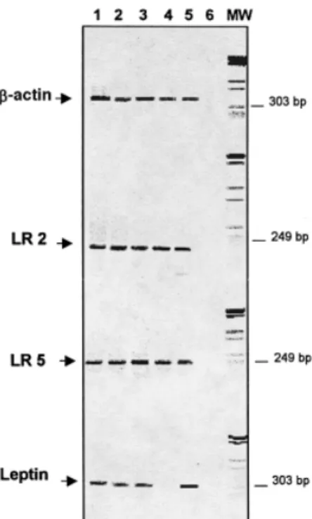
(5) Leptin and trophoblast invasion Gelatinolytic assays Zymography was performed as previously described (Bischof et al., 1991; Martelli et al., 1993). Zymograms were scanned in an ‘Apple Onescanner’ and the surface of the digestion bands measured by the NIH Image 1.60 program with a Power Macintosh 7100/66 computer. All zymograms were evaluated using the same preset standards and the activities of MMP-2 and MMP-9 were expressed in arbitrary units/ml. Hormone and protein assays fFN was measured by a commercially available enzyme immunoassay with a sensitivity of 50 ng/ml and a coefficient of variation of 7.5% (Adeza Biochemical, Sunnyvale, CA, USA). Total cell proteins were measured in the lysed cells using the Bio-Rad protein assay according to the manufacture’s instructions and using bovine serum albumin as the standard (Bio-Rad, Mu¨ nchen, Germany).. Statistical analysis To evaluate the effects of rhLept on the different trophoblastic parameters, the individual values were transformed into values per mg cell proteins and per day [(conc.day2 ⫹ conc.day4)/4] and expressed as a percentage of the respective controls (CTB in absence of rhLept). All experiments were run in duplicates and repeated with three different preparations of CTB. Statistical analyses were performed by one-way analysis of variance, and analyses of correlation using the Statview 4.5 program on a Power Macintosh 7100/66 computer.. Results Since the material from first trimester placentae only occasionally included substantial parts of the basal or chorionic plate our results concern the villous trees, cell islands and cell columns. However, the tissue probes from third trimester placentae covered representative parts of the whole placenta from the chorionic to the basal plate. Immunohistochemical analysis of leptin and leptin receptor Identical leptin receptor immunostaining patterns were observed with the antibodies recognizing the short and long form of leptin-R, suggesting that these forms are similarly expressed in the human placenta. The immunostaining patterns reported below were consistent across all samples. Chorionic villi Leptin immunoreactivity was present in the cytoplasm of the villous trophoblast (syncytiotrophoblast and cytotrophoblast), at all stages of pregnancy (Figures 1a,b). All the other villous components were negative for leptin. Leptin-R immuno-. Figure 2. Reverse transcription–polymerase chain reaction (RT–PCR) expression analysis of Leptin Receptors (LR) splice variants and leptin in human placenta. Lane 1 ⫽ first trimester placenta; lane 2 ⫽ third trimester placenta; lane 3 ⫽ umbilical cord; lane 4 ⫽ human ovary; lane 5 ⫽ human white adipose tissue; lane 6 ⫽ control reaction (without DNA); MW ⫽ molecular markers (low range, BIO-RAD). β-actin, LR2 (the short splice variant of leptin receptor), LR5 (the long splice variant of leptin receptor), and leptin are shown. All tissues were extracted and reverse transcribed at the same time.. Figure 3. Northern blotting for evaluating the expression of leptin mRNA in human placenta. Lane 1 ⫽ sample from human white adipose tissue; lanes 2–4 ⫽ samples from first trimester human placentas; lanes 5–7 ⫽ samples from third trimester human placentas and lanes 8 and 9 ⫽ samples from human umbilical cords. β-actin probe hybridization of the same blot is shown at the bottom.. Figure 1. Immunolocalization of leptin and leptin-R in human placenta of first and third trimester of gestation. (a) Immunohistochemical localization of leptin. The immunostaining is mainly located in the cytoplasm of villous trophoblast (syncytiotrophoblast and cytotrophoblast, ninth week of gestation). (b) Leptin immunostaining pattern during the 38th week of gestation. Syncytiotrophoblast and villous cytotrophoblastic cells (arrows) are positive. (c) Immunostaining for leptin-R at the eighth week of gestation. The positive reaction product is on the apical plasma membrane of the syncytiotrophoblast. Villous cytotrophoblastic cells are also stained. Note the immunostaining of endothelial cells of the fetal vessel (V) and of a Hofbauer cell (arrow). (d) Placental villi which are continuous with cell columns. In the cell columns the positive reaction product for leptin-R is in the extravillous cytotrophoblastic cells distally located (*) from the villous stroma (eighth week of gestation). (e) Basal plate. Immunostaining for leptin-R. This section is parallel to the one showing cytokeratin immunostaining depicted in part (f). Extravillous cytotrophoblastic cells (arrows) are positive for leptin-R. Note the decidual cells (arrowheads) which are positive for leptin-R and negative for cytokeratin in (f). (f) Basal plate at the 38th week of gestation. Immunostaining for cytokeratin. Section parallel to the one depicted in (e) and immunostained for leptin-R. Extravillous cytotrophoblastic cells (arrows) are positive for cytokeratin. Note the negative immunostaining of decidual cells (arrowheads). (a, e, f) scale bar ⫽ 23 µm; (b) scale bar ⫽ 14 µm. (c) scale bar ⫽ 9µm; (d) scale bar ⫽ 90 µm.. 955.
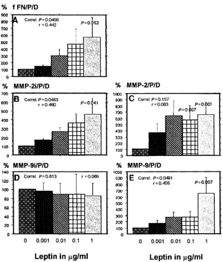
(6) M.Castellucci et al.. Figure 4. Effects of recombinant human leptin (rhLept) on the trophoblastic secretion of (A) fetal fibronectin (fFN), (B) of immunoreactive matrix metalloproteinse (MMP)-2 (D) and MMP-9, (C) of the gelatinolytic activity of MMP-2 and (E) MMP-9. Results are presented as mean ⫾ SEM per mg of cell protein and per day and expressed as percentage of control values (cell without leptin). Culture conditions are described in Materials and methods. % fFN/P/D ⫽ percentage fFN/mg cell protein per day; % MMP-2i/P/D ⫽ percentage immunoreactive MMP-2 per mg cell protein per day; % MMP-9i/P/D ⫽ percentage immunoreactive MMP-9 per mg cell protein and per day; % MMP-2/P/ D ⫽ percentage of zymographic activity of MMP-2 per mg cell protein per day; % MMP-9/P/D ⫽ percentage of zymographic activity of MMP-9 per mg cell protein per day.. reactivity was expressed in villous trophoblast at all stages of pregnancy (Figure 1c). The apical plasma membrane of the syncytiotrophoblast was intensely labelled (Figure 1c). In the first trimester of gestation the endothelial cells of various fetal vessels were positive for leptin-R (Figure 1c) whereas at term few blood vessels were stained for this antigen. Hofbauer cells also showed leptin-R immunostaining (Figure 1c). Cell columns and cell islands Leptin immunostaining was present in all extravillous cytotrophoblastic cells of cell columns and cell islands. Interestingly, leptin-R immunoreactivity was strongly expressed in the extravillous cytotrophoblastic cells distally located from the adherent villous (Figure 1d). Chorionic plate and basal plate In the third trimester of gestation, leptin immunostaining was found in both the extravillous cytotrophoblastic cells of the chorionic plate and in amniotic epithelial cells. Both cellular components were also positive for leptin-R (data not shown). Leptin and leptin-R (Figure 1e) were detectable in the majority of the extravillous trophoblastic cells (positive for cytokeratin; 956. Figure 1f) of the basal plate. However, some of these cytotrophoblastic cells were weakly stained and/or negative for this antigen. Decidual cells (negative for cytokeratin; Figure 1f), were positive for leptin and leptin-R (Figure 1e). Leptin and leptin receptor mRNA expression RT–PCR yielded bands of the expected sizes for ObRL (LR5) and ObRS (LR2) forms in all samples considered (Figure 2). Leptin mRNA was detected in first and third trimester placentae, umbilical and white adipose tissue. Human ovary was used as the negative control for leptin expression (Karlsson et al., 1997). The identities of the amplified fragments were confirmed by restriction enzyme cleavage. Northern blot analysis revealed leptin mRNA in all samples considered, in line with the RT–PCR data (Figure 3) and with a previous study (Senaris et al., 1997). Effects of leptin on cytotrophoblastic cells Positive immunostaining for leptin-R in both the invading extravillous cytotrophoblastic cells of cell islands and cell.
(7) Leptin and trophoblast invasion. columns and RT–PCR-positive results prompted us to investigate the influence of leptin on the enzymes responsible for trophoblast invasion. As shown in Figure 4, rhLept increased in a dose-dependent manner (P ⫽ 0.0456), the trophoblastic secretion of fFN (Figure 4A) and the concentration of immunoreactive MMP-2 (P ⫽ 0.0463, Figure 4B), but not of MMP9 (Figure 4D). However, the MMP-9 activity was increased in a dose-dependent manner (P ⫽ 0.0491, Figure 4E). Although rhLept did not stimulate MMP-2 activity in a dose-dependent manner (P ⫽ 0.157), the higher concentrations of rhLept (0.1 and 1 µg/ml) significantly increased MMP-2 activity (Figure 4C).. Discussion The functional roles of leptin and leptin-R in the human placenta have not yet been clarified. In this study we demonstrate for the first time that leptin and leptin-R are expressed in the extravillous cytotrophoblastic cells of cell columns which are structures of the placental villous tree that play a pivotal role in placental growth and in trophoblast invasion (Mu¨ hlhauser et al., 1995; Kaufmann and Castellucci, 1997). Immunostaining for leptin-R was peculiar in these structures, showing a clear gradient, i.e. weak expression in the more proximal (proliferating) elements and strong expression in the distal extravillous trophoblastic cells involved in the invasive processes. The functional significance of leptin and leptin-R expression in these cells could thus be related to the invasive processes, possibly involving an enhancement of the activity and/or synthesis of the molecules regulating trophoblastic invasion. Trophoblast invasion requires degradation of extracellular matrix molecules by the activity of specific proteases (Bischof and Martelli, 1992; Graham and Lala, 1992) as well as synthesis of fFN, a molecule that facilitates the migration of the extravillous cytotrophoblastic cells into the uterine wall (Bischof and Martelli, 1992; Graham and Lala, 1992). It has been previously demonstrated that MMP-2, MMP-9 and fFN (Feinberg et al., 1991; Castellucci et al., 1993; Huppertz et al., 1998) are expressed in the distal part of the extravillous cytotrophoblastic cells of cell columns where we detect a strong expression of leptin-R (this study). These data led us to consider leptin as a potential specific regulator of the production of these molecules. Our in-vitro studies support this hypothesis because leptin regulates MMP-2 and MMP-9, as well as the extracellular matrix molecule, fFN. We showed that leptin increases in a dose-dependent manner the secretion of immunoreactive MMP-2 and fFN and enhances the activity of MMP-9 in cytotrophoblastic cells. Interestingly, leptin does not affect the cytotrophoblastic secretion of immunoreactive MMP-9, although it significantly enhances MMP-9 activity. This enzyme is known to be essential for in-vitro and in-vivo invasion (Librach et al., 1991; Bischof et al., 1995a; Huppertz et al., 1998). This observation might indicate that leptin does not influence the production of MMP-9, but does stimulate its activation. It is tempting to speculate that leptin could regulate the synthesis of potential activators of MMP-9 (Ogata et al., 1992). In contrast to other regulators of trophoblast invasion, e.g.. leukaemia inhibitory factor (LIF) (Bischof et al., 1995b) and tumour necrosis factor α (Meisser et al., 1999), leptin stimulates fFN and MMP-2 secretion indicating that this hormone probably acts by a different mechanism. In the present study, leptin-stimulated matrix deposition (measured as fFN) as well as the proteolytic activity of cytotrophoblastic cells, thereby acting similarly to interleukin (IL)-1α (Meisser et al., 1999), a known stimulator of MMPs (Rawdanowicz et al., 1994). Extending previous observations (Masuzaki et al., 1997; Senaris et al., 1997; Mise et al., 1998), we show that leptinR (long and short forms) is expressed at the apical surface of the syncytiotrophoblast of the first and third trimester of gestation suggesting that plasma leptin may have an autocrine and/or paracrine role at this level. The function of leptin and its receptor in villous trophoblast might be related to the secretion of human chorionic gonadotrophin (HCG) since rhLept induces a dose-dependent stimulation of HCG secretion by primary cultures of cytotrophoblastic cells in vitro (Chatdonnens et al., 1999). Thus, the role of leptin-R may be considered similar to that of other molecules, e.g. c-erbB-2, which are expressed both on the syncytiotrophoblast and on extravillous cytotrophoblastic cells, but are considered to play different roles in these two locations (Mu¨ hlhauser et al., 1993). Recently, it has been demonstrated that leptin promotes angiogenesis in cultured human vein endothelial cells and in porcine aortic endothelial cells (Bouloumie´ et al., 1998; SierraHonigmann et al., 1998). Moreover, leptin exhibits a potent neovascularization effect in corneal assays conducted with normal rats (Sierra-Honigmann et al., 1998). Our findings showed leptin-R positivity in endothelial cells of various fetal blood vessels of chorionic villi during the first trimester of gestation. Leptin might therefore contribute to the development of placental vessels together with other growth factors such as vascular endothelial growth factor (Shiraishi et al., 1996), basic fibroblast growth factor (Mu¨ hlhauser et al., 1996) and platelet derived growth factor B (Holmgren et al., 1991).. Acknowledgements I.D. is a recipient of an Ernst Sharing–FIGO Scholarship. The authors wish to thank F.Verdenelli, L.Haenggeli and C.Wuillemin for their skillful technical assistance. The authors are indebted to Professor F.Carle for useful discussions. This study was supported by funds from the Swiss National Science Foundation (grant no. 32–49257.96) to B.P. and funds from the Italian Ministry of University and Scientific Research (MURST) and the University of Ancona to C.S. and C.M.. References Ahima, R.S., Prabakaran, D., Mantzoros, C., Qu, D. et al. (1996) Role of leptin in the neuroendocrine response to fasting. Nature, 382, 250–252. Bado, A., Levasseur, S., Attoub, S. et al. (1998) The stomach is a source of leptin. Nature, 20, 790–793. Benirschke, K. and Kaufmann, P. (1995) Pathology of the Human Placenta. Springer-Verlag, New York, USA. Bischof, P., Friedli, E., Martelli, M. et al. (1991) Expression of extracellular matrix-degrading metalloproteinases by cultured human cytotrophoblast cells: effects of cell adhesion and immunopurification. Am. J. Obstet. Gynecol., 165, 1791–1801. Bischof, P. and Martelli, M. (1992) Current topic: proteolysis in the penetration phase of the implantation process. Placenta, 13, 17–24.. 957.
(8) M.Castellucci et al. Bischof, P., Haenggli, L. and Campana, A. (1995a) Gelatinase and oncofetal fibronectin expression is dependent on integrin expression on human cytotrophoblasts. Hum. Reprod., 10, 734–742. Bischof, P., Haenggli, L. and Campana, A. (1995b) Effect of leukemia inhibitory factor on human cytotrophoblast differentiation along the invasive pathway. Am. J. Reprod. Immunol., 34, 225–230. Bodner, J., Ebenbichler, C.F., Wolf, H.J. et al. (1999) Leptin receptor in human term placenta: in situ hybridization and immunohistochemical localisation. Placenta, 20, 677–682. Bouloumie´ , A., Drexler, H.C., Lafontan, M. et al. (1998) Leptin, the product of Ob gene, promotes angiogenesis. Circ. Res., 16, 1059–1066. Bulmer, J.N., Morrison, L. and Johnson, P.M. (1988) Expression of the proliferation markers Ki67 and transferrin receptor by human trophoblast populations. J. Reprod. Immunol., 14, 291–302. Butte, N.F., Hopkinson, J.M. and Nicolson, M.A. (1997) Leptin in human reproduction: serum leptin levels in pregnant and lactating women. J. Clin. Endocrinol. Metab., 82, 585–589. Castellucci, M., Classen-Linke, I., Mu¨ hlhauser, J. et al. (1991) The human placenta: a model for tenascin expression. Histochemistry, 95, 449–458. Castellucci, M., Crescimanno, C., Schroeter, C.A. et al. (1993) Extravillous trophoblast: immunohistochemical localization of extracellular matrix molecules. In Genazzani, A.R., Petraglia, F., Genazzani, A.D. (eds) Frontiers in Gynaecologic and Obstetric Investigation. Parthenon Publishing Group, Carnforth, UK, pp. 19–25. Chatdonnens, D., Cameo, P., Aubert, M.L. et al. (1999) Modulation of human cytotrophoblastic leptin secretion by interleukin-1 alpha and 17betaoestradiol and its effect on HCG secretion. Mol. Hum. Reprod., 5, 1077–1082. Chen, H., Charlat, O., Tartaglia, L.A. et al. (1996) Evidence that the diabetes gene encodes the leptin receptor: identification of a mutation in the leptin receptor gene in db/db mice. Cell, 84, 491–495. Cinti, S., Frederich, R.C., Zingaretti, M.C. et al. (1997) Immunohistochemical localisation of leptin and uncoupling protein in white and brown adipose tissue. Endocrinology, 138, 897–904. Feinberg, R.F., Kliman, H.J. and Lockwood, C.J. (1991) Is oncofetal fibronectin a trophoblast glue for human implantation? Am. J. Pathol., 138, 537–543. Feng, Q., Liu, Y., Liu, K. et al. (2000) Expression of urokinase, plasminogen activator inhibitors and urokinase receptor in pregnant rhesus monkey uterus during early placentation. Placenta, 21, 184–193. Fisher, S.J., Leitch, M.S., Kantor, M.S. et al. (1985) Degradation of extracellular matrix by the trophoblastic cell of first-trimester human placentas. J. Cell Biochem., 27, 31–41. Fisher, S.J., Cui, T.Y., Zhang, L. et al. (1989) Adhesive and degradative properties of human placental cytotrophoblastic cells in vitro. J. Cell Biol., 109, 891–902. Glasow, A., Haidan, A., Hilbers, U. et al. (1998) Expression of Ob receptor in normal human adrenals: differential regulation of adrenocortical and adrenomedullary function by leptin. J. Clin. Endocrinol. Metab., 83, 4459–4466. Graham, C.H. and Lala, P.K. (1991) Mechanism of control of trophoblast invasion in situ. J. Cell Physiol., 148, 228–234. Graham, C.H. and Lala, P.K. (1992) Mechanisms of placental invasion of the uterus and their control. Biochem. Cell Biol., 70, 867–874. Holmgren, L., Glaser, A., Pfeifer-Ohlsonn S. et al. (1991) Angiogenesis during human extraembryonic development involves the spatio–temporal control of PDGF ligand and receptor gene expression. Development, 113, 749–754. Hsu, S.M., Raine, L. and Fanger, H. (1981) Use of avidine-biotine peroxydase complex (ABC) in immunoperoxydase technique: a comparison between ABC and unlabelled antibody (PAP procedure). J. Histochem. Cytochem., 29, 577–580. Hu, Z.Y., Liu, Y.X., Liu, K. et al. (1999) Expression of tissue type and urokinase type plasminogen activators as well as plasminogen activator inhibitor type-1 and type-2 in human and rhesus monkey placenta. J. Anat., 194, 183–195. Huppertz, B., Kertschanska, S., Demir, A.Y. et al. (1998) Immunohistochemistry of matrix metalloproteinases (MMP), their substrates, and their inhibitors (TIMP) during trophoblast invasion in the human placenta. Cell. Tiss. Res., 291, 133–148. Karlsson, C., Lindell, K., Svensson, E. et al. (1997) Expression of functional leptin receptors in the human ovary. J. Clin. Endocrinol. Metab., 82, 4144–4148. Kaufmann, P. and Castellucci, M. (1997) Extravillous trophoblast in the human placenta. Trophoblast Res., 10, 21–65. Lee, G.H., Proenca, R., Montez, J.M. et al. (1996) Abnormal splicing of the leptin receptor in diabetic mice. Nature, 379, 632–635.. 958. Librach, C.L., Werb, Z., Fitzgerald, M.L. et al. (1991) 92-kD type IV collagenase mediates invasion of human cytotrophoblasts. J. Cell. Biol., 113, 437–449. Liu, Y.X., Hu, Z.Y., Liu, K. et al. (1998) Localization and distribution of tissue type and urokinase type plasminogen activators and their inhibitors Type 1 and 2 in human and rhesus monkey fetal membranes. Placenta, 19, 171–180. Martelli, M., Campana, A. and Bischof, P. (1993) Secretion of matrix metalloproteinases by human endometrial cells in vitro. J. Reprod. Fertil., 98, 67–76. Masuzaki, H., Ogawa, Y., Sagawa, N. et al. (1997) Nonadipose tissue production of leptin: leptin as a novel placenta-derived hormone in humans. Nature Med., 3, 1029–1033. Matsuda, J., Yokota, I., Ilda, M. et al. (1997) Serum leptin concentrations in cord blood: relationship to birth weight and gender. J. Clin. Endocrinol. Metab., 82, 1642–1644. Meisser, A., Chardonnes, D., Campana, A. et al. (1999) Effects of tumor necrosis factors alpha, interleukin-1 alpha, macrophage colony stimulating factor and tranforming growth factor beta on trophoblastic matrix metalloproteases. Mol. Hum. Reprod., 5, 252–260. Mise, H., Sagawa, N., Matsumoto, T., et al. (1998) Augmented placental production of leptin in preeclampsia: possible involvement of placental hypoxia. J. Clin. Endocrinol. Metab., 83, 3225–3229. Morton, M.N., Emilsson, V., Liu, Y.-L., et al. (1998) Leptin action in intestinal cells. J. Biol. Chem., 278, 26194–26201. Mu¨ hlhauser, J., Crescimanno, C., Kaufmann, P. et al. (1993) Differentiation and proliferation patterns in human trophoblast revealed by c-erbB-2 oncogene product and EGF-R. J. Histochem. Cytochem., 41, 165–173. Mu¨ hlhauser, J., Crescimanno, C., Kasper, M. et al. (1995) Differentiation of human trophoblast populations involves alterations in cytokeratin patterns. J. Histochem. Cytochem., 43, 579–589. Mu¨ hlhauser, J., Marzioni, D., Morroni, M. et al. (1996) Codistribution of basic fibroblast growth factor and heparan sulfate proteoglycan in the growth zones of the human placenta. Cell Tiss. Res., 285, 101–107. Ogata, Y., Enghild, J.J. and Nagase, H. (1992) Matrix metalloproteinase 3 (stromelysin) activates the precursor for the human matrix metalloproteinase 9. J. Biol. Chem., 267, 3581–3584. Rawdanowicz, T.J., Hampton, A.L., Nagase, H. et al. (1994) Matrix metalloproteinase production by cultured human endometrial stromal cells: identification of interstitial collagenase, gelatinase-A, gelatinase-B, and stromelysin-1 and their differential regulation by interleukin-1 alpha and tumor necrosis-alpha. J. Clin. Endocrinol. Metab., 79, 530–536. Schubring, C., Kiess, W., Englaro, P. et al. (1997) Levels of leptin in maternal serum, amniotic fluid, and arterial and venous cord blood: relation to neonatal and placental weight. J. Clin. Endocrinol. Metab., 82, 1480–1483. Senaris, R., Garcia-Caballero, T., Casabiell, X. et al. (1997) Synthesis of leptin in human placenta. Endocrinology, 138, 4501–4504. Shiraishi, S., Nakagawa, K., Kinakawa, N. et al. (1996) Immunohistochemical localization of vascular endothelial growth factor in the human placenta. Placenta, 17, 111–121. Sierra-Honigmann, M.R., Nath, A.K., Murakami, C. et al. (1998) Biological action of leptin as an angiogenic factor. Science, 281, 1683–1686. Smith-Kirwin, S.M., O’Connor, D.M., Johnston, J. et al. (1998) Leptin expression in human mammary epithelial cells and breast milk. Endocrinology, 83, 1810–1813. Tartaglia, L.A. (1997) The leptin receptor. J. Biol. Chem., 272, 6093–6096. Tartaglia, L.A., Dembski, M., Weng, X. et al. (1995) Identification and expression cloning of a leptin receptor, OB-R. Cell, 83, 1263–1271. Ward, R.V., Hembry, R.M., Reynolds, J.J. et al. (1991) The purification of tissue inhibitor of metalloproteinases-2 from its 72 kDa progelatinase complex. Demonstration of the biochemical similarities of tissue inhibitor of metalloproteinases-2 and tissue inhibitor of metalloproteinases-1. Biochem. J., 15, 179–187. Zhang, Y., Proenca, R., Maffei, M. et al. (1994) Positional cloning of the mouse obese gene and its human homologue. Nature, 372, 425–431. Received on April 20, 2000; accepted on July 18, 2000.
(9)
Figure
Documents relatifs