doi:10.1093/intimm/dxp096 For permissions, please e-mail: [email protected]
Morbus Crohn—a disease of failing macroautophagy
in the immune system?
Sonja Meixlsperger and Christian Mu
¨ nz
Viral Immunobiology, Institute of Experimental Immunology, University Hospital of Zu¨rich, Winterthurerstrasse 190, CH-8057 Zu¨rich, Switzerland
Keywords: antigen presentation, Atg16L1, central tolerance, IRGM, macroautophagy, NOD2, Paneth cells, T cells, Toll-like receptors
Abstract
Mutations in genes involved in macroautophagy have been found to be associated with Morbus Crohn, also called Crohn’s disease (CD), an inflammatory bowel disease. Taking this disease as an example for pathogenesis due to altered macroautophagy, we discuss here how macroautophagy supports innate and adaptive immunity. This support ranges from maintenance of components of the immune system, antigen processing for presentation to the immune system, to education of the immune system in order to distinguish self from dangerous non-self. A better understanding of these mechanisms should allow us not only to develop therapeutical strategies for CD but also to utilize macroautophagy for enhanced immunity against pathogens and tumors.
Introduction
Autophagy was first described as starvation-induced path-ways in yeast that allow the cell to degrade cytoplasmic components, including whole organelles (1). Since the 1990s, the key autophagy machinery and its mammalian counterparts were identified as detailed in the article by Tamotsu Yoshimori in this issue. The most commonly studied of these pathways, on which we will also focus in this review, is called macroautophagy.
During macroautophagy, sheets of double membranes engulf part of the cytoplasm. On the molecular level, this requires two ubiquitin-like conjugation systems. The first one consists of a complex, encoded in yeast by autophagy-related gene 5 (atg5), atg12 and Atg16-like 1 gene (atg16L1); among these, Atg12 is the ubiquitin-like molecule. This complex facilitates the lipidation of the second ubiqui-tin-like molecule, Atg8, for which at least six mammalian homologues exist in higher eukaryotes like humans. These are microtubule-associated protein 1 light chains A–C (MAP1LC3A, MAP1LC3B and MAP1LC3C), c-aminobutyric acid (GABA) receptor-associated protein (GABARAP), GABARAP-like protein 1 [GABARAPL1; also known as glan-dular epithelial cell 1 [gec1] or Apg8/Atg8-like (APG8L)] and GABARAP-like protein 2 [GABARAPL2; also known as Golgi-associated ATPase enhancer of 16 kDa (GATE-16)].
Once coupled to its lipid ligand phosphatidylethanol-amine, Atg8 incorporates into the autophagic membrane and mediates membrane fusion (2) until the isolation membrane is
elongated enough to close to the fully mature autophagosome. The Atg5–Atg12–Atg16L1 complex and Atg8 on the outer membrane of this double-membrane-surrounded vesicle are recycled, whereas the Atg8 on the inner membrane stays luminal with it. These vesicles then fuse with late endosomes or lysosomes to form amphisomes or autolysosomes, respec-tively. Subsequently, hydrolases degrade the autophagosome content for recycling of nutrients and building blocks for macromolecules.
In addition to maintaining cellular homeostasis during times of starvation or stress, autophagy also plays a role in processes like aging, cancer, neurodegenerative disease and several aspects of innate and adaptive immunity (3, 4). Moreover, ge-nome-wide association studies of inflammatory bowel disease (IBD) identified several mutations of autophagy associated genes to be associated with Crohn’s disease (CD) (5, 6). These mutations affect atg16L1 (7, 8), immunity-related GTPase family M (IRGM) (9, 10) and nucleotide-binding oligo-merization domain containing 2 [NOD2, also known as cas-pase recruitment domain 15 (CARD15)] (11, 12), and they are exclusively associated with CD and not other forms of IBD.
First, the most prominent mutation in atg16L1 that is asso-ciated with CD is atg16L1T300A. The mutation seems to de-crease protein stability and possibly its interaction with the Atg5–Atg12 complex (13).
Second, mouse Irgm, but not human IRGM transcription, is up-regulated by IFN gamma (IFN-c) and in turn mediates
REVIEW
Correspondence to: C. Mu¨nz; E-mail: [email protected] Received 13 July 2009, accepted 2 September 2009 Advance Access publication 17 September 2009
IFN-c-dependent resistance to intracellular pathogens (14). For IRGM, single nucleotide polymorphisms associated with CD lie upstream of the gene and result in reduced expres-sion levels of IRGM. Whereas over-expresexpres-sion of IRGM efficiently up-regulates macroautophagy in macrophages (15), reduced levels of IRGM lead to inefficient anti-bacterial macroautophagy (9).
Finally, NOD2 is the pathogen-associated molecular pattern (PAMP) receptor for muramyl dipeptide, a component of bac-terial peptidoglycan. Ligand binding activates the nuclear factor jB and mitogen-activated protein kinase pathway resulting in the expression of pro-inflammatory cytokines (16). NOD2 3020insC, the mutation most common in CD, is an in-sertion that results in a premature stop codon. The truncated protein lacks the last leucine-rich repeat, which might affect li-gand binding (11). Although macroautophagy up-regulation in response to ligand binding by other PAMP receptors has been documented (17), such a connection has not yet been reported for NOD2. Nevertheless, these genetic links suggest the possibility that altered macroautophagy promotes CD.
In this review, we will discuss potential mechanisms whereby macroautophagy can protect from CD by virtue of its influence on innate and adaptive immune responses.
CD and the innate immune response
CD is a chronic IBD, for which cause and cure remain elu-sive. At present, the available data mainly support a model, in which an aberrant immune response to commensal bacte-ria in the gut causes the disease. Anti-inflammatory drugs and immunosuppression relieve the symptoms, but patients
frequently relapse and need surgery to remove the inflamed parts of the gut (18).
In the healthy gut, several innate immune mechanisms protect the gut from commensal and pathogenic bacteria (19). The epithelium forms a barrier that is further protected by a layer of mucus containing anti-microbial peptides (20). Paneth cells that are found near the base of the crypts of the small intestine play a major role in the secretion of anti-microbial peptides (Fig. 1). Additionally, macrophages and other innate as well as adaptive immune cells help keep the intestinal flora in check. In the inflamed gut of genetically predisposed CD patients, an aberrant immune response to commensal bacteria seems to cause chronic inflammation. Macroautophagy supports these mucosal innate immune mechanisms in various ways, as detailed below.
Macroautophagy in Paneth cells
First, we will discuss how CD-associated mutations and macroautophagy influence the biology of the Paneth cell. In mice with hypomorphic expression of atg16L1 or from patients homozygous for atg16L1T300A, Paneth cells show abnormal secretion of their granules (21). Furthermore, Paneth cells from both species express elevated amounts of adipocytokines. However, it is unclear how Atg16L1 inde-pendently or as part of the classical macroautophagy path-way interacts with the exocytosis pathpath-way and how it regulates transcription of genes that respond to gut injury.
The role of Paneth cells in gut inflammation is underscored by the finding that Paneth cells from patients carrying the CD-associated mutations in NOD2 secrete reduced amounts of a-defensins (22). By compromising the function of Paneth
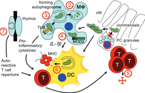
Fig. 1. Possible mechanisms by which mutations in macroautophagy genes could interfere with gut homeostasis and lead to IBD. Atg16L1 hypomorphic Paneth cells (PCs) secrete anti-microbial peptides less efficiently; (1) Atg16L1T300A-carrying PCs therefore might prevent commensal invasion less efficiently. (2) Macrophages (MU) with compromised macroautophagy might be attenuated in eliminating invading commensals; (3) they might fail to deliver commensal-derived PAMPs to TLRs for immune activation; (4) they might produce increased amounts of pro-inflammatory cytokines, like IL-1b, in response to cytosolic PAMP recognition. In addition to these innate immune recognition mechanisms, mutations in macroautophagy genes might also compromise adaptive immune responses. (5) T cell survival in the gut mucosa and therefore efficient restriction of invading commensals might be affected. (6) Macroautophagy-dependent endogenous MHC presentation of commensal-derived antigens by dendritic cells (DCs) could be compromised. (7) Finally, altered thymic T cell selection in patients with mutations in macroautophagy genes might favor a T cell repertoire with gut autoreactivity.
cells, mutations in the macroautophagy and NOD2 pathway affect the gut milieu, possibly allowing uninhibited expansion of resident microorganisms that could drive the inflammation seen in CD patients.
Macroautophagy in macrophages
The balance of the gut flora and the immune system may fur-ther be upset by the inability of innate immune cells to clear microorganisms. In macrophages, macroautophagy is criti-cally involved in efficient degradation of intracellular bacteria. Both pathogens that condition phagosomes for their replica-tion and those that escape into the cytosol can be restricted by macroautophagy. In murine macrophages infected with the parasite Toxoplasma gondii, Atg8 is recruited to the parasitophorous vacuole after CD40 stimulation, which in-duces fusion with lysosomes (23). In T. gondii as well as in Mycobacterium tuberculosis infection, macroautophagy can overcome bacterial evasion mechanisms and efficiently elim-inate the pathogen (24, 25). Similarly, group A streptococci that escape the endosome for replication in the cytosol can be restricted by macroautophagy (26).
These results suggest that CD-associated mutations in macroautophagy genes could affect the ability of macro-phages to clear intracellular bacteria. Indeed, human cell lines modified to only express Atg16L1T300A show impaired macroautophagy of Salmonella typhimurium (13). Diminished expression levels of IRGM also reduce macroautophagy of S. typhimurium in HeLa cells (9).
In addition to eliminating intracellular bacteria, components of the classical macroautophagy pathway might be involved in processing of extracellular pathogens after phagocytosis. If a particle activates Toll-like receptor (TLR) signaling while being phagocytosed, this recruits Atg8 to the phagosome in a murine macrophage cell line (27). Coating with Atg8 enhances fusion of the phagosomes with lysosomes after TLR2 activation and might have an impact on bacterial clear-ance. In this scenario, most organisms of the intestinal flora could be targeted by macroautophagy. Consequently, a de-fect in macrophage clearance of phagocytosed bacteria might contribute to an elevated bacterial load in the gut that could upset the gut equilibrium and result in inflammation. Macroautophagy and PAMP recognition
Independent of phagocytosis, there is crosstalk between PAMP recognition and the macroautophagy pathway. On the one hand, TLR signaling induces macroautophagy. For example, incubation of primary human macrophages or murine macrophage cell lines with the TLR4 ligand LPS induces macroautophagy in a myeloid differentiation factor 88 (MyD88)-independent, Toll–IL-1 receptor domain-containing adaptor-inducing IFN-b-dependent pathway (28). Ligand binding to TLR1, TLR3, TLR5, TLR6 or TLR7 also induces macroautophagy as measured by the increase of green fluo-rescent protein–Atg8 punctae in the same cell line (29), which was confirmed for TLR3 and TLR7 by another study (17). In contrast to Xu et al. (28), Shi and Kehrl (29) found macroautophagy induction to be MyD88-dependent and pro-vided mechanistic data on how the pathways intersect: TLR signaling frees Beclin-1 from the inhibitory complex with
Bcl-2 consequently inducing macroautophagy. In addition, TLR signaling positively influences macroautophagy by inducing transcription of Atg genes (27).
On the other hand, macroautophagy can deliver intracellu-lar TLR ligands to their respective endosomal TLRs. At least in plasmacytoid dendritic cells, efficient IFN-a production af-ter vesicular stomatitis virus infection was dependent on macroautophagy, which delivers cytosolic single-stranded RNA to TLR7 (30). Similarly, TLR9 ligand targeting to endo-somes by B cell receptors seems to be macroautophagy dependent (31).
In contrast to TLRs, much less is known about the influ-ence of cytosolic PAMP receptor engagement on macroau-tophagy. The NOD-like protein IPAF (IL-1b-converting enzyme protease-activating factor), however, seems to inhibit macroautophagy during Shigella infection (32). Vice versa, components of the macroautophagy machinery block signaling of RNA helicases, for example the retinoic acid-inducible gene-I molecules, and macroautophagy knockout cells produce more type I IFN in response to infection with single-stranded RNA viruses, which engage this cytosolic PAMP recognition pathway (33, 34). Mutations in macroautophagy genes could therefore diminish TLR signaling or enhance cytosolic PAMP recognition. These effects could either inhibit efficient commensal control or increase inflammatory cytokine secretion, respectively. Both mechanisms might contribute to gut inflammation during CD.
Inflammatory cytokines in CD
Indeed, CD patients have elevated levels of inflammatory cytokines like IL-1, IL-6 and tumor necrosis factor (TNF) in the intestine and the serum (35, 36). IL-17 is also up-regulated in CD patients (35). However, involvement of IL-17 in the pathogenesis in CD has recently been challenged by data showing a protective function of IL-17A in intestinal inflammation (37). Furthermore, macrophages from CD patients were shown to spontaneously produce IL-12, which could explain the bias toward Th1 cells in CD (38) and thereby the elevated levels of IFN-c (39). Expression of the Th2 cytokine IL-4 on the other hand is drastically reduced (38). Of note, transcription of IL-10, a cytokine known to down-regulate the expression of Th1 cytokines and inhibit in-flammation, is suppressed in patients carrying the CD-associated NOD2 mutation (40).
As a possible explanation for this pathogenic cytokine milieu, murine LPS-stimulated macrophages deficient for Atg16L1 produce elevated levels of the pro-inflammatory cytokine IL-1b and readily promote gut inflammation after dextran sulfate sodium treatment (41). However, these mice do not spontaneously develop IBD or recapitulate macroau-tophagic activity of patients carrying the CD-associated atg16L1T300A mutation. In Atg16L1-deficient mice, macro-autophagy is completely abrogated resulting in death soon after birth. The atg16L1T300A mutation, however, does not affect basal macroautophagy levels and causes more subtle effects than complete loss of function (13). It would therefore be interesting to study cytokine expression from macro-phages in Atg16L1 hypomorphic or atg16L1T300A knock-in mice (21) knock-in order to lknock-ink CD-associated mutations
with specific defects in macroautophagy and cytokine production. Nevertheless, macroautophagy might negatively regulate pro-inflammatory cytokine secretion in the gut and CD-associated mutations could compromise this regulation. CD and the T cell compartment
Clearly, adaptive immune responses also contribute to CD. In particular, T cells play a role in CD pathogenesis, which is accompanied by increased numbers of activated CD4+ T cells in the gut of CD patients (36) and remission of CD in HIV-infected patients (42). In addition, IBD can be induced by adoptive transfer of purified CD4+T cell populations (43). Disease-inducing CD4+T cells are predominantly T
h1 cells. T cell responses seem to be specific for a small number of antigens; however, the antigens vary from patient to patient (36). In the following part, we will discuss how mutations in the macroautophagy pathway could affect homeostasis, function and repertoire selection of T cells and thereby pre-dispose for CD.
T cell homeostasis
Murine and human T cells can up-regulate macroautoph-agy (44) and mainly need it for survival after development to single-positive thymocytes as well as after activation in pe-ripheral tissues. T cells from atg5 / chimeric mice develop normally, but T cell numbers are reduced in the thymus as well as in the periphery. This is due to increased apoptosis in peripheral CD8+ T cells and diminished proliferation of CD4+and CD8+T cells after TCR stimulation (44). In contrast to a pro-survival role of macroautophagy described by Pua et al. (44), Li et al. (45) found macroautophagy to be in-volved in increased cell death after growth factor withdrawal in murine Th2 cells. Furthermore, an autophagic death pro-gram can eliminate CD4+ T cells after IFN-c stimulation in the absence of Irgm1, the murine homologue of human IRGM (46).
With respect to CD4+ T cell homeostasis, CD patients again do not show a phenotype as pronounced as the ani-mal models discussed above. Nevertheless, these data sug-gest that mutations in CD-associated genes could affect survival of T cells, and it is tempting to speculate that sub-sets of T cells could be affected differently, for example with a survival effect on inflammation-promoting Th17 cells and a pro-apoptotic effect on regulatory T cells, including Th2-polarized IL-10-secreting cells.
Autophagy in antigen presentation
The role for autophagy in antigen presentation on MHC class II presentation is less speculative. We and others have shown that autophagy delivers antigens mainly from the cytoplasm into the MHC II-loading compartment in antigen-presenting cells (47–51). This results in enhanced antigen presentation of autophagy substrates on the cell surface, an increase in T cell activation and improved clearance of pathogens. Therefore, mutations in the macroautophagy pathway could affect how efficiently the adaptive immune system eliminates infections. Indeed, the atg16L1T300A mutation compromises MHC class II presentation of macroautophagy-targeted antigens (S. Meixlsperger and C. Mu¨nz, unpublished
data). Of note, mutations in macroautophagy genes could also inhibit extracellular antigen processing for MHC class II loading after phagocytosis (27). This could render priming of regulatory T cells and immune surveillance of commensal invasion more difficult.
In addition to the role of macroautophagy in MHC class II presentation, a recent report suggests that macroautophagy is also involved in the presentation of viral antigens on MHC class I molecules (52). English et al. noticed that, in late phases of infection, herpes simplex virus 1 (HSV-1)-infected murine macrophages were less capable of stimulating CD8+ T cell responses after treatment with the macroautophagy inhibitor 3-methyladenine or knock-down of Atg5. Mild heat shock or administration of the pyrogenic cytokine IL-1b up-regulated this pathway, which resembled classical mac-roautophagy with respect to the formation of Atg8-coated, vi-rus-containing vesicles that fuse with lysosomes. However, other aspects of this pathway that supports MHC class I presentation differed significantly from classical macroau-tophagy. Namely, the autophagosomal membranes origi-nated from the nuclear envelope and formed four-layered membrane structures as well as the classical double-membrane-surrounded autophagosomes.
Future experiments will have to show if this macroautoph-agy-dependent pathway contributes to the control of infec-tion in vivo and how its substrates gain access to the MHC class I antigen-processing machinery. Additionally, it will be important to analyze whether this pathway is specific for HSV-1 infection or also participates in the presentation of other antigens. Possibly, this additional, unconventional mac-roautophagy pathway might have evolved in adaptation to mechanisms used by HSV-1 to evade the classical macroau-tophagy pathway (53, 54). However, these data suggest that macroautophagy can also enhance MHC class I-restricted antigen presentation under certain conditions. Mutations in the macroautophagic machinery might therefore prohibit effi-cient MHC presentation for immune control of commensal in-vasion and for tissue-protective tolerance induction, failure of which could significantly contribute to CD.
Macroautophagy and T cell selection
One major mechanism of tolerance induction is based on the elimination of autoreactive T cells in the thymus. Re-cently, macroautophagy has been implicated in this central tolerance. Murine thymic epithelial cells (TECs), which select thymocytes that recognize MHC molecules and are self-tolerant, were described to show high levels of constitutive macroautophagy (55). This observation and the evidence for antigen presentation on MHC class II molecules after macroautophagy raised the possibility that macroautophagy delivers antigens to MHC class II molecules in TECs for pos-itive and negative selection, shaping the T cell repertoire to self-MHC recognition and self-tolerance, respectively.
To test this hypothesis, Nedjic et al. (48) transplanted atg5 / thymi into TCR transgenic or athymic recipient mice. atg5 / thymi supported positive selection of some but not all CD4+ T cell specificities and for all CD8+T cell specificities. In athymic nude mice, Atg5 deficiency shaped the T cell repertoire to-ward autoreactivity, resulting among other autoimmune signs in continuous gut inflammation. However, as most CD
patients develop symptoms only in their 20s, a general de-fect in T cell selection is unlikely. Also, the T cell repertoire seems to be normal in CD patients. Only within the lesions is the repertoire more restricted. Therefore, only distinct T cell specificities promoting bowel inflammation might not be deleted in thymi with mutated macroautophagy genes and promote disease only after recurrent re-stimulation. Possible treatment of CD by manipulating
macroautophagy
Macroautophagy is a central pathway for the function of in-nate and adaptive immune responses. As discussed, muta-tions of gene products in the pathway might affect multiple cell types and aspects of macroautophagy in CD. Therefore, manipulation of macroautophagy could have therapeutic merit for patients affected by this disease.
Rapamycin, a drug that is widely used to stimulate macro-autophagy in experimental settings and that has been approved as an immunosuppressant after organ transplanta-tion in humans, was recently used to treat a patient with CD after failure of standard medication (18). On a dose of 4 mg a day and in combination with an anti-TNF antibody, her symptoms improved markedly, abolishing the need for sur-gery. While this is very promising, the study needs to be ex-tended to more patients and additional in vitro analysis needs to show whether the effect is due to up-regulation of macroautophagy or to a general immunosuppressive effect of rapamycin. Experiments in mice support the possibility of an effect of rapamycin on the up-regulation of macroautoph-agy in vivo. When Jagannath and colleagues immunized mice with rapamycin-treated, bacille Calmette–Gue´rin-infected dendritic cells, the mice controlled infection with virulent M. tuberculosis better than control mice did (47). Furthermore, macroautophagy stimulation with rapamycin prevented toxicity of neurodegenerative protein aggregates in vivo (56).
However, more selective methods than rapamycin need to be developed to up-regulate macroautophagy in distinct cell lineages that are involved in altered innate and adaptive im-mune responses in CD. In any case, it will be important to monitor side effects closely, as macroautophagy regulates survival and apoptosis of various cell types as well as turn-over of proteins and organelles.
Conclusion
We are just beginning to understand what causes and main-tains inflammation in CD. In addition to environmental factors, there is a big genetic contribution to the pathogenesis of CD and probably only a combination of mutations results in disease. Some mutations are common to IBDs, while others are exclusively associated with CD, like polymorphisms of atg16L1, IRGM and NOD2. Especially, the first two affect the macroautophagy pathway. In addition, it is tempting to speculate that NOD2 mutations might compromise macroau-tophagy regulation upon bacterial constituent recognition, as has been shown for other PAMP receptors. Macroautophagy de-regulation affects the function of innate immune cells like Paneth cells and macrophages. It could also affect the T cell repertoire, antigen presentation and T cell homeostasis.
Most conclusions with respect to how mutations in macroau-tophagy genes could affect CD are based on the analysis of gene-deficient mice. However, these might only poorly recapit-ulate the situation in CD patients. For example, IRGM is a single gene in humans, but there are several Irgm family members in the mouse. Even though murine Irgm1 corresponds to hu-man IRGM, other Irgm family members might compensate for Irgm1 deficiency in the mouse. More importantly, the CD-associated mutations do not abrogate protein expression. They affect expression levels or impair certain functions of the affected proteins. It will therefore be important to generate and study knock-in mice for CD-associated mutations and to confirm findings from murine models on patient samples. Fur-thermore, it is of interest to analyze immune cells of the gut microenvironment as opposed to immune cells from the pe-riphery or cells differentiated from bone marrow precursors.
Understanding the molecular pathways inducing macroau-tophagy as well as knowing the downstream effectors will greatly enhance the possibility of manipulating the affected cells specifically. Atgs might also have functions distinct from the classical macroautophagy pathway (57) as has been suggested for Atg8 in phagocytosis (27). Since the macroautophagy pathway is associated with various other diseases, research into the role of macroautophagy in the pathogenesis of CD will not only benefit patients affected by this disease but also could affect treatment of neurodegen-erative disorders, muscle diseases and cancer.
Funding
National Cancer Institute (R01CA108609 and R01CA101741); Foundation for the National Institutes of Health (Grand Challenges in Global Health) to C.M.; German Research Foundation (Deutsche Forschungsgemeinschaft) fellowship to S.M.
Abbreviations
atg5 autophagy-related gene 5 atg16L1 Atg16-like 1 gene CD Crohn’s disease GABA c-aminobutyric acid
GABARAP GABA (A) receptor-associated protein HSV-1 herpes simplex virus 1
IBD inflammatory bowel disease IFN-c IFN gamma
IRGM immunity-related GTPase family M
MAP1LC microtubule-associated protein 1 light chain MyD88 myeloid differentiation factor 88
NOD2 nucleotide-binding oligomerization domain containing 2 PAMP pathogen-associated molecular pattern
TEC thymic epithelial cells TLR Toll-like receptor TNF tumor necrosis factor
References
1 Klionsky, D. J. 2007. Autophagy: from phenomenology to molecular understanding in less than a decade. Nat. Rev. Mol. Cell Biol. 8:931. 2 Nakatogawa, H., Ichimura, Y. and Ohsumi, Y. 2007. Atg8, a ubiquitin-like protein required for autophagosome formation, mediates membrane tethering and hemifusion. Cell 130:165. 3 Levine, B. and Deretic, V. 2007. Unveiling the roles of autophagy in
innate and adaptive immunity. Nat. Rev. Immunol. 7:767. 4 Levine, B. and Kroemer, G. 2008. Autophagy in the pathogenesis
5 Barrett, J. C., Hansoul, S., Nicolae, D. L. et al. 2008. Genome-wide association defines more than 30 distinct susceptibility loci for Crohn’s disease. Nat. Genet. 40:955.
6 Van Limbergen, J., Russell, R. K., Nimmo, E. R. and Satsangi, J. 2007. The genetics of inflammatory bowel disease. Am. J. Gastroenterol. 102:2820.
7 Hampe, J., Franke, A., Rosenstiel, P. et al. 2007. A genome-wide association scan of nonsynonymous SNPs identifies a susceptibility variant for Crohn disease in ATG16L1. Nat. Genet. 39:207.
8 Rioux, J. D., Xavier, R. J., Taylor, K. D. et al. 2007. Genome-wide association study identifies new susceptibility loci for Crohn disease and implicates autophagy in disease pathogenesis. Nat. Genet. 39:596.
9 McCarroll, S. A., Huett, A., Kuballa, P. et al. 2008. Deletion polymorphism upstream of IRGM associated with altered IRGM expression and Crohn’s disease. Nat. Genet. 40:1107.
10 Parkes, M., Barrett, J. C., Prescott, N. J. et al. 2007. Sequence variants in the autophagy gene IRGM and multiple other replicating loci contribute to Crohn’s disease susceptibility. Nat. Genet. 39:830.
11 Hugot, J. P., Chamaillard, M., Zouali, H. et al. 2001. Association of NOD2 leucine-rich repeat variants with susceptibility to Crohn’s disease. Nature 411:599.
12 Ogura, Y., Bonen, D. K., Inohara, N. et al. 2001. A frameshift mutation in NOD2 associated with susceptibility to Crohn’s disease. Nature 411:603.
13 Kuballa, P., Huett, A., Rioux, J. D., Daly, M. J. and Xavier, R. J. 2008. Impaired autophagy of an intracellular pathogen induced by a Crohn’s disease associated ATG16L1 variant. PLoS One 3:e3391.
14 Taylor, G. A., Feng, C. G. and Sher, A. 2004. p47 GTPases: regulators of immunity to intracellular pathogens. Nat. Rev. Immunol. 4:100.
15 Singh, S. B., Davis, A. S., Taylor, G. A. and Deretic, V. 2006. Human IRGM induces autophagy to eliminate intracellular mycobacteria. Science 313:1438.
16 Shaw, M. H., Reimer, T., Kim, Y. G. and Nunez, G. 2008. NOD-like receptors (NLRs): bona fide intracellular microbial sensors. Curr. Opin. Immunol. 20:377.
17 Delgado, M. A., Elmaoued, R. A., Davis, A. S., Kyei, G. and Deretic, V. 2008. Toll-like receptors control autophagy. EMBO J. 27:1110.
18 Massey, D. C., Bredin, F. and Parkes, M. 2008. Use of sirolimus (rapamycin) to treat refractory Crohn’s disease. Gut 57:1294. 19 Izcue, A., Coombes, J. L. and Powrie, F. 2009. Regulatory
lymphocytes and intestinal inflammation. Annu. Rev. Immunol. 27:313.
20 Kolls, J. K., McCray, P. B. Jr and Chan, Y. R. 2008. Cytokine-mediated regulation of antimicrobial proteins. Nat. Rev. Immunol. 8:829.
21 Cadwell, K., Liu, J. Y., Brown, S. L. et al. 2008. A key role for autophagy and the autophagy gene Atg16l1 in mouse and human intestinal Paneth cells. Nature 456:259.
22 Wehkamp, J., Harder, J., Weichenthal, M. et al. 2004. NOD2 (CARD15) mutations in Crohn’s disease are associated with diminished mucosal alpha-defensin expression. Gut 53:1658. 23 Andrade, R. M., Wessendarp, M., Gubbels, M. J., Striepen, B. and
Subauste, C. S. 2006. CD40 induces macrophage anti-Toxoplasma gondii activity by triggering autophagy-dependent fusion of pathogen-containing vacuoles and lysosomes. J. Clin. Invest. 116:2366.
24 Gutierrez, M. G., Master, S. S., Singh, S. B., Taylor, G. A., Colombo, M. I. and Deretic, V. 2004. Autophagy is a defense mechanism inhibiting BCG and Mycobacterium tuberculosis survival in infected macrophages. Cell 119:753.
25 Ling, Y. M., Shaw, M. H., Ayala, C. et al. 2006. Vacuolar and plasma membrane stripping and autophagic elimination of Toxoplasma gondii in primed effector macrophages. J. Exp. Med. 203:2063.
26 Nakagawa, I., Amano, A., Mizushima, N. et al. 2004. Autophagy defends cells against invading group A Streptococcus. Science 306:1037.
27 Sanjuan, M. A., Dillon, C. P., Tait, S. W. et al. 2007. Toll-like receptor signalling in macrophages links the autophagy pathway to phagocytosis. Nature 450:1253.
28 Xu, Y., Jagannath, C., Liu, X. D., Sharafkhaneh, A., Kolodziejska, K. E. and Eissa, N. T. 2007. Toll-like receptor 4 is a sensor for autophagy associated with innate immunity. Immunity 27:135. 29 Shi, C. S. and Kehrl, J. H. 2008. MyD88 and Trif target Beclin 1 to
trigger autophagy in macrophages. J. Biol. Chem. 283:33175. 30 Lee, H. K., Lund, J. M., Ramanathan, B., Mizushima, N. and
Iwasaki, A. 2007. Autophagy-dependent viral recognition by plasmacytoid dendritic cells. Science 315:1398.
31 Chaturvedi, A., Dorward, D. and Pierce, S. K. 2008. The B cell receptor governs the subcellular location of Toll-like receptor 9 leading to hyperresponses to DNA-containing antigens. Immunity 28:799.
32 Suzuki, T., Franchi, L., Toma, C. et al. 2007. Differential regulation of caspase-1 activation, pyroptosis, and autophagy via Ipaf and ASC in Shigella-infected macrophages. PLoS Pathog. 3:e111. 33 Jounai, N., Takeshita, F., Kobiyama, K. et al. 2007. The Atg5 Atg12
conjugate associates with innate antiviral immune responses. Proc. Natl Acad. Sci. USA 104:14050.
34 Tal, M. C., Sasai, M., Lee, H. K., Yordy, B., Shadel, G. S. and Iwasaki, A. 2009. Absence of autophagy results in reactive oxygen species-dependent amplification of RLR signaling. Proc. Natl Acad. Sci. USA 106:2770.
35 Fujino, S., Andoh, A., Bamba, S. et al. 2003. Increased expression of interleukin 17 in inflammatory bowel disease. Gut 52:65. 36 Romagnani, P., Annunziato, F., Baccari, M. C. and Parronchi, P.
1997. T cells and cytokines in Crohn’s disease. Curr. Opin. Immunol. 9:793.
37 O’Connor, W. Jr, Kamanaka, M., Booth, C. J. et al. 2009. A protective function for interleukin 17A in T cell-mediated intestinal inflammation. Nat. Immunol. 10:603.
38 Parronchi, P., Romagnani, P., Annunziato, F. et al. 1997. Type 1 T-helper cell predominance and interleukin-12 expression in the gut of patients with Crohn’s disease. Am. J. Pathol. 150:823. 39 Breese, E., Braegger, C. P., Corrigan, C. J., Walker-Smith, J. A.
and MacDonald, T. T. 1993. Interleukin-2- and interferon-gamma-secreting T cells in normal and diseased human intestinal mucosa. Immunology 78:127.
40 Noguchi, E., Homma, Y., Kang, X., Netea, M. G. and Ma, X. 2009. A Crohn’s disease-associated NOD2 mutation suppresses tran-scription of human IL10 by inhibiting activity of the nuclear ribonucleoprotein hnRNP-A1. Nat. Immunol. 10:471.
41 Saitoh, T., Fujita, N., Jang, M. H. et al. 2008. Loss of the autophagy protein Atg16L1 enhances endotoxin-induced IL-1beta produc-tion. Nature 456:264.
42 Pospai, D., Rene, E., Fiasse, R. et al. 1998. Crohn’s disease stable remission after human immunodeficiency virus infection. Dig. Dis. Sci. 43:412.
43 Coombes, J. L., Robinson, N. J., Maloy, K. J., Uhlig, H. H. and Powrie, F. 2005. Regulatory T cells and intestinal homeostasis. Immunol. Rev. 204:184.
44 Pua, H. H., Dzhagalov, I., Chuck, M., Mizushima, N. and He, Y. W. 2007. A critical role for the autophagy gene Atg5 in T cell survival and proliferation. J. Exp. Med. 204:25.
45 Li, C., Capan, E., Zhao, Y. et al. 2006. Autophagy is induced in CD4+T cells and important for the growth factor-withdrawal cell
death. J. Immunol. 177:5163.
46 Feng, C. G., Zheng, L., Jankovic, D. et al. 2008. The immunity-related GTPase Irgm1 promotes the expansion of activated CD4+
T cell populations by preventing interferon-gamma-induced cell death. Nat. Immunol. 9:1279.
47 Jagannath, C., Lindsey, D. R., Dhandayuthapani, S., Xu, Y., Hunter, R. L. Jr and Eissa, N. T. 2009. Autophagy enhances the efficacy of BCG vaccine by increasing peptide presentation in mouse dendritic cells. Nat. Med. 15:267.
48 Nedjic, J., Aichinger, M., Emmerich, J., Mizushima, N. and Klein, L. 2008. Autophagy in thymic epithelium shapes the T-cell repertoire and is essential for tolerance. Nature 455:396. 49 Schmid, D., Pypaert, M. and Mu¨nz, C. 2007. MHC class II antigen
loading compartments continuously receive input from autopha-gosomes. Immunity 26:79.
50 Zhou, D., Li, P., Lott, J. M. et al. 2005. Lamp-2a facilitates MHC class II presentation of cytoplasmic antigens. Immunity 22:571.
51 Mu¨nz, C. 2009. Enhancing immunity through autophagy. Annu. Rev. Immunol. 27:423.
52 English, L., Chemali, M., Duron, J. et al. 2009. Autophagy enhances the presentation of endogenous viral antigens on MHC class I molecules during HSV-1 infection. Nat. Immunol. 10:480.
53 Orvedahl, A., Alexander, D., Talloczy, Z. et al. 2007. HSV-1 ICP34.5 confers neurovirulence by targeting the Beclin 1 autophagy protein. Cell Host Microbe 1:23.
54 Talloczy, Z., Jiang, W., Virgin, H. W. IV et al. 2002. Regulation of starvation- and virus-induced autophagy by the eIF2alpha kinase signaling pathway. Proc. Natl Acad. Sci. USA 99:190.
55 Mizushima, N., Yamamoto, A., Matsui, M., Yoshimori, T. and Ohsumi, Y. 2004. In vivo analysis of autophagy in response to nutrient starvation using transgenic mice expressing a fluorescent autophagosome marker. Mol. Biol. Cell 15:1101.
56 Ravikumar, B., Vacher, C., Berger, Z. et al. 2004. Inhibition of mTOR induces autophagy and reduces toxicity of polyglutamine expansions in fly and mouse models of Huntington disease. Nat. Genet. 36:585. 57 Virgin, H. W. and Levine, B. 2009. Autophagy genes in immunity.