TRANSACTIONS OF THE ROYAL SOCIETY OF TROPICAL MEDICISE ANI, HYGIES~. (1989) 83, 389-393 389
Em2-ELISA for the follow-up
of alveolar echinococcosis
after complete
surgical resection
of liver lesions
B. Gottstein’, K. Tschudi*, J. Eckert’ and R. Ammann* ‘Institute of Parasitology, University of Ziirich, Zikich, Switzerland; 2Department of Internal Medicine, Division of Gastroenterologv, University Hospital, Ziirich, Switzerland
Abstract
Alveolar echinococcosis, a serious and often fatal human disease, can be efficiently cured only by complete surgical resection of the Echinococcus multi- locularis lesion. The present study showed that the determination in patients who had undergone surgery of antibody activity directed against the antigen Em2 reliably reflected complete or incomplete surgical resection. From 9 patients with pre-operative positive results in the Em2 enzyme-linked immunosorbent assay (Em2-ELISA) and successful surgical resection, 6 converted to negative within one year and the remaining 3 patients within 4 years after surgery. Six of 7 additional patients who showed recurrences in an average of 6 years after surgery despite assumed complete surgical resection, were positive by Em2- ELISA at the time of recurrence. Discrimination was not possible between these 2 groups of patients when using an ELISA employing crude antigen obtained from E. granulosus hydatid cyst fluid.
Introduction
Alveolar echinococcosis, caused by the larval stage of Echinococcus multilocularis, is one of the most lethal hehninth infections of humans. Only surgical com- plete removal of the entire parasite lesion offers a prospect for curative treatment. It has been reported that only 26% of cases were resectable in Alaska
(SCHANTZ et al., 1983). The development of new immunodiagnostic techniques for early detection of the infection (GOTTSTEIN et al., 1987) and improved methods for clinical diagnosis and surgical treatment (SCHR~DER & ROBOTTI. 1986: GILLET et al.. 1988) resulted in an increased.rate of radical resectabilitv: which is presently estimated to be about 40% regard: ing central European patients. The ‘radicality’ of the resection is very difficult to determine by the surgeons, as microlesions and root-like parasite protrusions (ECKERT et al.? 1983; MEHLHORN et al.,
1983) mav remain unseen m annarentlv healthv liver tissue. Iii this way, some of- these -patients may subsequently develop recurrences. Serological tests have been used, among others, for post-operative follow-up studies. In most studies, crude antigens derived from E. granulosus served to detect parasite- specific immunoglobulins (reviewed by SCHANTZ et al., 1983) or immunoglobulin classes (G~TTSTEIN et al., 1984; VUITTON-DROUHARD, 1985). Generally, a decrease of antibody concentration was observed after surgery. This decrease was more marked in parasite- Correspondence to Dr B. Gottstein, Institut fiir Parasitologie der Universitit Zurich, Winterthurerstrasse 266a, CH-8057
Ziirich, Switzerland.
specific immunoglobulin (Ig) E and IgA levels, compared to the parasite-specific IgG. Most of the patients investigated were under permanent meben- dazole or albendazole treatment, which may have influenced the course of serology. Nevertheless, antibodies were still detectable for long periods after surgical intervention. Based on the interpretation of the test results, clear statements regarding the actual status of the disease in individual patients were not possible in many cases. A recent study performed on 3 patients with alveolar echinococcosis indicated that antibodies detected by the use of a purified E. multilocularis antigen -(Em2 antigen) @OTTSTEIN,
1985) in an enzvme-linked imunosorbent assav (Em2- ELISA) declined dramatically within months‘ after radical operation (LANIER et al., 1987). In order to evaluate this method further as a follow-up technique, a study was carried out with 18 selected Swiss patients suffering from alveolar echinococcosis.
Patients and Methods
Patients
All 18 patients investigated were under long-term medical supervision by the ‘Swiss Echinococcosis Studv Groun’. The diagnosis of alveolar echinococ- cosis-had been confirmed clinically, histologically and serologically (WOODTLI et al., 1985; AMMANN et al .,
1989). All patients had received surgical treatment with subsequent long-term mebendazole chemother- apy (for details see AMMANN et al., 1989). The following groups of patients with confirmed alveolar echinococcosis of the liver were included in the study.
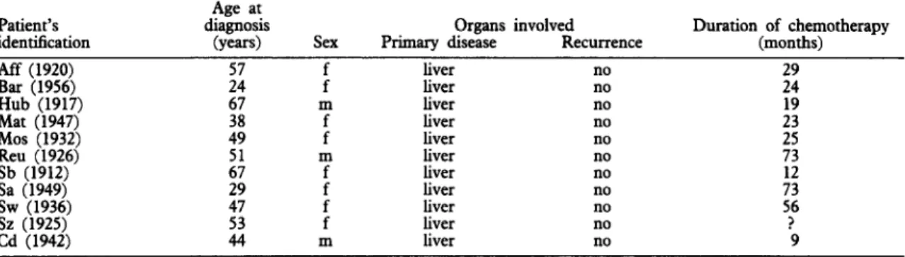
Group (a). Eleven persons with comnlete surgical resection of the parasite lesion. No recurrences-had been observed UD to the end of October 1988 (cut-off point). The average follow-up period after surgery was 7 years (range 2-11 years). More detailed information on the patients is given in Table 1.
Group (b). Seven persons with complete surgical resection of the visible parasite liver lesions reported by the surgeons. The 7 patients developed recurr- ences at time intervals of 3 years and 5 months to 11 years and 2 months (average 6 vears) after operation in spite of continuous~chen-&herapy: The average fol- low-up period was 13 years (range 10-21 years). More detailed information on these patients is given in Table 2.
Serological assays
Parasite-specific serum IgG was detected by EL- ISA. All serum samples had been kept frozen (- SO’C) and were examined at the same time and under uniform conditions in order to avoid intertrial varia- tions. Between 3 and 14 serum samples, during the
390
T;ksl. Information on the patients of group (a), showing no recurrence after complete surgical resection of the parasite
Patient’s identitication Aff (1920) Bar (1956) Hub (1917) Mat (1947j Mos (1932) Reu (1926) Sb (1912) Sa (1949) SW (1936) Sz (1925) Cd (1942) Age at diagnosis (VCXS) ._ :: 67 Organs involved
Sex Primary disease Recurrence : liver liver no no m liver no : liver liver no no m liver no : liver liver no no : liver liver no no m liver no Duration of chemotherapy (months) 29 24 :z :; :: 56 ? 9
Table 2. Information on the patients of group (b), showing recurrence after surgical resection of the parasite lesions
Patient’s identification
Age at diagnosis
(years) Sex
Organis involved Duration of chemotherapy Primary disease Recurrence (months) Eb (1918) Rm (1921) 57 55 :; m f m f liver liver liver liver liver, kidney, retroperitoneal, lungs liver liver liver coeliac olexus 77 1:; 117 Ah (1930) Pfr (1939) Rei (1945) 45 :: : f liver liver liver liver r liver liver 112 %
Table 3a. Follow-up by Em2-ELBA of patients of group (a), showing no recurrence after complete surgical resection. The last result for each patient corresponds to the serum sample at the cut-off point
Patient’s identification SwerYa AU at 1 2 3 8 9 10 Bar :: 2: x 0 0 0 0 0 Hub Mat 37 0 0 0 Mos Reu Sb
6i
108 x 18 8 8 rib
0 0 0 x 0 ndb 0 E 70 30 x ndb 0 0 : : 0 0 0 0 & 220 8 i : 0‘AU=antibody units, as percentage of a positive reference serum (see Patients and Methods); O=no antibodies detectable (negative).
bnd=Not done (no serum available).
periods indicated in Tables 3 a, b and 4 a, b, were 100% reference antibody reactivity and simultaneous- examined per patient. The ELISA was performed as ly to the absorbance at 404 run measured for this
described previously (GOTTSTEIN et al., 1984; GOTT-
STEIN, 1985), using the criteria for interpretation of antibodies could not be detected (AU=O). Two serum.) Patients were regarded as sero-negative when seropositivity reported by GOTTSTEIN et al. (1984).
The results are expressed in arbitrarily defined antigens were included in this study: a purified species-specifk antigen (Em2) derived from E. multi- antibody units (AU), as a percentage of the antibody
activity of a positive reference serum. (The reference bcularis and a crude E. granulosus antigen (EgHF) from hydatid cyst fluid of bovine origin (GOTTSTEIN serum was arbitrarily set at 100 AU, corresponding to et al., 1983, 1984).
391
Results
The results of serological follow-up of the patients from groups (a) and (b) are shown in Tables 3 and 4. Group (a) (radical resection, tw recuwence)
Em2-ELBA. Initially (before surgery), 9 of 11 patients had detectable anti-Err&antibodies. One
year after surgery, 6 of these 9 patients had already converted to negative (anti-En@. Of the remaining patients, 2 were negative (anti-Em2) 2 years after surgery and one 4 years after surgery. After conver- sion to negative, none of the patients showed any further anti-Em2-antibody activity up to the cut-off point (Table 3a).
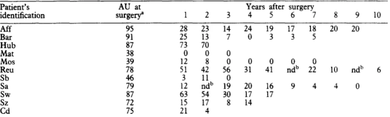
Table 3b. Follow-up by EgHF-ELBA of patients of group (a), showing no recurrence after complete surgical resection. The last result for each patient corresponds to the serum sample at the cut-off point
Patient’s AU at Years after surgery
identification surge# 12 3 4 5 6 7 8 9 10 Aff Bar Hub Mat Mos Reu ii i,w Cd :58 23 :i: 14 7 24 0 19 3 17 3 18 5 20 20 73 0 0 :? 428 56 i 3: 4: nodb 2: 10 nd’ 6 3 11 0 12 ndb 19 20 16 9 4 4 0 63 54 30 17 17 17 8 14 :: 4
“AU=antibody units, as percentage of a positive reference serum (see Patients and Methods); O=no antibodies detectable (negative).
bnd=Not done (no serum available).
Table 4a. Follow-up Em2-ELBA of patients of group (b), showing recurrence after surgical resection. The last result for each patient correponds to the serum sample at the cut-off point of the present study. The year 0 corresponds to the date of diagnosis of recurrence
Patient’s AU at times (in after
identification -7 -6 -5 -4 theJ11owing -2 -1 i;“‘” Pfor; and3 4 recyrrence) 6 7 8 9 10 2 18b nd’ I$‘ nd’ nd’ nd’ nd’ nd’ 6 14 nd’ 37 22 17 3 16 0 16 0 12 0 10 0 27 0 VW 5b 0 4 0 0 0 0 0 0 0 0 Ak Sb i Alt 1: 1: Pfr Rei Gd 4:: 1: :!I 0 0 0 1 11 0 :i 4; 3: ii : 0 0 0 5 0 “AU=antibody units, as a percentage of a positive reference serum (see Patients and Methods); O=no antibodies detectable
(negative).
bInitial values at the date of surgery. ‘nd=Not done (no serum available). dNo serum available of the date at surgery.
Table 4b. Follow-up by E&IF-ELBA of patients of group (b), showing recurrence alter surgical resection. The last result for each patient corresponds to the serum sample at the cut-off point of the present study. The year 0 corresponds to tbe date of diagnosis of recurrence
Patient’s identification 2 VW Ak AU at the_3following -2 time (in rrs lbefo2re and after -7 recurrence) -6 -5 -4 -1 3 4 5 6 7 8 9 10 96” ndb 123” ndb ndb ndb ndb ndb 63 nd* 79 d ;t 92 85 100 90 40 85 52 59 69 62 66 75 74 51 67’ 36 21 19 21 4 0 3 0 0 0 14s 9 4 7 0 0 2 47 Ah 48’ 38 2s 17 20 ii Pfr 47’ 37 55 46 44 25 24 28 31 31 20 Rei 45’ 63 70 61 55 28 36 24 18 10
“AU=antibody units, as percentage of a positive reference serum (see Patients and Methods); O=no antibodies detectable (negative).
‘Initial values at the date of surgery. bnd=Not done (no serum available). ‘No serum available of the date at surgery.
EgHF-ELISA. Anti-EgHF-antibodies were initial- ly detected in all of 11 patients. One year after surgery, only one patient had converted to negative. At the cut-off point, 7 patients still had anti-EgHF- antibodies detectable, while the 3 other patients had converted to negative (Table 3b).
Group (b) (radical resection, but with subsequent recurr- ence)
Em2-ELISA. Of 7 patients investigated, all demon- strated anti-En&antibody activity initially (before surgery). Two of them converted to negative within 2 years after surgery. One of the latter remained negative despite a recurrence, the other had a rise in anti-Em2-antibody concentration during recurrence. Overall, at the time of recurrence, 6 of 7 patients were En&positive and most of them maintained an&Em2- antibody activity for a period of years following recurrence. At the end of the follow-up period, 2 of 7 patients were still En&positive (Table 4a).
EgHF-ELISA. All 7 patients were initially positive by EgHF-ELISA, as well as at the time of recurrence. At the end of the follow-up period, 6 of 7 patients showed anti-EgHF-antibody activity (Table 4b). Discussion
The present study extends previous preliminary observations (LANIER et al., 1987) indicating that En&ELISA might prove useful for monitoring pa- tients with alveolar echinococcosis following surgical treatment. Our results indicate that 9 patients with anti-Em2 antibodies at the time of surgery converted to negative within 1 to 4 years after operation. In most of the oatients (7 of 9) antibodv levels had decreased to zero’ within the first year after operation, indepen- dent of the initial antibody concentration at the time of surgery. On the other hand, 6 of 7 patients with an assumed radical resection, but with recurrence de- monstrable on the average 6 years after surgery, showed anti-Em2-antibody activity at the time of recurrence. Although the number of patients selected for this study was relatively small, the present results, combined with those of LANIER et al. (1987), tend to indicate that after successful radical operation a significant decrease of anti-Em2 antibody concentra- tion may be expected, provided that no recurrence occurs. On the other hand, a persistence of anti-Em2 antibody concentration may indicate the persistence of parasite material and in this way indirectly also indicate the possibility of recurrence. The use of common serological tests employing crude antigens such as E. granulosus hydatid fluid (EgHF) does not permit such a discrimination, as most sera from both groups of patients remained positive for a long period after surgery. Nevertheless, a tendency of the anti- EgHF-antibody concentration to decrease was observed in the group of patients with successful resection, confirming similar observations published earlier (GOTTSTEIN et al., 1984). Several points must be considered concerning the results presented above. One concerns the observation that the presence of anti-En&antibodies correlates only with the presence of parasite material and not with its viability. This was shown by BAUSCH et al. (1987) who, conducting a sero-enidemioloaical survev bv Em2-ELISA. were able to detect 5 km2-positive patients, with lesions in which the larval E. multilocularis had died spon-
taneouslv at an early stage of infection. In all of these cases, the surgical resection of the dead lesions was followed bv a ranid decrease of anti-Em2antibodv concentration to zero (R. L. Rausch, personal com- munication). In the present study, because of these observations, we deliberately did not include patients with inoperable or partially operable lesions or who had received palliative surgery followed by mebenda- zole or albendazole therapy. Measurement of humoral immunity has been shown, up to now, to be of limited applicability for assessing the course of the disease during chemotherapy in such groups (MILLER et al.,
1982; SCHANTZ et al., 1983; G~TTSTEIN et al., 1984;
LANIER et al., 1987). We definitely need new im- munological tools which may reliably reflect the actual biological status of a treated, but not completely removed, parasite lesion. Some indications in this direction may have been given by measuring circulat- ing or immune-complexed parasite antigens (GOTT-
STEIN, 1984; CRAIG & NELSON, 1984), but such tests will have to be adapted specifically to problems of infections with E. multilocularis. Another point of discussion is the observation that all 15 patients whose serum was tested at the time of surgery were positive by EgHF-ELISA, whereas only 13 of them were positive by Em2-ELISA, indicating lower diagnostic sensitivitv of the latter test. This problem may be overcome by purifying new highly specific antigens which can be emaloved in addition to the Em2-
antigen for diagno&c purposes. To this end, we have recently synthesized a recombinant E. multilocularis antigen in Escherichiu coli (VOGEL et al., 1988), which proved to be useful for the purpose in question, as in both of the cases mentioned above antibodies were detected against the recombinant antigen.
Acknowledgements
Financial support by the Swiss National Science Founda-
tion (project No. 3.958-0.85) is gratefully acknowledged. We thank Miss J. Lauffer for her technical assistance. References
Ammann, R., Tschudi, K., von Ziegler, M., Meister, F., Cotting, J., Eckert, J., Witassek, F. & Freiburghaus, A. (1989). Langzeitverlauf bei 60 Patienten mit alveoliirer Echinokokkose unter Dauertherapie mit Mebendazol (197685). Klinische Wochenschrifi, 66, 1060-1073. Craie. P. S. & Nelson. G. S. (1984). The detection of
~~~~c&ulating antigen in human hydatid disease. Annals of Tropical Medicine and Parasitology, 78, 219-227.
Eckert, J., Thompson, R. C. A. & Mehlhom, H. (1983).
Proliferation and metastases formation of larval Echitw- coccus multiloculuris. I. Animal model, macroscopical and histological lindings. Parasitology Research, 69,737-748. Gillet, M., Miguet, J. P., Mantion, G., Bresson-Hadni, S., Becker, M. C., Rouget, C., Christophe, J. L., Roullier, M., Landecy, G., Guerder, L., Bechtel, P. & Vuitton- Drouhard, D. (1988). Orthotopic liver transplantation in alveolar echinococcosis of the liver: analysis of a series of six patients. Transplantation Procedures, 10, 573-576. Gottstein, B. (1984). An immunoassay for the detection of
circulating antigens in human echinococcosis. American Journal of Tropical Medicine and Hygiene, 33,1185-l 191. Gottstein, B. (1985). Purification and characterization of a
specific antigen from Echinococcus multilocularis. Parasite Immunology, 7, 201-212.
Gottstein, B., Eckert, J. & Frey, H. (1983). Serological differentiation between Echinococcw granulosus and E. multilocularis infections in man. Parasitology Research, 69, 347-356.
393
Gottstein, B., Eckert, J. & Woodtli, W. (1984). Determina- tion of parasite-specific immunoglobulins using the ELISA in patients with echinococcosis treated wtih mebendazole. Parasirolo~ Research, 70, 385-389.
Gottstein, B., Lengeler, C., Bachmann, P., Hagemann, P., Kocher, P., Brossard, M., Witassek, F. & Eckert? J. (1987). Sero-epidemiological survey for alveolar echmo- coccosis (by Em2-ELISA) of blood donors in an endemic area of Switzerland. Transactions of the Royal Sociery of Tropical Medicine and Hygiene, 81, 960-964.
Lanier, A. P., Trujillo, D. E., Schantz, P. M., Wilson, J. F., Gottstein, B. & McMahon, B. J. (1987). Comparison of serologic tests for the diagnosis and follow-up of alveolar hydatid disease. American Journal of Tropical Medicine and Hygiene, 37, 609-615.
Mehlhom, H., Eckert, J. & Thompson, R. C. A. (1983). Proliferation and metastases formation of larval Echino- coccus multilocularis. II. Ultrastructure. Parasitology Re- search, 69, 74%763.
Muller, E., Akovbiantz, A., Ammann, R. W., B&her, J., Eckert, J., Wissler, K., Witassek, F. & Wilthrich, B. (1982). Treatment of human echinococcosis with meben- dazole. Preliminary observations in 28 patients. Hepato- Gastroenterology, 29, 236-239.
Rausch, R. L., Wilson, J. F., Schantz, P. M. & McMahon, B. J. (1987). Spontaneous death of Echinococcus multilo- cularis: cases diagnosed serologically (by Em2-ELISA)
and clinical significance. American Journal of Tropical Medicine and Hygiene, 36, 576585.
Schantz, P. M., Wilson, J. F., Wahlquist, S. P., Boss, L. P. & Rausch, R. L. (1983). Serologic test for diagnosis and post-treatment evaluation of patients with alveolar hyda- tid disease (Echinococcus nndnk&w). American3oumal of Tropical Medicine and Hygiene, 32, 1381-1386. Schriider, R. & Robot& G. (1986). New aspects in the
management of alveolar echinococcosis involving the liver. World 3oumal of Surgery, 10, 968-973.
Vogel, M., Gottstein, B., Mtlller, N. & Seebeck, T. (1988). Production of a recombinant antigen of Echinococcus
maltilocularis with high immunodia~ostic sensitivity and specificity. Molecular and Biochemical Parasitology, 31,
117-126. _ - - - -
Vuitton-Drouhard, D. (1985). Aspects immunologiques de la relation h&e-parasite duns Pinfectim par Echinococcus multilocularis. Thesis, University of Lille, France. Woodtli, W., B&her, J., Witassek, F., Eckert, J., Wiieth-
rich, B. & Ammann, R. W. (1985). Effect of plasma mebendazole concentrations in the treatment of human echinococcosis. American 3oumal of Tropical Medicine and Hygiene, 34, 754-760.
Received 31 October 1988; accepted