Further studies of the pressure from the tongue on the teeth
in young adults
Katrin Frohlich, Bengt Ingervall, and Urs Thuer
Orthodontic Clinic, University of Bern, SwitzerlandSUMMARY The pressures from the tongue on the teeth were recorded simultaneously in four locations lingual to the upper and lower central incisors, and left first molars in 20 young adults with largely normal occlusion. Measurements in the rest position, and during chewing and swallowing were made with an extra-oral pressure transducer incorporated in a fluid-filled system with intra-oral mouthpieces. The size of the dental arches was determined from dental casts.
The median pressures in the rest position were low and negative at the upper incisors. Negative pressures at rest were recorded in a few subjects at all four points of measurement, most frequently at the upper incisors and least frequently at the lower molar. The pressures during swallowing were 2-4 times greater than those during chewing. There were no significant correlations between the pressures found and those recorded in the same individuals at an examination 2 years earlier.
Positive correlations were found between the pressures recorded in the four locations during the various functions. This was interpreted as being an effect of the size of the tongue. The relatively few correlations between the pressures and the parameters describing the dental arch size indicated an adaptive role of the tongue within the confines of the dental arches.
Introduction
In a previous article, we reported on the pressure from the tongue on the teeth in young adults in the rest position of the mandible and tongue, and during chewing and swallowing (Frohlich et al., 1991). The pressure was measured with an extra-oral pressure transducer incorporated in a fluid-filled system with intra-oral measuring points lingual to the upper and lower central incisors and first molars. This measuring system had many advantages over the intra-orally mounted pressure transducers used by many other investigators (Winders, 1958, 1962; Gould and Picton, 1963; Kydd et al., 1963; Proffit et al., 1964, 1969, 1975; Proffit, 1972; Wallen, 1974;Hensel, 1983, 1987; Archer and Vig, 1985). Our system was less bulky and, because of undistortable intra-oral parts, it could be used to record pressures from the tongue during chewing and, very important, it could record negative pressure. The results also showed that in many individuals the pressure in the rest position was negative at the incisors and at the upper molars. This was a new finding that had escaped previous investigators because their
measuring systems were unable to record nega-tive pressures.
We measured the pressure from the tongue on the teeth at the four intra-oral points (upper and lower incisors, and left first molars) one at a time. This had the disadvantage that the relationships between the pressures at the four locations in the individual subject could not be determined with certainty. In order to overcome this drawback, the measuring system was enlarged to render simultaneous recording from the four points of measurement possible. Another improvement concerned the design of the intra-oral part of the measuring system. Previously, the cannulae that connected the mouthpieces (points of measurement) with the extra-oral pressure transducer extended along the lingual surface of the teeth, passed the most distal tooth of the dental arch and passed the buccal surface of the posterior teeth. For the present investigation, the cannulae were made to pass from the lingual to the buccal surfaces of the teeth in the interproximal space between adjacent teeth. This arrangement is less bulky than the previous one and is a prerequisite for the simultaneous recording from four points of
measurement with less risk of the results being affected by the bulk of the measuring system.
The present study was undertaken to verify the results of the previous investigation of the pressures from the tongue on the teeth during various functions, especially to study the rela-tion between the pressures from the tongue on the teeth by simultaneous recording at various locations in the oral cavity. A further aim of the study was to reveal any correlations between the pressure from the tongue on the teeth and the size of the dental arches.
Subjects and methods
Twenty dental students (18 men and 2 women) aged 23-35 years (median age 26 years) particip-ated in the study. All of these subjects had also taken part in the previous investigation of the pressure from the tongue on the teeth (Frohlich
et al., 1991). The subjects had a complete
denti-tion (third molars excluded) with the excepdenti-tion of up to four missing premolars in three indi-viduals. These premolars had been extracted for orthodontic reasons or were congenitally miss-ing. One lower incisor was missing in one individual. The subjects had a neutral occlusion with an overjet varying between 0.8 and 3.8 mm (median 2.6 mm), and an overbite between 1.0 and 6.8 mm (median 3.6 mm). In two subjects one canine was in cross-bite. The overjet and the overbite were measured by the method of Lundstrom (1948).
The relative space of the dental arches was evaluated on dental casts in the canine-premolar segments and in the incisor segment. No case of spacing ( > 2 mm) in any segment was found. In eight subjects crowding ( > 2 mm) in one or more segments was recorded. Crowding was recorded in three upper, five lower incisor seg-ments and six lower canine-premolar segseg-ments.
•Measurement of the pressure from the tongue on the teeth
The pressure from the tongue on the lingual surfaces of the teeth was measured simultan-eously in four positions:
(1) in. the interdental space between the upper central incisors (upper incisor);
(2) in the'interdental space between the lower ' central incisors (lower incisor);
(3) in the interdental space between the upper left second premolar and first molar (upper molar);
(4) in the interdental space between the lower left second premolar and first molar (lower molar).
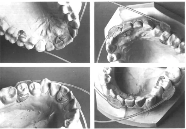
In these positions an open cannula (internal diameter 0.7 mm) was attached (Fig. 1). The cannula was embedded in a small custom-made acrylic shield (mouthpiece). The open end of the cannula was flush with the acrylic surface. The other end of the cannula was cut off 2 mm from the acrylic and was connected to a flexible tube (diameter 1.3 mm) which passed in the interdental space between two adjacent teeth to the buccal side of the teeth, where it passed through a hole in another acrylic shield. Both acrylic shields were bonded to the teeth. The tube continued from the buccal shield and passed between the lips at the corner of the mouth to an extra-oral pressure-measuring sys-tem. The placement of the measuring point (cannula) in the interdental space enabled the acrylic shields to be kept thin so that the measuring point projected at most 2 mm from the lingual surfaces of the adjacent teeth.
The extra-oral measuring system consisted of a bottle of water and compressed air, a pressure transducer, and a flow-limiting valve. The pres-sure caused a small, constant stream of water to escape through the cannula. When this was covered by the tongue, a resistance was offered to the escape of the water. Depending on the amount of pressure from the tongue on the mouthpiece, a varying pressure was built up in the water system which was recorded by the transducer. This reflects the pressure from the tongue on the mouthpiece with its open cannula. The water escaping through the cannula is swallowed by the person being tested.
Details of the pressure-measuring system, including the calibration procedure, are described in an article by Thiier et al. (1985). The application of the system for the measure-ment of pressure from the tongue on the teeth with a different design of the mouthpieces was reported by Frohlich et al. (1991).
The pressure from the tongue on the teeth was measured:
(1) in the rest position of the mandible and of the tongue;
(2) during chewing of 7 cm2 of crisp-bread;
(3) during swallowing of water.
Figure 1 Mouthpieces for the measurement of the pressures in the upper and lower jaws.
occasions 0-182 days apart (median time inter-val 7 days). At each recording session, simultan-eous recordings from the four mouthpieces were made in the following order:
(1) in the rest position; *
(2) recordings of two acts of swallowing (on command);
(3) new recording in the rest position; (4) recordings of two acts of chewing.
Electromyographic recording
Simultaneously, with the pressure registrations, the activity of the right anterior temporal muscle and of the muscles of the floor of the mouth was recorded with bipolar hook electrodes as described earlier (Frohlich et al., 1991).
The temporal muscle activity was recorded in order to evaluate the phase of the chewing cycle and to identify the act of swallowing. The activity of the muscles of the floor of the mouth was recorded to monitor the rest position of
the tongue which was attained after a command swallow.
Besides being recorded permanently with a writer, the electromyographic signal of the muscles of the floor of the mouth was also displayed on an oscilloscope which was placed in front of the subject being studied. The subject could thus follow the degree of activity of the muscles of the floor of the mouth (including the activity of the muscles of the tongue). This procedure helped the subject to keep the tongue at rest for the pressure recording.
Analysis, of the pressure recordings
The signals from the pressure transducers as well as the electromyographic signals were recorded on an electrostatic writer (GOULD ES 1000). The recordings were analysed on the paper strips of the writer. The measurement of the characteristic level of pressure at rest was done when the recording showed a straight pressure level with simultaneous minimal activ-ity of the muscles of the floor of the mouth for at least 5 seconds. The measurements of the
232 K. FROHLICH ET AL.
pressure level during the two recordings at rest were averaged.
The maximum pressure during the four chew-ing cycles in the middle of each of the two acts of chewing was measured and averaged. In
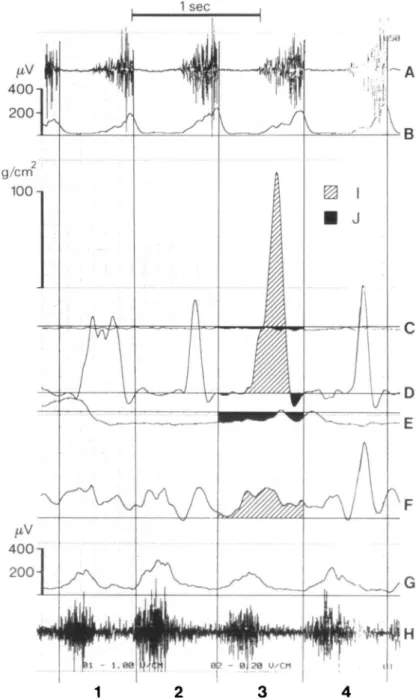
addition, the area under the pressure curve representing positive and negative pressures (Fig. 2) during the four chewing cycles was measured with a digitizer (MOP-AM 03, Kon-tron GMBH). The resulting net area (positive 1 sec g/crri 100 n
•k
\)
/V y~J\7
DFigure 2 Electromyographic and pressure recording of chewing. (A) Direct recording of anterior temporal muscle activity. (B) Mean voltage recording of anterior temporal muscle activity. (C) Pressure recorded at upper incisors. (D) Pressure recorded at upper molar. (E) Pressure recorded at lower incisors. (F) Pressure recorded at lower molar. (G) Mean voltage recording of activity of the muscles of the floor of the mouth. (H) Direct recording of activity of the muscles of the floor of the mouth. (I) Positive and (J) negative area under the pressure curve.
area minus negative area) per second of chewing was calculated. This is the mean pressure (in g/cm2) during the four chewing cycles. The
determination of the beginning and end of the chewing cycles, necessary for the measurements, was done from the electromyogram of the anterior temporal muscle.
The maximum pressure during the two acts of swallowing was measured and averaged. The net area under the pressure curve was measured, averaged, and calculated per second of swal-lowing, and thus expressed the mean pressure during an act of swallowing. The onset and end of an act of swallowing was evaluated from the electromyogram from the floor of the mouth.
Measurement of the size of the dental arches
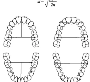
The widths and lengths of the upper and lower dental arches were measured on dental casts as shown in Fig. 3. The widths (at the canines and at the first molars) were measured with sliding calipers.to the nearest 0.10 mm. The lengths were measured with the special sliding calipers of Lundstom (1948) to the nearest 0.10 mm. The length measurements were made at both central incisors and averaged.
Statistical methods
Systematic differences between repeated record-ings were evaluated with Wilcoxon's matched-pairs signed-ranks test and accidental errors, si, (standard deviation of the single observation) calculated with the formula
si = d
2
V
In
Figure 3 Measurements of dental arch length and width
on casts.
where d is the difference between the duplicate measurements.
Differences between distributions were tested with Wilcoxon's matched-pairs signed-ranks test adjusted according to Bonferroni-Holm (Holm, 1979) in the case of multiple compar-isons. Correlations between variables were tested with Spearman's rank-correlation. In the text and in the tables a probability of 0.01 < /><0.05 is designated with*, a probability
of 0.00\<P<0.01 with**, and a probability of
P< 0.001 with***.
Results
Reproducibility of the pressure recordings
There were no significant systematic differences between the duplicate pressure recordings. The accidental errors of the method (including the intra-individual variation) were calculated from the duplicate pressure recordings and are given in Table 1. The table also shows the accidental errors (si) as a percentage of the pooled standard deviation of the two series of observations.
As is evident from Table 1, the intra-individual variation of the pressure recordings was great. Relative to the interindividual vari-ation (si in per cent), it seemed to be somewhat greater for the pressure recordings at rest than for those during chewing and swallowing.
Average pressure values
The median values and the range of variation for the pressures recorded are given in Table 2 and Fig. 4. The table is based on the means of the values recorded on the first and second occasions.
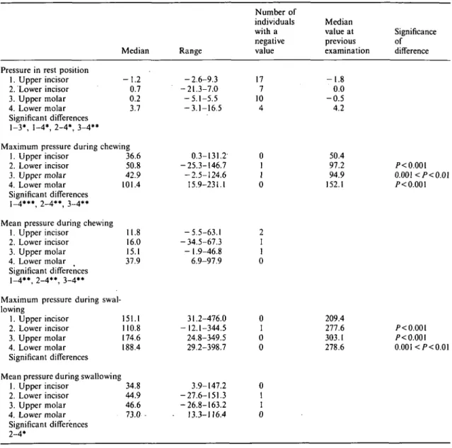
The median pressure in the rest position was negative at the upper incisors, but positive in the other locations (Fig. 5). Many individuals had, however, a negative resting pressure at the lower incisors and at the upper molar (Table 2). The resting pressure was significantly higher at the lower molar than in the other three locations.
Both the maximum and the mean pressures during chewing were significantly higher at the lower molar than in the other three locations. Negative pressures were only occasionally recorded during chewing. In one subject a nega-tive pressure during chewing was recorded at the upper incisors and at the upper molar, and in another subject at the lower incisors. In a
Table 1 Accidental errors of the method (sf) in
g/cm2 for duplicate recordings of tongue pressure in
20 subjects. The table also gives the vv a'u e 'n Pe r
cent of the pooled standard deviation of the two series of recordings.
Pressure in rest position Upper incisor Lower incisor Upper molar Lower molar
Maximum pressure during chewing Upper incisor
Lower incisor Upper molar Lower molar
Mean pressure during chewing Upper incisor
Lower incisor Upper molar Lower molar
Maximum pressure during swallowing Upper incisor
Lower incisor Upper molar Lower molar
Mean pressure during swallowing Upper incisor Lower incisor Upper molar Lower molar Si 3.55 6.14 2.62 4.55 28.31 44.09 30.26 33.81 13.37 15.96 11.88 11.34 68.32 47.48 73.00 60.40 31.29 23.61 35.45 16.84 S, in % 98 89 86 85 74 89 73 56 71 74 79 48 63 53 68 59 73 61 70 52
third subject a negative chewing pressure was found at the upper incisors.
The mean pressures during swallowing were significantly larger at the lower molar than at the lower incisors. In two individuals a negative pressure during swallowing was recorded, in one at the lower incisors, and in the other at the upper molar.
There were no significant differences between the pressures recorded in the rest position in the present investigation and those found in the same individuals at the recordings 2 years previ-ously (Frohlich et al,, 1991). The same was true for the pressures recorded at the upper incisors during chewing and swallowing. The pressures during chewing and swallowing at the lower incisors, and at the molars were, however, signi-ficantly smaller at the present examination com-pared with the previous findings (Table 2). The pressures recorded at the various locations and
during the various functions were tested for correlations with the pressures recorded in these same subjects at the same locations and during the same functions 2 years earlier. No significant correlations were found.
Correlations between the pressures recorded in the four locations during the same function
There were three significant positive coefficients of correlation between the pressures recorded in the four locations in the rest position, viz., lower incisors-upper molar, p = 0.58**, lower incisors —lower molar, p = 0.48*, upper molar-lower molar, p = 0.66**. Thus, subjects with a high pressure at rest at the lower incisors also tended to have a high pressure at the molars and subjects with a high pressure at the molar in one jaw also tended to have a high pressure at the molar in the other jaw.
The maximum and mean pressures in the same location during chewing and swallowing, respectively, were positively correlated (chewing
p = 0.78 -0.96***; swallowing p =
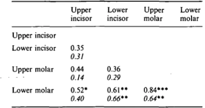
0.62**-0.88***). The coefficients of correlation between the pressures measured during chewing are given in Table 3 and those during swallowing in Table 4. The maximum pressures in the four locations during chewing were significantly intercorrelated, while for the mean pressures this was true only between the incisors and molar in the same jaw and between the upper and lower molars (Table 3). The coefficients of correlation were smaller between the mean pres-sures than between the maximum prespres-sures.
There were significant positive correlations between the maximum pressures recorded dur-ing swallowdur-ing at the upper and lower molars, at the lower incisors and lower molar, and at the upper incisors and lower molar. The mean pressures during swallowing were significantly correlated between the upper and lower molars and between the lower incisors and lower molar (Table 4).
Correlations between the pressures recorded in the same location during the different functions
Significant positive correlations were found at all four points of measurement between the maximum pressures during chewing and swal-lowing (p = 0.55* — 0.68**) as well as between the mean pressures during the two functions (p = 0.59** -0.85***).
Table 2 Median and range of variation (in g/cm2) of the pressures recorded at the various positions in 20 subjects. The table also gives the number of individuals with a negative pressure value as well as the median values recorded in the 20 subjects at a previous examination.
Pressure in rest position 1. Upper incisor 2. Lower incisor 3. Upper molar 4. Lower molar Significant differences 1-3*. 1-4*. 2-4*. 3-4** Median - 1 . 2 0.7 0.2 3.7 Range - 2 . 6 - 9 . 3 -21.3-7.0 - 5 . 1 - 5 . 5 -3.1-16.5 Number of individuals with a negative value 17 7 10 4 Median value at previous examination - 1 . 8 0.0 - 0 . 5 4.2 Significance of difference
Maximum pressure during chewing 1. Upper incisor 2. Lower incisor 3. Upper molar 4. Lower molar Significant differences 1-4***, 2-4**, 3-4** Mean pressure during chewing
1. Upper incisor 2. Lower incisor 3. Upper molar 4. Lower molar Significant differences 1-4**, 2-4**, 3-4**
Maximum pressure during swal-lowing 1. Upper incisor 2. Lower incisor 3. Upper molar 4. Lower molar Significant differences Mean pressure during swallowing
1. Upper incisor 2. Lower incisor 3. Upper molar 4. Lower molar Significant differences 2-4* 36.6 50.8 42.9 101.4 11.8 16.0 15.1 37.9 151.1 110.8 174.6 188.4 34.8 44.9 46.6 73.0 -0.3-131.2 -25.3-146.7 -2.5-124.6 15.9-231.1 -5.5-63.1 -34.5-67.3 -1.9-46.8 6.9-97.9 31.2-476.0 -12.1-344.5 24.8-349.5 29.2-398.7 3.9-147.2 -27.6-151.3 -26.8-163.2 - 13.3-116.4 0 1 1 0 2 1 1 0 0 1 0 0 0 1 I 0 50.4 97.2 94.9 152.1 209.4 277.6 303.1 278.6 P< 0.001 0.001<P<0.01 P< 0.001 P< 0.001 P< 0.001 0.00I</><0.01
and at the upper molar were not significantly correlated with the pressures during chewing or swallowing measured in these locations. The pressure at rest at the lower incisors was signi-ficantly correlated with the maximum and mean pressures during chewing (p = 0.54* and 0.46*, respectively) as well as with the mean pressure during swallowing (p = 0.51 *) measured in the same location. The pressure at rest at the lower
molar was correlated with the mean pressure during swallowing in this location (p = 0.56*). Correlations between the pressures and the overjet, overbite, and dental arch dimensions The pressure at rest at the upper incisors was negatively correlated with the overjet (p = - 0 . 5 0 * ) with the length of the upper dental arch (p= -0.58**), and with the width of the
236 g/cm 250 200 150 100 50 -50 g/cm' 5
Upper Incisor Lower Incisor Upper Molar Lower Molar | | Rest | [ Chewing | | Swallowing
Figure 4 Tongue pressures in the various locations during
the functions studied.
upper (p=-0.45*) and lower (p= -0.53*) dental arches at the molars. Thus, subjects with posteriorly wide dental arches, with a long upper dental arch and with a great overjet tended to have a small resting pressure at the upper incisors. No other significant coefficients of correlation between the pressures at rest and the dental arch dimensions or the overjet and overbite were found.
The following significant coefficients of cor-relation between the chewing and swallowing pressures and the dental arch dimensions were found:
(1) maximum and mean pressures at the lower incisors during chewing with the lengths of the upper and lower dental arches (p =
-0.49* 0.68**);
(2) mean pressures at the upper and lower incisors during swallowing with the widths of the upper and lower dental arches at the molars (p = 0.44 -0.51*);
Upper Incisor Lower Incisor Upper Molar Lower Molar Figure 5 Tongue pressures in the various locations in the rest position.
Table 3 Coefficients of correlation between the maximum and mean (italics) pressures recorded in the four locations during chewing.
Upper incisor Lower incisor Upper molar Lower molar Upper incisor 0.51* 0.32 0.64** 0.54* 0.67** 0.41 Lower incisor 0.57* 0.14 0.51* 0.47* Upper molar 0.74*** 0.54* Lower molar
(3) maximum pressure at the lower incisors during swallowing with the width of the lower dental arch at the molars (p = 0.47*); (4) mean pressure at the lower molar during swallowing with the width of the upper dental arch at the molars (p = 0.49*).
Table 4 Coefficients of correlation between the maximum and mean (italics) pressures recorded in the four locations during swallowing.
Upper incisor Lower incisor Upper molar Lower molar Upper incisor 0.35 0.31 0.44 0.14 0.52* 0.40 Lower incisor 0.36 0.29 0.61** 0.66** Upper molar 0.84*** 0.64** Lower molar Discussion
All 20 subjects of the present study also took part in the previous study by Frohlich et al. (1991), which included 25 individuals. The recording procedures were the same in the two investigations with the exception that in this study the recordings from the four points of measurement were made simultaneously and that the design of the intra-oral parts of the measuring system differed. As in the previous study, the reproducibility of the pressure record-ings was evaluated from repeated recordrecord-ings. In both studies, there were no systematic differ-ences between the repeated recordings. The accidental errors of the method for the record-ings during chewing and swallowing were gener-ally smaller in this study than in the previous investigation. This was also the case for the recording at the upper molar at rest, while the errors for the rest pressure recordings in the other three locations were greater in this study. It is believed that these errors are mainly biolo-gical in nature and are, therefore, unavoidable (Frohlich et al, 1991). To cope with this great intra-individual variation, repeated recordings, as in the present investigation, are necessary to establish a 'pressure level' for an individual during a certain function.
The median pressures in the rest position in this study agreed well with those found previ-ously in the same individuals (Frohlich et al, 1991). There was also good agreement in the proportion of individuals with a negative resting pressure with the exception that a negative pressure at the lower molar was found in four individuals in this study, but only one in the previous investigation. A negative pressure
dur-ing chewdur-ing and swallowdur-ing was found in a few individuals in this study, but was not recorded on the previous occasion.
In this study, smaller pressures during chew-ing and swallowchew-ing were found at the lower incisors and at the molars than in the same individuals at the previous investigation. We have no explanation for this difference. It may, however, be due to the less bulky design of the intra-oral part of the measuring system in this study. In our previous study, we found higher swallowing pressures at the lower incisors and at the upper and lower molars than reported by other authors. The swallowing pressures recorded in all four locations in this study, however, agree with those found by other authors (Kydd et al, 1963; Proffit, 1972; Wallen,
1974; Proffit et al, 1975).
The lack of correlation between the pressures recorded in this study and in the same indi-viduals two years earlier is noteworthy. This may signify that no long-term constancy in the pressures from the tongue on the teeth exists. It may also be due, however, to methodological problems, i.e. more recordings are needed to establish the true individual pressure level.
Considerably more significant coefficients of correlation between the pressures recorded at the four points of measurement during the various functions were found in this study than in the previous investigation. This is probably due to the simultaneous recording at all four points of measurement in this study. It is note-worthy that all these coefficients of correlation were positive, i.e. the tongue tended simultan-eously to exert either a high or a low pressure on the teeth at all points of measurement. One may speculate that this is an effect of the size of the tongue, a large tongue giving rise to a high pressure at many locations simultaneously and, conversely, a small tongue causing only a small pressure at the different locations. There was no indication of an effect of non-uniform placement of the tongue in the oral cavity; for example, a low tongue position giving rise to high pressures in the lower jaw, but small in the upper. Also, the correlations between the pressures recorded in the same location during the various functions were positive, which again may be an effect of the size of the tongue.
The negative correlations found between the size of the overjet and of the upper dental arch and the resting pressure at the upper incisors
confirm the hypothesis of an adaptive role of the tongue in the oral cavity, i.e. the tongue adapts to an existing morphological situation rather than actively moulding the dental arches. Thus, smaller pressures from the tongue were found in Australian aborigines than in Amer-icans although the dental arches are wider in the aborigines (Proffit, 1975; Proffit et al., 1975). Similarly, the vertical pressure from the tongue on the teeth during swallowing has been found to be smaller in individuals with open bite than in normal subjects (Wallen, 1974). A further indication of the adaptive role of the tongue is the finding of Proffit and Knight (1977) that individuals with maxillary protrusion had abnormally low swallowing pressures before anterior maxillary osteotomy, but that the pres-sures normalized as a result of the operation.
The pressures from the tongue on the teeth during chewing have not been studied by other authors. The negative correlations found between the chewing pressure at the lower incisors and the length of the dental arches can, therefore, not be compared with previous results. In this study, positive correlations were found between the pressures at the lower incisors during swallowing and the width of the dental arches at the molars. There was, however, no correlation between the width of the dental arch and the pressure measured at the molar in this arch. The results are, therefore, in line with those of previous investigators, who have failed to demonstrate any positive correlation between the oral cavity size or dental arch width, and the pressure from the tongue during swallowing (Proffit et al., 1969, 1975; McGlone and Proffit,
1972).
In this study, a new parameter, the mean pressure during chewing and swallowing, respectively, was introduced and used as an adjunct to the maximum pressure. The mean pressure, did not seem to contribute to the evaluation of the results. For the calculation of the mean pressure, it is necessary to determine the duration of the function studied. Especially for the swallowing function, it may be difficult to assess the onset and the end of the activity because there may be preparatory activity and a complex form with multiple peaks. For future studies, it therefore seems to be unnecessary to include the mean pressure as a parameter. The same information is obtained from the max-imum pressures, which are much easier to determine.
Address for correspondence Prof. Dr. Bengt Ingervall Klinik fur Kieferorthopadie Freiburgstrasse 7
CH-3010 Bern Switzerland
Acknowledgements
This study was supported by Schweizerischer Nationalfonds zur Forderung der wissensch-aftlichen Forschung, Grant No. 3.905-0.85.
References
Archer S Y, Vig P S 1985 Effects of head position on intraoral pressures in Class I and Class II adults. Amer-ican Journal of Orthodontics 87: 311-318
Frohlich K, Thiier U, Ingervall B 1991 Pressure from the tongue on the teeth in young adults. The Angle Ortho-dontist 61: 17-24
Gould M S E, Picton D C A 1963 An evaluation of a method of measuring forces exerted by the tongue on the teeth. British Dental Journal 114: 175-180
Hensel S 1983 Kopfhaltung und Weichteilfunktion-experimentelle Untersuchungen, Stomatologie der DDR 33: 249-259
Hensel S 1987 Metrische Untersuchungen des oro-fazialen Weichteilmilieus bei Patienten mit LKG-Spalten. Fortschritte der Kieferorthopadie 48: 11-20
Holm S 1979 A simple sequentially rejective multiple test procedure. Scandinavian Statistican 6: 65-70
Kydd W L, Akamine J S, Mendel R A, Kraus B S 1963 Tongue and lip forces exerted during deglutition in sub-jects with and without anterior open bite. Journal of Dental Research 42: 858-866
Lundstrom A 1948 Tooth size and occlusion in twins. Karger, Basel
McGlone R E, Promt W R 1972 Correlation between functional lingual pressure and oral cavity size. Cleft Palate Journal 9: 229-235
Promt W R 1972 Lingual pressure patterns in the transition from tongue thrust to adult swallowing. Archives of Oral Biology 17: 555-563
Promt W R 1975 Muscle pressures and tooth position: North American whites and Australian Aborigines. The Angle Orthodontist 45: 1-11
Promt W R, Knight J M 1977 Tongue pressures and tooth stability after anterior maxillary osteotomy. Journal of Oral Surgery 35: 798-801
Proffit W R, Kydd W L, Wilske G H, Taylor D T 1964 Intraoral pressures in a young adult group. Journal of Dental Research 43: 555-562
Proffit W R, Chastain B B, Norton L A 1969 Linguopalatal pressure in children. American Journal of Orthodontics 55: 154-166
Promt W R, McGlone R E, Barrett M J 1975 Lip and Wallen T R 1974 Vertically directed forces and malocclu-tongue pressures related to dental arch and oral cavity sion: a new approach. Journal of Dental Research 53: size in Australian Aborigines. Journal of Dental Research 1015-1022
54: 1161-1172 Winders R V 1958 Forces exerted on the dentition by the Thuer U, Janson T, Ingervall B 1985 Application in children Pe r i o r a l a n d l i neu a l musculature during swallowing. The
of a new method for the measurement of forces from the A nSl e Orthodontist 28: 226-235
lips on the teeth. European Journal of Orthodontics 7: Winders R V 1962 Recent findings in myometric research. 63-78 The Angle Orthodontist 32: 38-43