Detailed site distribution of melanoma and sunlight
exposure: aetiological patterns from a Swiss series
J.-L. Bulliard
1*, D. De Weck
2, T. Fisch
3, A. Bordoni
4& F. Levi
1,5,61
Unite´ d’e´pide´miologie du cancer, Institut universitaire de me´decine sociale et pre´ventive, Lausanne;2
Registre Valaisan des Tumeurs, Sion;
3
Krebsregister St.Gallen-Appenzell, St.Gallen **(currently Krebsregister Zu¨rich, Zu¨rich4
Registro Tumori del Cantone Ticino, Locarno;
5
Registre Vaudois des Tumeurs, Lausanne;6
Registre Neuchaˆtelois des Tumeurs, Neuchaˆtel, Switzerland
Received 26 January 2006; revised 2 June 2006 and 6 October 2006; accepted 7 December 2006
Background:
The relation between detailed cutaneous distribution of melanoma and indicators of sun exposure patterns has scantily been explored in moderately sun-sensitive populations.Patients and methods:
The precise site of 1658 primary malignant melanoma, registered from 1995 to 2002, in Switzerland were retrieved and clinically validated. Relative melanoma density (RMD) was computed by the ratio of observed to expected number of melanoma allowing for body site surface areas, and further adjusted for site-specific melanocyte density.Results:
Sites of highest risks were the face, shoulder and upper arm for both sexes, the back for men, and leg for women. Major features of this series were: (i) an unexpectedly high RMD for the face in women (5.6 versus 3.7 in men), (ii) the absence of a male predominance for melanoma on the ears and (iii) for the upper limbs, a steady gradient of increasing melanoma density with increasing proximity to the trunk, regardless of sex. Age and sex patterns of RMD parallelled general indicators of sun exposure and behaviour, except for the hand (RMD = 0.2).Conclusion:
RMD increased with (cumulative) site sun exposure, but a few notable exceptions support the impact of intermittent exposure in melanoma risk.Key words:
aetiology, anatomical site, melanoma, sun exposure, Switzerlandintroduction
The anatomical body site of cutaneous melanoma is of
importance because (i) it is an independent prognostic factor
[1], (ii) site-specific trends may be indicative of some impact of
early detection and preventive measures and (iii) mostly, it is
one of the best surrogates for assessing the pattern of sun
exposure (chronic versus intermittent) [2–5].
The uneven distribution of melanoma on the body, however,
cannot be explained by differences in sun exposure alone [6].
Melanocytes, the cells of origin for melanoma and which are
unequally distributed on the skin surface [7], might differ in
their response to UV insults and susceptibility to malignant
transformation according to anatomical region [3]. Emerging
evidence indicates that melanoma at different body sites might
arise through distinct causal pathways [8–11].
When comparing the propensity of different sites to produce
melanoma, availability of precise site information and
consideration of the surface area of body sites are paramount.
Most epidemiological series that documented the detailed
melanoma site focussed on highly sun-sensitive populations
[12–14] and relative skin surface areas used for adjustment
varied across studies.
On the basis of one of the largest detailed population-based
series on the cutaneous distribution of melanoma, this study
explores the relation between melanoma site and indicators of
sun exposure patterns. Data pertain to Switzerland, where
quality and completeness of ascertainment for skin cancer is
high [15–17], and whose population has a complexion and an
environmental UV exposure comparable to most western and
central European populations, for which details on melanoma
site distribution are sparse. A set of standard skin surface
proportions for 18 anatomical regions integrating sun exposure
patterns and common coding systems is proposed.
patients and methods
This study considered all primary malignant cutaneous melanoma (ICD-O T: 172.0–172.9, ICD-O M: 8720–8780), newly diagnosed from 1995 to 2002 in six Swiss cantons covered by a cancer registry and for which the detailed anatomical site was available (Neuchaˆtel, calendar years 1998–2001, n = 126; St-Gallen and both Appenzell 1995–2000, n = 529; Vaud 1999–2001, n = 407; Wallis 1998–2002, n = 225; Ticino 1996–2002, n = 429). For all registries but Ticino, the precise body site was obtained and validated as part of the Swiss Melanoma Study (SMS), which has been described fully elsewhere [18]. Briefly, a one-page questionnaire including a standardised body chart with site delineations was mailed to practitioners (mostly dermatologists) who biopsied the tumour. The diagram showed the anterior and posterior body sides, and the head was presented from front and side (left and right) positions to allow an unequivocal marking of the location of the lesion [18, 19].
original
article
*Correspondence to: Dr J.-L. Bulliard, Unite´ d’e´pide´miologie du cancer, Institut universitaire de me´decine sociale et pre´ventive, rue du Bugnon 17, 1005 Lausanne, Switzerland. Tel: +41 21-314-72-42; Fax:+41-21-314-73-73;
The SMS provided the detailed anatomical site of 1287 melanoma (90% of the 1428 questionnaires issued), for which demographic, epidemiological and clinical data were obtained by linkage with the cancer registries files. The sites originally specified and marked on the questionnaire seldom differed (about 5% of discrepant codes), and no statistically significant difference was observed between returned and unreturned questionnaires in the distribution of the main variables (body site, age, sex).
Procedure for melanoma ascertainment in Ticino includes a specific questionnaire filled by the dermatologist. Details on the body site are thereby recorded routinely in a comparable manner [20, 21] to that applied in the SMS, with a small fraction of cases with an unspecified body site (5%). The site distribution of melanoma did not differ materially across registries, thus analyses were carried out for all registries combined.
After exclusion of 33 lesions arising in unspecified, multiple or contiguous sites, and 25 melanoma whose site was insufficiently detailed [seven trunk not otherwise specified (NOS), 12 upper limbs NOS, six lower limbs NOS], this study included 1658 melanoma (89% of all incident cases registered in these cantons over the time period considered). By covering urban, rural and alpine regions, German- and Latin-speaking communities, participating Swiss registries (five out of nine centres) satisfactorily included geographical, socioeconomical and lifestyle factors associated with melanoma [18].
Division of the human body took into account (i) sun exposure and clothing patterns, (ii) anatomical regions for which the percent body surface areas (BSAs) were already measured or estimated [22–24] and (iii) current coding practices and coding systems used by cancer registries [21, 25]. Small areas with few cases were grouped for analyses, that is, supraclavicular area with neck, wrist with forearm, elbow and axilla with upper arm, knee with thigh and ankle with leg/calf (see Appendix). This stratification
enables the computation of the relative melanoma density (RMD), that is, the ratio of the observed to the expected number of cases by site, assuming an even distribution of melanoma over the whole body [24]) for 18 sites. For a few sites where proportional BSA were inconsistent across studies on the basis of commonly used sources [12–14], final estimates necessitated some minor adjustment (to ensure proportional surface areas for the head, trunk, upper and lower limbs of respectively 0.09, 0.32, 0.19 and 0.4 of the total body).
Anatomical sites were also aggregated for each sex into four categories of sun exposure [12], on the basis of clothing preferences of Swiss people at the relevant time period, as independently estimated by two of us (JLB and FL, see Appendix). RMD were calculated by type of sun exposure and broad age group. Concomitant adjustment for anatomical differences in melanocyte density [7] and surface area was computed by combining both sets of weights (details available from the authors). Chi-square tests were carried out to investigate associations between categorical variables and t-tests used to assess differences between sexes in RMD for any given site.
results
Table 1 presents the ratios of the observed to the expected number
of malignant melanoma for 18 anatomical sites. The highest
density of tumours occurred on the face, with RMD of 3.7 and 5.6
for males and females, respectively. The density of tumours on the
cheek and jaw was three-fold in women as compared with men
(P < 0.00001), whereas RMD for the ears and the nose indicated
a nonsignificant female excess (P = 0.44). Reversely, melanoma
occurrence was commoner among males than females for the
Table 1. Number of cases and RMD in Switzerland according to sex and 18 body sites
Body site BSA (%) Melanocyte
densitya
No. of cases RMD
Men Women Men Women
Ear 0.5 1400 13 19 3.26 4.29 NSb
Nose 0.2 1930 5 8 3.14 4.51 NS
Cheek, jaw 1.3 2310 27 79 2.61 6.86 **
Other parts of face 0.9 1940 41 38 5.72 4.77 NS
Face, totalc 2.9 2012 86 144 3.72 5.60 ** Scalp 3.7 1220 18 4 0.61 0.12 ** Neck 2.4 1165 25 16 1.31 0.75 NS Chest 10.0 890 81 39 1.02 0.44 ** Abdomen, flank 6.0 800 44 30 0.92 0.56 * Back 10.0 930 202 102 2.53 1.15 ** Buttocks 5.0 1260 23 20 0.58 0.45 NS
Perineum, groin, peri/anal areas 1.0 2380 3 4 0.38 0.45 NS
Shoulder 3.0 1210 84 59 3.51 2.22 **
Upper arm 5.0 1210 60 75 1.51 1.69 NS
Forearm, wrist 6.0 1100 36 49 0.75 0.92 NS
Hand (including fingers) 5.0 6 9 0.15 0.20
Hip, thigh 19.0 1000 57 106 0.38 0.63 **
Leg, calf, ankle 14.0 1510 40 178 0.36 1.44 **
Foot (including toes) 7.0 23 35 0.41 0.56 NS
aExpressed in average numbers of melanocyte per square millimetre [7]. Values for ‘other parts of face’, ‘face, total’ and ‘neck’ were obtained by averaging
relevant body site estimates. For anatomical sites which included regions with different sun exposure levels, such as the hand and foot (see Appendix), melanocyte density was separately considered for each region. For instance, melanocyte density values used for the foot were 610 (dorsum and heel), 1440 (sole) and 1290 (toes).
bP-value for the statistical comparison of RMD between men and women at this specific site (see patients and methods). cEar, nose, cheek, jaw and other or unspecified parts of face combined.
*P < 0.05; **P < 0.01.
scalp and neck areas, as well as the shoulder and most subregions
of the trunk. The back was the truncal site with the highest density
of melanoma (RMD of 2.5 for men and 1.2 for women). The
RMD was remarkably constant for other parts of the trunk for
females; this was not so for males. Melanoma density on the upper
limb increased in both genders with increasing proximity to the
trunk: between the shoulder and the hand, the RMD varied
23-fold for men and 11-23-fold for women. Apart from the leg in
women (RMD = 1.4), the RMD was below unity for anatomical
areas of the lower limbs. Males had systematically a lower density
of melanoma than females for the lower limbs.
While about 70% of melanoma occurred on intermittently
exposed sites (Table 2), lesions were more often associated with
a site of low intermittency of sun exposure in men (62.3% in
males and 23.9% in females, P < 0.001) and of high
intermittency in women (8.2% versus 45.6%, P < 0.001).
Lesions on usually covered sites were more frequent in males
than females (17.8% versus 13.1%, P = 0.008). For women, the
greater the site exposure the denser the occurrence of
melanoma. Density of melanoma by category of site exposure
was less contrasted for men, with nevertheless a three-fold
difference between least and most exposed body sites.
Table 2 also indicates that overall site variations in RMD were
only slightly reduced when differences in melanocyte density per
unit of skin surface were accounted for. The greatest change
occurred for maximally sun-exposed areas with a 50% decrease
in RMD for each sex. The global pattern of increasing density of
tumours with increasing (estimated) sun exposure, however,
persisted after this adjustment.
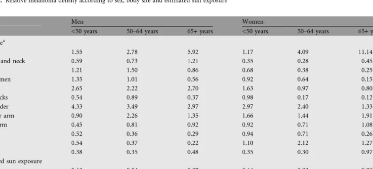
Since the age distribution of melanoma cases varies across
body sites, RMD were computed separately for three age
brackets (0–49, 50–64, 65+) by sex, site and estimated sun
exposure (Table 3). Under age 50, a raised density of melanoma
was observed on the back and the shoulder, and, for women
only, on the upper arm. At older ages, density above the unity
persisted for these sites in both genders, but the salient feature
was the high RMD for the face (5.9 and 11.1 for men and women
aged 65 or over). The known predominance of melanoma on the
lower limbs in females was most apparent in the 50- to 64-age
group (RMD = 2.1). The increase in tumour density with
increasing sun exposure became apparent after age 65 for men
and from age 50 for women. Sites associated with low and high
intermittency of sun exposure showed different patterns of
RMD with age, both within and between sexes.
Table 2. RMD, with and without correction for density of melanocytes, by sex and estimated gender-specific sun exposure in Switzerland
Estimated sun exposurea No. of cases RMD Adjusted RMDb
Men Women Men Women Men Women
Minimum 140 114 0.50 0.43 0.53 0.46
Low, intermittent 491 208 1.27 0.82 1.28 0.97
High, intermittent 65 397 0.98 1.35 1.01 1.23
Maximum 92 151 1.77 2.63 1.21 1.79
aSee Appendix for details of site exposure classification.
RMD, relative melanoma density.
bCorrected for melanocyte density.
Table 3. Relative melanoma density according to sex, body site and estimated sun exposure
Men Women
<50 years 50–64 years 65+ years <50 years 50–64 years 65+ years
Body sitea
Face 1.55 2.78 5.92 1.17 4.09 11.14
Scalp and neck 0.59 0.73 1.21 0.35 0.28 0.45
Chest 1.21 1.50 0.86 0.68 0.38 0.25 Abdomen 1.35 1.01 0.56 0.92 0.64 0.15 Back 2.65 2.22 2.70 1.63 0.97 0.80 Buttocks 0.54 0.89 0.37 0.98 0.17 0.12 Shoulder 4.33 3.49 2.97 2.97 2.40 1.33 Upper arm 0.90 2.26 1.35 1.66 1.44 1.91 Forearm 0.45 0.81 0.92 0.92 0.71 1.08 Thigh 0.52 0.36 0.29 0.94 0.71 0.26 Leg 0.54 0.37 0.22 1.10 2.12 1.27 Foot 0.38 0.35 0.48 0.35 0.30 0.97
Estimated sun exposure
Minimum 0.63 0.54 0.37 0.64 0.33 0.29
Low, intermittent 1.36 1.31 1.17 1.19 0.83 0.45
High, intermittent 0.59 0.83 1.36 1.24 1.56 1.30
Maximum 0.82 1.37 2.71 0.57 1.98 5.16
discussion
Results from this large, multicentric study corroborated a dual
association of melanoma with sun exposure. Overall, density of
melanoma increased with increasing (cumulative) site exposure.
A few notable exceptions were the hands and cases below age 50.
At younger age, RMD was highest on the intermittently
sun-exposed back and shoulder. This supports the apparently
greater impact of intermittent exposure in producing
melanoma.
From an aetiological point of view, the site distribution
generally fitted with the likely sun exposure, particularly in
regard to sex differences which matched differences in
general clothing patterns and hair cover. Thus, sites of highest
risks were the face, the shoulder and the upper arm for both
sexes, the back for men and the leg for women. In contrast,
the risk of melanoma was lowest for the buttocks, the foot
and the perianal, hip and thigh areas for both sexes, as well as
for scalp and neck, and the torso for females. The low RMD
for the hand (0.2 for each sex), at variance with the high UV
exposure of the back of the hand, confirmed observations
from other Caucasian populations [12, 26]. Melanomas of
the hand comprised a particularly heterogeneous group (two
palmar, five dorsum and eight fingers’ lesions) of varied
morphological types which could be related to different
aetiological pathways [27].
Being a relative (rather than absolute) measure, the RMD
allows direct comparisons between populations with different
melanoma incidence rates. In this respect, our series showed
some peculiarities: the density of facial melanoma was
unprecedentedly high in women (RMD = 5.6), significantly
exceeded that for men (RMD = 3.7) and no male predominance
was observed for the ears (RMD of 3.3 and 4.3, respectively).
Other Caucasian populations have shown a three to six-fold
male to female ratio in density of melanoma on the ears and
often a higher RMD for the face in men than women [12–14, 28,
29]. This unexpected finding, given the more frequent use of
facial cosmetic and sunscreens by women, did not appear to be
explained by substantial differences in histopathological
diagnosis or classification of lentigo malignant melanoma
(complementary analyses; data not shown). Swiss tend to be sun
exposed nearly all year round at altitudes of high UV irradiance.
The popularity of mountaineering, hiking and skiing has been
postulated to explain the comparatively high density of
melanoma of the head in Switzerland and neighbouring
Austrian Tyrol [30].
The distribution of lesions on the upper limbs is of particular
interest because (i) everyday clothing habits translate into an
increasing (cumulative) sun exposure with increasing distance
from the trunk and (ii) the exposure shifts from high intensity
and intermittency (shoulder) to chronicity (hand). The
gradually increasing density from the hand to the shoulder
(apart from women aged over 65) may underscore the greater
vulnerability to intense exposure of target cells on intermittently
exposed sites which are not shielded by permanent or all year
round UV-induced facultative pigmentation. To our
knowledge, such a steady gradient has not been observed before,
but few large series distinguished these four limb sections [31,
32]. Several factors may explain why some of our results differ
from earlier studies. The specific role of recent, differential
incidence trends by body site [33–36] or of some specificities
inherent to the Swiss population (high socioeconomical status
and fraction of indoor office workers, type and setting of
outdoor pursuits) cannot, however, be quantified with this
dataset.
Elwood and Gallagher [12] suggested to consider
melanocyte density and other skin features relevant to
melanoma development (nevi, amount of pigmentation) in
future investigations of the site distribution. The deviation
from a uniform body distribution was moderately reduced
after accounting for site-specific melanocyte density (Table 2).
If the number of melanocytes was essentially determined by the
amount of UV exposure, our correction would over-adjust
the RMD calculated by category of sun exposure. The
convergence of RMD towards one, especially for chronically
exposed sites, could support such an effect. The large
differences in RMD which subsisted after controlling for
melanocyte density, however, indicate that the probability of
epidermal melanocytes to become cancerous varies with the
type (or site) of exposure [3]. Hence, these descriptive data
lend support to a site-specific aetiology for melanoma, one
related to chronic exposure and the other to melanocyte
instability [8].
The advent of detailed body site coding systems, compatible
with the standard four-site classification (head, trunk, upper
and lower limbs) [20, 25] and recommended by the European
Network of Cancer Registries, should encourage and facilitate
the systematic recording of this information with affordable
effort by many cancer registries, especially when site
information could be obtained on pictorial support [19].
Historical codes, grouping for instance hand and shoulder
under upper limb [25], were not designed to allow inference on
intensity and intermittency of body site exposure. For example,
the age and sex pattern of RMD for the shoulder mirrored
that of the back (trunk) rather than that of other upper limb
sites, an observation consistent with sunlight exposure of the
shoulder.
Constitution and completeness of this series renders selection
bias unlikely (melanoma registration is 94%–99% complete in
Switzerland; F. Montanaro, Ticino Cancer Registry). Reliability
and precision of the site specification in this series, where
validation was directly provided by the practitioner who excised
the lesion, have been documented [19]. Along with the
remarkable consistency in RMD between contiguous sites of
apparently similar exposures (chest and abdomen, buttocks and
perianal area), we are confident that these results reflect the
characteristics of melanoma diagnosed in this moderately
sun-sensitive Swiss population.
This study has several limitations. Patterns and duration of
sun exposure were inferred from anatomical site and age,
respectively. Site grouping by estimated level of sun exposure
was arbitrary and could only reflect general patterns of dress.
Supine and prone limb sections were not distinguished even
though the dorsal part is generally more sun exposed—and
afford a more direct, visual inspection—than the ventral part
(e.g. the forearm). In absence of sounder data, the adjustment
for melanocytic density relied on rather old measures, obtained
for some sites from few donors [7].
Our relative BSA estimates for 18 sites integrated as much as
possible relevant measures in the most consistent and unified
manner. For sites where these estimates could be compared with
relative BSA derived from three-dimensional scans of human
bodies [37], the concordance was good. Given the
heterogeneous estimates of relative BSA applied across
epidemiological studies, sometimes derived from the same
source of measures, standard values on the basis of
anthropometric measurements of a sizable sample of subjects
are required. This would provide a sounder basis to interpret
differences for small area body sites, such as the ears, for which
estimates of the RMD are most sensitive to variation in relative
BSA. In the meantime, use of values provided in Table 1 should
enhance inter-studies comparisons.
appendix
acknowledgements
Practitioners are gratefully acknowledged for taking the time to
complete the questionnaires. We are indebted to the staff of the
cancer registries of Neuchaˆtel, St-Gall/Appenzell, Vaud, Wallis
and Ticino for their valuable collaboration. This study was
supported by a research grant from the Swiss Cancer League
(KFS-00925-09-1999) and performed during the tenure (by
JLB) of a fellowship from the Swiss National Science Foundation
(Nr. 32-63130.00).
references
1. Garbe C, Bu¨ttner P, Bertz J et al. Primary cutaneous melanoma. Prognostic classification of anatomic location. Cancer 1995; 75: 2492–2498. 2. Lee JAH, Merrill JM. Sunlight and the aetiology of malignant melanoma:
a synthesis. Med J Aust 1970; 2: 846–851.
3. Green A. A theory of site distribution of melanoma: Queensland, Australia. Cancer Causes Control 1992; 3: 513–516.
4. Franceschi S, Levi F, Randimbison L, La Vecchia C. Site distribution of different types of skin cancer: new aetiological clues. Int J Cancer 1996; 66: 1–5. 5. Bulliard J-L. Site-specific risk of cutaneous malignant melanoma and pattern of
sun exposure in New Zealand. Int J Cancer 2000; 85: 627–632.
6. Armstrong BK, Kricker A. Epidemiology of sun exposure and skin cancer. Cancer Surv 1996; 26: 133–153.
7. Szabo G. The regional anatomy of the human integument with special reference to the distribution of hair follicles, sweat glands and melanocytes. Philos Trans R Soc Lond 1967; 252: 447–485.
8. Whiteman DC, Parsons PG, Green AC. p53 expression and risk factors for cutaneous melanoma: a case-control study. Int J Cancer 1998; 77: 843–848. 9. Whiteman DC, Watt P, Purdie DM et al. Melanocytic nevi, solar keratoses, and
divergent pathways to cutaneous melanoma. J Natl Cancer Inst 2003; 95: 806–812. 10. Rivers JK. Is there more than one road to melanoma? Lancet 2004; 363:
728–730.
11. Maldonado JL, Fridlyand J, Patel H et al. Determinants of BRAF mutations in primary melanoma. J Natl Cancer Inst 2003; 95: 1878–1890.
12. Elwood JM, Gallagher RP. Body site distribution of cutaneous malignant melanoma in relationship to patterns of sun exposure. Int J Cancer 1998; 78: 276–280.
13. Green A, MacLennan R, Youl P, Martin NG. Site distribution of cutaneous melanoma in Queensland. Int J Cancer 1993; 53: 232–236.
14. Osterlind A, Hou-Jensen K, Moller Jensen O. Incidence of cutaneous malignant melanoma in Denmark 1978–1982. Anatomic site distribution, histologic types, and comparison with non-melanoma skin cancer. Br J Cancer 1988; 58: 385–391.
15. Parkin DM, Whelan SL, Ferlay J et al. Cancer Incidence in Five Continents. Lyon, France: International Agency for Research on Cancer, IARC Scientific Publication No 155 2002.
16. Levi F, Erler G, Te VC et al. Trends in skin cancer incidence in Neuchaˆtel, 1976– 98. Tumori 2001; 87: 288–289.
17. Levi F, La Vecchia C, Te VC, Mezzanotte G. Descriptive epidemiology of skin cancer in the Swiss Canton of Vaud. Int J Cancer 1988; 42: 811–816. 18. Bulliard J-L, De Weck D, Fisch T, Levi F. Population-based collection of
phenotypic and familial information on melanoma patients: a feasibility study. Schweiz Krebs Bull 2005; 25: 131–135.
19. Bulliard J-L, Levi F. Site misclassification of melanoma in the shoulder region: an issue not to be ‘shrugged at’? Arch Dermatol 2005; 141: 1047–1049. 20. World Health Organization. International Classification of Diseases for Oncology,
third edition. Geneva: WHO 2000.
21. World Health Organization. Tumor-Lokalisationsschlu¨ssel. International Classification of Diseases for Oncology, Topographischer Teil. In Wagner G (ed), 5th Edition. Berlin: Springer-Verlag 1993; 48–51.
22. Lund CC, Browder NC. The estimation of areas of burns. Surg Gynecol Obstet 1944; 79: 352–358.
Likely sun exposure and distribution of 1658 malignant cutaneous melanomas according to sex and detailed body site, Switzerland
Anatomical site Exposure levela No. of cases
Men Women
Lip 4 2 0
Eyelid 4 4 9
Ear, external auditory canal 4 13 19
Nose 4 5 8 Cheek, jaw 4 27 79 Forehead, eyebrows 4 14 9 Temple 4 16 10 Chin 4 0 5 Face, totalb 4 86 144 Scalp 2M/1F 18 4 Neck, front 3 9 9 Neck, back 3 16 7 Chest 2M/1F 81 39 Abdomen, flank 1 44 30 Back 2 202 102 Buttocks 1 23 20
Perineum, groin, peri/anal areas 1 3 4
Shoulder 2M/3F 84 59
Axilla, armpit 1 7 1
Upper arm 2M/3F 49 69
Elbow, antecubital space 3 4 5
Forearm, wrist 3 36 49
Back of hand 4 1 4
Palm of hand 1 0 2
Fingers 4 5 3
Hip, thigh 1M/2F 50 87
Knee, popliteal space 1M/2F 7 19
Leg, calf, ankle 2M/3F 40 178
Dorsum of foot, heel 2M/3F 15 11
Sole of foot 1 6 14
Toes 2M/3F 2 10
Total 788 870
a(1) minimal lifetime exposure, (2) low lifetime, mainly intermittent
exposure, (3) high lifetime, mainly intermittent exposure, (4) maximal lifetime (both chronic and intermittent) exposure.
23. Elwood JM, Gallagher RP. Site distribution of malignant melanoma. Can Med Assoc J 1983; 128: 1400–1404.
24. Pearl DK, Scott EL. The anatomical distribution of skin cancers. Int J Epidemiol 1986; 15: 502–506.
25. World Health Organization. International Classification of Diseases for Oncology. Geneva: WHO 1976.
26. Green A, McCredie M, Giles G, Jackman L. Re: ‘Occurrence of melanoma on the upper and lower limbs in eastern Australia’ [letter]. Melanoma Res 1997; 7: 437. 27. Naldi L, Altieri A, Imberti GL et al. Sun exposure, phenotypic characteristics, and
cutaneous malignant melanoma. An analysis according to different clinico-pathological variants and anatomic locations (Italy). Cancer Causes Control 2005; 16: 893–899.
28. Elder DE. Skin cancer. Melanoma and other specific nonmelanoma skin cancers. Cancer 1995; 75: 245–256.
29. Bulliard J-L, Cox B, Elwood JM. Comparison of the site distribution of melanoma in New Zealand and Canada. Int J Cancer 1997; 72: 231–235.
30. Moehrle M, Garbe C. Does mountaineering increase the incidence of cutaneous melanoma? A hypothesis based on cancer registry data. Dermatology 1999; 199: 201–203.
31. Green A, McCredie M, Giles G, Jackman L. Occurrence of melanoma on the upper and lower limbs in eastern Australia. Melanoma Res 1996; 6: 387–394.
32. van der Spek-Keijser LMT, van der Rhee HJ, Toth G et al. Site, histological type, and thickness of primary cutaneous malignant melanoma in western Netherlands since 1980. Br J Dermatol 1997; 136: 565–571.
33. Bulliard J-L, Cox B. Cutaneous malignant melanoma in New Zealand: trends by anatomical site, 1969–1993. Int J Epidemiol 2000; 29: 416–423.
34. Bulliard J-L, Cox B, Semenciw R. Trends by anatomic site in the incidence of cutaneous malignant melanoma in Canada, 1969–93. Cancer Causes Control 1999; 10: 407–416.
35. Chen YT, Zheng T, Holford TR et al. Malignant melanoma incidence in Connecticut (United States): time trends and age-period-cohort modeling by anatomic site. Cancer Causes Control 1994; 5: 341–350.
36. MacKie RM, Bray CA, Hole DJ et al. Incidence of and survival from malignant melanoma in Scotland: an epidemiological study. Lancet 2002; 360: 587–591. 37. Tikuisis P, Meunier P, Jubenville CE. Human body surface area: measurement
and prediction using three dimensional body scans. Eur J Appl Physiol 2001; 85: 264–271.