Echinococcus metacestodes as laboratory models for the
screening of drugs against cestodes and trematodes
A. HEMPHILL1*, B. STADELMANN1, S. SCHOLL1, J. MU¨ LLER1, M. SPILIOTIS1, N. MU¨ LLER1, B. GOTTSTEIN1and M. SILES-LUCAS2
1Institute of Parasitology, University of Berne, Laenggass-Strasse 122, CH-3012 Berne, Switzerland
2Parasitology Laboratory, Instituto de Recursos Naturales y Agrobiologı´a de Salamanca (CSIC), Cordel de Merinas 40-52,
37008 Salamanca, Spain
(Received 12 June 2009; revised 23 July 2009; accepted 25 July 2009; first published online 21 September 2009)
S U M M A R Y
Among the cestodes, Echinococcus granulosus, Echinococcus multilocularis and Taenia solium represent the most dangerous parasites. Their larval stages cause the diseases cystic echinococcosis (CE), alveolar echincoccosis (AE) and cysticercosis, respectively, which exhibit considerable medical and veterinary health concerns with a profound economic impact. Others caused by other cestodes, such as species of the genera Mesocestoides and Hymenolepis, are relatively rare in humans. In this review, we will focus on E. granulosus and E. multilocularis metacestode laboratory models and will review the use of these models in the search for novel drugs that could be employed for chemotherapeutic treatment of echinococcosis. Clearly, improved therapeutic drugs are needed for the treatment of AE and CE, and this can only be achieved through the development of medium-to-high throughput screening approaches. The most recent achievements in the in vitro culture and genetic manipulation of E. multilocularis cells and metacestodes, and the accessability of the E. multilocularis genome and EST sequence information, have rendered the E. multilocularis model uniquely suited for studies on drug-efficacy and drug target identification. This could lead to the development of novel compounds for the use in chemotherapy against echinococcosis, and possibly against diseases caused by other cestodes, and potentially also trematodes.
Key words : Cystic echinococcosis (CE), alveolar echinococcosis (AE), Echinoccoccus granulosus, Echinoccoccus multilocularis, in vitro culture, chemotherapy.
I N T R O D U C T I O N
The genus Echinococcus includes seven to nine described species (Nakao et al. 2007 ; Varcasia et al. 2008). Of these, Echinococcus multilocularis (the small fox tapewom) is the most pathogenic, and E. granu-losus (the small dog tapeworm) represents the most common species (Thompson, 1986). Both cause life-threatening diseases of serious public health and economic concern worldwide (McManus et al. 2003). Alveolar echinococcosis (AE), caused by E. multi-locularis, is largely restricted to the Northern hemi-sphere, such as Central Asia, Russia, Western China, Europe and Japan. Infections with E. granulosus, the causative agent of cystic echinococcosis (CE), occur globally, and mostly in the Mediterranean area, Central Europe, South America, Africa, Central Asia, and CE exists as an imported disease in Western Europe and the USA (Schantz et al. 1995 ; Eckert and Deplazes, 2004). 3.6 million disability-adjusted life years (DALYs) are lost due to CE (Craig
et al. 2007), showing that the impact of the disease is comparable to onchocercosis and African trypano-somosis (Budke et al. 2006). AE and CE are diseases of communities that often lack essential resources, thus the development of new drugs against these diseases has not been a major focus of the pharma-ceutical industry. First, because the population affected and the number of cases as such do not represent a promising market. Secondly, there has been an inherent lack of in vitro culture systems that would allow cost-effective high-throughput drug screening. However, the recent achievements in the establishment of in vitro cultivation systems, especially for E. multilocularis metacestodes, has now opened the door for medium-to-high-throughput drug screening possibilities. These developments could have considerable impact not only for the treatment of echinococcosis, but also for other dis-eases caused by cestodes such as Taenia, Hymeno-lepis, Mesocestoides and Spirometra and, potentially, trematodes.
E C H I N O C O C C U S:B I O L O G Y A N D D I S E A S E
E. multilocularis and E. granulosus share some dis-tinct features in their life cycle. The adult worms live
* Corresponding author : Andrew Hemphill, Institute of Parasitology Vetsuisse Faculty, University of Berne La¨nggass-Strasse 122, CH-3012 Berne, Switzerland.
Tel : +41 31 6312384. Fax: +41 31 6312477. Email:
in the intestine of their respective final host (dogs for E. granulosus, foxes, dogs and cats for E. multi-locularis) where sexual reproduction and subsequent egg production takes place. Faecal shedding spreads the eggs into the environment, where they are acci-dentally taken up by suitable intermediate hosts, such as small rodents for E. multilocularis, and cattle and sheep for E. granulosus. These eggs contain the first larval stage, the oncosphere, and during stomach passage, the oncosphere is activated and leaves the protective egg. It actively penetrates the intestinal lining, and migrates via blood and lymphatic vessels to the visceral organs. These are primarily the liver for E. multilocularis, and the liver, lung and other
target organs in the case of E. granulosus. There, these oncospheres develop into metacestodes, which represent the second larval stage. Within these me-tacestodes, protoscolex development takes place, and upon oral uptake by the respective final host, proto-scoleces attach to the intestinal epithelium and de-velop into adult worms, thus concluding the life cycle (Rausch, 1995).
Metacestodes of both species are fluid-filled vesicles, and represent the disease-causing stage. They are structured into an inner cellular and an outer acellular compartment (Fig. 1). The outer, acellular surface of the metacestode is formed by the laminated layer, a carbohydrate-rich structure
Fig. 1. SEM of E. granulosus (A, B) and E. multilocularis (C, D) metacestodes. A and C represent lower
magnification views of broken vesicles, exoposing both the acellular laminated layer (LL) and the germinal layer (GL) ; bars=1.5 mm. B and D represent higher magnification views of the germinal layer of E. granulosus and E. multilocularis, respectively ; bars=400 mm.
synthesized by the parasite. The laminated layer covers the entire metacestode surface, and is much more prominent in E. granulosus metacestodes (Morseth, 1967 ; Gottstein and Hemphill, 1997). The actual larval tissue is formed by the germinal layer, which itself is composed of a population of different cell types (Figs. 1 and 2). One part consists of the tegument, which is directly associated with the inner surface of the laminated layer. It is charac-terized by microvilli-like extensions, microtriches, which protrude into the matrix of the laminated layer and thereby increase the resorbing surface of the parasite (Fig. 2). In addition, the germinal layer contains highly differentiated cell types includ-ing connective tissue, muscle cells, and glycogen storage cells, as well as many undifferentiated cells (Fig. 2).
The fully mature E. granulosus metacestode (i.e. hydatid cyst) is a single-chambered or septate unilocular cyst that shows expansive growth and thereby causes compression of neighbouring tissue, tissue damage and organ dysfunction (Kern, 2006). Accidental or traumatic cyst rupture can result in release of cyst fluid and dissemination of proto-scoleces, leading to anaphylactic reactions and me-tastases (secondary echinococcosis) (Stey and Jost, 1993). E. granulosus metacestodes are surrounded by a host fibrous capsule, the adventitial layer, which is composed of host connective tissue.
In E. multilocularis infection, metacestode devel-opment exhibits different characteristics. There is no limiting host-tissue barrier comparable to the adventitial layer. The metacestode tissue represents a multivesicular structure that is reproducing
Fig. 2. TEM of E. multilocularis metacestodes cultured in vitro. A shows a low magnification view of a section through the metacestode wall, with laminated (LL) and germinal layer (GL) ; bar=7 mm. B shows a higher magnification view, demonstrating LL, GL-tissue with different cell types including undifferentiated cells with a large nucleus and nucleolus (uc), and the tegument with microtriches (arrows) ; bar=3.2 mm. C and D show higher magnification views of longitudinally sectioned (C ; bar=0.56 mm) and cross-sectioned (D; bar=0.43 mm) microtriches revealing actin microfilaments as main structural components. Also note the filamentous meshwork of electron-dense thin filaments that is embedded into the matrix of the LL.
asexually, by formation and budding of daughter vesicles, with progressive tumour-like growth (Ali-Khan et al. 1983). This leads to the formation of a large and heterogenous parasitic mass that is intermingled with host connective tissue, prolifer-ative in the periphery, and often necrotic in the centre (Gottstein and Hemphill, 1997). Metastases formation may occur in other organs due to the release of germinal layer cells into the blood or lymph system (Ali-Khan et al. 1983 ; Eckert et al. 1983 ; Mehlhorn et al. 1983). Thus, AE can resemble a benign malignant tumour (Vuitton, 2009). Proto-scolex development in humans has been rarely observed.
B E N Z I M I D A Z O L E S F O R T H E
C H E M O T H E R A P E U T I C T R E A T M E N T O F C Y S T I C E C H I N O C O C C O S I S ( C E ) A N D A L V E O L A R
E C H I N O C O C C O S I S ( A E )
For many years, the preferred treatment strategy for CE has been radical resection of the parasite mass (Kern, 2003, 2006). Other options include image-guide percutaneous treatment (PAIR=puncture, aspiration, injection, reaspiration) (Brunetti et al. 2004) and chemotherapy (El-On, 2002). Both sur-gery and PAIR are always accompanied by chemo-therapy and, for inoperable cases, chemochemo-therapy remains the only option. These cases include patients exhibiting cysts in crucial organs such as spine, pelvis and other sites that are not easily accessible, or patients with multiple cysts in several organs. The currently used drugs are mebendazole and albend-azole. These drugs clearly have the potential to kill metacestodes, and to cure the patients. Benzimid-azoles exert their action by binding to tubulin, in-ducing microtubule depolymerization and inhibiting polymerization of this essential cytoskeletal element (reviewed by Hemphill and Mu¨ ller, 2009). However, the efficacy of these benzimidazoles depends on size and type of cyst (benzimidazoles are more effective against smaller cysts), on the age of the patient (these drugs work better in younger than in older patients), and on which organ is affected (e.g. cysts localized in bones are less susceptible to chemotherapy than those in liver and lungs). The duration of treatment plays a crucial role, with prolonged drug adminis-tration producing the most favourable results for the patients (Horton, 1997 ; Franchi et al. 1999 ; Vutova et al. 1999). Praziquantel, a heterocyclic pyrazinoisoquinoline derivative, has been proposed to be used alongside benzimidazoles in CE-patients. Praziquantel is well tolerated, less toxic and better absorbed than albendazole. Praziquantel is used against the adult stages of Echinococcus and many other cestodes, and was shown to exhibit a high efficacy against protoscoleces (Morris et al. 1986) and metacestodes in animal experiments (Urrea-Paris et al. 1999, 2001). The mode of action of praziquantel
is a matter of debate. Respective studies were mostly carried out with schistosomes, where praziquantel represents the only drug that is currently marke-ted. Possibilities of praziquantel toxicity include its actions on nucleoside uptake (Angelucci et al. 2007), inhibition of phospho-inositide turnover (Wiest et al. 1992), binding to parasite actin (Tallima and El Ridi, 2007) and the parasite myosin light chain, and possibly inhibiting its functional activity (Gnanasekar et al. 2009), interference in glutathione-S-transferase activity (McTigue et al. 1995), and stimulation of Ca2+entry through voltage-operated
Ca2+channels (VOCCs) (Kohn et al. 2003 ; Jeziorski
and Greenberg, 2006). The most compelling evi-dence for the involvement of VOCCs was recently provided by Nogi et al. (2009) : they used the free-living flatworm Dugesia japonicum and investigated regeneration of fragments excised from this plan-arian that have the ability to reform a complete body plan, an ability that is driven by a totipotent popu-lation of stem cells called neoblasts. In the presence of praziquantel, however, this regeneration process yielded complete duplication of the entire antierior-posterior axis, resulting in two-headed organisms with duplicated nervous and organ systems. This effect of praziquantel was selectively ablated by in vivo RNAi of VOCC beta-subunits, but not by knock-down of alpha-subunits. At higher doses of praziquantel, knock-down of VOCC beta-subunit also conferred resistance to praziquantel, confirming the critical involvement of this beta subunit in the action of the drug.
A combined treatment regimen with albendazole and praziquantel given during the month prior to surgery of E. granulosus-infected patients increased the number of human patients with non-viable protoscoleces as compared to therapy with albend-azole alone (Cobo et al. 1998). Thus, praziquantel is regarded as useful in cases where cyst content is spilled during surgery.
For the treatment of AE, surgery and chemo-therapy are the two only treatment options (Ammann and Eckert, 1995). Spontaneous cure of AE, leading to calcified lesions, is possible, but it is not known how commonly this occurs (Gottstein and Hemphill, 1997 ; Vuitton, 2009). Radical surgery of viable le-sions is carried out if possible, but can be difficult to achieve, and only about one third of all cases are actually operated on. In contrast to E. granulosus metacestodes, E. multilocularis metacestodes are almost exclusively located in the liver. However, metastases can occur, also involving distant sites including lungs, spleen and brain. Chemotherapy should last for at least 2 years post-surgery, and monitoring of patients should be continued for 10 years. Inoperable cases must undergo long-term chemotherapy, often life-long (Reuter et al. 2000 ; 2004 ; Vuitton, 2009). The experiences with long-term use of benzimidazoles has had an enormous
impact on the use of surgery. Generally, palliative surgery should be avoided and replaced by benzi-midazole treatment alone or combined with percu-taneous or perendoscopic interventions to treat biliary or vascular complications (Bresson-Hadni et al. 2000, 2006 ; Vuitton, 2009). Extensive animal experimentation and clinical experience in human patients have both demonstrated that albendazole and mebendazole exhibit a parasitostatic rather than parasiticidal effect against E. multilocularis meta-cestodes (Reuter et al. 2004 ; Vuitton, 2009). Thus benzimidazoles only prevent parasite growth, and the recurrence rates after interruption of therapy are high. Nevertheless, clinical studies have shown that chemotherapy has significantly increased the 10-year survival rate of inoperable or non-radically operated AE patients from 6–25 % to 80–83 % (Hemphill et al. 2007).
Benzimidazoles are generally well tolerated, but problems can occur. Adverse reactions include hepatotoxicity, alopoecia, gastrointestinal disturb-ances and leukopaenia, which is sometimes severe and irreversible (Horton, 1997). Risks also include embryotoxicity and teratogenicity. In order to im-prove bioavailability, liposome-entrapped formu-lations and emulsions of albendazole have been tested (Wen et al. 1996 ; Chai et al. 2004). The use of cimetidine is discouraged, since it increases in-testinal absorption (Wen et al. 1996 ; Schipper et al. 2000), which in turn could result in toxic effects.
Major problems associated with benzimdazoles are the intra- and inter-individual variations of the pharmacokinetics, which make it necessary to measure albendazole sulphoxide and mebendazole plasma levels on a regular basis in order to adjust drug dosage and to avoid toxicity (Vuitton, 2009). This is only possible in specialized facilities, and thus precludes the use of benzimidazoles in those endemic areas where efficient drugs would be mostly needed. In addition, it is unknown whether there is a stage-specific response to exposure to benzimidazoles, and the actual concentration at the site where the drug exerts its action is not known. These pharmacological uncertainties, together with the difficulties in assess-ing metacestode viability in humans, show that novel and improved chemotherapeutical tools are needed in order to optimize the treatment of CE and AE.
T H E R O L E O F I N V I T R O C U L T U R E I N D R U G D I S C O V E R Y
In order to identify novel potential alternatives for chemotherapy against echinococcosis, the strategy used most commonly has been whole organism screening of E. multilocularis and E. granulosus metacestodes and/or protoscoleces. In most cases, researchers have focused on broad-spectrum anti-infective agents and anti-cancer compounds, many
of which also exhibited reasonable efficacy against Echinococcus (Hemphill and Mu¨ ller, 2009).
In vitro chemotherapy studies on CE have mostly, but not exclusively, been carried out on protoscoleces, since these are easily obtained from cysts of an infected animal. The metabolism, and thus drug-susceptibility, of E. granulosus proto-scoleces might however be different from the meta-cestode stage tissue. As an example, praziquantel is highly active against adult tapeworms and against protoscoleces, but only of limited efficacy against metacestodes. Conversely, albendazole is less active against protoscoleces, but of significantly higher efficacy against metacestodes (Taylor et al. 1989).
Infection of laboratory animals with E. granulosus protoscoleces results in the development of meta-cestodes, which mimics the process of secondary hydatid disease in humans, and this differentiation process can also be achieved in vitro (Walker et al. 2004). E. granulosus metacestodes do not proliferate in vitro but, as in vivo, show a marked increase in size. Thus, extensive animal experimentation is necessary for constant supply of metacestode material. In contrast, E. multilocularis metacestodes, besides in-creasing in size, also proliferate asexually and, pro-vided with the corresponding nutrients and growth factors, will form new vesicles either endogenously (from within the germinal layer) or exogenously (by budding of daughter metacestodes from older vesicles ; Fig. 3). Thus, the fact that E. multilocularis metacestodes proliferate in vitro has rendered this model suitable for the experimental assessment of the effects of chemotherapeutically interesting com-pounds.
Historically, the in vitro culture of E. multilocularis metacestodes was achieved by dissection of infected rodents and placing small pieces of infected tissue or vesicle suspension into a suitable culture medium at 37 xC. After a few weeks, newly formed me-tacestodes emerged, which were infective when re-introduced back into rodents (Hemphill and Gottstein, 1995). These were co-culture systems, as parasites could be maintained for several months in the presence of feeder cells, and in the absence of feeder cells, metacestodes degenerated within a few weeks. Jura et al. (1996) introduced a second in vitro co-culture system, which was based on co-incubation of homogenized parasite tissue obtained from in-fected rats with primary rat hepatocytes embedded in a collagen layer. Although useful in many instances (reviewed in Hemphill et al. 2002, 2007 ; Brehm and Spilitois, 2008 a), these co-culture systems provided only moderate yields and only a very limited number of drugs could be studied. Thus, the amount of metacestodes that could be generated was not suf-ficient for larger scale investigations.
Recently, Spiliotis and Brehm (2008) have over-come this problem and established a large-scale cultivation system which utilizes homogenized
metacestode tissue that is incubated in liquid culture together with rat hepatoma feeder cells. Using this system, high numbers of metacestodes with com-parable sizes can be generated within a few weeks of incubation. Thus, a major task of feeder cells ap-pears to be the provision and secretion of growth factors that are needed by the parasites to undergo proliferation, growth and differentiation. E. multi-locularis metacestodes possess several receptors that interact with human epidermal growth factor (EGF), insulin, transforming growth factor-beta and steroid hormones (Brehm et al. 2006 ; Brehm and Spiliotis, 2008 b), and these most likely allow the parasite to communicate with its host and adapt to alterations in living conditions, regulating gene expression to its favour. Of course, these receptors, and associated signalling pathways within the parasite, represent interesting targets for potential chemotherapeutical intervention (Brehm et al. 2006 ; Gelmedin et al. 2008). In addition, Spiliotis et al. (2004) developed the methodology to culture E. multilocularis meta-cestodes axenically, in the absence of feeder cells, under reducing and anaerobic conditions. In the presence of oxygen, these metacestodes degenerate within few weeks (Spilitois et al. 2004), illustrating the high sensitivity of E. multilocularis metacestodes to oxygen intermediates. The specific adaptation of the mitochondrial respiratory system to anaerobic environments has been shown also for E. multi-locularis protoscoleces (Matsumoto et al. 2008). Thus, another important task of feeder cells is to
remove toxic oxygen intermediates (Brehm and Spiliotis, 2008 a).
M E T H O D S F O R T H E A S S E S S M E N T O F A N T I-E C H I N O C O C C A L D R U G C A N D I D A T E S
By using animal experimentation, the primary as-sessment of anti-echinococcal drug candidates has often been performed in mice or gerbils, through evaluation of parasite mass and/or health parameters of the host. This has led to the extensive use of animal experimentation, but has often yielded inconclusive results (reviewed in Siles-Lucas and Hemphill, 2002). More recently, in vitro cultured Echinococcus metacestodes have been increasingly used for the primary assessment of drug susceptibilities (Hemphill et al. 2002, 2007).
A major problem associated with drug-efficacy assessment has always been the monitoring of the actual metacestode viability following in vitro drug treatment. In a primary evaluation, the effects of in vitro drug treatment can be assessed mainly by visual inspection of morphological alterations and light-microscopy. SEM and TEM have also been used extensively to investigate tissue damage in more detail (Ingold et al. 1999 ; Stettler et al. 2003 ; Naguleswaran et al. 2006 ; Spicher et al. 2008 a ; see also Fig. 2E, F), and nuclear magnetic resonance spectroscopy (NMR) has been applied to investigate metabolic changes imposed upon the parasites by drug treatments (Ingold et al. 1999). Others have
Fig. 3. Exogenous and endogenous proliferation of E. multilocularis metacestodes in vitro. Exogenous outgrowth of a newly formed vesicle (arrows in A–B) from a pre-exisiting parent vesicle appears to be followed by budding off of the daughter vesicle (C). Vesicles are also formed within older metacestodes, most likely by emerging out of the germinal layer, and these smaller metacestodes are released upon desintegration of the older metacestode (D, E).
attempted to assess parasite viability and growth by the quantification of the expression of mol-ecular marker genes such as 14-3-3 and II/3-10 (Matsumoto et al. 2006). Visual inspection relies on subjective observations and thus requires experi-enced personnel, while other techniques such as SEM, TEM, NMR and RT-real time PCR are in-trinsically time-consuming and expensive. Although these approaches have lead to the identification of several compounds with reasonable activities in the micromolar range in vitro (see below), it has not been possible to implement these techniques in a cost-effective manner at a larger scale, and they have not always produced conclusive results. For instance, in vitro culture of E. multilocularis metacestodes in the presence of albendazole sulphoxide for 14 days resulted in altered composition of vesicle fluid metabolites as assessed by NMR, and complete destruction of the metacestode structural integrity as visualized by SEM and TEM (Ingold et al. 1999). However, bioassay (re-inoculation of treated parasite material into mice) subsequently showed that the parasite had not been killed completely (Stettler et al. 2003). This was confirmed by Reuter et al. (2006), and illustrates the difficulties in viability assessment following drug treatment.
Although several potentially interesting com-pounds were identified, and combinations of some of these drugs with albendazole led to slightly improved treatment efficacy in experimentally infected mice or gerbils, none of these compounds exhibited im-proved activities compared to albendazole (reviewed in Hemphill et al. 2007). Thus, many more drugs and/or compound classes should be investigated, and there is an urgent need for a reliable, but also easy-to-handle and rapid in vitro drug screening assay for the identification of chemotherapeutically inter-esting compounds in vitro.
Attempts to develop screening assays based on the use of vital dyes employed for the assessment of cell viability, such as Alamar blue and Trypan blue, were undertaken. They worked well for protoscolex viability asessment (Walker et al. 2004), but they were not practical when used with metacestodes, as these dyes tend to bind to components of the laminated layer (M. Spicher et al. unpublished observations). The MTT viability assay, which is based on the activity of mitochondrial reductase that reduces yellow 3-(4,5-dimethylthiazol-2-yl)-2,5-diphenyltetrazoliumbromide to purple formazan in living cells, worked well for E. multilocularis me-tacestodes in an experimental setting (Emery et al. 1995), but was not found to be useful for the assess-ment of larger numbers of compounds (A. Hemphill, unpublished observations).
Another biomarker-based approach to develop such a screening assay could be to identify specific enzyme activities in culture supernatant obtained from drug-treated parasites, which release vesicle
fluid components once they are damaged or struc-turally impaired upon exposure to an active com-pound. Such biomarkers indicating damage are not present in culture supernatants of untreated meta-cestodes. E. multilocularis metacestodes are basically fluid-filled vesicles that are surrounded by an outer, acellular laminated layer, and the inner surface of this laminated layer is delineated by the actual parasite tissue, the germinal layer. The germinal layer secretes the components of the laminated layer towards the metacestode periphery into the lami-nated layer, and also secretes and/or releases meta-bolites into the vesicle fluid. One enzyme that represents an intrinsic component of the vesicle fluid, and which is also found on the laminated layer, is alkaline phosphatase (AP ; Sarciron et al. 1991 ; Lawton et al. 1997). The detection of AP activity in medium supernatants of drug-treated metacestode cultures has been proposed as a method to screen for active drugs (Stettler et al. 2001). AP-activity was indeed increased in culture supernatants of metacestodes treated with nitazoxanide (Stettler et al. 2003), 2-methoxyestradiol and artemisinin-derivatives (Spicher et al. 2008 a, b), but the sensi-tivity of this assay was not always satisfactory (Gelmedin et al. 2008 ; A. Hemphill, unpublished observations). Thus, detection of AP activity does not represent a reliable measure for drug screening in vitro. Another enzyme, phosphoglucose isomer-ase, released by dying E. multilocularis metacestodes, was found to be a promising marker that allows assessment of parasite viability of drug-treated E. multilocularis metacestodes. The potential of this screen for high-throughput assays will be published soon (B. Stadelmann et al. unpublished observations). Parasite-derived biomarkers could also play a major role for monitoring drug efficacy in patients suffering from echinococcosis during drug trials. For instance, it was previously shown that the serological response of patients against E. multilocularis alkaline phosphatase could reflect viability following surgery and/or chemotherapy (Sarciron et al. 1997). Such immunological tests are dependent on the availability and specificity of antigens and do not always provide clear-cut results ; the same applies to molecular tests, such as PCR, which require specific primers. Thus, more recently, disease-specific biomarkers have been identified by metabolic profiling emplyoing1H nuclear magnetic resonance spectroscopy (NMR). This technology delivers an overview of the meta-bolic composition of biofluids and tissues in diseased versus non-diseased individuals, and such studies have provided the means for detection and differen-tiation of e.g. coronary heart disease and schizo-phrenia (Ordovas and Moser, 2006 ; Holmes et al. 2006). Metabolic profiling has been carried out in rodents infected with S. mansoni (Wang et al. 2004), S. japonicum (Wang et al. 2006), T. spiralis (Martin et al. 2006), Trypanosoma brucei brucei (Wang et al.
2008), Plasmodium berghei (Li et al. 2008), and Echinostoma caproni (Saric et al. 2008), and could be of high interest for other infections where good markers for disease progression and/or regression are still missing. For instance, a recent study (Hosch et al. 2008) has shown that there is a good correlation between metabolic viability assessment achieved through 1H NMR of E. granulosus cyst fluid measured ex vivo from 50 patients with different degrees of disease classification and the classical ultrasound/light microscopy disease assessment, demonstrating the potential benefit of the use of metabolic biomarkers in disease and parasite viability assessments, e.g. during chemotherapy.
D R U G C A N D I D A T E S F O R T H E T R E A T M E N T O F E C H I N O C O C C O S I S
Cystic echinococcosis (CE)
A selection of drugs and potential drug candidates against CE is provided in Table 1. In many in-stances, in vitro studies on compounds active against E. granulosus have employed protoscoleces. Exper-imental prophylactic therapy of E. granulosus protoscoleces was carried out as a model that would mimic spillage during surgery, by treating proto-scoleces with praziquantel (Urrea-Paris et al. 2001) or a combination of praziquantel and albendazole (Casado et al. 2001) prior to injection into mice. The combination of albendazole and praziquantel has been used successfully in the treatment of human CE (Cobo et al. 1998). Other promising compounds with in vitro protoscolicidal actions against E. granulosus were cetrimide (Frayha et al. 1981) and the iono-phore monensin (Rogan and Richards, 1986), but these drugs were rather ineffective against metaces-todes. Levamisole and ivermectin, which are classi-cally used against nematode infections, exhibited in vitro activities similar to benzimidazoles (Martinez et al. 1999 ; Elissondo et al. 2009). The direct injec-tion of ivermectin into E. granulosus cysts in laparo-tomised patients has also shown parasiticidal effects (Ochieng’-Mitula and Burt, 1996). The combination of ivermectin plus albendazole has shown synergistic effects in infected mice (Moreno et al. 2002). Benzi-midazoles vary considerably with regard to their protoscolicidal action. Albendazole and albendazole sulphoxide have been shown to be active against E. granulosus protoscoleces in vitro (Chinnery and Morris, 1986 ; Morris et al. 1987 ; Perez-Serrano et al. 1994), but the in vitro protoscolicidal action of these drugs is rather slow and requires a longer incubation period compared to mebendazole (Morris et al. 1987 ; Rodriguez-Caabeiro et al. 1989) or oxfendazole, the major fenbendazole sulphoxide metabolite. Against E. granulosus infection in rodents, a combination of fenbendazole and netobimin (Garcia-Llamazarez et al. 1997) showed synergistic effects, allowing the
Table 1. A selection of drug candidates against cystic hydatid disease caused by Echinoco ccus granulosus . Compound In vitro activity In vivo activity Activity in humans References Mebendazole, albendazole Yes – cysts, protoscoleces Yes – rodents, sheep Yes Rev. in Siles-Lucas and Hemphill, 2002 Oxfendazole Not assessed Yes – sheep, goats Under license Blanton et al . 1988 ; Dueger et al . 1999 ; Gavidia et al . 2009 Flubendazole Yes – cysts, protoscoleces Yes – rodents Yes Ellissondo et al . 2006 ; 2007 Albendazole + praziquantel Yes – protoscoleces Not assessed Yes Cobo et al . 1998 ; Casado et al . 2001 Albendazole + ivermectin Yes – cysts, protoscoleces Yes – rodents Not assessed Moreno et al . 2002 ; Elissondo et al . 2009 Fenbendazole + netobimin Not assessed Yes – rodents Not assessed Garcia-Llamazarez et al . 1997 Cetrimide Yes – protoscoleces Yes – sheep, cattle (intracyst) Not assessed Frayha et al . 1981 Monensin Yes – protoscoleces Not assessed Not assessed Rogan and Richards, 1986 Levamisole Yes – protoscoleces Not assessed Not assessed Martı ´nez et al . 1999 Ivermectin Yes – cysts, protoscoleces No – rodentsYes – sheep (intracyst) Not assessed Ochieng’-Mitula and Burt, 1996 ; Martinez et al . 1999 ; Moreno et al . 2002 ; Elissondo et al . 2009 Nitazoxanide Yes – cysts, protoscoleces No-sheep Yes – active against progressive disease Walker et al . 2004 ; Gavidia et al . 2009 ; Winning et al . 2009 Thymol Yes -protoscoleces No – rodents Not assessed Kammerer and Perez-Esandi, 1975 ; Elissondo et al . 2008 Cyclosporin A Yes – protoscoleces Yes – rodents Not assessed Hurd et al . 1993 ; Colebrook et al . 2004 Genistein and derivates Yes – protoscoleces Not assessed Not assessed Naguleswaran et al . 2006 Dihydroartemisinin, artesunate Yes – cysts Not assessed Not assessed Spicher et al . 2008 b
administration of lower drug dosages. Oxfendazole, like albendazole, is a benzimidazole, used in veter-inary medicine for the treatment of nematode infec-tions, and has a similar antimicrobial spectrum but a longer half-life. Experimental treatments of naturally E. granulosus-infected sheep and goats suggested that oxfendazole may be as efficacious as albendazole, but does not require daily uptake of the drug because of its prolonged bioavailability (Blanton et al. 1988 ; Dueger et al. 1999). Nitazo-xanide, a nitro-thiazole-analogue (Hemphill et al. 2006) induced severe damage to E. granulosus protoscoleces and the germinal layer of E. granulosus metacestodes within few days of in vitro culture (Walker et al. 2004). Nitazoxanide treatment was not effective against experimental E. granulosus infection in sheep, but oxfendazole treatment, and a combi-nation of oxfendazole and nitazoxanide, significantly decreased the number of fertile cysts and increased the number of degenerated cysts (Gavidia et al. 2009). On the other hand, a recent case report has suggested beneficial effects of nitazoxanide in the treatment of refractory bony hydatid disease in a human patient (Winning et al. 2009). This patient had been suffering from progressive disease despite treatment with albendazole and praziquantel, and the clinical response on nitazoxanide treatment showed marked improvement in the soft tissue cysts, with stable disease in the bony pelvis. Although further studies are required, this report suggests that nita-zoxanide may be an effective treatment option in CE, particularly in patients with progressive disease who are receiving conventional treatment (Winning et al. 2009).
Profound protoscolicidal activity was also reported for another benzimidazole-derivative, flubendazole (Ellissondo et al. 2006, 2009). Further studies sub-sequently showed that flubendazole also exhibited anti-E. granulosus metacestocidal activities in vitro and in vivo in experimentally infected mice (Elissondo et al. 2007). The same authors, together with Kammerer and Perez-Esandi (1975), showed that thymol, one of the major components of the essential oils of Thymus, was found to exhibit sub-stantial protoscolicidal activity (Elissondo et al. 2008).
Cyclosporin A, employed mainly as an immuno-suppressant during the management of organ trans-plants, also exhibits anti-echinococcal activity. The administration of cyclosporin A in five consecutive daily doses, beginning 2 days prior to the infection of mice with E. granulosus protoscoleces, resulted in a significant reduction in cyst numbers and cyst masses measured at 20 weeks after infection. However, no changes in cyst mass and numbers were recorded when the drug was administered 18 weeks after infection, but the wet weight was decreased by 42 % compared with untreated controls. Ultrastruc-tural examination of the germinal membrane and
laminated layer of late-treated E. granulosus revealed abnormalities in all cysts studied whereas control and early-treated hydatids were normal (Hurd et al. 1993). Cyclosporin A has also been shown to affect E. granulosus protoscoleces in vitro (Colebrook et al. 2004).
Alveolar echinococcosis (AE)
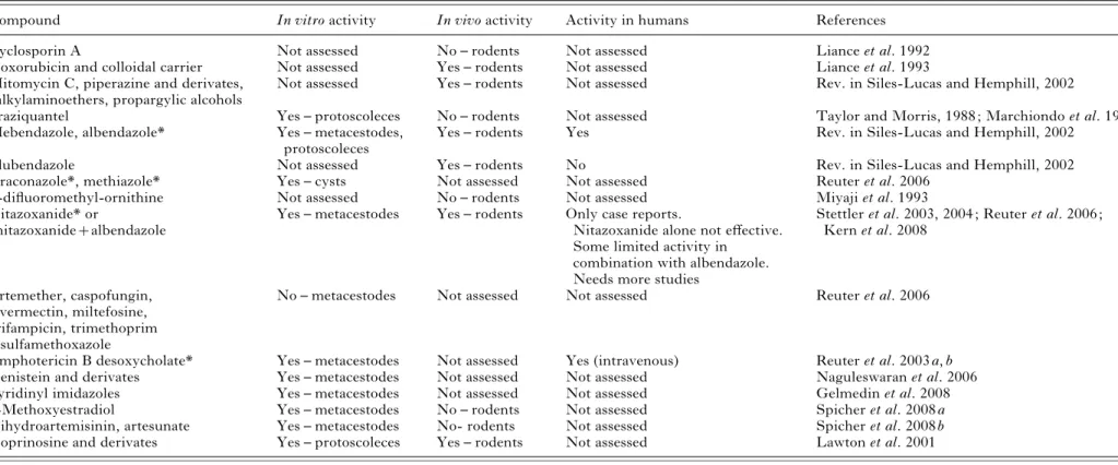
A selection of drugs and potential drug candidates for the treatment of AE is provided in Table 2. Cyclosporin A, contrary to what was found in CE, lacked anti-parasitic activity against E. multilocularis infection in experimentally infected mice (Liance et al. 1992). Doxorubicin, or hydroxyldaunorubicin, a DNA-interacting drug used widely in the treat-ment of a wide range of cancers (Launchbury and Habboubi, 1993), was bound to poly-isohexylcyanoacrylate nanoparticles (a colloidal biodegradable drug carrier) and applied in E. mutli-locularis-infected mice, resulting in the reduction of the development of the parasite in the liver and a reduced viability of the metacestode. Free doxo-rubicin or unbound nanoparticles had no antiparasitic activity (Liance et al. 1993). Animal experimentation in rodents demonstrated parasitostatic effects of mytomicin C, piperazine and quinolone derivates, alkylaminoethers and propargylic alcohols, either at a lower level or comparable to benzimidazoles (reviewed in Siles-Lucas and Hemphill, 2002). The efficacy of praziquantel was inadequate (Marchiondo et al. 1994), although showing some effects on pro-toscoleces in vitro (Taylor and Morris, 1988). Also, the treatment of E. multilocularis-infected mice with alpha-difluoromethylornithine was not successful (Miyaji et al. 1993).
More recently, studies on chemotherapeutically interesting compounds have employed in vitro cultured parasites. Nitazoxanide, a broad-spectrum anthelminthic also used for treatment against enteric bacteria, Giardia and Cryptosporidium (Hemphill et al. 2006), was identified as a compound inducing significant distortion of the germinal layer in vitro, and nitazoxanide-treated E. multilocularis meta-cestodes were non-viable when introduced into susceptible mice (Stettler et al. 2003). Reuter et al. (2006) investigated the in vitro efficacy of a series of compounds against E. multilocularis metacestodes, including albendazole, artemether, caspofungin, itraconazole, ivermectin, methiazole, miltefosine, nitazoxanide, rifampicin and trimethoprim/sulfa-methoxazole. They found that albendazole, itra-conazole, methiazole and nitazoxanide effectively destroyed parasite vesicles in vitro. However, after drug discontinuation, re-growth of vesicles occurred, indicating a parasitostatic effect only. Combination treatment with albendazole/nitazoxanide at con-centrations between 1 and 10mg/ml for 3 weeks yielded no re-growth of parasites during 8 months of
Table 2. A selection of drug candidates against alveolar echinococcosis caused by Echinococcus multilocularis
Compound In vitro activity In vivo activity Activity in humans References
Cyclosporin A Not assessed No – rodents Not assessed Liance et al. 1992
Doxorubicin and colloidal carrier Not assessed Yes – rodents Not assessed Liance et al. 1993
Mitomycin C, piperazine and derivates, alkylaminoethers, propargylic alcohols
Not assessed Yes – rodents Not assessed Rev. in Siles-Lucas and Hemphill, 2002
Praziquantel Yes – protoscoleces No – rodents Not assessed Taylor and Morris, 1988 ; Marchiondo et al. 1994
Mebendazole, albendazole* Yes – metacestodes,
protoscoleces
Yes – rodents Yes Rev. in Siles-Lucas and Hemphill, 2002
Flubendazole Not assessed Yes – rodents No Rev. in Siles-Lucas and Hemphill, 2002
Itraconazole*, methiazole* Yes – cysts Not assessed Not assessed Reuter et al. 2006
a-difluoromethyl-ornithine Not assessed No – rodents Not assessed Miyaji et al. 1993
Nitazoxanide* or
nitazoxanide+albendazole
Yes – metacestodes Yes – rodents Only case reports.
Nitazoxanide alone not effective. Some limited activity in combination with albendazole. Needs more studies
Stettler et al. 2003, 2004 ; Reuter et al. 2006 ; Kern et al. 2008
Artemether, caspofungin, ivermectin, miltefosine, rifampicin, trimethoprim /sulfamethoxazole
No – metacestodes Not assessed Not assessed Reuter et al. 2006
Amphotericin B desoxycholate* Yes – metacestodes Not assessed Yes (intravenous) Reuter et al. 2003 a, b
Genistein and derivates Yes – metacestodes Not assessed Not assessed Naguleswaran et al. 2006
Pyridinyl imidazoles Yes – metacestodes Not assessed Not assessed Gelmedin et al. 2008
2-Methoxyestradiol Yes – metacestodes No – rodents Not assessed Spicher et al. 2008 a
Dihydroartemisinin, artesunate Yes – metacestodes No- rodents Not assessed Spicher et al. 2008 b
Isoprinosine and derivates Yes – protoscoleces Yes – rodents Not assessed Lawton et al. 2001
* Parasitostatic A. Hemphill and others 578 . https://doi.org/10.1017/S003118200999117X https:/www.cambridge.org/core
. University of Basel Library
, on
30 May 2017 at 15:06:47
drug discontinuation, and the subsequent evaluation in a bioassay in gerbils did also not result in viable parasite infections. In this respect, Stettler et al. (2004) showed that nitazoxanide, applied orally to E. multilocularis infected mice, either alone or in combination with ABZ, exhibited a profound anti-parasitic efficacy, with the albendazole/nitazoxanide combination yielding the most promising outcome in terms of reducing parasite weight. The pharmaco-kinetic analysis of corresponding serum levels in mice showed that the application of albendazole in combination with nitazoxanide increased consider-ably the levels and the half-life of albendazole sul-foxide (Stettler et al. 2004). Therefore, the increased efficacy observed in mice could be the result of an increased availability of albendazole sulfoxide in mice receiving the combination treatment. Despite these promising results, neither nitazoxanide mono-therapy nor nitazoxanide-albendazole combination therapies were highly effective in human patients sufffering from AE (Kern et al. 2008).
Amphotericin B desoxycholate (cAMB), an anti-fungal compound, effectively inhibited the growth of E. multilocularis metacestodes, first in vitro, and subsequently in human patients in vivo (Reuter et al. 2003 a, b). A major limitation of cAMB is its mode of administration (intra-venous), which makes it unsuitable for prolonged use, except for salvage treatment (Reuter et al. 2003 b). Also, the action of cAMB is only parasitostatic and, since the drug is nephrotoxic, its widespread use is limited. Never-theless, prolonged application of cAMB for months to years may be feasible in some cases, as side effects are mild and serious organ damage does not appear to occur (Reuter et al. 2003 b).
In vitro studies on E. multilocularis metacestodes and E. granulosus protoscoleces have shown that genistein, representing a major component of soya and the most prominent isoflavonoid, as well as a number of genistein derivatives, are also highly effective against these parasites. The molecular basis of the efficacy of genistein and its derivative Rm6423 have not yet been elucidated, but these compounds could interfere in signalling, for instance, through an inhibition of the tyrosine kinase activity associated with the epidermal growth factor receptor identified in E. multilocularis (see Brehm et al. 2006). Recently, Gelmedin et al. (2008) identified pyridinyl imida-zoles as ATP-competitive inhibitors of a p38-like mitogen-activated protein kinase (MAPK) of E. multilocularis by adding them to in vitro cultures, demonstrating death of parasite vesicles at con-centrations that did not affect cultured mammalian cells.
An endogenous metabolite of oestrogen with both anti-angiogenic and anti-tumour effects, 2-methoxyestradiol (2-ME) (reviewed by Schumacher and Neuhaus, 2001) was shown to induce severe damage to E. multilocularis metacestodes in vitro in a
dose-dependent manner (Spicher et al. 2008 a). However, 2-ME-treatment of experimentally in-fected mice did not result in a reduction in parasite weight compared to the control, demonstrating that in vitro and in vivo situations are not always com-parable. Best results were achieved with a treatment using a combination of 2-ME and albendazole, which lead to a reduction in parasite weight compared to albendazole treatment alone, but results did not show statistical significance (Spicher et al. 2008 a). In vitro treatment of E. multilocularis and E. granulosus larval stages with the antimalarials dihydroarte-misinin and artesunate exhibited promising results, while 6 weeks of in vivo treatment of mice with in-fected E. multilocularis metacestodes had no effect. Again, combination treatments of both drugs with albendazole led to a substantial but statistically not significant reduction in parasite weight compared to results with albendazole alone (Spicher et al. 2008 b). The in vitro effect of isoprinosine and its derivates has also been demonstrated against protoscoleces of E. multilocularis (Lawton et al. 2001).
R E L E V A N C E O F E C H I N O C O C C U S M E T A C E S T O D E S C R E E N I N G M O D E L S F O R O T H E R C E S T O D E S A N D T R E M A T O D E S
Numerous studies have demonstrated that many of those drugs that were active in E. granulosus drug screening models were also of significant relevance for E. multilocularis and vice versa (see above). In addition, there is ample evidence that most compounds with good in vitro and in vivo effi-cacy against Echinococcus metacestodes are also of relevance for combatting infections by other cestode larval stages. For instance, cysticerci of T. taeniae-formis were highly sensitive to praziquantel (Becker et al. 1981), and the same applied to T. solium and T. pisiformis (Garcia-Dominguez et al. 1991 ; Martinez Zedillo et al. 1992). Albendazole and mebendazole (or modified derivatives) were also active against T. taeniaeformis cysticerci in exper-imentally infected mice (Verheyen et al. 1978 ; Jain et al. 1989). To date, albendazole and praziquantel, taken as short-term treatments (8–15 days) are the main drugs of choice for chemotherapeutic treatment of neurocysticercosis in humans (Garcia et al. 2003 ; Shandera and Kass, 2006). A recent in vitro study on T. crassiceps indicates that a combination of nitazoxanide and albendazole could be used for the treatment of cysticercosis infections (Palomares-Alonso et al. 2007), but in vivo evidence is still lacking. The effects of other benzimidazoles, such as flubendazole, were evaluated in T. solium-infected swine, with promising results (Tellez-Giron et al. 1981) that were subsequently confirmed in human patients (Tellez-Giron, 1984).
The activity of benzimidazole derivatives against Hymenolepis larvae was evaluated in the intermediate
host Tribolium confusum, demonstrating clear effects with regard to larval development (Novak and Blackburn, 1985). However, in the rodent model it was shown that benzimidazole drugs were only effective against H. nana oncosphere infection, but already developed cysticercoids were difficult to cure (Gupta et al. 1981 ; Maki and Yanagisawa, 1985), and neither flubendazole nor thiabendazole could clear H. nana cysticercoid infections in mice. Mebendazole and a series of modified benzimidazole derivatives were effective against H. nana and H. diminuta cysticercoids (Dubey et al. 1985).
In vitro studies on the effects of exposure of Mesocestoides corti tetrathyridia to anti-parasitic drugs revealed that liposomized praziquantel and albendazole had a deleterious effect on the parasite morphology and development (Hrckova et al. 1998 ; Britos et al. 2000 ; Saldana et al. 2001). The efficacy and mode of action of praziquantel were studied in the mouse model, showing that application of prazi-quantel had an adverse effect on the tetrathyridia burden in the liver and peritoneum (Hrckova and Velebny, 1995, 1997). Following the oral adminis-tration of mebendazole to M. corti-infected mice, a parasiticidal effect was observed (Heath et al. 1975 ; Eckert and Pohlenz, 1976). This is in contrast to other cestode larvae, against which mebendazole exerts a parasitostatic effect only. Deleterious actions of other drugs on Mesocestoides tetrathyridia, in-cluding cyclosporin A (Chappell et al. 1989) and albendazole (Terenina et al. 1998), have also been reported.
Potentially, the Echinococcus screening models could also have some, albeit limited, relevance for a number of trematode species, including Schistosoma japonicum, S. manoni, Chlonorchis sinensis, Fasciola hepatica and Opistorchis viverrini. For instance, praziquantel is the drug of choice for the treatment of schistosomiasis (King, 2007). The drug is highly effective against the adult worm (as for cestodes), but has only a minor activity against the larval schisto-somula. Another class of drugs that show similarities between Echinococcus and trematodes are the arte-misinins and synthetic trioxolanes. These anti-malarial drugs possess a broad spectrum of activity against trematodes, causing profound damage in vitro and substantially reducing the worm burden in experimentally infected mice (reviewed by Keiser and Utzinger, 2007 a, b). Echinoccoccus metacestodes were also susceptible to artesunate and dihydro-artemisinin in vitro, while the schistosomula were found to be particularly susceptible to artemether and artesunate (Utzinger et al. 2002 ; Spicher et al. 2008 b). The promising activity of mefloquine, another anti-malarial drug, in mice experimentally infected with S. japonicum and S. mansoni, has been reported (Keiser et al. 2009), and recent studies in our laboratory also revealed that mefloquine has a profound impact on in vitro cultured E. multilocularis
metacestodes (A. Hemphill et al. unpublished ob-servations).
On the other hand, albendazole and mebendazole, the main anti-echinococcal drugs, have no impact on S. mansoni (Schmidt, 1998). In contrast, flubend-azole, a mebendazole-derivative, was active against S. mansoni in experimentally infected mice (Nessim et al. 2000 ; Williams et al. 2003), and another benzimdazole, triclabendazole, was active against Fasciola hepatica (Robinson et al. 2001). As for Echinococcus, F. hepatica was susceptible to nitaz-oxanide treatment in vitro, and clinical studies showed that nitazoxanide could be used for the treatment of fasciolasis in children and adults (re-viewed in Hemphill et al. 2006).
W H E R E T O G O F R O M H E R E?
As outlined in this review, considerable efforts have been undertaken to improve the therapeutic options for the treatment of CE and AE (reviewed in Vuitton, 2009). These efforts have largely concen-trated on the establishment of procedures on how to manage the diseases and by setting up guidelines for treatment and classifications of disease status. Although most successful to a large extent, the cur-rent benzimidazole-based chemotherapy is far from optimal and, owing to the limited efficacy of this class of compounds, their side-effects and their costs, alternative drugs or drugs that could be integrated into a combination treatment are clearly needed. Thus, considerably more input and support is needed from academic institutions as well as pharma-ceutical and biotechnological industries and govern-mental agencies to provide solutions for these neglected diseases. A recent survey on the financial resources going into research and development funding in 2007 has shown, that HIV/AIDS received 1.1 billion US dollars, malaria and tuberculosis obtained over 400 million US dollars each, and kinetoplastid diseases and diarrhoeal diseases were granted over 125 and 114 million dollars each. On the other hand, helminth infections as a whole, encom-passing nematodes, cestodes and trematodes, re-ceived only 51.6 million dollars (Voelker, 2009).
But finances alone will not provide novel possi-bilities. Until recently, Echinococcus drug discovery has been based on rather anecdotal reports, where a limited number of drugs belonging to a certain compound class have been investigated. However, the Echinococcus drug discovery process has been lacking several important aspects that are compul-sary for successfully identifying the best and most interesting compounds. First, we need to gain access to comprehensive compound libraries, and we need to be able to screen these libraries. This can only be done through the implementation of easy-to-handle and reliable medium-to-high-throughput in vitro assays that allow screening of larger numbers of
anti-parasitic drugs in an efficient manner. Pre-ferentially, these assays do not rely on subjective microscopic evaluation, but on objective criteria, such as specific markers (e.g. enzyme activities) that indicate parasite viability/intactness or non-viability/ damage, and which could be detected preferentially in an (at least) semi-automated system. The devel-opment of such assays is ongoing, but should be intensified.
Secondly, large-scale drug screening activities are only possible if sufficient numbers of parasite organisms can be generated in vitro. This prerequi-site has been fullfilled due to the pioneering work of Brehm and co-workers (see Spiliotis and Brehm, 2008), who developed an E. multilocularis metaces-tode in vitro culture system that allows the generation of massive numbers of metacestodes out of a rela-tively small quantity of parasite tissue. This does not only enable researchers to carry out numerous drug-screening assays, but also provides the basis for bio-chemical studies, including the identification of drug targets for specific compounds by affinity chroma-tography and mass spectrometry-based sequencing.
Thirdly, it is important to have access to genomic and EST databases. In 2008, shotgun sequencing of the E. multilocularis genome was completed (http://www.sanger.ac.uk/Projects/Echinococcus/). This opens the door for increased use of in silico approaches for drug target identification, similar to the discovery of the anti-metacestodicidal activity of clarithromycin reported by Mathis et al. already in 2005. In addition, the availiability of a genome database permits drug target identification by affinity chromatography of parasite extracts on drug-coupled matrices and subsequent protein identifi-cation by mass spectroscopy (Mu¨ ller et al. 2008 a, b). Furthermore, a system is required to verify puta-tive drug targets and investigate their functional role, preferentially by genetic means, such as over-expression or silencing of genes of interest. The basis for this was set in 2008, when a method for the long-term in vitro cultivation and proliferation of primary cells isolated from axenically grown E. multilocularis metacestodes was established by Spiliotis et al. (2008). Isolated E. multilocularis cells were transiently transfected with a plasmid carrying the gene coding for the cyano-fluorescent-protein (CFP), and the corresponding gene product was expressed and detected by Western blot analysis. When co-cultured with hepatocytes, cultured E. multilocularis cells form aggegates, and eventually undergo complete in vitro regeneration of meta-cestode vesicles. Prospectively, this could well lead to the development of transgenic larval stages, and even adult E. multilocularis worms, and genetic tools can be exploited to elucidate the exact functional role of putative drug targets in different stages of development (reviewed in Brehm and Spiliotis, 2008 a).
Finally, the in vitro activity of a compound that exhibits outstanding performance can be verified in vivo, initially preferentially in a relevant small laboratory animal model. The murine or gerbil models for primary and secondary AE (Stettler et al. 2004 ; Reuter et al. 2006) represent reliable tools for such in vivo studies.
C O N C L U S I O N S
Approximately 2 billion helminth infections occur in humans worldwide, and these involving the larval stages of the three cestodes E. multilocularis, E. granulosus and T. solium are among the most serious and life-threatening ones (Brehm et al. 2006). From a practical point of view, the E. multilocularis model, in contrast to other cestodes, clearly fulfills the criteria that would allow for intensified drug-screening processes. It displays advantages such as rapid growth and proliferation in vitro, access to comprehensive genomic information and EST-databases, the possibility to maintain parasite isolates routinely in laboratory mice, and to verify in vitro results in a relevant in vivo model. Currently, the monitoring of therapy effectiveness in humans is based methods based on PET scan using18 F-deoxy-glucose (Reuter et al. 2004). After optimization, such methods could be used for the follow-up of suitable drug candidates in mice or gerbils thereby further reducing the number of animals per study. Com-pounds that not only act parasitostatic but also parasitocidal against Echinococcus in vivo have not been discovered to date, but it is conceivable that such compounds exist, and that they would be a very useful addition to the arsenal of anti-parasitic drugs against cestodes and trematodes.
A C K N O W L E D G E M E N T S
We apologize to those authors whose contributions could not be cited in this paper. This work was supported through the National Science Foundation (31-111780), the Novartis Research Foundation, Helvetia Sana Foundation, and the Swiss Life Jubila¨umsstiftung. JM is a recipient of a research fellowship provided by Novartis Animal Health, and BS has been supported by the Karl Enigk Stiftung.
R E F E R E N C E S
Ali-Khan, Z., Siboo, R., Gomersall, M. and Faucher, M. (1983). Cystolytic events and the possible role of germinal cells in metastasis in chronic alveolar hydatidosis. Annals of Tropical Medicine and Parasitology77, 497–512.
Ammann, R. and Eckert, J. (1995). Clinical diagnosis and treatment of echinococcosis in humans. In Echinococcosis and Hydatid Disease (ed. Thompson, R. C. A. and Lymbery, A. J.), pp. 411–430. CAB International, Wallmingford.
Angelucci, F., Basso, A., Bellelli, A., Brunori, M., Pica Maattoccia, L. and Valle, C. (2007). The
anti-schistosomal drug praziquantel is an adenosine antagonist. Parasitology134, 215–1221.
Becker, B., Mehlhorn, H., Andrews, P. and Thomas, H. (1981). Ultrastructural investigations on the effect of praziquantel on the tegument of five species of cestodes. Zeitschrift fu¨r Parasitenkunde 64, 257–269. Blanton, R. E., Wachira, T. M., Zeyhle, E., Njoroge,
E. M., Magambo, J. K. and Schantz, P. M. (1988). Oxfendazole treatment of cystic hydatid disease in naturally infected animals. Antimicrobial Agents and
Chemotherapy42, 601–605.
Brehm, K. and Spiliotis, M. (2008 a). Recent advances in the in vitro cultivation and genetic manipulation of Echinococcus multilocularis metacestodes and germinal cells. Experimental Parasitology119, 506–515.
Brehm, K. and Spiliotis, M. (2008 b). The influence of host hormones and cytokines on Echinococcus multilocularis signalling and development. Parasite15, 286–289.
Brehm, K., Spiliotis, M., Zavala-Gongora, R., Konrad, C. and Frosch, M. (2006). The molecular mechanisms of larval cestode development : first steps into an unknown world. Parasitology International55, S15–S21. Bresson-Hadni, S., Delabrousse, E., Blagosklonov, O.,
Bartholomot, B., Koch, S., Miguet, J. P., Andre´
Mantion, G. and Ange` le Vuitton, D. (2006). Imaging
aspects and non-surgical interventional treatment in human alveolar echinococcosis. Parasitology International55 (Suppl.), S267–S272.
Bresson-Hadni, S., Vuitton, D. A., Bartholomot, B., Heyd, B., Godart, D., Meyer, J. P., Hrusovsky, S., Becker, M. C., Mantion, G., Lenys, D. and Miguet, J. P. (2000). A twenty year history of alveolar
echinococcosis : analysis of a series of 117 patients from eastern France. European Journal of Gasteroenterology
and Hepatology12, 327–336.
Britos, L., Dominguez, L., Ehrlich, R. and Marin, M. (2000). Effect of praziquantel on the strobilar
development of Mesocestoides corti in vitro. Journal of Helminthology74, 295–299.
Brunetti, E., Troia, G., Garlaschelli, A. L., Gulizia, R. and Filice, C. (2004). Twenty years of percutaneous treatments for cystic echinococcosis : a preliminary assessment of their use and safety. Parassitologia46, 367–370.
Budke, C. M. (2006). Global socioeconomic impact of cystic echinococcosis. Emerging Infectious Diseases12, 296–303.
Chappell, L. H., Wastling, J. M. and Hurd, H. (1989). Action of cyclosporin A on the tapeworms Hymenolepis microstoma, H. diminuta and Mesocestoides corti in vivo. Parasitology98, 291–299.
Casado, N., Urrea-Paris, M. A., Moreno, M. J. and Rodriguez-Caabeiro, F. (2001). Combined praziquantel and albendazole chemoprophylaxis in experimental hydatidosis. Parasitology Research87, 787–789.
Chai, J., Menghebat Wie, J., Deyu, S., Bin, L., Jincao, S., Chen, F., Xiong, L., Yiding, M., Xiuling, W., Dolikun, Guliber, Yanchun, W., Fanghua, G. and Shuhua, X. (2004). Observations on clinical efficacy of albendazole emulsion in 264 cases of hepatic cystic echinococcosis. Parasitology International53, 3–10.
Chinnery, J. B. and Morris, D. L. (1986). Effects of albendazole sulphoxide on viability of
hydatid protoscoleces in vitro. Transactions of the Royal Society of Tropical Medicine and Hygiene80, 815–817.
Cobo, F., Yarnoz, C., Sesma, B., Fraile, P., Aizcorbe, M., Trujillo, R., Diaz-de-Liano, A. and Ciga, M. A. (1998). Albendazole plus praziquantel versus
albendazole alone as preoperative treatment in intra-abdominal hydatidosis caused by Echinococcus granulosus. Tropical Medicine and International Health3, 462–466.
Colebrook, A. L., Jenkins, D. J., Jones, M. K., Tatarczuch, L. and Lightowlers, M. W. (2004). Effect of cyclosporin A on the survival and ultrastructure of Echinococcus granulosus protoscoleces in vitro. Parasitology129, 497–504.
Craig, P. S., McManus, D. P., Lightowlers, M. W., Chabalgoity, J. A., Garcia, H. H., Gavidia, C. M., Gilman, R. H., Gonzalez, A. E., Lorca, M., Naquira, C., Nieto, A. and Schantz, P. M. (2007). Prevention and control of cystic echinococcosis. Lancet Infectious Diseases7, 385–394.
Dubey, R., Abuzar, S., Sharma, S., Chatterjee, R. K. and Katiyar, J. C. (1985). Synthesis and anthelmintic activity of 5(6)-(benzimidazol-2-ylcarbamoyl) and (4-substituted piperazin-1-yl)benzimidazoles. Journal of Medical Chemistry28, 1748–1750.
Dueger, E. L., Moro, P. L. and Gilman, R. H. (1999). Oxfendazole treatment of sheep with naturally acquired hydatid disease. Antimicrobial Agents and
Chemotherapy43, 2263–2267.
Eckert, J. and Deplazes, P. (2004). Biological,
epidemiological, and clinical aspects of echinococcosis, a zoonosis of increasing concern. Clinical Microbiology Reviews17, 107–135.
Eckert, J. and Pohlenz, J. (1976). On the effect of mebendazole on metacestodes of Mesocestoides corti and Echinococcus multilocularis. Tropenmedizin und Parasitologie27, 247–262.
Eckert, J., Thompson, R. C. A. and Mehlhorn, H. (1983). Proliferation and metastases formation of larval Echinococcus multilocularis. I. Animal model,
macroscopical and histological findings. Zeitschrift fu¨r Parasitenkunde69, 737–748.
Elissondo, M. C., Albani, C. M., Gende, L., Eguaras, M. and Denegri, G. (2008). Efficacy of thymol against Echinococcus granulosus protoscoleces. Parasitology International57, 185–190.
Elissondo, M. C., Ceballos, L., Alvarez, L., Sa´nchez Bruni, S., Lanusse, C. and Denegri, G. (2009). Flubendazole and ivermectin in vitro combination therapy produces a marked effect on Echinococcus granulosus protoscoleces and metacestodes. Parasitology Research. doi. 10.1007/s00436-009-1469-y
Elissondo, M., Ceballos, L., Dopchiz, M., Andresiuk, V., Alvarez, L., Sanchez Bruni, S., Lanusse, C. and Denegri, G. (2007). In vitro and in vivo effects of flubendazole on Echinococcus granulosus metacestodes. Parasitology Research100, 1003–1009.
Elissondo, M. C., Dopchiz, M. C., Ceballos, L., Alvarez, L., Sanchez Bruni, S., Lanusse, C. and Denegri, G. (2006). In vitro effects of flubendazole
on Echinococcus granulosus protoscoleces. Parasitology Research98, 317–323.
El-On, J. (2002). Benzimidazole treatment of cystic echinococcosis. Acta Tropica85, 243–252. Emery, I., Bories, C., Liance, M., and Houin, R.
(1995). In vitro quantitative assessment of Echinococcus multilocularis metacestode viability after in vivo and in vitro maintenance. International Journal for Parasitology25, 275–278.
Franchi, C., DiVico, B. and Teggi, A. (1999). Long term evaluation of patients with hydatidosis treated with benzimidazole carbamates. Clinical Infectious Diseases 29, 304–309.
Frayha, G. J., Bikhazi, K. J. and Kachachi, T. A. (1981). Treatment of hydatid cysts (Echinococcus granulosus) by Cetrimide. Transactions of the Royal Society of Tropical Medicine and Hygiene75, 447–450. Garcia, H. H., Gonzales, A. E., Evans, C. A. W. and
Gilman, R. H. (2003). Taenia solium cysticercosis.
The Lancet361, 547–556.
Garcia-Dominguez, C., Correa, D., Rabiela, M. T. and Flisser, A. (1991). Praziquantel treatment of muscle Taenia solium cysticercosis. 4. Reversible in vitro effect. Parasitology Research77, 691–696.
Garcia-Llamazares, J. L., Alvarez-de-Felipe, A. I., Redondo-Cardena, P., Voces-Alonso, J. A. and Prieto-Fernandez, J. G. (1997). In vivo inhibition of the regenerative capacity of hydatid material after treatment with netobimin. Parasitology Research83, 105–108.
Gavidia, C. M., Gonzalez, A. E., Lopera, L., Jayashi, C., Angelats, R., Barron, E. A., Ninaquispe, B., Villareal, L., Garcia, H. H., Verastegui, M. R. and Gilman, R. (2009). Evaluation of nitazoxanide and oxfendazole efficacy against cystic echinococcosis in naturally infected sheep. American Journal of Tropical
Medicine and Hygiene80, 367–372.
Gelmedin, V., Caballero-Gamiz, R. and Brehm, K. (2008). Characterization and inhibition of a p38-like mitogen-activated protein kinase (MAPK) from Echinococcus multilocularis : Antiparasitic activities of a
p38 MAPK inhibitors. Biochemical Pharmacology76,
1068–1081.
Gnanasekar, M., Salunkhe, A. M., Mallia, A. K., He, Y. X. and Kalyanasundaram, R. (2009). Prazquantel affects the regulatory myosin light chain of Schistosoma mansoni. Antimicrobial Agents and
Chemotherapy53, 1054–1060.
Gottstein, B. and Hemphill, A. (1997). Immunopathology of echinococcosis. Chemical
Immunology66, 177–208.
Gupta, S., Katiyar, J. C. and Sen, A. B. (1981). Effect of mode of infection on the development and
chemotherapeutic response of Hymenolepis nana in rats. Journal of Helminthology55, 101–107.
Heath, D. D., Christie, M. J. and Chevis, R. A. (1975). The lethal effect of mebendazole on secondary
Echinococcus granulosus, cysticerci of Taenia pisiformis and tetrathyridia of Mesocestoides corti. Parasitology70, 273–285.
Hemphill, A. and Gottstein, B. (1995). Immunological and morphological studies on the proliferation of in vitro cultivated Echinococcus multilocularis metacestodes. Parasitology Research81, 605–614.
Hemphill, A. and Kern, P. (2008). Special issue : experimental studies in echinococcosis. Experimental Parasitology119, 437–438.
Hemphill, A. and Mueller, J. (2009). Alveolar and cystic echinococcosis : towards novel chemotherapeutical treatment options. Journal of Helminthology83, 99–111.
Hemphill, A., Mueller, J. and Esposito, M. (2006). Nitazoxanide, a broad-spectrum thiazolide
anti-infective agent for the treatment of gastrointestinal infections. Expert Opinion in Pharmacotherapy7, 953–964.
Hemphill, A., Spicher, M., Stadelmann, B., Mueller, J., Naguleswaran, A., Gottstein, B. and Walker, M. (2007). Innovative chemotherapeutical treatment options for alveolar and cystic echinococcosis. Parasitology134, 1657–1670.
Hemphill, A., Stettler, M., Walker, M., Siles-Lucas, M., Fink, R. and Gottstein, B. (2002). Culture of Echinococcus multilocularis metacestodes ; an alternative to animal use. Trends in Parasitology18, 445–449. Holmes, E., Tsang, T. M., Huang, J. T., Leweke,
F. M., Koethe, D., Gerth, C. W., Nolden, B. M., Gross, S., Schreiber, D., Nicholson, J. K. and Bahn, S. (2006). Metabolic profiling of CSF : evidence that early intervention may impact on disease
progression and outcome in schizophrenia. PloS Medicine3, e327. doi :10.1371/journal.pmed.0030327. Horton, R. J. (1997). Albendazole in the treatment
of human cystic echinococcosis : 12 years of experience. Acta Tropica64, 79–93.
Hosch, W., Junghanss, T., Stojkovic, M., Brunetti, E., Heye, T., Kauffmann, G. W. and Hull, W. E. (2008). Metabolic viability assessment of cystic echinococcosis using high field1
H MRS of cyst contents. NMR in Biomedicine21, 734–754.
Hrckova, G. and Velebny, S. (1995). Effects of free and liposomized praziquantel on worm burden and antibody response in mice infected with Mesocestoides corti tetrathyridia. Journal of Helminthology69, 213–221.
Hrckova, G. and Velebny, S. (1997). Effect of praziquantel and liposome-incorporated praziquantel on peritoneal macrophage activation in mice infected with Mesocestoides corti tetrathyridia (Cestoda). Parasitology114, 475–482.
Hrckova, G., Velebny, S. and Corba, J. (1998). Effects of free and liposomized praziquantel on the surface morphology and motility of Mesocestoides vogae tetrathyridia (syn. M. corti ; Cestoda : Cyclophyllidea) in vitro. Parasitology Research84, 230–238.
Hurd, H., Mackenzie, K. S. and Chappell, L. H. (1993). Anthelmintic effects of cyclosporin A on protoscoleces and secondary hydatid cysts of Echinococcus granulosus in the mouse. International Journal for Parasitology23, 315–320.
Ingold, K., Bigler, P., Thormann, W., Cavaliero, T., Gottstein, B. and Hemphill, A. (1999). Efficacies of albendazole sulfoxide and albendazole sulfone against in vitro cultivated Echinococcus multilocularis
metacestodes. Antimicrobial Agents and Chemotherapy 43, 1052–1061.
Jain, M. K., Gupta, S., Katiyar, J. C., Maitra, S. C., Singh, J. and Bhakuni, D. S. (1989). Methyl