Cannabis affects people differently: inter-subject
variation in the psychotogenic effects of
D
9
-tetrahydrocannabinol: a functional magnetic
resonance imaging study with healthy volunteers
Z. Atakan1*, S. Bhattacharyya1, P. Allen1, R. Martı´n-Santos2, J. A. Crippa3, S. J. Borgwardt4, P. Fusar-Poli1, M. Seal5, H. Sallis6, D. Stahl6, A. W. Zuardi3, K. Rubia7and P. McGuire1 1Section of Neuroimaging, Department of Psychosis Studies, Institute of Psychiatry, King’s College London, London, UK 2Institut Municipal Investigacio´ Me`dica IMIM Hospital del Mar, Barcelona, Spain
3Departamento de Neuropsiquiatria e Psicologia Me´dica, Faculdade de Medicina de Ribeira˜o Preto, Universidade de Sa˜o Paulo, Brazil 4Psychiatric Outpatient Department, University Hospital Basel, Basel, Switzerland
5Murdoch Children’s Research Institute, The Royal Children’s Hospital, Parkville, Australia 6Department of Biostatistics, Institute of Psychiatry, King’s College London, London, UK
7Section of Developmental Neuropsychology and Neuroimaging, Institute of Psychiatry, King’s College London, London, UK
Background. Cannabis can induce transient psychotic symptoms, but not all users experience these adverse effects.
We compared the neural response to D9-tetrahydrocannabinol (THC) in healthy volunteers in whom the drug did or
did not induce acute psychotic symptoms.
Method. In a double-blind, placebo-controlled, pseudorandomized design, 21 healthy men with minimal experience
of cannabis were given either 10 mg THC or placebo, orally. Behavioural and functional magnetic resonance imaging measures were then recorded whilst they performed a go/no-go task.
Results. The sample was subdivided on the basis of the Positive and Negative Syndrome Scale positive score
following administration of THC into transiently psychotic (TP ; n=11) and non-psychotic (NP ; n=10) groups. During the THC condition, TP subjects made more frequent inhibition errors than the NP group and showed differential activation relative to the NP group in the left parahippocampal gyrus, the left and right middle temporal gyri and in the right cerebellum. In these regions, THC had opposite effects on activation relative to placebo in the two groups. The TP group also showed less activation than the NP group in the right middle temporal gyrus and cerebellum, independent of the effects of THC.
Conclusions. In this first demonstration of inter-subject variability in sensitivity to the psychotogenic effects of THC,
we found that the presence of acute psychotic symptoms was associated with a differential effect of THC on activation in the ventral and medial temporal cortex and cerebellum, suggesting that these regions mediate the effects of the drug on psychotic symptoms.
Received 17 February 2012 ; Revised 20 July 2012 ; Accepted 20 July 2012 ; First published online 1 October 2012
Key words: Cerebellum, fMRI, middle temporal gyri, parahippocampus, response inhibition, THC.
Introduction
Epidemiological research points towards a link be-tween the use of cannabis and the increased risk of developing a psychotic illness, in a dose-dependent manner (Arseneault et al. 2002 ; Zammit et al. 2002 ; Moore et al. 2007). However, cannabis affects in-dividuals differently and not everyone who uses it develops psychosis. The basis of this variable
sensitivity is unclear, as is the location where D9
-tetrahydrocannabinol (THC), the main compound of the plant, mediates its psychotogenic effects. Individuals with a predisposition to psychosis who might be particularly vulnerable to its adverse effects were indicated by a positive family history of psy-chosis (McGuire et al. 1995), a schizotypal personality (Stirling et al. 2008), the presence of subclinical psy-chotic features (Henquet et al. 2004), being at ultra-high risk for psychosis (Peters et al. 2009) or carrying specific genes (Caspi et al. 2005 ; van Winkel et al. 2011 ; Bhattacharyya et al. 2012a).
Elucidating which behavioural and biological fac-tors confer greater risk for psychosis in cannabis users * Address for correspondence : Z. Atakan, M.D., Department of
Psychosis Studies, PO67, Section of Neuroimaging, Institute of Psychiatry, King’s College London, DeCrespigny Park, London, SE5 8AF, UK.
(Email : [email protected]) doi:10.1017/S0033291712001924
is crucial, because as the availability of plants with higher THC content has increased, so have the related health risks (Degenhardt et al. 2010 ; Cascini et al. 2011). Although relatively few individuals develop a full-blown psychotic illness after cannabis use, a larger number (between 15 and 51 %) experience transient psychotic symptoms lasting from a few hours to a few days, as a result of cannabis use (Thomas, 1996 ; Green et al. 2003 ; D’Souza et al. 2004, 2009 ; Morrison et al. 2009). It is not yet known if there is a continuum of risk between those who become transiently psychotic (TP) and those who develop an enduring psychotic illness in relation to cannabis use. However, it would be both logical and ethically feasible to study the effects of THC in healthy individuals by comparing those who experience transient psychotic symptoms due to can-nabis intoxication with those who do not. Findings may inform research on the mechanisms underlying psychotic symptoms per se, as well as examining behavioural and neurobiological mechanisms that increase the potential risk to an individual.
Although there are a growing number of neuro-imaging studies that have examined the acute effects of THC administration on brain function (Martı´n-Santos et al. 2010), including those from our group (Borgwardt et al. 2008 ; Fusar-Poli et al. 2009; Bhattacharyya et al. 2009, 2010; Winton-Brown et al. 2011), to date none of these studies has examined the effects of THC according to psychotic symptom outcome.
Response inhibition, the ability to suppress irrel-evant acts, is a function that is impaired in cannabis users, since they make more inhibitory errors (Hester et al. 2009 ; Ramaekers et al. 2009 ; Battisti et al. 2010a). It is also relevant to patients with schizophrenia who are reported to perform slowly in various response inhi-bition tasks (Enticott et al. 2008 ; Huddy et al. 2009) and have poor error awareness (Turken et al. 2003). Furthermore, this group shows abnormal fronto-striatal activation during inhibition tasks (Rubia et al. 2001a). A well-established response inhibition para-digm used in imaging studies is the go/no-go task, which involves the activation of the inferior frontal cortex (IFC), dorsolateral prefrontal cortex (DLPFC), inferior parietal cortices and anterior cingulate gyrus (ACG) (Rubia et al. 2001b ; Simmonds et al. 2008).
The neuroimaging findings regarding the effect of cannabis on response inhibition are inconclusive due to methodological variations. Two studies report sig-nificantly lower activation in regular cannabis users, relative to non-users, within the ACG and diffuse bilateral activity in the DLPFC (Gruber & Yurgelun-Todd, 2005 ; Hester et al. 2009). Another study reports increased response in the right DLPFC, bilateral medial frontal, inferior and superior parietal lobules in cannabis users even after 28 days of monitored
abstinence (Tapert et al. 2007). In our previous study on response inhibition in participants who had seldom used cannabis, but were challenged with oral THC relative to placebo, THC was shown to attenuate activation in the right IFC and ACG and precuneus bilaterally (Borgwardt et al. 2008).
In the present study, we supplemented our pre-vious sample by recruiting additional participants, using exactly the same criteria and methodology, to investigate brain activation in those who experienced transient psychotic symptoms after THC adminis-tration, compared with those who did not. The ad-ministration of cannabidiol, in addition to THC and placebo, is not included in this paper, as it is not rel-evant to the investigation in question. We hypothe-sized that participants who developed transient psychotic symptoms with THC would show differen-tial activation relative to those that did not experience psychotic symptoms in brain regions that have pre-viously been implicated in the pathophysiology of psychosis, such as the prefrontal, medial temporal and ventral temporal cortex.
Method Design
This was a double-blind, placebo-controlled within-subject study, with a 1-month interval between scans. The order of drug administration was pseudo-randomized so that equal numbers followed each drug sequence. The Joint South London and Maudsley National Health Service and Institute of Psychiatry Research Ethics Committee approved the protocol. Each subject provided informed consent and was given extensive written and verbal information about the effects of cannabis, including psychotic symptoms. Participants
All 21 participants were healthy, native English-speaking, right-handed males. The majority (90.5 %) were white British. Their ages ranged from 20 to 42 years. All of them had used cannabis on no more than 25 occasions in their lifetime and none had used cannabis in the previous 3 months.
Criterion for inclusion into the TP group was made post hoc, on the basis of those who scored 3 or more on at least three items of the Positive and Negative Syndrome Scale (PANSS) positive subscale (Kay et al. 1987) at 2-h measurements. D’Souza et al. (2004), in their THC challenge study, had used the same criterion previously. Participants who scored below these thresholds were classed as ‘ non-psychotic ’ (NP). We identified 11 who met the criteria for transient
psychosis. All completed the scanning procedure, ex-cept for one who became too anxious to stay in the scanner. Therefore, the behavioural and symptomatic data are based on 11 TP participants and the imaging data on 10.
Participants were carefully screened and the details of the procedures can be found in the supplementary material. They were asked to abstain from any illicit drug use during the study period, from alcohol and coffee 24 and 12 h before, respectively, and cigarettes on the morning of each session, as well as receiving a urine drug screening prior to scans.
Procedure
Participants were examined at the start of each session and their pulse and blood pressure were monitored. They were given identical-looking red gelatine cap-sules of either 10 mg of THC (99.6 % pure ; THC-Pharm, Germany) or placebo (flour). Both participants and researchers were blind to the content of the cap-sules. The dose of THC was selected on the basis of previous research (Chesher et al. 1990 ; Curran et al. 2002 ; Gray et al. 2008) to produce an effect on region-al brain activation without prominent intoxication. Even though oral administration is known to indicate an erratic absorption and inter-subject variability (Grotenhermen, 2003), it was the preferred method in this study in order to produce a slow peaking plasma level for the duration of the imaging session (Lemberger et al. 1971 ; Ohlsson et al. 1980).
Behavioural ratings
The behavioural effects were evaluated at baseline (before drug administration), +1 h (immediately be-fore scanning),+2 h (immediately after scanning) and at +3 h time points by using the Visual Analogue Mood Scale (VAMS), State-Trait Anxiety Inventory (STAI), Addiction Research Centre Inventory (ARCI),
Analogue Intoxication Scale (AIS), Cambridge
Depersonalization Scale and PANSS. Further infor-mation on these scales is available in the supplemen-tary material.
As the focus of this paper is to explore the differ-ences between those who become TP under THC and those who do not, we mainly evaluated the baseline and 2-h measurements, when the peak intoxication is experienced following oral administration. The 3-h measurements are also presented in the graphs.
Researchers stayed with the participants until all their symptoms disappeared. In all cases symptoms had resolved spontaneously within 2–3 h. No psycho-pathological symptoms were reported in follow-up checks the next day, and at 1 week and 1 month later.
Functional MRI paradigm – go/no-go
Participants practised the go/no-go task prior to scanning to ensure familiarity. The task involves motor response inhibition and selective attention. Subjects are required to either execute or inhibit a motor response according to the visual cues presented on a screen. The task is described in detail in the sup-plementary material.
Behavioural analyses
Data were recorded on SPSS version 20.0 (SPSS, Inc., USA) and analysed using Stata 11 (StataCorp LP, USA). Descriptive statistics were used to summarize the baseline variables. Age, years of education, and cannabis, cigarette, alcohol and other drug use were compared between the two groups using t tests (or equivalent non-parametric Mann–Whitney U tests or Fisher’s exact test). A multilevel model was used to assess the effect of THC on each outcome measure, with subject included as a random effect and time as a fixed effect. A second multilevel model assessed the difference between the TP and NP groups. The distri-bution of each measure was assessed and no gross violations of normality were found, thus making transformations of the data unnecessary. Non-parametric methods are not advisable in this situation as they are unable to handle missing data. The multi-level models used in our analysis are less restrictive regarding missingness assumptions.
When investigating task performance, two further multilevel models were run. The first included the main effect of drug only, while group effect and its interaction with drug were also added in the second. Image acquisition
Images were acquired on a 1.5-T Signa (GE, USA) system at the Maudsley Hospital, London. T2*-weighted images were acquired with a repetition time (TR) of 1.8 s, echo time (TE) of 40 ms, flip angle 90x in 16 planes (7 mm thick), parallel to the anterior com-missure–posterior commissure line. To facilitate ana-tomic localization of activation, a high-resolution inversion recovery image dataset was also acquired, with 3-mm contiguous slices and an in-plane resol-ution of 3 mm (TR 16000 ms, inversion time 180 ms, TE 80 ms).
Data processing and analysis
A complete description of image analysis including pre-processing and non-parametric statistical model-ling can be found in the supplementary material. A non-parametric approach (XBAM v4 ; http://www. Variation in the psychotogenic effects of D-tetrahydrocannabinol 1257
brainmap.co.uk) was used to analyse the imaging data, as this method does not assume that the pop-ulation distribution is Gaussian. It is difficult to test this assumption with neuroimaging data in small groups, and, when tested, is often found to be violated (Rabe-Hesketh et al. 1997 ; Thirion et al. 2007). Instead, this approach uses median statistics to control outlier effects and employs permutation rather than normal theory-based inference as recommended by Hayasaka & Nichols (2003). The test statistic is computed by standardizing for individual difference in residual noise before embarking on second-level, multi-subject testing, using robust permutation-based methods, employing a mixed-effects method. The group activation maps for each task condition were com-puted for THC and placebo by determining the me-dian sum of squares ratio at each voxel and then compared using non-parametric repeated-measures analysis of co-variance, with a voxelwise threshold of p=0.05. The clusterwise threshold was set such that the total number of false-positive clusters per brain volume was<1 per map and the p value at which this occurred is reported.
Results
Of the 12 participants receiving THC in the first ses-sion and placebo in the second, six were classified as being in the TP group. The order of drug adminis-tration was reversed in the remaining nine partici-pants, of whom five were subsequently included in the TP group. There was no evidence of an order effect, and no significant group differences with respect to age or years of education (all p>0.1). Out of 21, 13 did not smoke. A total of eight participants were current tobacco smokers, but only two smoked more than 10 cigarettes per day and both of these were in the NP group. Fisher’s exact test showed no significant dif-ference between the two groups in terms of cigarette smoking, cannabis, alcohol and other drug use (all p>1.00) (Table 1).
Symptom data
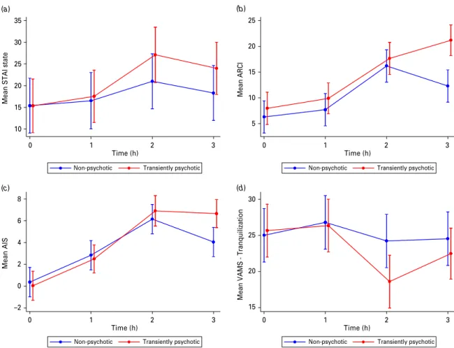
In all participants, a significant change in the level of the following outcome measures was observed 2 h after the administration of THC : STAI (p<0.001), ARCI (p<0.001), VAMS tranquillization subscale (p=0.007), AIS (p<0.001) and each of the PANSS subscales (pf0.001) (Fig. 1). For each of these meas-ures an increase in score was observed, with the ex-ception of VAMS tranquillization, which was lower at 2 h. The differences observed between baseline and 2 h were only significant when participants received THC, rather than placebo.
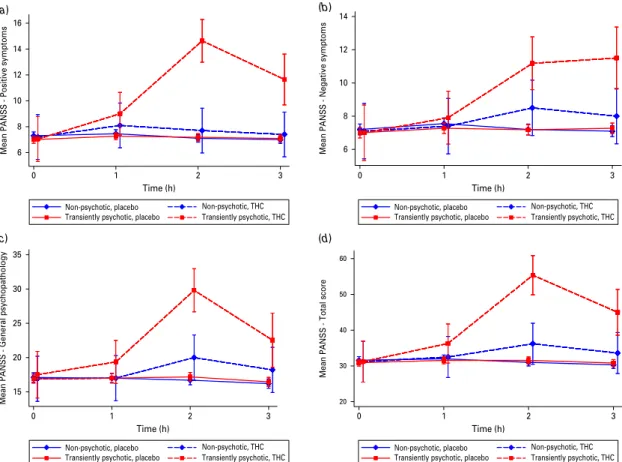
There was no significant difference between the TP and NP groups on any symptom measure at baseline or after placebo administration. However, 2 h after the administration of THC, there was a significant differ-ence between the groups for VAMS tranquillization (p=0.031), PANSS negative (p=0.020) PANSS posi-tive, general and total subscales (all pf0.001); no sig-nificant difference was found for the other behavioural scales (Table 2, Fig. 2).
Physiological measures
Under the THC condition, there was no evidence of a difference in heart rates between the two groups either at baseline or 2 h after drug administration. However, when looking at the effect of the drug across all parti-cipants, heart rate was significantly increased at 2 h after administration of either THC (pf0.001) or pla-cebo (p=0.002). There were no significant differences between either systolic or diastolic blood pressure in the two groups either at baseline or 2 h after adminis-tering THC. Graphs of physiological measures are provided in the supplementary material.
Task performance
There was a non-significant trend suggesting that THC increased inhibition errors among all partici-pants (p=0.066). A significant interaction was found between group and drug condition (p=0.002). Inhibition errors were significantly higher in the TP group than in the NP group (p<0.001), but only when participants received THC. No significant differences were found for mean reaction time to ‘ go ’ trials be-tween the THC and placebo conditions. A table on task performance is provided in the supplementary ma-terial.
Neuroimaging results Task effect
Under the placebo condition, no-go relative to oddball trials were associated with activation in the right ACG, prefrontal cortex and right middle temporal gyrus (MTG) independent of group, but with a less con-servative significance threshold contrast (p<0.025; uncorrected for<1 false-positive cluster).
Main effect of drug
During no-go compared with oddball trials, across all subjects, THC increased activation in the hippocam-pus, the tail of the caudate nucleus and the insula in the right hemisphere, relative to placebo. There were no areas where THC was associated with reduced ac-tivation relative to placebo.
Main effect of group
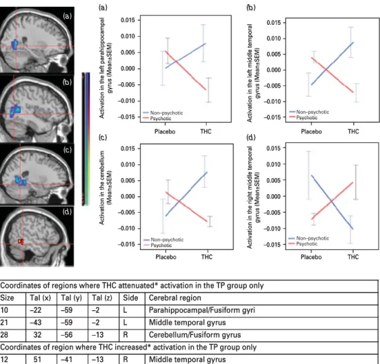
During no-go compared with oddball trials, indepen-dent of drug, the TP group showed less activation than the NP group in the right MTG (p<0.005; corrected for <1 false-positive cluster) and the vermis of the cerebellum (p<0.005; corrected for <1 false-positive cluster). There were no areas where the TP group showed greater activation than the NP group (Fig. 3).
Grouprdrug interaction
There was a significant interaction (p<0.01; corrected for <1 false-positive cluster) between the effects of drug and group in the left parahippocampal gyrus (PHG), MTG, superior temporal gyrus (STG) and in the region spanning the right cerebellum and adjacent fusiform gyrus. In all of these regions, relative to pla-cebo, THC significantly attenuated activation in the TP group, whereas it increased it in the NP group.
Relative to placebo, THC also increased activation in the right MTG in the TP group (p<0.01; corrected for <1 false-positive cluster), but it attenuated activation in the NP group (Fig. 4).
Discussion
We used functional magnetic resonance imaging to investigate differential response to oral THC in a group of healthy, seldom cannabis users and com-pared the behavioural and imaging findings of those who developed transient psychotic symptoms with those who did not. We found significant differences between the two groups in the effects of THC in the left PHG, STG, MTG and cerebellum, where THC de-creased activation in TPs, but inde-creased it in NPs. In the right MTG the reverse happened ; THC increased activation in the TPs and decreased it in the NPs. This was accompanied by a higher error rate in the TPs, relative to the NP group, during THC condition. TPs also showed less activation than the NPs in the right
Table 1.Participants’ sociodemographic and substance-use comparisonsa
Transiently psychotic (n=11)
Non-psychotic (n=10)
THC in first session, n 6 6
Mean age, years (S.D.) 26.76 (5.00) 25.70 (6.27)
Mean education, years (S.D.) 15.33 (3.64) 16.78 (4.15)
Employment, n ( %) Employed 8 (72.7) 3 (30.0) Unemployed 0 (0) 1 (10.0) Student 2 (18.2) 5 (50.0) No details 1 (9.1) 1 (10.0) Cannabis useb, n ( %) Experimental 7 (63.6) 5 (50.0) Occasional 4 (36.4) 5 (50.0) Cigarette use, n ( %) Non-smoker 7 (63.6) 6 (60.0) Smoker 4 (36.4) 4 (40.0) Alcohol usec, n ( %) Occasional 6 (54.6) 5 (50.0) Moderate 5 (45.4) 5 (50.0) Other drugs, n ( %) Not used 8 (72.7) 7 (70.0) Used 3 (27.2) 3 (30.0) THC, D9-Tetrahydrocannabinol ;
S.D., standard deviation ; df, degrees of freedom.
aNo order effect (x2=0.06, df=1, p=0.80) and no significant group differences with
respect to age (Mann–Whitney U test : Z=x0.78, p=0.44), years of education (t=0.79, df=16, p=0.44) and with Fisher’s exact test : use of cigarette smoking (p=1.00), cannabis (p=0.67), alcohol (p=1.00) and other drug use (p=1.00).
bExperimental cannabis use=less than 10 times. Occasional use=10–25 times, in
lifetime.
cOccasional alcohol use=drinking at weekends, social events. Moderate
use=drinking at least three times per week.
MTG and the vermis of the cerebellum, independent of THC.
Symptomatic effects of THC
As the groups were defined in terms of their psychotic experiences following THC, it is not surprising that they differed in their PANSS scores. Due to the small sample size, however, formal corrections for multiple testing were not possible. Instead we lowered the sig-nificance level from 5 % to 1 % at which the PANSS positive, general and total scores remained significant, and the negative subscale showed a trend (p=0.02). Therefore the negative scale result needs to be treated with caution. Even though THC significantly affected most measures in all participants, there were remark-ably few significant differences in the levels of mood, anxiety and intoxication between the two groups. This may suggest that the differential sensi-tivity to the effects of THC was particularly and
specifically related to psychotic symptoms. We cannot exclude the possibility that the absence of differences between the two groups in NP symptoms was due to limited statistical power. However, that seems un-likely, as the groups differed significantly not just on psychotic symptom severity, but also in terms of another behavioural measure : response inhibition errors.
In terms of the acute effects of THC, our findings are in line with other challenge studies in which healthy volunteers who received THC, either orally or intravenously, experienced a broad range of transient positive psychotic, negative psychotic and cognitive symptoms (Curran et al. 2002 ; D’Souza et al. 2004 ; Morrison et al. 2009). These studies also found that psychotic symptoms were not correlated with anxiety symptoms following THC. Significant in-creases in pulse rate occurred both in THC and pla-cebo conditions, possibly due to the experimental conditions. (a) 10 15 20 25 30 35
Mean STAI state
0 1 2 3 0 1 2 3
Time (h) Time (h)
0 1 2 3 0 1 2 3
Time (h) Time (h)
Non-psychotic Transiently psychotic Non-psychotic Transiently psychotic
(c) –2 0 2 4 6 8 Mean AIS (d) 15 20 25 30
Mean VAMS - Tranquilization
Non-psychotic Transiently psychotic Non-psychotic Transiently psychotic
(b) 5 10 15 20 25 Me an A RCI
Fig. 1.Comparison of behavioural measures over time, in the D9-tetrahydrocannabinol (THC) condition : (a) Spielberger’s
State-Trait Anxiety Inventory ; (b) Addiction Research Centre Inventory ; (c) Analogue Intoxication Scale ; and (d) Visual Analogue Mood Scale (VAMS) tranquillization category. Data are means, with standard errors represented by vertical bars. Measurements were taken just before drug administration at baseline (0) and repeated 1, 2 and 3 h after drug administration. When conditioning on THC, a comparison of transiently psychotic and non-psychotic at 2 h after drug administration showed a significant difference in the VAMS tranquillization category only (p=0.03).
Task performance
We found a process-specific effect of THC on the main inhibitory measure of the task (commission/inhibition errors), but not on the executive process of the task (mean reaction time to ‘ go ’ trials). Across all partici-pants THC increased inhibition errors at a trend-level of significance. Furthermore, there was a significant groupr drug interaction on this measure, where TPs made significantly more commission errors than NPs. This finding cannot be related to performance differ-ences, as we modelled only the correct trials. The findings show, that the effect of THC on impairing go/ no-go task performance is specific to the inhibitory process and that this effect is more pronounced in those who develop transient psychosis. Our partici-pants seldom used cannabis, whilst previously both occasional and heavy users of cannabis have been
shown to have increased reaction time with
the stop signal task, which is considered to be indica-tive of poor impulse control to single doses of THC (Ramaekers et al. 2009). The finding of inhi-bition deficits in TPs is comparable with those
reported in people with schizophrenia and bipolar disorder during the go/no-go task (Kiehl et al. 2000 ; Fleck et al. 2011). Higher impulsivity, inability to sup-press irrelevant acts and being unaware of making errors are likely to originate from a poorly coordinated response inhibition system and may be associated with the formation of some of the psychotic symp-toms.
Neural effects of go/no-go task
Even though in our previous study (Borgwardt et al. 2008) relative to placebo, THC attenuated activation in the right inferior frontal gyrus and the ACG, here we found that the activation of the specific motor response inhibition network occurred only with a lenient threshold. However, consistent with the results of previous studies (Borgwardt et al. 2008 ; Bhattacharyya et al. 2010), we have again found that THC significantly increased activation in the right hippocampus, tail of the caudate and insula. While the latter two are key areas of inhibition, the hippocampus is not (Chambers et al. 2009). These findings suggest
Table 2.Comparison of symptom scales between TP and NP groups at both baseline and 2 ha
Placebo THC Time Mean difference between TP and NP (95 % CI) Z p Mean difference between TP and NP (95 % CI) Z p
STAI state Baseline x1.10 (x8.07 to 5.87) x0.31 0.76 x0.05 (x8.73 to 8.63) x0.01 0.99
2 h 0.31 (x6.71 to 7.33) 0.09 0.93 6.12 (x2.69 to 14.92) 1.36 0.17
ARCI Baseline 2.83 (x0.94 to 6.60) 1.47 0.14 1.67 (x2.68 to 6.02) 0.75 0.45
2 h 2.59 (x1.14 to 6.33) 1.36 0.17 1.47 (x2.88 to 5.82) 0.66 0.51
AIS score Baseline x0.06 (x1.70 to 1.59) x0.07 0.94 x0.34 (x2.20 to 1.52) x0.36 0.72
2 h 0.24 (x1.34 to 1.83) 0.30 0.76 0.76 (x1.14 to 2.66) 0.78 0.43
VAMS
tranquillization
Baseline x0.96 (x5.48 to 3.56) x0.42 0.68 0.65 (x4.46 to 5.75) 0.25 0.80
2 h x0.43 (x4.88 to 4.02) x0.19 0.85 x5.62 (x10.73 to x0.52) x2.16 0.03*
PANSS positive Baseline x0.30 (x0.70 to 0.10) x1.48 0.14 x0.15 (x2.55 to 2.24) x0.13 0.90
2 h 0.08 (x0.32 to 0.48) 0.40 0.69 6.94 (4.59–9.28) 5.81 <0.001*
PANSS negative Baseline x0.20 (x0.64 to 0.24) x0.88 0.38 x0.10 (x2.41 to 2.21) x0.08 0.93
2 h x0.02 (x0.46 to 0.43) x0.08 0.94 2.68 (0.42–4.94) 2.33 0.02*
PANSS general Baseline x0.19 (x1.11 to 0.73) x0.41 0.68 0.60 (x4.04 to 5.25) 0.25 0.80
2 h 0.48 (x0.44 to 1.40) 1.03 0.31 9.82 (5.35–14.28) 4.31 <0.001*
PANSS total Baseline x0.69 (x2.05 to 0.67) x0.99 0.32 x2.10 (x7.94 to 7.94) 0.10 1.00
2 h 0.55 (x0.82 to 1.91) 0.78 0.43 19.16 (11.40–26.92) 4.84 <0.001*
TP, Transiently psychotic ; NP, non-psychotic ; THC, D9-tetrahydrocannabinol ; CI, confidence interval ; STAI, State-Trait
Anxiety Inventory ; ARCI, Addiction Research Centre Inventory ; AIS, Analogue Intoxication Scale ; VAMS, Visual Analogue Mood Scale ; PANSS, Positive and Negative Syndrome Scale.
aDue to small sample size, multilevel model analyses were performed separately for THC and placebo. The only significant
difference between the groups was seen in VAMS tranquillization (p=0.03) and all PANSS subscales : PANSS negative (pf0.02) and the PANSS positive, general and total subscales (all pf0.001).
* p<0.05.
increased brain-processing effort during an inhibition task in a more widespread manner involving brain regions other than the specific response inhibition network, as has been reported previously in subjects who use cannabis on a regular basis (Tapert et al. 2007 ; Roberts & Garavan, 2010). Our findings extend those previous findings by showing that the up-regulation effect of these areas is already observed in people who use cannabis seldomly and further support the view that THC may be disrupting the neural mechanisms involved with this task. Alternative neuroanatomic recruitment such as involvement of the STG, MTG and cerebellum have also been reported in a number of studies carried out on patients with bipolar disorder and schizophrenia during response inhibition tasks (Fleck et al. 2011 ; Hughes et al. 2012).
Group effect
The two groups differed inherently in terms of their task-related activation in the right MTG and the
vermis of the cerebellum, independent of THC, which were reduced in the TPs. This is an interesting finding which implies a trait difference between the groups. As we excluded those with personal and family his-tory of psychosis, it is unlikely that this finding reflects these factors. Additionally, the task we used does not normally involve the right MTG or the cerebellum. We can tentatively suggest that the differences we found may reflect a more general difference in participants’ vulnerability to transient psychosis or to inhibitory dyscontrol and could be related to variations in single nucleotide polymorphisms that are associated with an increased risk of psychosis. However, our sample was not large enough to investigate this. Some recent studies focusing on early identification of psy-chosis have reported that the right MTG is implicated in at-risk or high-risk groups (Fusar-Poli et al. 2010 ; Meijer et al. 2011). Grey matter loss in the cerebellum amongst first-onset psychosis patients has also been shown in a recent meta-analysis (Fusar-Poli et al. 2011). Other supporting evidence for the involvement 6 8 10 12 14 16
Mean PANSS - Positive symptoms
(a) 0 1 2 3 Time (h) 0 1 2 3 Time (h) 15 20 25 30 35
Mean PANSS - General psychopathology
(c) 20 30 40 50 60
Mean PANSS - Total score
(d) 6 8 10 12 14
Mean PANSS - Negative symptoms
(b) 0 1 2 3 Time (h) 0 1 2 3 Time (h) Non-psychotic, THC Non-psychotic, placebo
Transiently psychotic, placebo Transiently psychotic, THC
Non-psychotic, THC Non-psychotic, placebo
Transiently psychotic, placebo Transiently psychotic, THC
Non-psychotic, THC Non-psychotic, placebo
Transiently psychotic, placebo Transiently psychotic, THC
Non-psychotic, THC Non-psychotic, placebo
Transiently psychotic, placebo Transiently psychotic, THC
Fig. 2.Comparison of Positive and Negative Syndrome Scale (PANSS) subscales, between the transiently psychotic and
non-psychotic groups under D9-tetrahydrocannabinol (THC) and placebo conditions : (a) positive symptoms ; (b) negative
symptoms ; (c) general psychopathology ; and (d) total score. Data are means, with standard errors represented by vertical bars. A comparison of transiently psychotic and non-psychotic participants 2 h after drug administration showed significant differences in the PANSS negative subscale (p=0.02) and a highly significant difference in all other subscales (all pf0.001). Note that the y-axes have different scales in the graphs.
of this region to genetic vulnerability to psychosis is found in a recently reported study, when a significant three-way interaction between two susceptibility genes implicated in glutamate transmission (G72 and DAAO) and the diagnosis of psychosis was detected at the right MTG (Mechelli et al. 2012).
Differential neurophysiological processing of THC Our other main finding was that, as hypothesized, THC had a different effect on brain function in parti-cipants who developed transient psychotic symptoms from those who did not. These effects were evident in the left PHG, an area that has been implicated in the pathophysiology of psychosis in post-mortem (McDonald et al. 2000), neuropsychological (Marvel et al. 2007), volumetric (Witthaus et al. 2009), functional (Wolf et al. 2007) and neurochemical (Stone et al. 2010) imaging studies. Effects in this region in relation to THC-induced psychosis are of particular interest be-cause of the evidence that chronic cannabis use can impair memory (Battisti et al. 2010b). Our group had previously reported that THC increased para-hippocampal activation bilaterally during an encoding task (Bhattacharyya et al. 2009) and attenuated it dur-ing an attentional salience task (Bhattacharyya et al. 2012b). Furthermore, structural and functional chan-ges in the parahippocampal region are frequently identified in relation to cannabis use (Lorenzetti et al. 2010 ; Martı´n-Santos et al. 2010). The finding that atte-nuated left parahippocampal activity is observed only
in the TPs, but not in the NPs, provides further sup-port that this region may be implicated in psychoses.
Additional differences were evident in the left middle/superior temporal cortices and in the cer-ebellum, areas that are implicated as key regions in schizophrenia (for reviews, see Honea et al. 2005 ; Smieskova et al. 2010 ; Jardri et al. 2011). The STG, as well as the cerebellum, has been implicated in inhibitory control (Rubia et al. 2007). The increased inhibition error rate in the TP group together with the increased activation in these two inhibition-related areas may suggest that the TP group had to work harder to maintain their inhibitory capacity, which was still below the level of that in the NP group. Conversely, THC increased activation in the right MTG in the TPs, whilst it attenuated it in the NPs. It is interesting that this area is differentially activated be-tween the groups whether or not THC was present. It is difficult to interpret the two findings in relation to one another as they involve different analyses involv-ing the same region.
In all of these regions, the effect of THC on acti-vation in the group that experienced psychotic symp-toms was in the opposite direction to that in the group that did not develop psychotic symptoms. The underlying processes for this dissociated effect will require further research and replication. Interestingly, a ketamine challenge study with healthy volunteers also reported a compelling consistency between the task, region, symptom associations and those reported in patients with schizophrenia (Honey et al. 2008).
Tal (x) Tal (y) Tal (z) Side Cerebral region
58 –22 15 Right Right middle temporal gyrus
–7 –67 –24 – Vermis of cerebellum
(a) (b)
Fig. 3.Trait differences. Analysis of all subjects, independent of drug condition, showed significant differences between the two
groups in two regions. (a) Crosshair showing that activation in the right middle temporal gyrus is attenuated in the transiently
psychotic (TP) group in comparison with the non-psychotic (NP) group (TP<NP, p<0.007, corrected for <1 false-positive
cluster). (b) Crosshair showing that activation in the vermis of the cerebellum is attenuated in the TP group in comparison with
the NP group (TP<NP, p<0.005, corrected for <1 false-positive cluster). The left side of the brain is shown on the left side of
the images. All coordinates in Talairach (Tal) space.
To our knowledge, the present study is the first to demonstrate neurobiological differences that may contribute to the differential sensitivity to the psycho-togenic effects of cannabis in healthy participants. Our findings imply that there is an association between individual variability in brain response and sub-sequent transitory psychotic symptom formation. Even though THC only transiently produced psy-chotic symptoms in some, the brain regions that were up-regulated are also those critically implicated in schizophrenia. Whilst acknowledging that transient psychosis is not the same as a full-blown psychosis, there may be varying degrees of risk in response to the psychotogenic effects of THC. How THC modulates specific brain regions can also provide information on symptom formation. Given the size of the problem
universally, similar studies with larger samples are required to understand the basis of differential neural responses to THC to inform the ongoing public health debate about the risks of cannabis use, as well as leading to the development of interventions designed to reduce its use, particularly targeting those most at risk.
Limitations
This study has a modest sample size. Studies of this type are logistically difficult when participants, who seldom use cannabis, are asked to attend more than one study session. However, we have used non-para-metric, repeated-measures analyses to obtain more robust findings in order to compensate for the low numbers (Brammer et al. 1997 ; Bullmore et al. 1999).
Coordinates of regions where THC attenuated* activation in the TP group only
Size Tal (x) Tal (y) Tal (z) Side Cerebral region
10 –22 –59 –2 L Parahippocampal/Fusiform gyri
21 –43 –59 –2 L Middle temporal gyrus
28 32 –56 –13 R Cerebellum/Fusiform gyrus
Coordinates of region where THC increased* activation in the TP group only
12 51 –41 –13 R Middle temporal gyrus
0.015
0.010
0.005
0.000
Activation in the left parahippocampal
gyrus (Mean±SEM) –0.005 –0.015 –0.010 0.015 0.010 0.005 0.000 –0.005 –0.010 –0.015
Activation in the cerebellum
(Mean±SEM)
0.015
0.010
0.005
0.000
Activation in the left middle temporal
gyrus (Mean±SEM) –0.005 –0.010 –0.015 0.015 0.010 0.005 0.000 –0.005 –0.010 –0.015
Activation in the right middle temporal
gyrus (Mean±SEM) Placebo THC Placebo THC Placebo THC Placebo THC Non–psychotic Psychotic Non–psychotic Psychotic Non–psychotic Psychotic Non–psychotic Psychotic (a) (a) (b) (c) (d) (b) (c) (d)
Fig. 4.Interaction between the transiently psychotic (TP) and non-psychotic (NP) groups and drug conditions [D9
-tetrahydrocannabinol (THC) versus placebo]. Plots (a), (b) and (c) show that the administration of THC attenuated activation in the left parahippocampal gyrus/fusiform gyrus (a crosshair), left middle temporal gyrus/superior temporal sulcus (b crosshair) and right cerebellum/fusiform gyrus (c crosshair) in the TP group, whilst it increased activation in the same region in the NP group (p=0.01). Plot (d) shows that THC modulated activation by increasing it in the right middle temporal gyrus (d crosshair) in the TP group, whilst it attenuated it in the NP group (p=0.01). Data are means indexed by the mean sum of squares ratio, with standard errors represented by vertical bars. The left side of the brain is shown on the left side of the images. All coordinates in
The use of PANSS is another limitation, as this scale is not designed for transient psychosis, even though our participants experienced frank hallucinations and de-lusions temporarily.
Supplementary material
For supplementary material accompanying this paper visit http://dx.doi.org/10.1017/S0033291712001924.
Acknowledgements
The present study was supported by a Joint Medical Research Council/Priory clinical research training fellowship to S.B. and support from the Psychiatry Research Trust, UK. We are grateful to Glynis Ivin (Department of Pharmacology, the Maudsley Hospital), for the storing, blinding procedure and dispensing of the THC and placebo.
Declaration of Interest None.
References
Arseneault L, Cannon M, Murray R, Poulton R, Caspi A,
Moffitt TE(2002). Cannabis use in adolescence and risk for
adult psychosis : longitudinal prospective study. British Medical Journal 325, 1212–1213.
Battisti RA, Roodenrys S, Johnstone SJ, Pesa N,
Hermens DF, Solowij N(2010a). Chronic cannabis users
show altered neurophysiological functioning on Stroop task conflict resolution. Psychopharmacology 212, 613–624. Battisti RA, Roodenrys S, Johnstone SJ, Respondek C,
Hermens DF, Solowij N(2010b). Chronic use of cannabis
and poor neural efficiency in verbal memory ability. Psychopharmacology 209, 319–330.
Bhattacharyya S, Atakan Z, Martı´n-Santos R, Crippa JA, Kambeitz J, Prata D, Williams S, Brammer M, Collier DA,
McGuire PK(2012a). Preliminary report of biological basis
of sensitivity to the effects of cannabis on psychosis : AKT1 and DAT1 genotype modulates the effects of D-9-tetrahydrocannabinol on midbrain and striatal function. Molecular Psychiatry. Published online 31 January 2012. doi :10.1038/mp.2011.
Bhattacharyya S, Crippa JA, Allen P, Martı´n-Santos R, Borgwardt S, Fusar-Poli P, Rubia K, Kambeitz J, O’Carroll C, Seal ML, Giampietro V, Brammer M,
Zuardi AW, Atakan Z, McGuire PK(2012b). Induction of
psychosis by D9-tetrahydrocannabinol reflects modulation of prefrontal and striatal function during attentional salience processing. Archives of General Psychiatry 69, 27–36. Bhattacharyya S, Fusar-Poli P, Borgwardt S, Martı´n-Santos R, Nosarti C, O’Carroll C, Allen P, Seal ML, Fletcher PC, Crippa JA, Giampietro V, Mechelli A, Atakan Z,
McGuire P(2009). Modulation of mediotemporal and
ventrostriatal function in humans by
D9-tetrahydrocannabinol : a neural basis for the effects of Cannabis sativa on learning and psychosis. Archives of General Psychiatry 66, 442–451.
Bhattacharyya S, Morrison PD, Fusar-Poli P, Martı´n-Santos R, Borgwardt S, Winton-Brown T, Nosarti C, O’Carroll CM, Seal M, Allen P, Mehta MA, Stone JM, Tunstall N, Giampietro V, Kapur S, Murray RM, Zuardi AW, Crippa JA, Atakan Z,
McGuire P(2010). Opposite effects of
D-9-tetrahydrocannabinol and cannabidiol on human brain function and psychopathology. Neuropsychopharmacology 35, 764–774.
Borgwardt SJ, Allen P, Bhattacharyya S, Fusar-Poli P, Crippa JA, Seal ML, Fraccaro V, Atakan Z,
Martı´n-Santos R, O’Carroll C, Rubia K, McGuire PK (2008). Neural basis of D-9-tetrahydrocannabinol and cannabidiol : effects during response inhibition. Biological Psychiatry 64, 966–973.
Brammer MJ, Bullmore ET, Simmons A, Williams SCR, Grasby PM, Howard RJ, Woodruff PWR, Rabe-Hesketh S (1997). Generic brain activation mapping in fMRI : a non-parametric approach. Magnetic Resonance Imaging 15, 763–770.
Bullmore ET, Suckling J, Overmeyer S, Rabe-Hesketh S,
Taylor E, Brammer MJ(1999). Global, voxel and
cluster tests, by theory and permutation, for a difference between two groups of structural MR images of the brain. IEEE Transactions on Medical Imaging 18, 32–42.
Cascini F, Aiello C, Di Tanna G(2011). Increasing
delta-9-tetrahydrocannabinol (D-9-THC) content in herbal cannabis over time : systematic review and meta-analysis. Current Drug Abuse Reviews 5, 32–40.
Caspi A, Moffitt TE, Cannon M, McClay J, Murray R, Harrington H, Taylor A, Arseneault L, Williams B,
Braithwaite A, Poulton R, Craig IW(2005). Moderation of
the effect of adolescent-onset cannabis use on adult psychosis by a functional polymorphism in the catechol-O-methyltransferase gene : longitudinal evidence of a
gener environment interaction. Biological Psychiatry 57,
1117–1127.
Chambers CD, Garavan, Bellgrove MA(2009). Insights into
the neural basis of response inhibition from cognitive and clinical neuroscience. Neuroscience and Biobehavioural Reviews 33, 631–646.
Chesher GB, Bird KD, Jackson DM, Perrignon A,
Starmer GA(1990). The effects of orally administered delta
9-tetrahydrocannabinol in man on mood and performance measures : a dose–response study. Pharmacology,
Biochemistry and Behavior 35, 861–864.
Curran HV, Brignell C, Fletcher S, Middleton P, Henry J (2002). Cognitive subjective dose–response effects of acute
oral D9-tetrahydrocannabinol (THC) in infrequent cannabis
users. Psychopharmacology 164, 61–70.
Degenhardt L, Coffey C, Carlin JB, Swift W, Moore E,
Patton GC(2010). Outcomes of occasional cannabis
use in adolescence : 10-year follow-up study in Victoria, Australia. British Journal of Psychiatry 196, 290–295.
D’Souza DC, Perry E, MacDougall L, Ammerman Y, Cooper TB, Wu YT, Braley G, Gueorguieva R, Krystal JH (2004). The psychotomimetic effects of intravenous delta-9-tetrahydrocannabinol in healthy individuals :
implications for psychosis. Neuropsychopharmacology 29, 1558–1572.
D’Souza DC, Sewell RA, Ranganathan M(2009). Cannabis
and psychosis/schizophrenia : human studies. European Archives of Clinical Neurosciences 259, 413–431.
Enticott PG, Ogloff JR, Bradshaw JL(2008). Response
inhibition and impulsivity in schizophrenia. Psychiatry Research 157, 251–254.
Fleck DE, Kotwal R, Eliassen JC, Lamy M, DelBello MP, Adler CM, Durling M, Cerullo MA, Strakowski SM (2011). Preliminary evidence for increased
frontosubcortical activation on a motor impulsivity task in mixed episode bipolar disorder. Journal of Affective Disorders 133, 333–339.
Fusar-Poli P, Broome MR, Matthiasson P, Woolley JB, Johns LC, Tabraham P, Bramon E, Valmaggia L,
William SC, McGuire P(2010). Spatial working memory
in individuals at high risk for psychosis : longitudinal fMRI study. Schizophrenia Research 123, 45–52.
Fusar-Poli P, Crippa JA, Bhattacharyya S, Borgwardt SJ, Allen P, Martı´n-Santos R, Seal M, Surguladze SA,
O’Carroll C, Atakan Z, Zuardi AW, McGuire PK(2009).
Distinct effects of D9-tetrahydrocannabinol and cannabidiol on neural activation during emotional processing. Archives of General Psychiatry 66, 95–105.
Fusar-Poli P, Radua J, McGuire P, Borgwardt S(2011).
Neuroanatomical maps of psychosis onset : voxel-wise meta-analysis of antipsychotic-naı¨ve VBM studies. Schizophrenia Bulletin. Published online 17 November 2011. doi :10.1093/schbul/sbr134.
Gray KM, Hart CL, Christie DK, Upadhyaya HP(2008).
Tolerability and effects of oral D9-tetrahydrocannabinol in older adolescents with marijuana use disorders.
Pharmacology, Biochemistry and Behavior 91, 67–70.
Green B, Kavanagh D, Young R(2003). Being stoned : a
review of self-reported cannabis effects. Drug and Alcohol Review 22, 453–460.
Grotenhermen F(2003). Pharmacokinetics and
pharmacodynamics of cannabinoids. Clinical Pharmacokinetics 42, 327–360.
Gruber SA, Yurgelun-Todd DA(2005). Neuroimaging of
marijuana smokers during inhibitory processing : a pilot investigation. Brain Research. Cognitive Brain Research 23, 107–118.
Hayasaka S, Nichols TE(2003). Validating cluster size
inference : random field and permutation methods. Neuroimage 20, 2343–2356.
Henquet C, Krabbendam L, Spauwen J, Kaplan C, Lieb R,
Wittchen HU, van Os J(2004). Prospective cohort study of
cannabis use, predisposition for psychosis, and psychotic symptoms in young people. British Medical Journal 330, 11–14.
Hester R, Nestor L, Garavan H(2009). Impaired error
awareness and anterior cingulate cortex hypoactivity in chronic cannabis users. Neuropsychopharmacology 4, 2450–2458.
Honea R, Crow TJ, Passingham D, Mackay CE(2005).
Regional deficits in brain volume in schizophrenia : a meta-analysis of voxel-based morphometry studies. American Journal of Psychiatry 162, 2233–2245.
Honey GD, Corlett PR, Absolam AR, Lee M, Pomarol-Clotet E, Murray GK, McKenna PJ,
Bullmore ET, Menon DK, Fletcher PC(2008). Individual
differences in psychotic effects of ketamine are predicted by brain function measure under placebo. Journal of Neuroscience 28, 6295–7303.
Huddy VC, Aron AR, Harrison M, Barnes TR, Robbins TW,
Joyce EM(2009). Impaired conscious and preserved
unconscious inhibitory processing in recent onset schizophrenia. Psychological Medicine 39, 907–916.
Hughes ME, Fulham WR, Johnston PK, Michie PT(2012).
Stop-signal response inhibition in schizophrenia : behavioural, event-related potential and functional neuroimaging data. Biological Psychiatry 89, 220–231.
Jardri R, Pouchet A, Pins D, Thomas P(2011). Cortical
activations during auditory verbal hallucinations in schizophrenia : a coordinate-based meta-analysis. American Journal of Psychiatry 168, 73–81.
Kay SR, Fiszbein A, Opler LA(1987). The Positive and
Negative Syndrome Scale (PANSS) for schizophrenia. Schizophrenia Bulletin 13, 261–276.
Kiehl KA, Smith AM, Hare RD, Liddle PF(2000). An
event-related potential investigation of response inhibition in schizophrenia and psychopathy. Biological Psychiatry 48, 210–221.
Lemberger L, Axelrod J, Kopin IJ(1971). Metabolism
disposition of D9-tetrahydrocannabinol in man.
Pharmacological Review 23, 371–380.
Lorenzetti V, Lubman DI, Whittle S, Solowij N, Yucel M (2010). Structural MRI findings in long-term cannabis users : what do we know ? Substance Use and Misuse 45, 1787–1808.
Martı´n-Santos R, Fagundo AB, Crippa JA, Atakan Z, Bhattacharyya S, Allen P, Fusar-Poli P, Borgwardt S,
Seal M, Busatto GF, McGuire P(2010). Neuroimaging in
cannabis use : a systematic review of the literature. Psychological Medicine 40, 383–398.
Marvel CL, Turner BM, O’Leary DS, Johnson HJ, Pierson
RK, Pnoto LL, Andreasen NC(2007). The neural correlates
of implicit sequence learning in schizophrenia. Neuropsychology 21, 761–777.
McDonald B, Highley JR, Walker MA, Herron BM,
Cooper SJ, Esiri MM, Crow TJ(2000). Anomalous
asymmetry of fusiform and parahippocampal gyrus gray matter in schizophrenia : a postmortem study. American Journal of Psychiatry 157, 40–47.
McGuire PK, Jones P, Harvey I, Williams M, McGuffin P,
Murray RM(1995). Morbid risk of schizophrenia for
relatives of patients with cannabis-associated psychosis. Schizophrenia Research 15, 277–281.
Mechelli A, Fusar-Poli P, Papagni SA, Tognin S, Kambeitz J, Fu C, Picchioni M, Walshe M,
Toulopoulou T, Bramon E, Murray R, McGuire P(2012).
Genetic vulnerability to psychosis and cortical function : epistatic effects between DAAO and G72. Current Pharmacological Design 18, 510–517.
Meijer JH, Schmitz N, Nieman DH, Becker HE, van Amelsvoort TA, Dingemans PM, Linszen DH,
de Haan L(2011). Semantic fluency deficits and
reduced grey matter before transition to psychosis : a voxelwise correlational analysis. Psychiatry Research 194, 1–6.
Moore THM, Zammit S, Lingford-Hughes A, Barnes TRE,
Jones PB, Burke M, Lewis G(2007). Cannabis use and risk
of psychotic or affective mental health outcomes : a systematic review. Lancet 370, 319–328.
Morrison PD, Zois V, McKeown DA, Lee TD, Holt DW,
Powell JF, Kapur S, Murray RM(2009). The acute effects of
synthetic intravenous D9-tetrahydrocannabinol on
psychosis, mood and cognitive functioning. Psychological Medicine 39, 1607–1616.
Ohlsson A, Lindgren JE, Wahlen A, Agurell S, Hollister LE,
Gillespie HK(1980). Plasma delta-9 tetrahydrocannabinol
concentrations clinical effects after oral intravenous administration smoking. Clinical Pharmacology and Therapeutics 28, 409–416.
Peters BD, de Koning P, Dingemans P, Becker H,
Linszen DH, de Haan L(2009). Subjective effects of
cannabis before the first psychotic episode. Australian and New Zealand Journal of Psychiatry 43, 1155–1162.
Rabe-Hesketh S, Bullmore ET, Brammer MJ(1997). The
analysis of functional magnetic resonance images. Statistical Methods in Medical Research 6, 215–237. Ramaekers JG, Kauert G, Theunissen EL, Toennes SW,
Moeller MR(2009). Neurocognitive performance during
acute THC intoxication in heavy and occasional cannabis users. Journal of Psychopharmacology 23, 266–277.
Roberts GM, Garavan H(2010). Evidence of increased
activation underlying cognitive control in ecstasy and cannabis users. Neuroimage 15, 429–435.
Rubia K, Russell T, Bullmore ET, Soni W, Brammer MJ, Simmons A, Taylor E, Andrew C, Giampietro V,
Sharma T(2001a). An fMRI study of reduced left
prefrontal activation in schizophrenia during normal inhibitory function. Schizophrenia Research 52, 47–55. Rubia K, Russell T, Overmeyer S, Brammer MJ,
Bullmore ET, Sharma T, Simmons A, William SC,
Giampetro V, Andrew CM, Taylor E(2001b). Mapping
motor inhibition : conjunctive brain activations across different versions of go/no-go and stop tasks. Neuroimage 13, 250–261.
Rubia K, Smith AB, Brammer MJ, Taylor E(2007). Temporal
lobe dysfunction in medication-naı¨ve boys with attention-deficit/hyperactivity disorder during attention allocation and its relation to response variability. Biological Psychiatry 62, 999–1006.
Simmonds DJ, Pekar JJ, Mostofsky SH(2008). Meta-analysis
of go/no-go tasks demonstrating that fMRI activation associated with response inhibition task is task-dependent. Neuropsychologia 46, 224–232.
Smieskova R, Fusar-Poli P, Allen P, Bendfeldt K, Stieglitz RD, Drewe J, Radue EW, McGuire PK,
Riecher-Ro¨ssler A, Borgwardt SJ(2010). Neuroimaging
predictors of transition to psychosis – a systematic review and meta-analysis. Neuroscience and Biobehavioral Reviews 34, 1207–1222.
Stirling J, Barkus EJ, Nabosi L, Irshad S, Roemer G,
Schreudergoidheijt B, Lewis S(2008). Cannabis-induced
psychotic-like experiences are predicted by high schizotypy. Confirmation of preliminary results in a large cohort. Psychopathology 41, 371–378.
Stone JM, Howes OD, Egerton A, Kambeitz J, Allen P, Lythgoe DJ, O’Gorman RL, McLean MA, Barker GJ,
McGuire P(2010). Altered relationship between
hippocampal glutamate levels and striatal dopamine function in subjects at ultra high risk of psychosis. Biological Psychiatry 68, 599–602.
Tapert SF, Schweinsburg AD, Drummond SPA, Paulus MP,
Brown SA, Yang TT, Frank LR(2007). Functional MRI of
inhibitory processing in abstinent adolescent marijuana users. Psychopharmacology 194, 173–183.
Thirion B, Pinel P, Tucholka A, Roche A, Ciuciu P,
Mangin JF, Poline JB(2007). Structural analysis of fMRI
data revisited : improving the sensitivity and reliability of fMRI group studies. IEEE Transactions on Medical Imaging 26, 1256–1269.
Thomas H(1996). A community survey of adverse effects of
cannabis use. Drug and Alcohol Dependence 42, 201–207. Turken AU, Vuilleumier P, Mathalon DH, Swick D,
Ford JM(2003). Are impairments of action monitoring and
executive control true dissociative dysfunctions in patients with schizophrenia ? American Journal of Psychiatry 160, 1881–1883.
van Winkel R ; Genetic Risk and Outcome of
Psychosis (GROUP) Investigators(2011). Family-based
analysis of genetic variation underlying psychosis-inducing effects of cannabis : sibling analysis and proband follow-up. Archives of General Psychiatry 68, 148–157. Winton-Brown TT, Allen P, Bhattacharrya S, Borgwardt SJ,
Fusar-Poli P, Crippa JA, Seal M, Martı´n-Santos R,
Ffytche D, Zuardi AW, Atakan Z, McGuire PK(2011).
Modulation of auditory and visual processing by delta-9-tetrahydrocannabinol and cannabidiol : an fMRI study. Neuropsychopharmacology 36, 1340–1348.
Witthaus H, Kaufmann C, Bohner G, Ozgurdal S, Gudlowski Y, Gallinat J, Ruhrmann S, Brune M, Heinz
A, Klingebiel R, Juckel G(2009). Gray matter
abnormalities in subjects at ultra-high risk for schizophrenia and first-episode schizophrenic patients compared to healthy controls. Psychiatry Research 173, 163–169.
Wolf DH, Gur RC, Valdez JN, Loughead J, Elliott MA,
Gur R, Ragland JD(2007). Alterations of fronto-temporal
connectivity during word encoding in schizophrenia. Psychiatry Research 154, 221–232.
Zammit S, Allebeck P, Andreasson S, Lundberg I, Lewis G (2002). Self reported cannabis use as a risk factor for schizophrenia in Swedish conscripts of 1969 : historical cohort study. British Medical Journal 325,
1199–1201.