Emergency stent-graft placement for hemorrhage control in
acute thoracic aortic rupture
q
Serguei Melnitchouk
a,*, Thomas Pfammatter
c, Alexander Kadner
a, Hitendu Dave
a,
Herbert Witzke
a, Otmar Trentz
b, Marko Turina
a, Mario Lachat
aaClinic for Cardiovascular Surgery at Zurich University Hospital, Zurich, Switzerland b
Department of Trauma Surgery at Zurich University Hospital, Zurich, Switzerland
c
Department of Radiology at Zurich University Hospital, Zurich, Switzerland Received 3 November 2003; received in revised form 7 March 2004; accepted 8 March 2004
Abstract
Objective: To report mid-term results of stent-graft (SG) implantation in acute thoracic aortic rupture as alternative to conventional open surgery with its associated high morbidity and mortality rates. Methods: Out of a series of 69 patients undergoing thoracic aortic SG implantation since 1998, 24 (mean age 57 ^ 19 years, range 20 – 85-years-old) patients were treated on an emergency basis for hemorrhage control. The indication for SG placement was acute traumatic aortic rupture in 15 patients, type B dissection with contained rupture in 3 patients, penetrating aortic ulcer with periaortic hematoma in 3 patients, and thoracic aortic aneurysm rupture in 3 patients. Preoperative assessment was done by computed tomography (CT) scanning and echography. Patients were treated in the angiography suite by implantation of Excluder ðn ¼ 18Þ; Talent ðn ¼ 4Þ; Corvita ðn ¼ 1Þ; and Vanguard ðn ¼ 1Þ self-expanding grafts. Local anesthesia was the most frequently used anaesthesiologic technique. Results: Technical success rate of SG deployment was 100%. The early postoperative mortality was 12.5% (3 of 24). One patient suffered temporary paraplegia (4%). There was no intervention-related mortality during the mean follow-up of 34.1 months. Two secondary endoleaks were successfully treated with additional SG placement at 2 and 12 months postoperative, respectively. Conclusions: Emergency SG repair to control hemorrhage in patients with an acute thoracic aortic rupture is a less-invasive attractive and rational treatment option, especially if associated lesions or co-morbidity may interfere with the surgical outcome. Long-term follow-up results will be helpful to clarify procedure durability bounded by material failure and postoperative aneurysm or aortic wall remodelling.
q2004 Elsevier B.V. All rights reserved.
Keywords: Thoracic aorta; Acute rupture; Endovascular; Stent-graft
1. Introduction
Acute thoracic rupture, whether due to a deceleration trauma of the aorta, thoracic aortic aneurysm, aortic type B dissection or a penetrating ulcus of the aorta pose a major risk of death to the patient and remain a therapeutic challenge. Hemorrhage control and prevention of fatal bleeding are primary treatment goals in this medical emergency. Seventy to 90% of patients with acute aortic
rupture following blunt trauma die at the scene of injury or before reaching the operating room [1 – 3]. Those who survive usually have a contained aortic rupture and require mostly emergent intervention to control mediastinal hemor-rhage. Early postoperative mortality of surgically treated patients is estimated to range from 7.7 [4] to 28% [5]. Ruptured thoracic aortic aneurysm, type B aortic dissection, penetrating aortic ulcer is more common in older patients and have a high early postoperative mortality, ranging from 24 [6] to 42.1% [7]. In this subgroup of elderly patients, mortality has been reported to be 50%, when operation was performed on the emergency basis[8].
An increasing number of reports on successful endovas-cular treatment of the thoracic aorta lesions by stent-grafting (SG) has been published so far[9 – 11]. Our report presents
www.elsevier.com/locate/ejcts
1010-7940/$ - see front matter q 2004 Elsevier B.V. All rights reserved. doi:10.1016/j.ejcts.2004.03.005
q
Presented at the joint 17th Annual Meeting of the European Association for Cardio-thoracic Surgery and the 11th Annual Meeting of the European Society of Thoracic Surgeons, Vienna, Austria, October 12 – 15, 2003.
* Corresponding author. Address: 89 Longwood Ave Apt 6, Brookline, MA 02446, USA. Tel.: þ 1-617-320-0018.
results of the medium-term follow-up of 24 patients treated with SG for hemorrhage control in ruptured thoracic aorta.
2. Methods 2.1. Patients
Between February 1998 and April 2003, 69 patients received endoaortic SGs in the descending thoracic aorta at our institution. Out of this group, 24 patients presented an acute thoracic aortic rupture and had to be treated on the emergency basis for hemorrhage control. Nineteen men (79.2%) and 5 women (20.8%) ranged in age from 20 to 85 years (mean (^ SD), 57 ^ 19 years). The underlying aortic disease were traumatic aortic rupture (62.5%) in 15 patients, penetrating aortic ulcer with periaortic hematoma in 3 patients, ruptured aortic type B dissection in three, and ruptured thoracic aortic aneurysm (each 12.5%) in three. Median delay between the event and the procedure was 2 days. This seemingly long delay is mostly due to very few hemodynamically stable patients who underwent SG placement only after prolonged recovery from associated life-threatening lesions. However, in the vast majority of cases the procedure was done either on the same or the event
following day and very often the procedure had to be performed on hemodynamically unstable patients. The preoperative patient characteristics and comorbidities are shown inTable 1.
Preinterventional diagnostic work-up included chest X-ray, spiral contrast-enhanced computing tomography, and echography in all patients. At the time of SG insertion, intravascular ultrasound (IVUS) examination was per-formed in six cases in addition to calibrated catheter predeployment aortography performed by all the procedures.
In 15 trauma patients, the location of primary tear at the aortic isthmus ranged from 0.5 to 3 cm distally of the left subclavian artery origin (mean 1.9 ^ 0.5 cm) and consisted of contained circular ðn ¼ 1Þ; semi-circum-ferential ðn ¼ 5Þ transsection, and intimal tear with localized dissection ðn ¼ 9Þ: In 3 patients with type B aortic dissection, the primary entry-tear location was 2 – 4 cm below the left subclavian artery origin with the dissection extended downward. Ruptured thoracic aortic aneurysms, ranging in diameter from 5.5 to 8.0 cm, were located in distal ðn ¼ 2Þ or extended from middle to distal portion of the thoracic aorta ðn ¼ 1Þ: Penetrating aortic ulcers were found in the mid ðn ¼ 1Þ or distal ðn ¼ 2Þ portion of the thoracic aorta.
Table 1
Preoperative characteristic of 24 patients with aortic rupture undergoing stent-graft placement
No. Age Sex Type of rupture Comorbidities // preoperative state
1 65 M Traum Aort Rupt HTN // CCtr, hemopneumothorax, lung cont, facial bone/rib Fr 2 51 M Traum Aort Rupt COPD // CCTr, rib Fr, aneurysma spurium
3 24 M Traum Aort Rupt CCTr, hemothorax, myocard/lung cont, liver/kidney lac, facial bone/Rib /UE Fr
4 65 M Traum Aort Rupt CCTr, hemopneumothorax, lung cont, liver lac, skull base/rib/bilat UE Fr 5 80 F Penetr Aort Ulcus HTN, CAD, PAD // periaortic hematoma
6 41 M Traum Aort Rupt Multiple UE Fr, aneurysma spurium
7 20 M Traum Aort Rupt CCTr, Hemothorax, myocard/lung cont, liver/kidney lac, pelvic/ vertebr Fr 8 68 M Traum Aort Rupt Mediastinal hematoma, hemopneumothorax, lung cont, rib Fr
9 81 M Penetr Aort Ulcus HTN, CAD, PAD, COPD // hemothorax 10 70 M Ruptured TAA htn, copd // hemothorax
11 75 M Rupt B-Diss Periaortic and mediastinal hematoma, hemothorax 12 48 M Traum Aort Rupt Hemothorax, rib/carpal Fr
13 46 M Traum Aort Rupt CCTr, Liver lac, lung cont, rib/pelvic/le fr, contained aortic rupture 14 70 M Penetr Aort Ulcus HTN, CAD, PAD // hemothorax
15 85 F Rupt B-Diss HTN, renal failure // mediastinal hematoma, hemothorax
16 54 M Traum Aort Rupt Hemopneumothorax, lung cont, liver/kidney lac, rib/lumbal/vertebr Fr 17 44 M Traum Aort Rupt HTN, DM // CCTr, hemothorax, facial bone/rib/pelvic/LE Fr 18 46 M Traum Aort Rupt Hemopneumothorax, facial bone/rib/bilat LE Fr
19 74 F Ruptured TAA HTN, severe COPD, history of CVA // hemothorax
20 22 M Traum Aort Rupt Hemothorax, lung cont, liver/spleen/kidney Lac, rib/pelvic/LE Fr 21 75 F Rupt B-Diss HTN, CAD, PAD, DM, infrarenal AAA repair 12 years prior // hemothorax 22 41 M Traum Aort Rupt CCTr, hemopneumothorax, lung cont, spleen lac, rib/mult LE Fr 23 38 M Traum Aort Rupt HTN // hemothorax, myocard/lung cont, facial bone/rib/bilateral UE Fr 24 76 F Ruptured TAA HTN, PAD, ARF, chronic mesenteric vascular occlusion // hemothorax
TAA, thoracic aortic aneurysm; HTN, arterial hypertension; CAD, coronary artery disease; PAD, peripheral arterial disease; COPD, chronic obstructive pulmonary disease; DM, diabetes mellitus; CVA, history of cerebro-vascular accident; ARF, acute renal failure; CCTr, craniocerebral trauma; Fr, fracture; UE, upper extremity; LE, lower extremity; Lac, laceration; Cont, contusion; B-Diss, Type B aortic dissection.
2.2. Stent-grafts
Patients were treated by implantation of commercially available standard self-expanding endovascular SGs: Excluder Endograft (Gore and Assoc., Flagstaff, AZ) ðn ¼ 18Þ; Talent Stent-Graft (Medtronic, Sunnyvale, CA) ðn ¼ 4Þ; Corvita Endovascular Graft (Schneider Corp./ Boston Scientific Corp., Natick, MA) ðn ¼ 1Þ; and Vanguard Stent-Graft (Boston Scientific Corp., Natick, MA) ðn ¼ 1Þ: The SGs used for the repairs of type III endoleak in patient 3 (12 months after the first procedure) and of aneurysm at the distal end of the prosthesis in patient 5 (2 months after the first procedure) were Talent and Vanguard, respectively (Table 2).
The decisions on SG dimensions were based on measurements by contrast enhanced volumetric computed tomography (CT) and quantitative aortography with a calibrated catheter. The endovascular prostheses were oversized by 10 – 15% relative to the aortic diameter. The mean diameter of the SGs was 31 ^ 5 mm (range 20 – 40 mm) and their mean length was 12.8 ^ 4.7 cm (range 5 – 20 cm).
2.3. Procedure
All procedures were performed in the angiography suite with its capabilities for high-quality imaging in
multiple projections. Patients were positioned supine, prepped and draped for primarily intended femoral arteriotomy, allowing also potential retroperitoneal iliac artery approach. In 13 cases local anesthesia was used. General anesthesia was performed in the nine trauma patients who had been already intubated prior to the procedure and in two cases to enable mandatory retro-peritoneal access. In the majority of cases, the right common femoral artery (CFA) was surgically exposed and used for puncture, followed by guidewire and sheath (range 22 – 28-French) placement. Considering the indi-vidual bleeding risk, especially in polytrauma patients, no heparin at all ðn ¼ 3Þ; or maximum 100 U/kg intravenous heparin was administered and completely reversed after SG delivery. Aortography was done with a 5- or 6-French calibrated angiographic pigtail catheter advanced through either the left brachial artery ðn ¼ 8Þ or the contraleteral CFA into the ascending aorta. In all cases, it was possible to accurately identify the primary entry tear site. Under fluoroscopic control, and additional IVUS in 6 patients, SG was delivered through the sheath and deployed in the thoracic aorta. Final angiogram was made in each case to confirm the correct position of the prosthesis and exclusion of the lesion.
Following the monitoring in the intensive care unit (ICU), patients underwent spiral CT and in selected cases angiography prior to discharge. All patients underwent strict
Table 2
Implanted stent-grafts and postoperative follow-up of 24 patients
No. Age Stent-graft type (Size in mm) Follow-up (months) Complications/follow-up
1 65 Excluder 34 £ 120 45 No complications, nearly 4 years after interv died due to a lung cancer 2 51 Excluder 31 £ 100 60 No complications, alive and well
3 24 Corvita 20 £ 80 60 12 Months after interv talent 24 £ 63 to fix endoleak III, alive and well 4 65 Talent 36 £ 120 56 No complications, alive and well
5 80 Vanguard 26 £ 50 54 2 Months after interv Vanguard 26 £ 50 to fix distal aneurysm, alive and well
6 41 Excluder 30 £ 70 48 No complications, alive and well 7 20 Excluder 28 £ 74 43 No complications, alive and well
8 68 Talent 36 £ 130 Died Died 1 day after intervention due to intractable bleeding 9 81 Excluder 34 £ 200 37 No complications, alive and well
10 70 Excluder 34 £ 200 36 Postinterv Paraplegia Th 4, resumed ambulation after 5 months, alive and well
11 75 Excluder 34 £ 150 36 No complications, alive and well 12 48 Excluder 28 £ 100 33 No complications, alive and well 13 46 Excluder 28 £ 100 32 No complications, alive and well 14 70 Excluder 40 £ 200 32 No complications, alive and well 15 85 Excluder 24 £ 200 30 No complications, alive and well 16 54 Excluder 34 £ 150 28 No complications, alive and well 17 44 Excluder 28 £ 100 27 No complications, alive and well 18 46 Excluder 28 £ 100 21 No complications, alive and well
19 74 Excluder 34 £ 150 13 13 months after interv died due to a cardiac arrest 20 22 Excluder 28 £ 150 12 No complications, alive and well
21 75 Excluder 40 £ 200 Died Died 5 days after interv due to intestinal infarction (non-embolic cause) 22 41 Excluder 31 £ 150 7 No complications, alive and well
23 38 Talent 28 £ 60 7 No complications, alive and well
24 76 Talent 28 £ 116 Died Died 1day after interv due to mesenterial infarction (thromboembolic cause)
follow-up protocol comprising of a contrast enhanced spiral CT and a complete clinical examination at 6 and 12 months after the SG implantation and then yearly thereafter. During these follow-up visits, quality of life was measured based on functional and physical status assessment and global health-related quality of life questionnaire.
3. Results
The endovascular SG deployment within the true lumen of the aorta had an immediate technical success of 100%. The origin of the left suclavian artery was overstented partially in 5 patients; however, subclavian artery bypass was not needed in any of the cases. Series of images (patient 15, Table 1) depicted in Figs. 1 and 2 shows successful SG of the ruptured type B dissection. In one patient, 12 months post implantation a persisting type III endoleak caused by material failure occurred (the SG is not available anymore). This has been treated by an additional SG implantation. In another 80-year-old patient, an aneurysm formed at the distal end of the prosthesis and had to be excluded by an additional SG 2 months after
primary intervention. Temporary paraplegia occurred in one patient who resumed ambulation after 5 months. Early mortality was 12.5% (3/24 patients) with various causes. One 68-year-old patient with a semi-circumferential traumatic rupture of the aorta died 12 h after the SG placement due to a severe and intractable bleeding. The autopsy suggested an undetected incomplete proximal sealing as a cause of bleeding. Another 75-year-old polymorbid NYHA IV patient (Table 1) with a severe arteriosclerosis and a history of infrarenal abdominal aortic aneurysm (AAA) repair 12 years prior to the ruptured type B dissection of the aorta developed postinterventionally a mesenteric infarction. This was treated with a partial resection of the small bowel, however, patient died from a septic shock and bilateral pneumonia in the ICU. Post-mortem examination could not identify any thrombo-embolic material or dissection membrane in the mesenteric vessels. One further patient, a 76-year-old patient with ruptured thoracic aortic aneurysm, generalized arterio-sclerosis, and preexisting chronic occlusion of the celiac trunk, died 1 day postinterventionally following a mural thrombus dislodgement in the aorta and the thromboem-bolic occlusion of the superior mesenteric artery (Table 2).
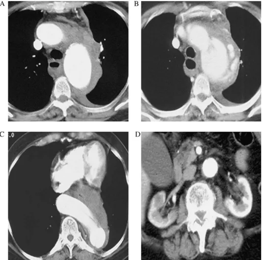
Fig. 1. Periaortic hematoma with left-sided hemothorax (A); dissection and aortic rupture at the distal arch level (B); huge kinking of the distal thoracic aorta (C); and disease-free infrarenal aorta (D).
The overall mean ICU stay was 3.5 days. However, excluding polytrauma patients, ICU stay averaged 1.5 days. The overall mean hospital stay was 15 days, also prolonged due to multiple associated injuries in trauma patients. After discharge from the hospital all 21 survivors were subjected to a strict follow-up protocol consisting of CT scans and complete physical examination at 6 and 12 months post intervenetion and then yearly thereafter. Postinterventional CT scans showed a complete exclusion of the aortic rupture locations and no stent migration was documented over the follow-up period. Ruptured aortic type B dissection in one patient showed complete thrombosis of the false lumen. In another asymptomatic patient with ruptured type B dissec-tion, a partial thrombosis of the false lumen with no need for further intervention was observed. No further procedure related complications occurred and in no case was aortic reoperation needed.
Final follow-up for this study was determined in June 2003 and was 100% complete. The mean follow-up period was 34.1 ^ 15.9 months (ranging from 6.6 to 60.0 months). All patients have found to enjoy an excellent quality of life. An actuarial survival curve is shown inFig. 3.
4. Discussion
Current stand on the most efficient treatment of acute aortic rupture remains still a controversial issue. Associated lesions in polytrauma patients with traumatic rupture of the aorta and a wide spectrum of comorbidities in the high-risk patients with ruptured thoracic aortic aneurysm or ruptured type B aortic dissection make open surgery a therapy with a relatively high postoperative morbidity and mortality rates.
The natural course of traumatic aortic injury has been reported to be very poor, with an initial survival rate ranging from 10 to 30%. In-hospital mortality rates increases from 32% on the first day, to 61% within the first week, and 74% after 2 weeks[1]. When survived without intervention, more than 30% of chronic traumatic thoracic aortic aneurysms
were ruptured in the late phase [12]. Actually, there is no study that allows to differentiate intimal injuries that will progress to hemorrhage from those with a more benign course. On the other hand, those patients undergoing surgical treatment have an early postoperative mortality rate ranging from 7.7[4]to 28%[5]. The series of 263 aortic blunt trauma patients published by Attar et al.[13], reported a 24% operative mortality rate and a 13% incidence of paraplegia. Timing of surgery in these polytrauma patients is usually complicated by associated lesions, where necessary heparinization would exacerbate early mortality rate. In addition, aortic cross-clamping might cause increased intracranial pressure and increase the risk of postoperative paraplegia, especially in patients with cranio-cerebral trauma [14]. In our subgroup of 15 patients with traumatic aortic ruptures, there was one early postoperative death resulting in an early mortality rate of 6.7%.
Following the excellent results of our earlier experience in SG of traumatic aortic rupture [15], we expanded the indication for hemorrhage control to any cases of acute aortic rupture, as for instance acute type B aortic dissection, penetrating aortic ulcer, and thoracic aortic aneurysm. This meant including older and sicker patients with much higher
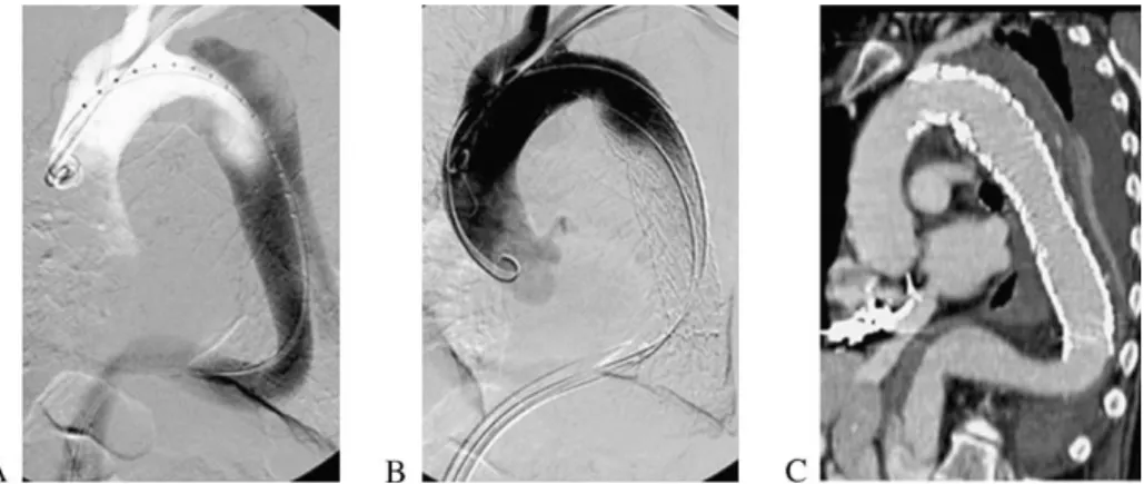
Fig. 2. Stent-graft procedure. First step consisting of angiography with calibrated catheter, located within the true lumen. Note the second pigtail catheter introduced through the left brachial artery (A); gore stent-graft deployed with overstenting of the left subclavian artery origin (B); and post-procedural Angiography CT scan reconstruction showing a complete sealing and thrombosis of the false lumen (C).
Fig. 3. Acturial survival curve. Time scale is based on number of months. Number of patients remaining at risk for main intervals analyzed are depicted above the time scale.
preoperative risk factor index and also explains higher overall mortality in the present study than in our first published series, consisting only of traumatic aortic rupture patients.
Severely diseased and atherosclerotic aorta in elderly patients, presenting whether ruptured thoracic aortic aneurysm or ruptured type B aortic dissection remains unchanged a major therapeutic challenge to clinicians, no matter which approach would be chosen. In this high-risk polymorbid group of elderly patients, endovascular approach has been demonstrated to be readily justifiable. Several groups published encouraging results after implan-tating endografts electively in patients with thoracic aortic aneurysms and type B dissections [9,10,16 – 22]. Conven-tional open surgery, using cardiopulmonary bypass, has a high mortality rate, and ranges from 17 to 29% in elective cases, but can be as high as 50%, when surgery is performed on an urgent basis[7,23,24]. Two of the 9 patients treated for a non-traumatic descending aortic rupture died within 30 days (22% early mortality rate). This is clearly below the expected mortality rate of the surgical approach.
Mortality rate of the 24 patients treated by SG showed a lower mortality as it would be expected from the preoperative EuroSCORE [25]. The group of patients suffering from traumatic aortic rupture ðn ¼ 15Þ presented a preoperative EuroSCORE of 6.7 ^ 1.9. This would predict a calculated mortality rate of 9.5 ^ 4.3%. Patients with ruptured thoracic aorta, penetrating aortic ulcer or ruptured type B dissection (overall n ¼ 9) have shown a much higher EuroSCORE due to associated comorbidities and higher age—12.1 ^ 1.6 with calculated mortality rate of 34.0 ^ 12.4%.
As in most of the cases, SG implantation can be performed through the femoral arteries, the procedure can be performed under local anesthesia. This has several advantages especially in the elderly and/or polymorbid population suffering from coronary artery-, cerebral-, mesenteric-, renal arteries disease. Pulmonary complication rate is reduced, as patients do not require an intubation/ ventilation. Moreover, hypotension caused by the induction of general anesthesia is minimized, which is detrimental especially in multifocally atherosclerotic diseased patients. Both, open surgery and endoluminal treatment of thoracic aortic lesions bears a significant risk of post-procedural paraplegia. Especially, intra-operative hypoten-sion and cross-clamp time longer than 30 min significantly increases postoperative paraplegia incidence [13,14]. In a study comprising 263 patients with traumatic aortic rupture, Attar et al.[13] reports a 13% incidence of postoperative paraplegia after an open graft repair and von Oppell et al. in their 20-year meta-analysis on traumatic aortic rupture report a 11.1% incidence. Paraplegia following emergent open repair of thoracic aortic aneurysm can be as high as 22% [6]. On the other hand, endoluminal SG of the descending thoracic aorta is associated with a post-interventional paraplegia rate ranging from 0 to 7%
[9 – 11,17,19,20]. In the present study, only one
post-interventional paraplegia occurred (1/24, 4%) and resolved within 5 months. Retrospectively, the length of recovery could have been possibly shortened, had cere-brospinal fluid drainage been instituted in the very early postoperative phase.
Secondary endoleaks after SG remains an issue. In the present study, two secondary endoleaks occurred. One patient presenting a coating failure was treated by implantation of an additional SG 12 months after the primary procedure. Another patient needed 2 months after the first procedure a distal SG extension to treat an attachment endoleak. No other reinterventions were needed and no graft migration was noted on any of the patients during the follow-up period.
In conclusion, emergent SG for hemorrhage control in acute thoracic aortic rupture is a safe and less-invasive alternative to the open graft repair. In addition to a shorter procedure time and local anesthesia benefits, it avoids the necessity of extracorporeal circulation and aortic cross-clamping with its risk of paraplegia and feared side effects of systemic heparinization. Mid-term results are so far excellent, however, long-term results are required.
References
[1] Parmley L, Mattingly T, Manion W, Jahnke E. Non-penetrating traumatic injury of the aorta. Circulation 1958;17:1086 – 101. [2] Hunt JP, Baker CC, Lentz CW, Rutledge RR, Oller DW, Flowe KM,
Nayduch DA, Smith C, Clancy TV, Thomason M, Meredith W. Thoracic aorta injuries: management and outcome of 144 patients. J Trauma 1996;40:547– 56.
[3] Preˆtre´ R, Chilcott M. Blunt trauma to the heart and great vessels. N Engl J Med 1997;336:626– 32.
[4] Gammie JS, Shah AS, Hattler BG, Kormos RL, Peitzman AB, Griffith BP, Pham SM. Traumatic aortic rupture: diagnosis and management. Ann Thorac Surg 1998;66:1295– 300.
[5] Cowley RA, Turney SZ, Hankins JR, Rodriguez A, Attar S, Shankar B. Rupture of thoracic aorta caused by blunt chest trauma: a fifteen-year experience. J Thorac Cardiovasc Surg 1990;100:652– 61. [6] Crawford ES, Hess KR, Cohen ES, Coselli JS, Safi HJ. Ruptured
aneurysm of the descending thoracic and thoracoabdominal aorta: analysis according to size and treatment. Ann Surg 1991;213: 417 – 26.
[7] Mastroroberto P, Chello M. Emergency thoracoabdominal aortic aneurysm repair: clinical outcome. J Thorac Cardiovasc Surg 1999; 118:477 – 82.
[8] Huynh TTT, Miller CC, Estrera AL, Porat EE, Safi H. Thoracoab-dominal and descending thoracic aortic aneurysm surgery in patients aged 79 years or older. J Vasc Surg 2002;36:469– 75.
[9] Grabenwo¨ger M, Hutschala D, Ehrlich MP, Cartes-Zumelzu F, Thurnher S, Lammer J, Wolner E, Havel M. Thoracic aortic aneurysms: treatment with endovascular self-expandable stent grafts. Ann Thorac Surg 2000;69:441– 5.
[10] Dake MD, Kato N, Mitchell S, Semba CP, Razavi MK, Shimono T, Hirano T, Takeda K, Yada I, Miller DC. Endovascular stent-graft placement for the treatment of acute aortic dissection. N Engl J Med 1999;340:1546 – 52.
[11] Marty-Ane´ C-H, Berthet J-P, Branchereau P, Mary H, Alric P. Endovascular repair for acute traumatic rupture of the thoracic aorta. Ann Thorac Surg 2003;75:1803– 7.
[12] Finkelmeier BA, Mentzer RM, Kaiser DL, Tegtmeyer CJ, Nolan S. Chronic traumatic thoracic aneurysm: influence of operative treatment on natural history—an analysis of reported cases. J Thorac Cardiovasc Surg 1982;84:257– 66.
[13] Attar S, Cardarelli MG, Downing SW, Rodriguez A, Wallace DC, West RS, McLaughlin JS. Traumatic aortic rupture: recent outcome with regard to neurologic deficit. Ann Thorac Surg 1999;67:959 – 65. [14] Oppell UOv, Dunne TT, Groot MKD, Zilla P. Traumatic aortic rupture: twenty-year metaanalysis of mortality and risk of paraplegia. Ann Thorac Surg 1994;58:585– 93.
[15] Lachat M, Pfammater T, Witzke H, Bernard E, Wolfensberger U, Ku¨nzli A, Turina M. Acute traumatic aortic rupture: early stent-graft repair. Eur J Cardiothorac Surg 2002;21:959– 63.
[16] Lansman SL, Hagl C, Fink D, Galla JD, Spielvogel D, Ergin MA, Griep RB. Acute type b aortic dissection: surgical therapy. Ann Thorac Surg 2002;74:S1833 – 5.
[17] Lepore V, Lo¨nn L, Delle M, Bugge M, Jeppsson A, Kjellman U, Radberg G, Risberg B. Endograft therapy for diseases of the descending thoracic aorta: results in 43 high-risk patients. J Endovasc Ther 2002;9:829– 37.
[18] Pierre A, Jean-Philippe B, Branchereau P, Veerapen R, Marty-Ane´ CH. Endovascular repair for acute rupture of the descending thoracic aorta. J Endovasc Ther 2002;9:II-51– 9.
[19] Grabenwoger M, Fleck T, Czerny M, Hutschala D, Ehrlich M, Schoder M, Lammer J, Wolner E. Endovascular stent graft placement in patients with acute thoracic aortic syndromes. Eur J Cardiothorac Surg 2003;23:788– 93.
[20] Ehrlich M, Grabenwoeger M, Cartes-Zumelzu F, Grimm M, Petzl D, Lammer J, Thurnher S, Wolner E, Havel M. Endovascular stent graft repair for aneurysms on the descending thoracic aorta. Ann Thorac Surg 1998;66:19– 25.
[21] Bell R, Taylor P, Aukett M, Sabharwal T, Reidy J. Results of urgent and emergency thoracic procedures treated by endoluminal repair. Eur J Vasc Endovasc Surg 2003;25:527– 31.
[22] Gambria RP, Brewster DC, Lauterbach SR, Kaufman JL, Geller S, Fan C-M, Greenfield A, Hilgenberg A, Clouse WD. Evolving experience with thoracic aortic stent graft repair. J Vasc Surg 2002; 35:1129 – 36.
[23] Safi HJ, Miller CC, Subramaniam MH, Campbell MP, Iliopoulos DC, O’Donnell JJ, Reardon MJ, Letsou GV, Espada R. Thoracic and thoracoabdominal aortic aneurysm repair using cardiopulmonary bypass, profound hypothermia, and circulatory arrest via left side of the chest incision. J Vasc Surg 1998;28:591 – 8.
[24] Crawford ES, Crawford JL, Safi HJ, Coselli JS, Hess KR, Brooks B, Norton HJ, Glaeser DH. Thoracoabdominal aortic aneurysms: preoperative and intraoperative factors determining immediate and long-term results of operations in 605 patients. J Vasc Surg 1986;3: 389 – 404.
[25] Nashef S, Roques F, Michel P, Gauducheau E, Lemeshow S, Salamon R. European system for cardiac operative risk evaluation (Euro-SCORE). Eur J Cardiothorac Surg 1999;16:9 – 13.
Appendix A. Conference discussion
Dr A. Graffigna (Trento, Italy): I would like to know from you, how do you deal with those cases of traumatic aortic rupture where rupture is very close to a subclavian artery? How do you face the riddle whether to cover
the subclavian artery with the stent or to address these patients through traditional surgery?
Dr Melnitchouk: Well, following blunt injury to the aorta, the aorta usually disrupts where the ligamentum arteriosum attaches to the aorta at the aortic isthmus and it is approximately 2 cm below. We had 1.9-cm median value of where the disruption was. And of course, it is very close to the subclavian artery.
But in the cases where disruption is very close to the subclavian artery, it can be safely overstented, except for the cases where a patient has a LIMA to LAD graft, then you would have to perform the carotid subclavian bypass.
Also you would have to monitor the blood pressure in the left arm. If the blood pressure after the overstenting of the subclavian artery is below 50, then you would have to go ahead and do the carotid subclavian bypass. We had some cases that had to be overstented, but in none of the cases there was a steal syndrome in the left arm.
Dr J. Bachet (Paris, France): I was a little surprised that compared to surgery, your results are not really convincing. I mean, they are, but not as I expected they should be. That’s the first comment.
And second question: what is the policy of your group concerning patients with acute traumatic rupture who are not threatened immediately from exsanguination? Do you still send them to surgery or do you switch systematically to the endoprosthesis treatment?
Dr Melnitchouk: Well, we had very good experience with stent-graft placement for traumatic aortic rupture. And for the high-risk patients with severe comorbidities, high age and very high preoperative EUROScore, I think endovascular approach to treat these aortic lesions is very elegant and a very good way. High-risk patients tend to be sick and they tend to go a certain direction. No matter what you do, you decide to operate on them or you decide to stent them, they are just high-risk patients and there is quite a high mortality in these patients.
Dr M. Turina (Zurich, Switzerland): If I may add, the present policy due to this one death case, which was totally surprising to us—because patient had a good EAP deployment and good sealing of the rupture, has been changed. Every patient with a substantial periaortic hematoma will go to surgery, unless there is a major contraindication like a severe cerebral trauma, liver rupture or something similar. Small periaortic hematomas, intimal lesions and flaps receive EAP. And the policy about the immediate surgery is governed by other organ trauma and by the cerebral state of these patients, so that we will be waiting for a day or two if there is a brain edema or similar like this.
Dr S. Aziz (Takoma Park, Maryland, USA): We actually had a case of traumatic aortic tear in the usual location near the left subclavian artery. The patient also had multiple other injuries to the head and abdomen. He was not an open candidate for open surgical repair. We were able to use the Gore thoracic Excluder stent. After placement of the device we noted that we still had a leak from the proximal side. So we put another stent within the stent. This covered the tear and there was no evidence of further leakage.
Dr Melnitchouk: Yes. Especially earlier grafts were of not very good quality, but we have very good experience with the next generation of stent-grafts.
Dr M. Krason (Zabrze, Poland): We have had about 15 cases in total series of stent-grafting of descending thoracic aorta dissections and aneurysm. And we have closed successfully in five or six cases subclavian artery with no surgical treatment afterwards and the patients were doing successfully quite well with no ischemia.