Nephrol Dial Transplant (1995) 10: 39-46
Original Article
Nephrology
Dialysis
Transplantation
Light-chain-induced renal tubular acidosis: effect of sodium bicarbonate
on sodium-proton exchange
H. P. Reusch
1, J. F. E. Mann
1, M. J. Mihatch
2, W. Siffert
3and F. C. Luft
1'
4department of Internal Medicine-Nephrology University of Erlangen-Nurnberg, Germany; institute for Pathology, University of Basel, Switzerland; 3Max Planck Institute for Biophysics Frankfurt a.M., Germany; 4Franz Volhard Clinic, Rudolf Virchow Univ. Hosps. Max Delbriick Center for Molecular Medicine, Free University of Berlin, Berlin, Germany.
Abstract. We measured sodium-proton (Na+/H+) exchange in lymphocytes and platelets of a 46-year-old woman with the adult Fanconi syndrome before, during, and after treatment with NaHCO3. Kappa
light chains in her urine and unique but rarely observed crystalline structures confirmed the presence of light-chain nephropathy. Her glomerular nitration rate was only moderately impaired at 72 ml/min. NaHCO3 at
1, 3, and 5 mmol/kg/day for 5 days increased her serum HCO3 and pH from 17 to 21 mmol/1 and 7.28
to 7.39 respectively. Plasma renin and aldosterone values were decreased by NaHCO3. Na+/H+ exchange
(ciHi/min) was measured with the fluorescent marker BCECF after acidification of lymphocytes and platelets with sodium propionate at five (10-50mM) doses. Na+/H+ exchange was accelerated in this patient compared to normal controls. NaHCO3 treatment
significantly decreased Na+/H+ exchange in lympho-cytes, but not in platelets. These findings suggest that Na+/H+ exchange can be influenced by NaHCO3
ingestion at doses that only modestly affect systemic pH. Since Na+/H+ exchange is involved in stimulus response coupling, cell growth regulation, cell differen-tiation, and perhaps the progression of nephrosclerosis, these observations may have clinical relevance.
Key words: renal tubular acidosis; Fanconi syndrome;
Bence Jones protein; light chain nephropathy; sodium-proton exchange; sodium bicarbonate; multiple myel-oma; electron-microscopy
Introduction
Renal Fanconi syndrome in adults is an uncommon cause of proximal renal tubular acidosis and is gener-ally associated with multiple myeloma, light-chain pro-teinuria, or amyloidosis [1,2]. We were recently referred
Correspondence and offprint requests to: Friedrich C. Luft, MD,
Franz Volhard Clinic, Univ. Hosps. Rudolf Virchow, Wiltberg Strasse 50, 13122 Berlin, Germany.
an asymptomatic woman in whom glycosuria and proteinuria had been serendipitously identified. Elevated amounts of kappa light chains in her urine and multiple electron-dense, cytoplasmic, crystalline inclusions on renal biopsy secured the diagnosis. Sodium bicarbonate (NaHCO3) loading, up to
5 mmol/kg/day, caused only a modest increase in her plasma pH and plasma HCO3 concentrations; however,
it did decrease the accelerated sodium-proton (Na+/H+) exchange in her lymphocytes. This patient allowed us to corroborate our earlier observations, that Na+/H+ exchange in lymphocytes is increased in patients with renal acidosis compared to values at a more normal systemic pH [3]. We now present evidence that Na+/H+ exchange can be influenced by clinically attainable alkalinization.
Subjects and methods
Case Report
A 46-year-old woman was referred because of glycos-uria and proteinglycos-uria on routine urinalysis. She denied any symptoms or lack of wellbeing. There was no family history of hypertension, renal disease, diabetes, or malignancy. She weighed 59 kg, was 161cm tall, and had a blood pressure of 140/85 mmHg. The rest of her physical examination was entirely normal. The haemoglobin was 14.4 g/dl, haematocrit 44 vol%, white blood cell count 6800, and platelet count 247,000/mm3. Tests of liver function and thyroid function were completely normal. Serologies for collagenoses and tests for cryoglobulins were negative. Multiple blood sugar values and an oral glucose tolerance test were normal. The sodium was 139 mmol/1, chloride 111 mmol/1, potassium 4.2 mmol/1, calcium 2.26 mmol/1, and phosphate 0.7 mmol/1. The blood urea nitrogen was 32 mg/dl, and uric acid concentra-tion was 1.1 mg/dl. An arterial blood sample disclosed a plasma pH of 7.35, PO2 100 mmHg, PCO2 33 mmHg,
and plasma HCO3 18 mmol/1. The serum protein
elec-trophoresis disclosed only a marginal decrease in
1
gammaglobulins. Plasma vitamin D3 values, plasma
parathyroid hormone, plasma renin activity, and plasma aldosterone were within normal limits.
Roentgenograms of the thorax, axial skeleton and hands were unremarkable. An ultrasound examination showed kidneys which were normal in size and consist-ency. No abnormal calcifications or calculi were found. The plasma creatinine was 1.27 mg/dl, creatinine clear-ance 76 ml/min, 24-h urine protein excretion ranged between 2.3 and 3.8 g/day. The urinalysis revealed + + proteinuria and + -I- glycosuria. Granular casts were identified in the urinary sediment. A urine disc protein electrophoresis showed a mixed, tubular pat-tern. The urinary excretion of amino acids was gener-ally increased with values for individual amino acids 4-10 times above the normal range. A fourfold increase in the normal urinary excretion of fi2 microglobulin
was found. Urinary immunoelectrophoresis disclosed the presence of kappa light chains. A percutaneous renal biopsy and bone marrow biopsy were performed. The patient consented to a bicarbonate loading test after the University of Erlangen committee on human subjects had given approval. The protocol consisted of observation for 5 days, ingestion of 1 mmol/kg, 3 mmol/kg, and 5 mmol/kg NaHCO3 daily each for 5
days, and an additional 10-day recovery period. On the last day of each period, 24-h urine samples were collected. Arterialized venous blood was obtained for electrolytes, pH, PCO2, HCO3, and N a+/ H+ exchange
in lymphocytes and platelets three times during the baseline phase, at the end of each treatment phase, and twice during the recovery phase.
Histology
Renal tissue was obtained for light- and electron-microscopy. A bone marrow biopsy was also obtained. The tissue preparation was done according to standard techniques. The material for light microscopy was fixed in 4% buffered formalin, embedded in paraplast, cut in 3-um-thick sections and stained with haematoxylin, PAS, PASM, and trichrome stains. The material for electron-microscopy was fixed in 3% buffered glutar-aldehyde, embedded in Epon and ultrathin sections were stained with lead citrate and uranyl acetate, and evaluated with a Phillips EM 200. Snap-frozen tissue was used for immunofluorescence.
Preparation of platelet-rich plasma and isolation of lymphocytes
The blood was anticoagulated by the addition of 20% (vol/vol) of acid citrate dextrose [3]. Platelet-rich plasma was prepared by centrifugation of blood at 200 g for 15 min at room temperature. The upper two-thirds of the supernatant was used for the prepar-ation of 2'-7'-bis(carboxyethyl)5,6-carboxyfluorescein (BCECF)-loaded platelets and the remaining pellet was resuspended 1:1 with Hepes-buffered RPMI 1640 medium, pH 7.4. Lymphocytes were prepared after centrifugation of blood on a Ficoll gradient.
Measurement ofpH{
Cytosolic pH was determined using the fluorescent pH indicator BCECF. Pelleted platelets were resuspended in Hepes buffer consisting of 140 mM NaCl, 5 mM KCI, 5 mM KH2PO4, 1 mM MgSO4, 10 mM Hepes
(free acid), and 5 mM glucose, pH 6.5 (at 37°C). 10 uM BCECF-AM (final concentration; Molecular Probes Inc., Eugene, OR) was added, and the cells were incubated for 30 min at 37°C. Thereafter, 1 uM PGI2 was added and the cells were washed twice in
Hepes buffer, pH 6.5, by repeated centrifugation. The final platelet pellet was resuspended in Hepes buffer, pH 7.4, at a concentration of 5 x 10⁹ cells/ml. 100-jxl aliquots of these suspensions were transferred to 2 ml of Hepes buffer in a cuvette and prewarmed at 37°C. All measurements were conducted within 1 h after loading. During this time leakage of BCECF did not exceed 10% as assessed by comparison of fluorescence of platelets and corresponding supernatants and was, therefore neglected.
Lymphocytes and platelets were incubated with 10 uM BCECF-AM for 30 min at 37°C in RPMI 1640 medium and washed twice in this medium by repeated centrifugation. Before prewarming to 37°C, aliquots of lymphocytes were briefly spun down in an Eppendorf centrifuge and then resuspended into Hepes buffer before being used for fluorescence measurements (final concentration 1 x 10³ cells/ul). This procedure efficiently removes extraneous dye. Leakage of dye during prewarming was < 10% and was therefore not corrected. The fluorescence of BCECF was recorded under constant stirring using a KONTRON SFM 24 spectrofluorimeter (Kontron, Diisseldorf, FRG) equipped with a thermostatted cuvette holder. Wavelengths for excitation and emission were set to 495 and 530 nm respectively. Calibration of the BCECF signals in terms of pH; was performed using the K+/nigericin method [3].
Determination ofNa+/H+ exchange activity
Na+/H+ exchange was activated by addition of various amounts of Na+-propionate (final concentrations 10-50 mM) from a 1-M stock solution, pH 7.4 [3]. Recovery of pH; in lymphocytes and platelets was inhibited by >90% at 10 uM of the specific inhibitor 5-(N-ethyl-N-isopropyl)-amiloride. Both findings sug-gest that pHj recovery was almost exclusively mediated by Na+/H+ exchange. Osmotic activation of the anti-port by 50 mM NaCl failed to affect pHj (data not shown). Initial rates of EIPA-sensitive pHj recovery were calculated as described in detail [3] and are expressed as <5pHj/min. The relationship between pH;
and pHj recovery could best be described by a sig-moidal function as stated earlier [3].
Exact characterization of the antiport's kinetic para-meters can only be achieved by the nigericin pH; clamp
method or by the NH4C1 prepulse method [3].
Determination of true vmqax requires acidification to
pHj 6.0, whereas with propionate acidification beyond pHj 6.6 (at pHo 7.4) cannot be achieved. Further, pH;
Light chain RTA and Na+/H+-exchange
recovery rates are dampened and the original baseline pH is not re-established due to the continuous influx of propionic acid. However, we wished to examine antiport activity in two cell specimens from one indi-vidual at the same occasion, which requires fast experi-mental procedures. This renders application of the nigericin pH; clamp method impossible, since this technique is time consuming. Further, prolonged stor-age of blood may affect antiport activity. Finally, neither the nigericin pH; clamp technique nor the
NH4CI prepulse method can be applied to platelets as these techniques require one or more centrifugation steps. In platelets this is possible only in the presence of agents that raise cAMP, or an acidic extracellular pH, in order to prevent any preactivation or aggrega-tion of these cells. Since these manipulaaggrega-tions might have caused unforeseen effects on antiport activity, we preferred to use acidification by propionate, which enabled us to apply the same procedure to both cell types. Finally, all these potential confounders are pre-sent to a similar extent in all cell preparations under all conditions. This notion is also supported by the evaluation of the immediate effects of propionate addi-tion on pHj in platelets and lymphocytes under the various states investigated. Identical amounts of Na+ propionate produced similar acidification of the cells (buffering capacity), whereas only pH; recovery rates
were different (see results).
Statistical analysis
We conducted a repeated measures analysis of variance on the N a+/ H+ exchange data. Since each observation was derived from a different set of cells, the data could be analysed as independent observations. We tested for the dpHj/min after each the five doses of propionate under control, under HCO3 administration, and under
recovery conditions. We also compared the slopes of the relationships between dpHJmin and pH;. A P value
<0.05 was accepted as significant.
Results
Renal biopsy
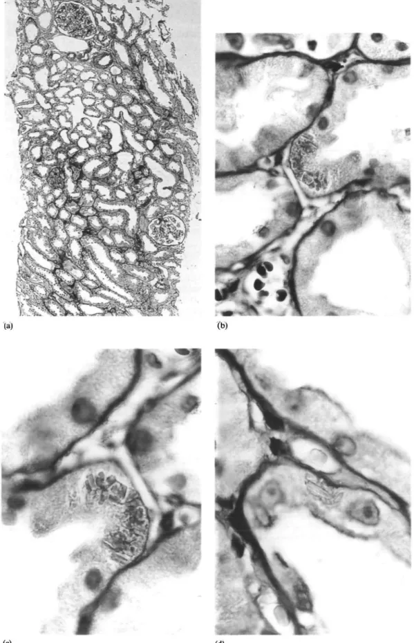
The renal tissue for light-microscopy contained 12 normal glomeruli and two completely sclerosed glo-meruli. The latter were surrounded by scattered lymphocytic infiltrates. The tubules were slightly dilated and the lumina did not contain casts. The proximal tubular epithelium exhibited minimal vacuol-ization (Figure 1). Crystalline structures were present within individual cells. No evidence of light-chain deposit disease, amyloidosis, or cast nephropathy was found. Immunofluorescence for immunoglobulins (IgG, IgM, IgA, complement factors C3 and C4 was negative. By electron-microscopy, the normal architec-ture of the glomeruli was confirmed. Proximal and distal tubules contained numerous highly osmiophilic protein crystals without suprastructure. These
crystal-41
line structures were suggestive of light chains. Identical crystals were found in the bone marrow (Figure 2).
Na+/H+ exchange
Plasma Na, Cl, and K barely changed while plasma HCO3 increased incrementally from 17 to 21 mmol/1 and plasma pH increased incrementally from 7.28 to 7.39 with NaHCO3 loading. UNaV increased from 102
to 275 mmol/24 h at the highest dose of NaHCO3,
while UC1 and K excretion increased modestly. Ca excretion showed no consistent change. Urine pH increased from 6.1 at baseline to 8.3 at the highest dose of NaHCO3 and decreased to 5.7 thereafter.
Plasma renin activity decreased from 2.95 to 0.84 ng/Ang I/ml/h, while plasma aldosterone decreased from 589 to 294 ng/ml with NaHCO3
loading.
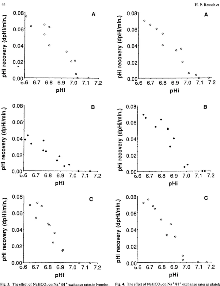
In Figure 3 are shown pH; recovery rates in lympho-cytes before (panel A), during (panel B), and after (panel C) NaHCO3 therapy. The lymphocytes
obtained during NaHCO3 therapy displayed reduced
pHj recovery rates after propionate; the <5pHj/min at each dose of propionate differed across treatments
(P<0.05) compared to values obtained before or after
NaHCO3 therapy. The parameters from the data in
Figure 3 yielded an apparent vmax of 0.071 <5pH/min
before and 0.067 (5pHi/min afterwards. During NaHCO3 therapy, on the other hand, the rate was
reduced to 0.041 SpHJmin. The apparent half-maximum activation occurred at pHj 6.9 before, pH;
6.87 during, and pH; 6.85 after the treatment. Thus
the increase in apparent vmax in lymphocytes was not
accompanied by an alkaline shift in the Na+/H + exchanger activation curve. Moreover, the slope of the line generated by the relationship between <5pHj/min and pHj with NaHCO3 therapy differed from that
obtained either before or after NaHCO3 treatment
(P<0.05).
In contrast, no significant differences in vmax were
observed in the platelets (Figure 4). The parameters varied between 0.070 <5pHj/min before (panel A), 0.066 (5pH;/min during (panel B) and 0.068 <5pHj/min after
(panel C) NaHCO3 treatment. The pHj values at which
the apparent half-maximum activation of the anti-porter occurred, varied only in a narrow range between 6.9 and 6.86.
Finally, neither a change in basal pHj of the lympho-cytes nor of the platelets was seen during the entire study. The values averaged 7.07 + 0.04, 7.09x0.05, and 7.06 + 0.03 for lymphocytes and 7.13 + 0.04, 7.12±0.03, and 7.14 + 0.01 for platelets on day 0, 15, and 25 respectively. In addition to the constant pH;
values, enhancement of the lymphocyte antiport could not be attributed to changes in the buffering capacity for H+, since the addition of 50 mM propionate caused the same degree of immediate acidification of the cells on all days (0.043 + 0.04 on day 0, 0.045 + 0.09 on day 15 (maximal NaHCO3 therapy) and 0.042 ±0.03 after
10 days of recovery. Similar values were recorded for the platelets.
(c)
Fig. 1. Panel A shows an overview by lower power (x70, PAS stain). Tubular dilatation is evident. Panels B, C, and D show individual tubules with crystalline structures (x 850). These were elucidated further by electron-microscopy.
Light chain RTA and Na+/H +-exchange 43
(d)
Fig. 2. Panels A, B, and C show the presence of protein crystals in tubular epithelial cells by transmission electron-microscopy. Panel D shows the presence of protein crystals in a specimen of bone marrow. The magnifications are 5x957, 5x 1305, 5x 1972, and 5x957 respectively.
Discussion
This patient presented with an unusual, but well-described proximal renal tubular acidosis resulting from the effects of deposited light chains [1,2]. She had all the clinical features of the adult Fanconi syndrome, as well as kappa light chains in her urine. The renal biopsy showed none of the typical renal
complications of multiple myeloma, i.e. amyloidosis, cast nephropathy or light-chain deposit disease [9]. Rather, slight vacuolization of proximal tubular cells and intracellular osmiophilic crystals compatible with light chain crystals were found. Thus our patient is unusual. The overall incidence of crystals in kidneys of patients with plasma cell dyscrasia accounts for about 6%; crystals in large amounts are found in only
X Q. 0.08
^
0.06 Q_>i 0.04
o og
0.02 0.00 o o o D.6 6.7 6.8 6.9 7.0 7.1 7.2 pHi 0.081I 0.06
Q. <5 ox
Q. 0.04 0.02 H. P. Reusch el al. Ao oo
J•—*—°^
• D.6 6.7 6.8 6.9 7.0 7.1 7.2 pHi c E X Q . > ore
c
X Q . 0 0. 0. 0. /•»u.
08 06 04 02 • •oo
D.6 B • • • • • 6.7 6.8 6.9 7.0 7.1 7.2 pHiI
X a. o X a. ~ 0.08x
°-
06
0.040.021
0.00 °o D.6 6.7 6.8 6.9 7.0 7.1 7.2 pHi cE
Q. •a o u a>x
Q. O.O81 0.06 0.04 0.02 B ° ' ° D . 6 6.7 6.8 6.9 7.0 7.1 7.2 pHi _0.08
d
|0.06
a.^
0.04£
0.02x
a
0.00 D.6 6.7 6.8 6.9 7.0 7.1 7.2 pHiFig. 3. The effect of NaHCO3 on Na+/H+ exchange rates in lympho- Fig. 4. The effect of NaHCO3 on Na+/H+ exchange rates in platelets
cytes. On the ordinate are shown the pooled pH, recovery rates (see Figure 3). The slopes of the regression relationships are not (3pHj/min) during control (panel A), day 15 of NaHC03 ingestion different, indicating no effect of NaHCO3 treatment on Na+/H +
(panel B), and 10 days after NaHCO3 treatment (panel C). On the exchange rates in platelets,
abscissa is shown then pHi values obtained by acidifying the cells with sodium propionate. The slopes of the regression relationships are different (/><0.05); that in panel B after NaHCO3 treatment is
Light chain RTA and Na+/H +-exchange
1% of cases. In cases of cast nephropathy, crystals in tubular casts and/or tubular epithelium were reported in 18 of 24 patients [10], and in eight of 12 patients with concomitant Fanconi's syndrome [1]. Cases of tubular vacuolization with intracytoplasmic crystals associated with Fanconi's syndrome without cast nephropathy are highly uncommon. To our knowledge only three cases were reported in the literature [1,11,12]. Two were associated with kappa chain [1,11], one with lambda light-chain excretion [12]. We believe our light- and electron-photomicrographs of both bone marrow and renal tissue are an excellent example of this rare condition.
We administered NaHCO3 to our patient in order
to document that the threshold for HCO3 was
drastic-ally reduced [13]. Our patient had difficulty tolerating the 5 mmol/kg/day dose because of gastrointestinal side-effects. We encountered urine pH values above 5.5. Thus it is quite possible that our patient also had some degree of distal tubular dysfunction. We failed to determine the pCO2 of her urine, which would
admittedly have clarified matters.
Na+/H+ exchange or antiport is important in regula-tion of pHi; cell volume, stimulus response coupling,
and cell proliferation [14]. The finding that Na+/H + exchange is increased in hypertension and diabetes raises the possibility that Na+/H+ exchange may be relevant to the pathogenesis of these conditions or their complications [15,16]. Since Nath et al. [17] showed that NaHCO3 treatment ameliorated the
course of chronic renal failure in rats with 5/6 nephrec-tomy, particularly in terms of reducing interstitial proliferative changes which may be related to ammonia production [18], it occurred to us that perhaps the acidosis of chronic renal disease is associated with accelerated Na+/H+ exchange. In an earlier study [3], we observed such an increase in Na+/H+ exchange in lymphocytes of patients with chronic renal disease compared to normal subjects as well as in normal subjects given NH4C1 for 5 days. Patients with chronic
renal disease had a mean pH; recovery (5pH/min) of
0.08 (range 0.07-0.11), compared to a mean 0.05 observed in normal subjects [3]. Our patient's <5pHi/min value was 0.08. We were also able to show by means of quantitative, reverse transcription poly-merase chain reaction [19] that NHE-1 mRNA was increased under the condition of metabolic acidosis, suggesting that additional antiporter protein was pro-duced. A recent study of erythrocytes from patients with renal insufficiency supports this view [20].
The Na+/H+ exchanger in lymphocytes and platelets is not identical with the N a+/ H+ exchanger present on renal tubular brush border epithelium. Krapf et al. studied the effects of metabolic acidosis induces on Na+/H+ exchange in renal tissue and were able to show an increase in antiport mRNA in renal tubular cells [21,22]. Moe et al. [8,23] were able to confirm that mouse renal cortical tubule cells and an opossum kidney cell line responded to a 24-h in vitro metabolic acidosis with an almost twofold increase in Na+/H+ exchange activity. This increase in antiport activity
45
was accompanied by a threefold increase in antiport mRNA. These results could be confirmed by feeding rats with a diet that induced metabolic acidosis. The increase in antiport activity could also be inhibited by the addition of cycloheximide to the culture medium. These findings could apply to Na+/H+ exchange in lymphocytes and platelets.
If one accepts the hypothesis of an as yet unidentified pHrsensor in these cells, an effect on antiport mRNA
and de-novo regulation of the corresponding protein synthesis in lymphocytes appears conceivable. Platelets, on the other hand, are non-nucleated 'cells' which have only a minor capability of protein synthesis, if any. It is possible that the relatively short-term NaHCO3
treatment was insufficient to assure adequate numbers of new circulating platelets with decreased antiport activity. Since we could only measure the overall platelet Na+/H+ exchange activity, minor differences were most likely under the detection limits of our assay. It is also possible that circulating agonists influenced Na+/H+ exchange in the lymphocytes of our patient. Although her renin and aldosterone values were normal prior to the intervention, the administration of NaHCO3 had a suppressing effect on renin and
aldos-terone not dissimilar from that reported after sodium citrate by Sharma et al. [24]. Aldosterone enhances N a+/ H+ exchange activity in human lymphocytes in
vitro [25-27]. Angiotensin is also able to stimulate
Na+/H+ exchange [28]. Thus we cannot rule out the possibility that the decrease in Na+/H+ exchange we observed after NaHCO3 in lymphocytes was mediated
by hormones instead of a direct pH effect.
It is possible that acidosis per se may be important in the progression of chronic renal disease. Protein degradation is enhanced and growth is impaired by acidosis in uraemia [29]. Na+/H+ exchange may be involved in these effects. In contrast to our findings and those of Corry et al. [20], Greiber and Mitch found that the antiport was decreased in thymocytes of uraemic rats [30]. Additional studies will be neces-sary to elucidate these issues. Finally, the above-mentioned findings by Nath et al. [17] have not received the attention they deserve. Although reduced protein intake is an accepted preventative strategy in chronic renal failure management, control of acidosis when applied at all is primarily conducted to prevent bone disease. We suggest that control of renal acidosis may be directly relevant to the progression of chronic renal disease, perhaps by influencing Na+/H + exchange.
Acknowledgements. These studies were supported by grants in aid
from the Verband, Deutscher Mineralbrunnen, Siemens Erben, and Sandoz AG.
References
1. Maldonado JE, Velosa JA, Kyle RA, Wagoner RD, Holley ICE, Salassa RM. Fanconi syndrome in adults: a manifestation of a latent form of myeloma. Am J Med 1975; 58: 354-364 2. Smithline N, Kassirer JP, Cohen JJ. Light-chain nephropathy:
renal tubular dysfunction associated with light-chain proteinuria.
N EngI J Med 1976; 294: 71-74
3. Reusch HP, Reusch R, Rosskopf D, Siffert W, Mann JFE, Luft FC. Na+/H+ exchange in human lymphocytes and platelets in chronic and subacute metabolic acidosis. J Clin Invest 1993; 92: 858-865
4. Peper RJ, Tina WZ, Mickelson MM. Purification of lymphocytes and platelets by gradient centrifugation. J Lab Clin Med 1968; 72: 842-848
5. Rink TJ, Tsien RY, Pozzan T. Cytoplasmic pH and free Mg in lymphocytes. J Cell Biol 1988; 95(1): 189-96
6. Siffert W, Siffert G, Scheid P, Akkerman JWN. Na+/H + Exchange modulates Ca Mobilization in human platelets stimu-lated by ADP and the thromboxane mimetic U 46619. J Biol
Chem 1990; 264: 719-725
7. Siffert W, Jakobs KH, Akkerman JWN. Sodium fluoride pre-vents receptor-and protein kinase C-mediated activation of the human platelet Na+/H+ exchanger without inhibiting its basic pH,-regulating activity. J Biol Chem 1990; 265: 15441-15448 8. Moe OW, Miller RT, Horie S, Cano A, Preisig PA, Alpern
RJ. Differential regulation of Na+/H+ antiporter by acid in renal epithelial cells and fibroblasts. J Clin Invest 1991; 88: 1703-1708
9. Schubert GE. Die Plasmozystniere. I. Haufigkeit pathologisch-anatomischer Veranderungen. Klin Wochenschr 1974; 52: 763-770
10. Pirani CL, Silva F, D'Agati V. Renal lesions in plasma cell dyscrasia: Ultrastructural observations. Am J Kidney Dis 1987; 10: 208-221
11. Lee DBN, Drinkard JPD. The adult Fanconi syndrome: Observations on etiology, morphology, renal function and min-eral metabolism in three patients. Medicine 1972; 51: 107-124 12. Thorner PS, Bedard YC, Fernandes BJ. Lambda-light-chain
nephropathy with Fanconi's syndrome. Arch Pathol Lab Med 1983; 107: 654-657
13. Kurtzman NA. Acquired distal renal tubular acidosis. Kidney
Int 1983; 24: 807-819
14. Seifter JL, Aronson PS. Properties and physiological roles of the plasma membrane sodium-hydrogen exchanger. J Clin Invest 1986; 78: 859-864
15. Rosskopf D, Dusing R, Siffert W. Membrane Na/H exchange and primary hypertension. Hypertension, (in press)
16. Huot SJ, Aronson PS. Na-H exchanger and its role in essential hypertension and diabetes mellitus. Diabetes Care 1991; 14: 521-535
17. Nath KA, Hostetter MK, Hostetter TH. Pathophysiology of
chronic tubulo-interstitial disease in rats. Interactions of dietary acid load, ammonia and complement component C3. J Clin
Invest 1985; 76: 667-675
18. Tolins JP, Hostetter MK, Hostetter TH. Hypokalemic nephro-pathy in the rat: role of ammonia in chronic tubular injury.
J Clin Invest 1987; 79: 1447-1458
19. Quednau B, Rosskopf D, Reusch HP, Luft FC, Siffert W. Enhanced Na+/H+-exchanger activity and raised Na+/H+ -exchanger mRNA levels in human lymphocytes during metabolic acidosis. Am J Physio/ 1994; 266: C480-C488
20. Corry DB, Tuck ML, Nicholas S, Weinmann EJ. Increased Na/H antiport activity and abundance in uremic red blood cells.
Kidney Int 1993; 44: 574-578
21. Krapf R, Pearce D, Lynch C, Xi X-P, Reudelhuber TL, Pouyssegur J, Rector FC. Expression of rat renal Na+/H +
antiporter mRNA levels in response to respiratory and metabolic acidosis. J Clin Invest 1991; 87: 747-751
22. Krapf R, Solioz M, Fehlmann C. Na+/H+ antiporter mRNA expression in single nephron segments of rat kidney cortex.
J Clin Invest 1991; 88: 783-788
23. Horie S, Moe O, Tejedor A, Alpern RJ. Preincubation in acid medium increases Na -f/H + antiporter activity in cultured renal proximal tubule cells. Proc Natl Acad Sci USA 1991; 87: 4742-4745
24. Sharma AM, Schattenfroh S, Thiede HM, Oelkers W, Distler A. Effects of sodium salts on pressor reactivity in salt-sensitive men. Hypertension 1992; 19: 541-548
25. Wehling M, Kasmayr J, Theisen K. Rapid effects of mineralocor-ticoids on sodium-proton exchanger. Genomic or nongenomic pathway. Am J Physiol 1991; 260: E719-E726
26. Wehling M, Kasmayr J, Theisen K. Fast effects of aldosterone on electrolytes in human lymphocytes are mediated by the sodium-proton exchange of the cell membrane. Biochem Biophys
Res Commun 1984; 164: 961-967
27. Wehling M, Kasmayr J, Theisen K. The Na-H exchanger is stimulated and cell volume increased in lymphocytes from patients with essential hypertension. / Hypertens 1991; 9: 519-524
28. Saccomani G, Mitchell KD, Navar LG. Angiotensin II stimula-tion of Na-H exchange in proximal tubular cells. Am J Physiol 1990; 258: F1188-F1195
29. Maniar S, Laouri D, Caldas A, Kleinknecht C. Protein synthesis and growth in uremic rats with and without chronic metabolic acidosis. Miner Electrolyte Metab 1992; 18: 250-252
30. Greiber S, Mitch WE. Mechanisms for protein catabolism in uremia: metabolic acidosis and activation of proteolytic path-ways. Miner Electrolyte Metab 1992; 18: 233-236
Received for publication 19.5.94 Accepted 10.8 94