Br. J. Anaesth. (1987), 59, 1044-1051
DISTRIBUTION AND KINETICS OF
14C-VECURONIUM IN
RATS AND MICE
P. G. WASER, H. WIEDERKEHR, A. CHANG SIN-REN AND
E. KAISER-SCHONENBERGER
Vecuronium-bromide is the monoquaternary analogue of pancuronium (Savage, Sleigh and Carlyle, 1980). Thus one might anticipate little change in activity because of this structural similarity or, alternatively, there could be a substantial decrease in neuromuscular blocking activity as with other monoquaternary com-pounds. Since the anaesthetist tends to use short-acting non-depolarizing neuromuscular blockers, vecuronium will be used extensively. As a result, its distribution to the organs, and its pathways of elimination, are of clinical importance.
MATERIALS AND METHODS
Animal preparation, drug application and sectioning
Twenty male rats were anaesthetized by the i.p. injection of 20% urethane solution (1.8mg/g animal weight), a tracheostomy undertaken and artificial ventilation instituted via a tracheal tube. The ECG was monitored throughout. 14
C-Vecuronium bromide 6.76 |ig g"1 (specific
ac-tivity 2.39 mCi mmol litre"1 = 3.7 nCi mg~l) or
0.025 nCi/g animal weight, synthesized in our isotope laboratory by a special selective methy-lation technique (fig. 1), was injected via the tail vein. This dose is equal to seven times the LD100.
During the period of complete muscle paralysis artificial ventilation (48 b.p.m.: 95 % oxygen: 5 % carbon dioxide 4 ml) was maintained (Harvard rodent ventilator). At time intervals of 2, 5,20, 60 and 120 min after injection, four animals from each of the five groups were killed by immediate
PHTKR G. WASHR, MJX, PH.D.; ALOIS CHANG S I N - R E N , PH.D.; HEINZ WIEDERKEHR, DIPL. PHARM.; ELSBBTH
KAISER-SCHON-ENBERGER; Institute of Pharmacology, University of Zurich, Gloriastrasse 32, CH-8006 Zurich, Switzerland. Accepted for Publication: May 6, 1986.
Correspondence to P. G. W.
SUMMARY
The distribution and kinetics of 1AC-vecuronium
were studied in rats and mice. llC-Vecuronium
accumulated rapidly in the liver. Both unchanged and metabolized vecuronium were excreted with the bile into the intestines and stomach. Re-absorption in the gut was probably responsible for an enterohepatic increase in radioactivity in the liver after one hour. Excretion through the kidneys increased continuously from low values after the initial peak. Binding in compartments with acidmucopo/ysaccharides such as cartilage, connective tissue etc., was less important. Blood-brain barrier and placenta were permeable only to a small degree.
immersion in a mixture of hexane/solid carbon dioxide (temperature — 70 °C). The frozen ani-mals were embedded in a gel of carboxymethyl cellulose (water-Na-CmC) and submerged in the cooling mixture and frozen to a solid block over 20 min. At six different levels, sagittal sections were cut (Cryo-microtome Type PMV-450, LKB, Stockholm) from the block, which was covered by broad scotch tape (3M). The under-cutting produced tape-mounted sections of 20 |im
o
O—C-CHo
CH3 J ^
CHo—C—O H
FIG. 1. Chemical structure of vecuronium. *Carbon-14 label.
thickness. These were first dried for 3 days in the deep freeze, and autoradiographs were obtained. Corresponding sections were kept for histological staining and localization of radioactive tracers. The rest of the animal was stored in plastic bags in the deep freeze for radioactivity analysis by liquid-scintillation counting.
Whole-body autoradiography (WBAR)
The 20-|im mounted sections were covered by x-ray films (NS-25-No-Screen films, Eastman Kodak), and exposed for 16 weeks at — 20 °C in the dark. The films were developed under standardized conditions. The densitometric eval-uation of the degree of film blackening was achieved with a densitometer (TD-504 by Mac-beth Kollmorgen). The evaluation of the degree of blackening of the films utilized a calibration scale which was obtained by simultaneous exposure of
14C-glucose impregnated emulsion sheets on the
films (Cross, Groves and Hesselbo, 1974). An exponential function for correcting the saturation of total film blackening was applied (Keller and Waser, 1982).
Liquid scintillation counting (LSC)
Samples of tissues or body fluids (20-200 mg) were taken out of the frozen animals, weighed and mixed with Soluene-100 1 ml (Packard) in vials, shaken on a 50 °C waterbath until completely dissolved after 3-5 h. Isopropanol 0.5 ml and 30% HjO, 0.5 ml were added and, after the exo-thermic reaction, mixed with Insta-Gel (Packard) and hydrochloric acid 0.5 mol litre"1 in a ratio of
9:1. These tissues were measured in a counter (Tricarb 3375, Packard). The correlation between ratio (R) and efficiency (E) was determined experimentally by determination of a quenching curve. As radioactive standard, I4C-hexane
(Amer-sham International Ltd) with a specific activity of 1.123 x 10⁸ d min"1 g"1 was used.
Comparison between the results of densito-metric determination (D) and liquid scintillation counting (L) of radioactivity was expressed as the ratio between the two relative standard deviations:
en o/
Experiments with mice
Twelve male mice of average weight (24-27 g) were anaesthetized by i.p. injection of 20% urethane solution. I4C-Vecuronium bromide
0.02-0.2 uCi g"1 was administered via a tail vein.
The animals' lungs were ventilated artificially
(95 % oxygen, 5 % carbon dioxide) via a tracheal cannula. The ECG was monitored and body temperature maintained at 36-37 °C. The animals were killed 2, 7 and 10 min after the i.v. injection, and immediately frozen. Eight pregnant mice, weighing 60-70 g, received 14C-vecuronium 0.02
or 0.04 uCi g"1. They were killed 2 or 5 min after
the i.v. injection and frozen. The microtome sections were processed as described above, and the exposed films evaluated qualitatively but not quantitatively, because in this pilot experiment the doses given were very high, in order to produce positive autoradiographs.
RESULTS
Whole-body autoradiography (rats)
An immediate accumulation of radioactivity (after 2-5 min) occurred in the kidneys and liver (fig. 2). The liver concentration then decreased rapidly to one-third its orignal value by 20 min.
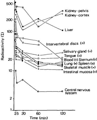
500 © Kidney-pelvis Kidney-cortex Liver Intervertebral discs (°) Salivary gland U) Tongue (o)
Blood (0 Sternum (o) Lung (•) Spleen (o) Skeletal muscle (») Intestinal mucosa (») Central nervous system 25 20 60 Time (min) 120
FIG. 2. Distribution of "C-vecuronium in rat organs at different times. Densitometric measurement of whole-body autoradiographs. Ordinate: radioactivity compared with con-centrations in blood at 2 min = 100%. The elimination organs (liver, kidney) have most, the nervous system least radio-activity; intermediate concentrations of radioactivity in
1046 BRITISH JOURNAL OF ANAESTHESIA
K
2 min
In
2 min
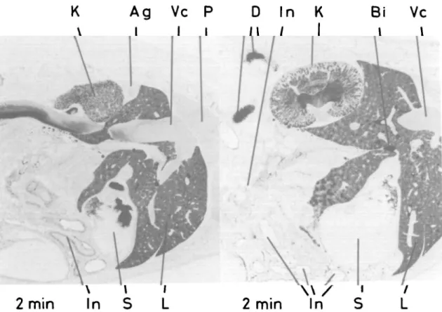
FIG. 3. Autoradiographs of section through liver, kidney, stomach, intestines, vena cava 2 min after i.v. injection of 14C-vecuronium 0.025 uCi g"1. The liver shows lobular structure with high radioactivity in
the centre (central vein), and in the bile (ductus choledochus) entering the duodenum and stomach. The mucous membrane of the small intestine has little radioactivity. The kidneys excrete a large amount through their glomerulae and tubules into the kidney pelvis. Adrenal gland—no radioactivity. Ag = adrenal gland; Bi = bile; Bl = bladder; Br — brain; F = fetus; Gb = gall bladder; H = heart; ID = intervertebral disc; In = intestine; K •= kidney; Kc — kidney cortex; Km = kidney medulla; Kp = kidney pelvis; L = liver; La = larynx; P = lungs; PI = placenta; S •» stomach; Sc = spinal cord;
Sp = spleen; St = sternum; Vc •= vena cava.
The autoradiography of liver tissue had a lobular aspect (figs 3, 4). The central veins of the lobules were mostly black for more than 20 min, and the liver cells remained uniformly dark for 2 h. At the same time the bile ducts were filled with highly radioactive bile.
The radioactivity of the renal cortex and medulla decreased simultaneously. The urine in the pelvis of the kidneys had a high concentration of radioactivity during the first 20 min which decreased slowly over the next 20-60 min.
A second period of increased radioactivity in the liver and kidneys followed after 20 min (fig. 5). The amount of 14C-metabolites in the liver
decreased after 60 min. Ample radioactivity was noted in the intestine. Radioactivity in the renal
cortex, and in the urine in the pelvis of the kidney increased, over 60-120 min, demonstrating active excretion in the urine, measured in the kidney pelvis.
Much less radioactivity was found in the lungs, spleen, myocardium, skeletal muscle, bones, salivary and adrenal glands, and thymus, with a first peak 2 min after injection. The cortex of the adrenals had more radioactivity than the medulla. Very little radioactivity was observed in the central nervous system.
The blood 14C-concentration followed an
ex-ponential decrease with three different phases (<*) P) y), in the same range as the mentioned organ activities (fig. 6). The bile—highly radio-active immediately after the injection—flows via
In K Ag In
5
min
100-o 1C I n L
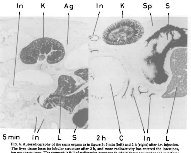
FIG. 4. Autoradiography of the same organs as in figure 3, 5 min (left) and 2 h (right) after i.v. injection. The liver tissue loses its lobular structure after 2 h, and more radioactivity has entered the intestines, but not the mucosa. The stomach is full of radioactive compounds; the kidneys are unchanged as before;
adrenal gland is free of radioactivity. For key to abbreviations, see figure 3.
the bile duct to the duodenum, the intestines and the stomach. The radioactivity remained within the different parts of the intestines during the experiment and much of it was bound to the surface of mucous membrane and, perhaps, even absorbed.
In some tissues as cartilage of the sternum, intervertebral discs, snout, tendons and connec-tive tissues, there was an immediate accumulation of considerable radioactivity—at first similar to that in the kidneys (fig. 7). However, the radioactivity decreased rapidly to average values within 20 min.
Distribution of radioactivity in mice
The distribution of radioactivity in the organs of mice was different than in rats. As mice are more sensitive to vecuronium than rats, the injected dose for autoradiography was relatively high, but proved useful for testing the permeability of the blood-brain and placental barriers. Two minutes after the i.v. injection of vecuronium the blood
Salivary gland Skeletal muscle Blood Lung 2520 60 120 Time (min)
FIG. 5. Liquid scintillation counting of organ probes of the same rats show a similar distribution: high concentrations of radioactivity in kidneys and liver over 2 h, little in skeletal
1048 100000 i 10000 8 1000 100 10
BRITISH JOURNAL OF ANAESTHESIA 5004 Bile I Blood 251020 40 60 90 120 Time (min) 150 180 25 20 FIG. 6. The radioactivity ( ± S E M ) of vecuronium and
metabolites in the excretion fluids is much greater than in the blood. The concentration in the bile is nearly 1000-fold; in the
urine, as measured in the kidney pelvis, 10-fold.
60 Time (min)
FIG. 7. As with other quaternary neuromuscular blockerj, accumulation of positively charged drug molecules is detec-table in the cartilage of bones in the vertebral discs, sternum, etc. It is less marked with vecuronium than with pancuronium.
Vertical bars represent SEM.
Bl In
FIG. 8. Whole-body autoradiography of I4C-vecuronium in a mouse, 10 min after i.v. injection of
0.025 uCi g~'. The distribution in the organs is similar to that in the rat. Much radioactivity is already concentrated in the intervertebral discs, the sternum, the trachea and larynx. For key to abbreviations,
SC Br
In
H St
La
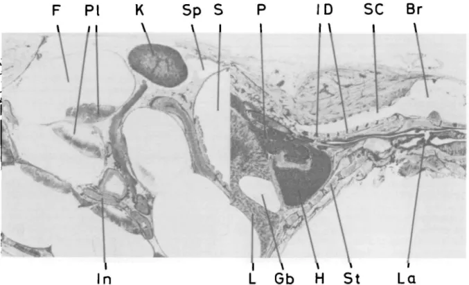
FIG. 9. Distribution in pregnant mouse 5 min after i.v. injection of 0.04 (iCi g~'. Even with a high dose, radioactivity is not detectable in the fetus, but is present in the placenta, with similar densities in the kidney, a renal vein and the intestinal mucosa. Intense radioactivity in the spinal column, sternum and
larynx. For key to abbreviations, see figure 3.
was more radioactive than the liver tissue of male and pregnant animals. However, as radioactivity accumulated in the liver, both tissues had similar concentrations after 10 min. The bile contained little radioactivity and the gall bladder remained free of radioactivity. In contrast, the kidneys and the urine in the pelvis and in the bladder were darker than the liver, and the gastrointestinal tract contained (after 10 min) traces of radioactive material—mainly in the mucous membrane and partly in its lumen. There was no radioactivity in nervous tissues: only some of the blood vessels in the brain were blackened. The cartilage of joints, intervetebral discs, larynx and connective tissue became increasingly black within 10 min (fig. 8).
The placentae of pregnant animals were radio-active, similar to the intestines, but there were only slight traces of radioactivity in the fetuses (similar to the brain) (fig. 9). Even the livers of the embryos did not produce positive auto-radiographs.
Liquid scintillation analysis of rat organs and mouse fetuses
The activities of a few typical uniform tissues and organs, measured by this method during the time course of the study, correspond largely with the densitometric measurements of the auto-radiographs. However, no details of distribution in discrete small areas are recognizable. The comparison of the relative (%) values shows the close coincidence of the measurements by the two analytical methods.
Thirty minutes after i.v. injection (0.49 nCi/g animal weight) of 14C-vecuronium to pregnant
mice, the fetuses contained only 2.1% of the radioactivity of the injected dose per g tissue weight.
DISCUSSION
Based on the timing peaks of radioactivity in the various organs, we calculate that the distribu-tion of 14C-vecuronium is over 2 min after i.v.
1050 BRITISH JOURNAL OF ANAESTHESIA injection. Blockade of neuromuscular
trans-mission—with the 7 times LD100 dose (high,
because this radioactivity is required for positive autoradiographs)—starts immediately but lasts, on average, only 10 min when one considers ventilatory movements. Thus onset time and duration of relaxation are shorter than with pancuronium (Durant, Houwertjes and Crul,
1980).
Vecuronium is metabolized in the liver and one metabolite, the 3-deactylated Org 7268, is ex-creted in bile and urine. We found only traces of this metabolite in the plasma. Therefore, the radioactivity in blood and organs, except the liver, is produced mostly by WC-vecuronium (Waser
and Wiederkehr, in preparation).
Much radioactivity is extracted within the first 2 min by the liver and excreted through the bile into the intestines. The radioactivity in the stomach is probably the result of reflux from the duodenum. The radioactivity accumulated in the liver is 5 times greater than the blood con-centration. Pancuronium, with an activity in the liver only 2 times greater than that in plasma after 60 min (Waser, 1973), is markedly different in its distribution, probably because of the more lipophilic character of vecuronium and its rapid metabolism in the liver cells. The second peak of radioactivity in the liver (between 20 and 60 min) may be attributable to reabsorption of 14
C-vecuronium excreted with the bile into the intestine, whereas the more hydrophilic metabo-lite will be excreted through the kidneys.
Lower concentrations of radioactivity were found in the kidneys at the start of distribution. After 5 min they were similar to those in the liver. The urine in the renal pelvis and in the bladder was strongly radioactive at the beginning of the excretion phase, then diminished and increased again after 60 min. This second wave of excretion followed the second liver peak. The total elimi-nation through the kidney was not prominent at the start, but became important after 60 min.
There was an immediate uptake of vecuronium into different tissues containing acid mucopoly-saccharides, such as cartilage, connective tissue, tendons. The uptake occurred immediately after the first pass of 14C-vecuronium through the local
circulation of these tissues (nucleus pulposus). Then the accumulation decreased rapidly—within the first 5 min—and then slowly in two stages over the next 60 min. The difference between the movement of other short-acting
neuromus-cular blockers (pancuronium (Waser, 1973), alcuronium (Waser and Luthi, 1966)) into this storage compartment and that of vecuronium is evident, since with vecuronium it occurs early and is of short duration. The liberation of non-metabolized drug from these compartments into the blood stream will not prolong the neuro-muscular blockade as this second inflow to the blood will be taken up immediately by the liver, where it will be partly metabolized and excreted through the bile.
The three kinetic phases of the radioactivity in plasma (fig. 10) can be explained as follows: a-phase—after the distribution by uptake into the liver and other organs or compartments as well as elimination through the kidney; p-phase—re-appearance from the skeletal muscles and the mucopolysaccharide binding sites, again uptake into liver and metabolism producing more polar water-soluble metabolites; y-phase—by the rest of vecuronium returning from the organs and water soluble metabolites being excreted now mainly through the kidneys.
During the early phases the extraction by the liver and the elimination of vecuronium and its metabolites with the bile are the most important of its kinetics. It is much greater than excretion
100
r
•a 8 X) c 2" 10 o CO o ~»_ iWBAR -« LSC 25 20 60 Teme (min) 120FIG. 10. The radioactivity ( ± S D ) in the blood of " C -vecuronium and metabolites) measured with whole-body autoradiography and liquid scintillation counting diminishes
through the kidneys and in the urine. With pancuronium, a bisquaternary highly polar com-pound, this proportion is reversed and in favour of elimination through the kidneys (Upton et al., 1982). The shifting of 14C-vecuronium to the mucopolysaccharide-containing compartments (cartilage, connective tissue) is less important than with pancuronium. The relatively short duration of muscle relaxation produced by vecuronium is mostly the result of its rapid elimination, plasma binding (60-80%) and high extraction into the liver.
The pilot experiments in mice demonstrated some differences in the distribution of radio-activity compared with the rats. Some minutes after the injection of 14C-vecuronium, the clear-ance of blood from the liver was smaller, and the kidneys and the urine contained more radio-activity. This may be because of the high injected doses. After 10 min this relation was partly reversed, and in the pregnant animals became at least equal. The intestines contained large amounts of radioactive bile, but the urine con-tinued to be the main pathway of elimination. Possibly, in pregnancy, the elimination pathway through the liver-bile system is used less than in normal animals. There is a difference between rats and mice, as there was little radioactivity in the bile ducts of the latter, and the gall bladders were free of radioactivity.
Finally, the central nervous system with plenty of lipids in its membranes and neurones, contained
very little 14C-vecuronium. The passage through the blood-brain barrier, as through the placental barrier, is very limited; indeed, it is virtually zero (Demetriou et al., 1982).
REFERENCES
Cross, S. A. M., Groves, A. D., and Hesselbo, T. (1974). A quantitative method for measuring radioactivity in tissues sectioned for whole-body autoradiography. Int. J. Appl.
Radiat. Isotopes, 25, 381.
Demetriou, M., Depoix, J.-P., Diakite, B., Fromentin, M., and Duvaldestin, P. (1982). Placenta] transfer of Org NC 45 in women undergoing Caesarean section. Br.J. Anaesth., 54, 643.
Durant, N. N., Houwertjes, M. C , and Crul, J. F. (1980). Comparison of the neuromuscular blocking properties of Org NC 45 and pancuronium in the rat, cat and rhesus monkey. Br. J. Anaesth., 52, 723.
Keller, F., and Waser, P. G. (1982). Quantification in macroscopic autoradiography with carbon-14, an evaluation of the method. Int. J. Appl. Radiat. Isotopes, 33, 1427. Savage, D. S., Sleigh, T., and Carlyle, I. (1980). The
emergence of Org NC 45, l-[2,3a,5a) 16,17)-3,17-bis(ace-tyloxy)-2-(l-piperidinyl)-androstan-16-yl]-l-methyl piperidinium-bromide, from the pancuronium series. Br. J.
Anaesth., 52, 3S.
Upton, R. A., Nguyen, T.-L., Miller, R. D., and Castagnoli, N. (1982). Renal and bilary elimination of vecuronium (Org NC 45) and pancuronium in rats. Anesth. Analg., 61, 313. Waser, P. G. (1973). Localization of 14C-pancuronium by histo- and whole-body autoradiography in normal and pregnant mice. Naunyn Schmiedeberg's Arch. Pharmacol., 279, 399.
Liithi, U. (1966). Verteilung, Metabolismus und Elimination von *H-Diallyl nor Toxiferin (Alloferin) bei Katzen. Helv. Physiol. Pharmacol., A24, 259.