The PGE
2
-Stat3 interaction in doxorubicin-induced
myocardial apoptosis
Miguel A. Frias
1*, Sarin Somers
2, Christine Gerber-Wicht
1, Lionel H. Opie
2, Sandrine Lecour
2,
and Ursula Lang
11
Division of Endocrinology, Diabetology and Nutrition, University Hospital, 24, rue Micheli-du-Crest, CH-1211 Geneva 14, Switzerland and2Hatter Cardiovascular Research Institute, Faculty of Health Science, University of Cape Town, Cape Town, South Africa
Received 28 November 2007; revised 12 June 2008; accepted 16 June 2008; online publish-ahead-of-print 20 June 2008 Time for primary review: 18 days
Aims Both cyclooxygenase-2 (COX-2) and the transcription factor signal transducer and activator of transcription 3 (Stat3) are involved in adaptive growth and survival of cardiomyocytes. In ventricular cardiomyocytes, prostaglandin E2(PGE2), a major COX-2 product, leads to adaptive growth via Stat3
activation, but whether this transcription factor acts as a signalling molecule in PGE2-induced cell
survival is unknown. Therefore, the purpose of this study was to determine whether PGE2counteracts
cardiac apoptosis induced by doxorubicin (DOX), and if so, whether Stat3 plays a critical role in this cardioprotective effect.
Methods and results Neonatal rat ventricular cardiomyocytes were incubated with DOX (0.5 mM) and/or PGE2(1 mM). Apoptosis was assessed by determining caspase3 activation and apoptotic DNA
fragmenta-tion. The role of Stat3 was evaluated in vitro and in vivo by transfecting cardiomyocytes with siRNA targeting rat Stat3 and by using cardiomyocyte-restricted Stat3 knockout (Stat3 KO) mice, respectively. Incubation of ventricular cardiomyocytes with PGE2 led to a time-dependent decrease in the
DOX-induced caspase3 activation, reaching a maximal inhibition of 70 + 5% after 4 h. Similarly, PGE2
inhibited DOX-induced DNA fragmentation by 58 + 5% after 24 h. This antiapoptotic action of PGE2
was strongly reduced by the ERK1/2 inhibitor, U0126, whereas the p38 MAP kinase inhibitor, SB203580, had no effect. Depleting Stat3 expression by 50–60% in isolated ventricular cardiomyocytes markedly reduced the protective effect of PGE2on DOX-induced caspase3 activation and DNA
fragmen-tation. Likewise, the stable PGE2analogue, 16,16-dimethyl-PGE2, was unable to counteract cardiac
apoptosis induced by DOX in Stat3 KO mice.
Conclusion Our results demonstrate that PGE2prevents myocardial apoptosis induced by DOX. This
protection requires the activation of the prosurvival pathways of Stat3 and ERK1/2. KEYWORDS
Prostaglandins; Apoptosis; Signal transduction
1. Introduction
The anthracycline doxorubicin (DOX) is one of the most effec-tive anticancer drugs and frequently used to treat solid tumours and haematological malignances. Unfortunately, its use is limited by cumulative dose-related cardiotoxicity.1,2
The pathogenesis of DOX has not yet been clearly identified. The presence of a large amount of apoptotic cells in the myocardium of DOX-treated patients could contribute to the dilated cardiomyopathy and heart failure induced by this anthracycline.3,4 Among the mechanisms involved in DOX-induced cardiotoxicity, there is formation of reactive oxygen radicals,5,6leading to apoptosis in cardiomyocytes.7,8 Multiple studies indicate that cyclooxygenase-2 (COX-2) and its downstream products such as prostaglandin E2
(PGE2) and prostacyclin (PGI2) play a cardioprotective
role, in particular counteracting DOX treatment9–11 and ischaemia/reperfusion (I/R) injury12–15 whose deleterious effects are mediated in a similar manner as DOX-induced injury, including the generation of reactive oxygen species.16
Indeed, COX-2 inhibition aggravates DOX-mediated cardio-myocyte injury including apoptosis in vitro and in vivo.9–11
Consistently, the prostacyclin analogue, iloprost, was found to reduce cardiac cell apoptosis and to ameliorate the cardiac function in DOX-treated mice,11but the
mech-anisms of protection are still unclear.
There is increasing evidence that the signal transducer and activator of transcription 3 (Stat3) plays a cardioprotective role in the heart.17–21 Indeed, conditional knockout (KO) mice harbouring a cardiomyocyte-restricted deletion of Stat3 KO (Stat3 KO) showed increased susceptibility to cardiac injury caused by myocardial ischaemia, inflammation, or
*Corresponding author. Tel: þ41 22 3729322; fax: þ41 22 3729326. E-mail address: [email protected]
Published on behalf of the European Society of Cardiology. All rights reserved.&The Author 2008. For permissions please email: [email protected].
drug toxicity.18,22Stat3 has also been suggested as a critical intermediate to provide cardioprotection from DOX.22–23
At the present time, there is little information concerning the effect of PGE2on Stat3. In ventricular cardiomyocytes,
we have recently demonstrated that PGE2 leads to an
increase in cell size and protein synthesis via a Stat3-dependent pathway involving ERK1/2.24However, whether Stat3 acts as a signalling molecule in PGE2-induced cell
survival is unknown. Therefore we determined whether PGE2 counteracts the apoptotic effect of DOX in neonatal
rat ventricular cardiomyocytes and whether Stat3 and/or ERK1/2 are involved. To further evaluate the interaction between PGE2 and Stat3, we also investigated the role of
Stat3 in the antiapoptotic effect of PGE2 in vivo using
Stat3 KO and wild-type (WT) mice.
2. Methods
2.1 Cell culture
This investigation conforms with the Guide for the Care and Use of Laboratory Animals published by US National Institues of Health (NIH Publication No. 85–23, revised 1966). Neonatal ventricular cardiomyocytes were isolated from 1–2-day-old Wistar rat ventri-cles by digestion with trypsin-EDTA and type 2 collagenase as we have previously described.24For all experiments described herein, cells were used the third or fourth day of culture, after 16–24 h in DMEM medium depleted in FCS.
2.2 Small interfering RNA transfection
Stat3 was silenced by using a siRNA targeting rat Stat3 (Stat3-siRNA) with a mix of the following sequences: 50-GGCUGAUCAUUUAUAU
AAA-30 and 50- GAGGGUCUCGGAAAUUUAA-30. These siRNAs which
were provided by Qiagen AG (Hombrechtikon, Switzerland), were annealed according to manufacturer’s instructions and stored at 2208C. Cardiomyocytes were transfected using lipofectamine 2000 (Invitrogen) reagent, following the manufacter’s protocol. Briefly, 24 h after cardiomyocytes were plated, transfection was proceeded with a mix containing lipofectamine 2000 reagent and Stat3-siRNA (300 pmol) in OptiMEMþGlutaMAX medium (Invitrogen). Cardiomyocytes were stimulated, with the different drugs 24–36 h after transfection. For control, cells were transfected as described for siRNA experiments with non-silencing siRNA (300 pmol) (50-UUCU
CCGAACGUGUCACGU-30) which is ineffective in rat cells since it has
no mammalian target.
2.3 In vivo experiments
WT or Stat3 cardiomyocytes restricted KO (Stat3 KO) mice (13–16 weeks old and weighting 20–40 g) were used in this study. Genotype was confirmed by DNA extraction and PCR analysis as previously described.25 DOX was administered as a single intraperitoneal
(i.p.) injection at a dose of 12 mg/kg. The stable PGE2analogue,
16,16-dimethyl-PGE2 (dmPGE2) was administered (i.p.) at a dose
of 10 mg/kg trice daily from 2 days before DOX and continued until 5 days after DOX. The mice were divided into eight groups: (i) WT as control, (ii) Stat3 KO as control, (iii) WTþdmPGE2, (iv)
Stat3 KOþdmPGE2, (v) WTþDOX, (vi) Stat3 KOþDOX, (vii) WTþ
dmPGE2þDOX, (viii) Stat3 KOþdmPGE2þDOX. When the animals
were sacrificed, the hearts were removed and cardiac tissue processed for determination of apoptotic DNA fragmentation. The doses of dmPGE2and DOX were based on previous work.11,26
2.4 Western blotting
Cardiomyocytes were starved of serum overnight and subjected to treatment as indicated in the figure legends. After stimulation in
serum-free DMEM, cells were washed, lysed, and analysed by western blotting as previously described.24 Briefly, total cell
proteins were separated by SDS–PAGE and blotted onto nitrocellu-lose membrane. Afterwards, membranes were probed against cleaved caspase3 (17 kDa fragment) or phosphorylated Stat3 (Cell Signaling Technology, Denvers, MA, USA), and reprobed against glyceraldehyde-3-phosphate dehydrogenase (Chemicon Inter-national Inc., Hampshire, UK) or Stat3 total (Upstate Biotechnology, Lake Placid, NY, USA).
2.5 DNA fragmentation
DNA fragmentation was quantified by measuring the content of intracellular nucleosomes. Neonatal rat ventricular cardiomyocytes were cultured in 6-well plates for 2 days (106cells per well).
There-after, cardiomyocytes were starved of serum for 24 h and subjected to treatment as indicated in the figure legends. After stimulation in serum-free DMEM, cells were washed with ice-cold PBS (phosphate-buffered saline) and lysed with 60 ml of the same buffer used for western blotting.24Concerning the in vivo studies, minced cardiac
tissue was homogenized in the same lysis buffer. The cellular and tissue lysates were used for protein determination and for quanti-tative evaluation of histone-associated DNA fragments by photo-metric enzyme immunoassay (Cell Death Detection ELISAPLUS,
Roche Diagnostics, Germany) according to the manufacturer’s instructions. Results are reported as arbitrary absorbance units normalized to milligram of proteins.
2.6 Determination of cardiomyocyte viability
Cell viability was evaluated by colorimetric MTT assay (3-[4,5-dimethylthiazol-2-yl]-2,5-diphenyl tetrazolium bromide) which is based on the reduction of MTT into a blue formazan dye by functional mitochondria of viable cells. Briefly, at the end of treatments, cardiomyocytes were washed twice with PBS prior to incubation with 5 mg/mL MTT for 3 h at 378C in an atmosphere of 5% CO2. Thereafter, cells were again washed twice with PBSbefore lysing cell membranes with dimethyl sulfoxane. The amount of MTT formazan was quantified by determining the absorbance at 540 and 570 nm. Cell viability was also evaluated by staining cardiomyocytes with trypan blue (0.04% in PBS).
2.7 Chemicals
PGE2and dmPGE2were purchased from Sigma-Aldrich GmbH (Buchs,
Switzerland), dmPGE2was prepared as previously described.27MTT,
SB203580, AG490, and piceatannol were obtained from Calbiochem (Dietikon, Switzerland), while U0126 was from Biomol Research Laboratories (Plymouth Meeting, PA, USA).
2.8 Statistical analysis
All values are expressed as mean + SEM with n referring to the number of experiments. Linear regression analysis was made using the Origin software (Microcal Software, Inc., Northhampton, MA, USA). Differences between groups were determined using either two-tailed unpaired Student’s t-tests or ANOVA, followed by Bonferroni’s post hoc test where applicable. P , 0.05 was accepted as statistically significant.
3. Results
3.1 Effect of prostaglandin E
2on
doxorubicin-induced myocardial apoptosis
and decrease of cell viability
To investigate whether PGE2 can counteract DOX-induced
apoptosis in ventricular cardiomyocytes, we incubated cells with or without PGE2(1 mM) for 30 min before adding
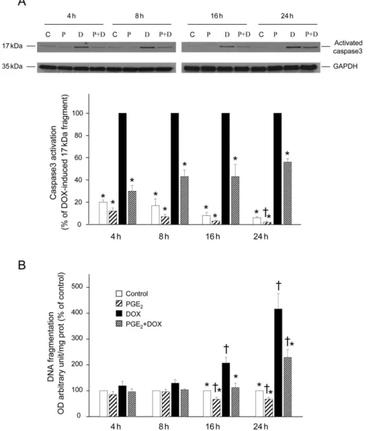
DOX (0.5 mM) for 4–24 h (Figure 1). Thereafter, we analysed activation of caspase3 by measuring the expression of the 17 kDa fragment of cleaved caspase3. As is illustrated in Figure 1A, PGE2 caused a significant decrease in the
acti-vation of caspase3 induced by DOX after 4 h (70 + 5%), 8 h (57 + 6%), 16 h (57 + 11%), or 24 h (44 + 3%). DOX also increased apoptotic DNA fragmentation by 109 + 20 and 319 + 54% after 16 and 24 h of treatment, respectively, but had no significant effect after 4 and 8 h (Figure 1B). The presence of PGE2 abolished the DNA fragmentation
induced after 16 h of incubation with DOX and inhibited that after 24 h of treatment by 49 + 7%.
Cell viability was determined by using the MTT assay. Figure 2A illustrates that DOX decreased cell viability in a time- and concentration-dependent manner. As for apoptotic
DNA fragmentation, PGE2 (1 mM) abolished the decrease in
cell viability observed after 16 h of incubation with DOX (0.5 mM), and markedly inhibited that after 24 h of treatment with DOX (Figure 2B). Similar results were obtained with the trypan blue exclusion method (data not shown).
To compare DOX-induced DNA fragmentation with cell viability, linear regression analysis was used to fit the data described in Figures 1B and 2B (see Figure 2C). This analysis shows a high linear correlation between apoptotic DNA frag-mentation and cell viability in cardiomyocytes treated or not with DOX (0.5 mM) for 4, 16, and 24 h in the presence and absence of PGE2(1 mM).
Our results indicate that in ventricular cardiomyocytes, PGE2counteracts DOX-induced apoptosis and the associated
decrease in cell viability.
Figure 1 Prostaglandin E2inhibits doxorubicin-induced caspase3 activation and apoptotic DNA fragmentation in ventricular cardiomyocytes. Cells were
pre-treated or not (control, C) with prostaglandin E2(P, 1 mM) for 30 min prior to incubation with doxorubicin (D, 0.5 mM) during different time periods (4, 8, 16,
and 24 h). (A) After these incubation periods, caspase3 activation was analysed in cellular extracts by determining the level of the catalytically active 17 kDa fragment by western blotting. Representative blots are shown at the top. Equal gel loading was assessed using an anti-GAPDH antibody. Specific bands corresponding to activated caspase3 were quantified by densitometry and expressed as percentage of doxorubicin-induced formation of the 17 kDa fragment. (B) After the incubation periods with doxorubicin, DNA fragmentation was analysed by measuring histone-associated DNA fragments. Results were calculated as arbitrary absorbance units normalized to mg of proteins and are expressed as percentage of control. *P , 0.05 compared with values from doxorubicin-treated cells,†P , 0.05 compared with control values (n ¼ 4). GAPDH, glyceraldehyde-3-phosphate dehydrogenase.
3.2 Critical role of ERK1/2 in the antiapoptotic
effect of prostaglandin E
2We have previously reported that in ventricular cardiomyo-cytes, ERK1/2 is critical for PGE2-induced Stat3 activation
as assessed by nuclear tyrosine phosphorylation and DNA-binding activity. Both ERK1/2 and Stat3 have been shown to play an essential role in PGE2-stimulated cell growth.24 In
this study, we investigated the role of ERK1/2 and p38
MAPK in the antiapoptotic responses induced by PGE2 in
cardiomyocytes exposed to DOX (0.5 mM) for 4 or 24 h. To this purpose we used the specific inhibitors of the ERK1/2 and p38 MAPK signalling pathways, U0126 and SB203580, respectively.28 U0126 suppresses the activation of MEK1/2 leading to the inhibition of its downstream target ERK1/2.28
SB203580 specifically inhibits a and b p38 MAP kinases, the main isoforms expressed in cardiomyocytes,28,29 but it has
Figure 2 Effect of doxorubicin and prostaglandin E2on myocardial cell viability. (A) Cardiomyocytes were treated or not with the indicated concentration of
doxorubicin (0.05–5 mM) for 4, 16, and 24 h, respectively. Thereafter, cell viability was determined by using the MTT assay. Each line represents mean + SEM of three separate experiments (n ¼ 3 wells in each experiment). Results are expressed as percentage of the corresponding control. (B) Cells were pretreated or not with prostaglandin E2(1 mM) for 30 min prior to incubation with doxorubicin (0.5 mM) during different time periods (4, 16, and 24 h). After these incubation
periods, cell viability was determined by using the MTT assay. Results are expressed as percentage of the control. *P , 0.05 compared with control values (n ¼ 3–4). (C ) The apoptotic DNA fragmentation observed after 4, 16, or 24 h of incubation with 0.5 mM doxorubicin in the presence or absence of 1 mM PGE2
no effect on g and d p38 MAP kinases.30As shown in Figure 3A, U0126, but not SB203580, abolished the PGE2-induced Stat3
activation which was observed after 90 min of stimulation by determination of cellular Stat3 tyrosine phosphorylation.
To evaluate the role of ERK1/2 and p38 MAPK pathways on DOX-induced apoptosis in the presence and absence of PGE2, cells were incubated for 30 min with U0126 (10 mM)
or SB203580 (10 mM). Thereafter, cardiomyocytes were
stimulated for 30 min with PGE2(1 mM) before DOX (0.5 mM)
was added. Activation of caspase3 was determined after 4 h of DOX treatment, while generation of DNA fragmentation was assessed after 24 h of incubation with DOX. As shown in Figure 3B, U0126 strongly reduced the antiapoptotic effect of PGE2 on DOX-induced caspase3 activation, whereas
SB203580 had not significant effect. Similarly, the presence of U0126, but not SB203580, abolished the protective effect
Figure 3 ERK1/2 plays a role in the inhibitory effect of prostaglandin E2on doxorubicin-induced activation of caspase3 and DNA fragmentation. Cardiomyocytes
were incubated for 30 min with U0126 (10 mM) or SB203580 (SB, 10 mM), prior to stimulation with prostaglandin E2(1 mM). (A) After 90 min of prostaglandin E2
stimulation, Stat3 tyrosine phosphorylation (Stat3-P) was analysed in cellular extracts by western blotting. A representative blot is shown: similar results were obtained in four separate experiments. (B) After 30 min of prostaglandin E2stimulation, cells were incubated with doxorubicin (0.5 mM) during 4 h. At the end of
this incubation period, caspase3 activation was analysed in cellular extracts by determining the level of the catalytically active 17 kDa fragment by western blotting. A representative blot is shown at the top of (B) (C, control, P, prostaglandin E2, D, doxorubicin). Equal gel loading was assessed using an anti-GAPDH
antibody. Specific bands corresponding to activated caspase3 were quantified by densitometry and expressed as percentage of doxorubicin-induced formation of the 17 kDa fragment. (C ) After 30 min of prostaglandin E2stimulation, cells were incubated with doxorubicin (0.5 mM) during 24 h. At the end of this incubation
period, DNA fragmentation was analysed by measuring histone-associated DNA fragments. Results were calculated as arbitrary absorbance units normalized to mg of proteins and are expressed as percentage of doxorubicin-induced DNA fragments. *P , 0.05 compared with values from doxorubicin-treated cells (n ¼ 4).
of PGE2 on DOX-induced DNA fragmentation (Figure 3C). It
appears that ERK1/2 but not p38 MAPK is involved in the protective influence of PGE2 counteracting apoptosis in
cardiomyocytes exposed to DOX.
3.3 Critical role of signal transducer and activator
of transcription 3 in the antiapoptotic effect of
prostaglandin E
2in vitro
To evaluate the role of Stat3 in the antiapoptotic effect of PGE2 in vitro, we transfected cardiomyocytes with siRNA
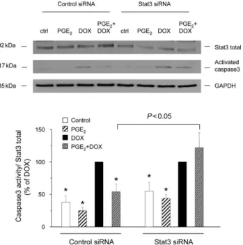
targeting rat Stat3. This treatment decreased Stat3 expression by 50–60%, 40 h after transfection of cardiomyo-cytes when compared with cells transfected with non-silencing RNA (control siRNA) (Figure 4, top). This Stat3 knock-down effect remained sustained until 48 h after transfection with anti-Stat3 siRNA (data not shown). After incubation with DOX, caspase3 activation, normalized to Stat3 expression, was increased in both control and anti-Stat3 siRNA transfected cells (Figure 4). PGE2
signifi-cantly decreased DOX-induced caspase3 activation in the control siRNA transfected cardiomyocytes, whereas it had no effect in the anti-Stat3 siRNA transfected cells. Exposure of cardiomyocytes to DOX for 24 h also increased apoptotic fragmentation of DNA in both control and anti-Stat3 siRNA transfected cells (Figure 5A). As for caspase3 activation, PGE2 decreased DOX-induced DNA fragmentation in the
control siRNA transfected cells. In contrast, this antiapoptotic
effect of PGE2was virtually abolished in the anti-Stat3 siRNA
transfected cardiomyocytes. Since the transfection technique appears to increase basal DNA fragmentation (compare Figure 1), resulting in apparently smaller effects of DOX and PGE2, we also investigated the role of Stat3 in the
antiapoptotic effect of PGE2 on DOX-induced DNA
frag-mentation by using the Stat3 pathway inhibitors AG49031
Figure 4 Small interfering RNA targeting Stat3 blocks the inhibitory effect of prostaglandin E2on doxorubicin-induced activation of caspase3. Thirty-six
hour after transfection of cardiomyocytes with control small interfering RNA or small interfering RNA targeting Stat3 (Stat3 siRNA), cells were pretreated or not (ctrl) with prostaglandin E2(1 mM) for 30 min prior to incubation with
doxorubicin (0.5 mM) during 4 h. Thereafter, the expression of Stat3 as well as that of the active caspase3 fragment of 17 kDa was analysed in cellular extracts by western blots. Representative blots are shown at the top. Specific bands corresponding to activated caspase3 were quantified by densitometry and normalized to Stat3 expression. Equal gel loading was assessed using an anti-GAPDH antibody. Results are expressed as percentage of doxorubicin-induced formation of the 17 kDa fragment. *P , 0.05 compared with values from doxorubicin-treated cells (n ¼ 5).
Figure 5 Small interfering RNA targeting Stat3 and pharmacological inhi-bition of Stat3 reduce the inhibitory effect of prostaglandin E2 on
doxorubicin-induced apoptotic DNA fragmentation. (A) 24 h after transfection of cardiomyocytes with control siRNA or siRNA targeting Stat3 (Stat3 siRNA), cells were pretreated or not (control, ctrl) with prostaglandin E2(1 mM) for
30 min prior to the incubation with doxorubicin (0.5 mM) during 24 h. There-after, DNA fragmentation was analysed. (B and C ) Cells were incubated for 1 h with the Stat3 inhibitors piceatannol (10 mM) or AG490 (10 mM) prior to stimu-lation with prostaglandin E2 (1 mM). (B) After 90 min of prostaglandin E2
stimulation, Stat3 tyrosine phosphorylation (Stat3-P) was analysed in cellular extracts by western blotting. A representative blot is shown: similar results were obtained in three separate experiments. (C ) After 30 min of prostaglan-din E2stimulation, cells were incubated with doxorubicin (0.5 mM) during
24 h. At the end of this incubation period, DNA fragmentation was analysed. *P , 0.05 compared with values from doxorubicin-treated cells;†*P , 0.05
and piceatannol.32 Both these inhibitors strongly inhibited PGE2-induced Stat3 phosphorylation (Figure 5B) as well as
the antiapoptotic effect of PGE2on the DNA fragmentation
induced by DOX (Figure 5C).
3.4 Critical role of signal transducer and activator
of transcription 3 in the antiapoptotic effect of
prostaglandin E
2in vivo
In order to evaluate the role of Stat3 in the antiapoptotic effect of PGE2 in vivo, we used Stat3 KO mice
25
and their littermate controls (WT). Stat3 KO and WT mice were each divided in four groups: control, þdmPGE2, þDOX, and þ
dmPGE2þDOX. The survival rate of animals was 90–100% in
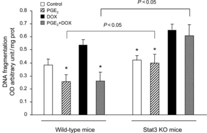
all groups. DOX treatment did not impair survival rate, neither in WT animals nor Stat3 KO mice. Likewise, the heart weight/body weight ratio was similar in all animal groups. After treatment, cardiac tissue (n ¼ 5–7 animals per group) was assessed for apoptosis by measuring DNA fragmentation. As shown in Figure 6, no difference was observed in myocardial DNA fragmentation between WT and Stat3 KO mice at baseline. Treatment with DOX induced an increase in cardiac DNA fragmentation which reached a higher significance level in Stat3 KO mice than in WT animals. Our results confirm previous data from the literature reporting that Stat3 KO mice are more sensitive to DOX treatment than WT animals.22 As expected, the increase in DOX-induced cardiac apoptosis was markedly attenuated by dmPGE2 injections in WT mice. In contrast,
the same treatment remained without effect in Stat3 KO mice. Of note, in dmPGE2-treated animals, cardiac
apop-tosis was significantly different between WT and Stat3 KO mice, whether or not animals had been injected with DOX. Our investigations, both in vitro and in vivo, demonstrate that the protective effect of PGE2 against DOX-induced
cardiac apoptosis is Stat3-dependent.
4. Discussion
Our study revealed that in ventricular cardiomyocytes, PGE2
exerts antiapoptotic activities which are mediated by Stat3 involving ERK1/2. Indeed, PGE2 was found to counteract
DOX-induced caspase3 activation and apoptotic DNA frag-mentation which was associated with impaired survival in ventricular cardiomyocytes. By transfecting cardiomyocytes with siRNA targeting specifically rat Stat3, we obtained 50– 60% inhibition of Stat3 expression. In these Stat3 silenced cells, the antiapoptotic effect of PGE2 on DOX-induced
caspase3 activation and DNA fragmentation was significantly reduced. Likewise, the Stat3 pathway inhibitors, piceatan-nol and AG490, abolished the antiapoptotic effect of PGE2
on DNA fragmentation generated by DOX. The role of Stat3 in PGE2-induced antiapoptotic activity was further
con-firmed in vivo using WT and Stat3 KO mice. While treatment with the stable analogue dmPGE2significantly inhibited the
DOX-induced increase in myocardial apoptotic DNA fragmen-tation in WT mice, it had no effect in Stat3 KO mice.
According to a recently published, fundamental hypoth-esis,33 the myocardium adaptation to stress depends on the nature of the signalling stimulus. The cardiomyocyte can either survive leading to beneficial or adaptive hypertro-phy34or undergo apoptosis, which induces left ventricular failure and dilation (maladaptive hypertrophy).35Including observations from a previous study,24 our results suggest that PGE2is beneficial for the heart, favouring an adaptive
pattern of hypertrophy by its dual action on cell survival and hypertrophic growth, via the activation of Stat3.
COX-2 has been shown to mediate PGE2production in both
neonatal and adult cardiac myocytes.36,37Moreover, several in vitro and in vivo studies reported that COX-2 and its pros-tanoid products as well as Stat3 similarly counteract DOX- and oxidative stress-induced myocardial damage in both neonatal and young adult ventricular cardiomyocytes.9–11,19,21–23 However, there is little information concerning the effect of PGE2 or other COX-2-derived products on myocardial Stat3
activation and its cardioprotective role. Very recently, Qian et al.38using cardiac EP4-deficient mice, reported that this prostaglandin receptor plays a role in hypertrophy via acti-vation of Stat3 which seems cardioprotective in mice with myocardial infarction.
There is growing evidence that Stat3 plays an important role in cardiac remodelling, particularly by promoting cardiomyocyte survival and hypertrophy.19,20,23 Moreover, it has been demonstrated that Stat3 activation confers cardioprotection at the time of reperfusion in response to ischaemic preconditioning and TNFa.15,17,25Interestingly, a recent study reported a decrease in myocardial Stat3 levels in the aged mouse heart associated with a loss of cardioprotection induced by ischaemic post-conditioning.39
Consistent with our results, the prostacyclin analogue iloprost was shown to prevent the release of lactate dehy-drogenase induced by DOX in ventricular cardiomyocytes, while COX-2 inhibition increased this deleterious response.9 In vivo, it has been shown that inhibition of COX-2 aggra-vates DOX-induced cardiac injury as detected by cardiomyo-cyte apoptosis and the release of lactate dehydrogenase and cardiac troponin C.10Moreover, Neilan et al.11observed that DOX-induced cardiac apoptosis and dysfunction was significantly higher in COX-2 KO than in WT mice, and that this DOX-induced myocardial toxicity was attenuated
Figure 6 Stat3 plays a role in the inhibitory effect of 16,16-dimethyl pros-taglandin E2on doxorubicin-induced myocardial DNA fragmentation in vivo.
Wild-type and Stat3 knockout mice were treated or not (control) with the stable prostaglandin E2 analogue 16,16-dimethyl prostaglandin E2 (PGE2)
for 2 days before and 5 days after a single injection of doxorubicin (DOX) (12 mg/kg, i.p.). Thereafter, mice were killed, hearts were removed and cardiac tissue processed for DNA fragmentation analysis. Apoptotic DNA frag-mentation in cardiac tissue was assessed by measuring histone-associated DNA fragments. Results are expressed as arbitrary absorbance units normal-ized to mg of proteins. *P , 0.05 compared with values from doxorubicin-treated mice (n ¼ 5–7 animals per group).
by the stable prostacyclin analogue iloprost which was administered 2 days before and 5 days after DOX injection. The latter finding is in agreement with the myocardial antiapoptotic effect of dmPGE2 we observed in vivo, using
a similar experimental protocol.
Interestingly, Delgado et al.40showed in a murine model, that when COX-2 inhibition treatment was initiated after heart failure had been established by prolonged DOX admin-istration, it attenuated further progression of this cardio-myopathy. The authors reasoned that in their experimental setting, the effects of COX-2 inhibition cannot be related to an interaction with the effects of DOX. They further speculate that the role of COX-2 is initially adaptive, attenu-ating the deleterious effects during DOX treatment but later, after removal of this stress, it becomes maladaptive, leading to the progression of heart failure.
Confirming the cardioprotective role of COX-2, several studies have demonstrated that this enzyme and its products reduce I/R injury12–15and mediate the late phase of precon-ditioning.15,41Indeed, both prostaglandin receptor EP3 and EP4 signalling have been reported to protect the heart from I/R injury.13,14 Moreover, Xuan et al.15 have shown that activation of Stat3 leading to up-regulation of COX-2 underlies the protective effect of late preconditioning against myocardial I/R injury. From the point of view of a positive feed back phenomenon, the stimulatory role of PGE2 in Stat3 activation which we demonstrated in our
work, could contribute to this cardioprotective process in cardiac myocytes.
In this study, we also found a role for ERK1/2 in the Stat3-dependent, cardioprotective effect of PGE2, counteracting
DOX-induced apoptosis in cardiomyocytes. Indeed, inhibition of the ERK1/2, but not of the p38 MAPK pathway, strongly reduced the antiapoptotic effect of PGE2 on DOX-induced
caspase3 activation and DNA fragmentation in ventricular cardiomyocytes. These observations are in agreement with our previous results showing that in ventricular cardiomyo-cytes, PGE2-induced Stat3 activation and hypertrophic
growth involves ERK1/2 but not p38 MAPK.24 Consistent with our present results, it has been shown that ERK1/2 is implicated in myocardial survival signalling, while p38 MAPK appears to have pro- or antiapoptotic effects depending on the experimental model.35 In neonatal ventricular cardio-myocytes, ERK1/2 was found to prevent apoptosis in cells exposed to the anthracycline daunomycin, whereas p38 MAPK appeared to be involved in the induction of apoptosis.42 Taken together, our data demonstrate that PGE2prevents
ventricular cardiomyocytes from apoptosis induced by DOX, and that Stat3 activation involving ERK1/2 plays a key role in this protective effect. Our in vivo studies confirm the importance of Stat3 in the protective role of PGE2 against
DOX-induced cardiac apoptosis. Thus, our investigations underline the potential significance of the PGE2–Stat3
inter-action for the development of novel therapeutic strategies offering increased cardioprotection.
Conflict of interest: none declared.
Funding
This work was supported by the Swiss National Science Foundation (grant 310000-108342/1), the Swiss University Conference
Foundation, the South African National Research Foundation and the South African Medical Research Council.
References
1. Singal PK, Iliskovic N. Doxorubicin-induced cardiomyopathy. N Engl J Med 1998;339:900–905.
2. Takemura G, Fujiwara H. Doxorubicin-induced cardiomyopathy from the cardiotoxic mechanisms to management. Prog Cardiovasc Dis 2007;49: 330–352.
3. Keizer HG, Pinedo HM, Schuuruis GJ, Joenje H. Doxorubicin (adriamycin): a critical review of free radical-dependent mechanisms of cytotoxicity. Pharmacol Ther 1990;47:219–231.
4. Singal PK, Li T, Kumar D, Danelisen D, Iliskovic N. Adriamycin-induced heart failure: mechanism and modulation. Mol Cell Biochem 2000;207: 77–86.
5. Iliskovic N, Singal PK. Lipid lowering: an important factor in preventing adriamycin-induced heart failure. Am J Pathol 1997;150:727–734. 6. Siveski-Iliskovic N, Kaul N, Singal PK. Probucol promotes endogenous
antioxidants and provides protection against adriamycin-induced cardiomyopathy in rats. Circulation 1994;89:2829–2835.
7. Childs AC, Phaneuf SL, Dirks AJ, Phillips T, Leeuwenburgh C. Doxorubicin treatment in vivo causes cytochrome C release and cardiomyocyte apoptosis, as well as increased mitochondrial efficiency, superoxide dismutase activity, and Bcl-2:Bax ratio. Cancer Res 2002;62:4592–4598. 8. Kluza J, Marchetti P, Gallego MA, Lancel S, Fournier C, Loyens A et al. Mitochondrial proliferation during apoptosis induced by anticancer agents: effects of doxorubicin and mitoxantrone on cancer and cardiac cells. Oncogene 2004;23:7018–7030.
9. Adderley SR, Fitzgerald DJ. Oxidative damage of cardiomyocytes is limited by extracellular regulated kinase 1/2-mediated induction of cyclooxygenase-2. J Biol Chem 1999;274:5038–5046.
10. Dowd NP, Scully M, Adderley SR, Cunningham AJ, Fitzgerald DJ. Inhibition of cyclooxygenase-2 aggravates doxorubicin-mediated cardiac injury in vivo. J Clin Invest 2001;108:585–590.
11. Neilan TG, Doherty GA, Chen G, Deflandre C, McAllister H, Butler RK et al. Disruption of COX-2 modulates gene expression and the cardiac injury response to doxorubicin. Am J Physiol Heart Circ Physiol 2006; 291:H532–H536.
12. Camitta MG, Gabel SA, Chulada P, Bradbury JA, Langenbach R, Zeldin DC et al. Cyclooxygenase-1 and -2 knockout mice demonstrate increased cardiac ischemia/reperfusion injury but are protected by acute precondi-tioning. Circulation 2001;104:2453–2458.
13. Xiao CY, Yuhki K, Hara A, Fujino T, Kuriyama S, Yamada T et al. Prostaglan-din E2 protects the heart from ischemia-reperfusion injury via its receptor subtype EP4. Circulation 2004;109:2462–2468.
14. Martin M, Kaber G, Jacoby C, Flogel U, Schrader J, Schror K et al. Cardi-ospecific overexpression of the prostaglandin EP3 receptor attenuates ischemia-induced myocardial injury. Circulation 2005;112:400–406. 15. Xuan YT, Guo Y, Zhu Y, Han H, Langenbach R, Dawn B et al. Mechanism of
cyclooxygenase-2 upregulation in late preconditioning. J Mol Cell Cardiol 2003;35:525–537.
16. Zhao ZQ. Oxidative stress-elicited myocardial apoptosis during reperfusion. Curr Opin Pharmacol 2004;4:159–165.
17. Lecour S, Suleman N, Deuchar GA, Somers S, Lacerda L, Huisamen B et al. Pharmacological preconditioning with tumor necrosis factor-alpha activates signal transducer and activator of transcription-3 at reperfusion without involving classic prosurvival kinases (Akt and extracellular signal-regulated kinase). Circulation 2005;112:3911–3918.
18. Hilfiker-Kleiner D, Hilfiker A, Fuchs M, Kaminski K, Schaefer A, Schieffer B et al. Signal transducer and activator of transcription 3 is required for myocardial capillary growth, control of interstitial matrix deposition, and heart protection from ischemic injury. Circ Res 2004;95:187–195. 19. Hilfiker-Kleiner D, Hilfiker A, Drexler H. Many good reasons to have STAT3
in the heart. Pharmacol Ther 2005;107:131–137.
20. Negoro S, Kunisada K, Tone E, Funamoto M, Oh H, Kishimoto T et al. Activation of JAK/STAT pathway transduces cytoprotective signal in rat acute myocardial infarction. Cardiovasc Res 2000;47:797–805. 21. Negoro S, Kunisada, Fujio Y, Funamoto M, Darville MI, Eizirik DL et al.
Activation of signal transducer and activator of transcription 3 protects cardiomyocytes from hypoxia/reoxigenation-induced oxidative stress through the upregulation of manganese superoxide dismutase. Circula-tion 2001;104:979–981.
22. Jacoby JJ, Kalinowski A, Liu MG, Zhang SSM, Gao Q, Chai GX et al. Cardiomyocyte-restricted knockout of STAT3 results in higher sensitivity
to inflammation, cardiac fibrosis, and heart failure with advanced age. Proc Natl Acad Sci USA 2003;100:12929–12934.
23. Kunisada K, Negoro S, Tone E, Funamoto M, Yamauchi-Takihara K. Signal transducer and activator of transcription 3 in the heart transduces not only a hypertrophic signal but a protective signal against doxorubicin-induced cardiomyopathy. Proc Natl Acad Sci USA 2000;97: 315–319.
24. Frias MA, Rebsamen MC, Gerber-Wicht C, Lang U. Prostaglandin E2 activates Stat3 in neonatal rat ventricular cardiomyocytes: A role in cardiac hypertrophy. Cardiovasc Res 2007;73:57–65.
25. Smith RM, Suleman N, Lacerda L, Opie LH, Akira S, Chien KR et al. Genetic depletion of cardiac myocyte STAT-3 abolishes classical precondi-tioning. Cardiovasc Res 2004;63:611–616.
26. Hansen-Petrik MB, McEntee MF, Jull B, Shi H, Zemel MB, Whelan J. Prostaglandin E2 protect intestinal tumors from nonsteroidal anti-inflammatory drug-induced regression in APCMin/þ Mice. Cancer Res 2002;62:403–408.
27. Wilson JW, Potten CS. The effect of exogenous prostaglandin adminis-tration on tumor size and yield in Min/þ mice. Cancer Res 2000;60: 4645–4653.
28. Davies SP, Reddy H, Caivano M, Cohen P. Specifity and mechanism of action of some commonly used protein kinase inhibitors. Biochem J 2000;351:95–105.
29. Jiang Y, Gram H, Zhao M, New L, Gu J, Feng L et al. Characterization of the structure and function of the fourth member of p38 group mitogen-activated protein kinase p38d. J Biol Chem 1997;272:30122–30128. 30. Lee JC, Kassis S, Kumar S, Badger A, Adams JL. P38 Mitogen-activated
protein kinase inhibitors—mechanisms and therapeutic potentials. Pharmacol Ther 1999;82:389–397.
31. Meydan N, Grunberger T, Dadi H, Shaha M, Arpaia E, Lapidot Z et al. Inhibition of acute lymphoblastic leukaemia by a Jak-2 inhibitor. Nature 1997;379:645–648.
32. Su I, David M. Distinct mechanisms of Stat phosphorylation via the interferon-a/b receptor. Selective inhibition of Stat3 and Stat5 by piceatannol. J Biol Chem 2000;275:12661–12666.
33. Opie LH, Commerford PJ, Gerh BJ, Pfeffer MA. Controversies in ventricu-lar remodelling. Lancet 2006;367:356–367.
34. Sugden PH. Mechanotransduction in cardiomyocyte hypertrophy. Circula-tion 2001;103:1375–1377.
35. Baines CP, Molkentin JD. STRESS signaling pathways that modulate cardiac myocyte apoptosis. J Mol Cell Cardiol 2005;38:47–62. 36. Rebsamen MC, Perrier E, Gerber-Wicht C, Benitah JP, Lang U. Direct and
indirect effects of aldosterone on COX-2 and IL-6 expression in rat cardiac cells in culture and after myocardial infarction. Endocrinoloyg 2004;145:3135–3142.
37. LaPointe MC, Mendez M, Leung A, Tao Z, Yang XP. Inhibition of cyclooxygenase-2 improves cardiac function after myocardial infarction in the mouse. Am J Physiol Heart Circ Physiol 2004;286:H1416–H1424. 38. Qian JY, Harding P, Liu Y, Shesely E, Yang XP, LaPointe MC. Reduced
cardiac remodeling and function in cardiac-specific EP4 receptor knock-out mice with myocardial infarction. Hypertension 2008;51:560–566. 39. Boengler K, Buechet A, Heinen Y, Roeskes C, Hilfiker-Kleiner D, Heusch G
et al. Cardioprotection by ischemic postconditioning is lost in aged and Stat3-deficient mice. Circ Res 2008;102:131–135.
40. Delgado RM, Nawar MA, Zewail AM, Kar B, Vaughn WK, Wu KK et al. Cyclooxygenase-2 inhibitor treatment improves left ventricular function and mortality in a murine model of doxorubicin-induced heart failure. Circulation 2004;109:1428–1433.
41. Bouchard J-F, Chouinard J, Lamontagne D. Participation of prostaglandin E2 in the endothelial protective effect of ischaemic preconditioning in isolated rat heart. Cardiovasc Res 2000;45:418–427.
42. Zhu W, Zou Y, Aikawa R, Harada K, Kudoh S, Uozumi H et al. MAPK super-family plays an important role in daunomycin-induced apoptosis of cardiac myocytes. Circulation 1999;100:2100–2107.