. . . .
. . . .
Endothelial mineralocorticoid receptor activation
mediates endothelial dysfunction in diet-induced
obesity
Nicola Scha¨fer
1,2,3, Christine Lohmann
1,2, Stephan Winnik
1,2,
Lambertus J. van Tits
1,2, Melroy X. Miranda
1,2, Athanasios Vergopoulos
4,
Frank Ruschitzka
2,3, Ju¨rg Nussberger
5, Stefan Berger
6, Thomas F. Lu¨scher
1,2,3,
Franc¸ois Verrey
1,3,†, and Christian M. Matter
1,2,3,†,*
1
Institute of Physiology, University of Zurich, Winterthurerstrasse 190, CH-8057 Zurich, Switzerland;2
Cardiovascular Research and Cardiology, University of Zurich, Winterthurerstrasse 190, CH-8057 Zurich, Switzerland;3
Center of Integrative Human Physiology, University of Zurich, Winterthurerstrasse 190, CH-8057 Zurich, Switzerland;
4
Institute for Clinical Chemistry, University Hospital Zurich, Zurich, Switzerland;5
Division of Internal Medicine, University of Lausanne, Lausanne, Switzerland; and6
German Cancer Research Center, Heidelberg, Germany
Received 22 July 2012; revised 29 January 2013; accepted 4 March 2013; online publish-ahead-of-print 17 April 2013
This paper was guest edited by Prof. Filippo Crea, University Hospital Rome, Italy See page 3475 for the editorial comment on this article (doi:10.1093/eurheartj/eht158)
Aims Aldosterone plays a crucial role in cardiovascular disease. ‘Systemic’ inhibition of its mineralocorticoid receptor (MR)
decreases atherosclerosis by reducing inflammation and oxidative stress. Obesity, an important cardiovascular risk factor, is an inflammatory disease associated with increased plasma aldosterone levels. We have investigated the role of the ‘endothelial’ MR in obesity-induced endothelial dysfunction, the earliest stage in atherogenesis.
Methods and results
C57BL/6 mice were exposed to a normal chow diet (ND) or a high-fat diet (HFD) alone or in combination with the MR antagonist eplerenone (200 mg/kg/day) for 14 weeks. Diet-induced obesity impaired endothelium-dependent relaxation in response to acetylcholine, whereas eplerenone treatment of obese mice prevented this. Expression ana-lyses in aortic endothelial cells isolated from these mice revealed that eplerenone attenuated expression of pro-oxidative NADPH oxidase (subunits p22phox, p40phox) and increased expression of antioxidative genes (glutathione peroxidase-1, superoxide dismutase-1 and -3) in obesity. Eplerenone did not affect obesity-induced upregulation of cyclooxygenase (COX)-1 or prostacyclin synthase. Endothelial-specific MR deletion prevented endo-thelial dysfunction in obese (exhibiting high ‘endogenous’ aldosterone) and in ‘exogenous’ aldosterone-infused lean mice. Pre-incubation of aortic rings from aldosterone-treated animals with the COX-inhibitor indomethacin restored endothelial function. Exogenous aldosterone administration induced endothelial expression of p22phox in the pres-ence, but not in the absence of the endothelial MR.
Conclusion Obesity-induced endothelial dysfunction depends on the ‘endothelial’ MR and is mediated by an imbalance of oxida-tive stress-modulating mechanisms. Therefore, MR antagonists may represent an attracoxida-tive therapeutic strategy in the increasing population of obese patients to decrease vascular dysfunction and subsequent atherosclerotic complications.
-Keywords Obesity † Endothelial † Aldosterone † Mineralocorticoid receptor
†Equal contribution.
*Corresponding author. Tel:+41 44 635 6467, Fax: +41 44 635 6827, Email:[email protected]
&The Author 2013. Published by Oxford University Press on behalf of the European Society of Cardiology.
This is an Open Access article distributed under the terms of the Creative Commons Attribution License (http://creativecommons.org/licenses/by-nc/3.0/), which permits non-commercial use, distribution, and reproduction in any medium, provided that the original authorship is properly and fully attributed; the Journal, Learned Society and Oxford University Press are attributed as the original place of publication with correct citation details given; if an article is subsequently reproduced or disseminated not in its entirety but only in part or as a derivative work this must be clearly indicated. For commercial re-use, please contact [email protected]
Introduction
Obesity has reached pandemic dimensions.1 In association with
insulin resistance, hypertension, and dyslipidaemia, obesity forms
the ‘metabolic syndrome’, a hallmark of cardiovascular risk.2The
‘white’ adipose tissue (WAT) is increased in both obese mice
and humans3and is characterized by an increase in the
inflamma-tory cytokines and oxidative stress.4Furthermore, adipocytes can
induce aldosterone synthesis in the adrenocortical gland in an
endocrine fashion.5As a result, visceral obesity is associated with
increased plasma aldosterone levels.6 Many of its effects are
mediated by activation of its nuclear receptor, the mineralocortic-oid receptor (MR).
Aldosterone itself plays a crucial role in cardiovascular dis-eases. Mineralocorticoid receptor antagonists reduce morbidity
and mortality in patients with congestive heart failure.7,8In
corre-sponding mouse models, the beneficial effects of MR antagonism were associated with decreased expression of inflammatory
mediators.9 Atherosclerosis is initiated at sites of endothelial
dysfunction which may be caused by enhanced generation of COX-derived vasoconstricting prostanoids or reactive oxygen
species (ROS).10,11The increased oxidative stress is mainly due
to a disturbed expression of ROS-producing enzymes such as NADPH oxidase and enzymes involved in antioxidant defense such as glucose-6-phosphate dehydrogenase (G6PDH) and
gluta-thione peroxidase (GPx).12–14
Pharmacological MR antagonism decreases atherosclerosis in
mice by diminishing oxidative stress and inflammation.15
Moreover, MR blockade attenuates endothelial dysfunction in
animal models of heart failure.16 MR activation induces
expression of NADPH oxidase subunits, thereby contributing
to generation of oxidative stress.17 Furthermore, aldosterone
alters expression of prostanoid-producing enzymes (enhanced) and G6PDH (reduced) that are associated with endothelial
dysfunction.18,19
Mineralocorticoid receptor is expressed in both endothelial cells and adipocytes. While it may induce an array of mediators involved
in endothelial dysfunction20in the former, it acts in adipocytes as a
pro-adipogenic transcription factor.21Pharmacological MR
block-ade using eplerenone reduces insulin resistance, macrophage infil-tration, expression of pro-inflammatory factors, and ROS release
in WAT.22,23 Of note, the effects of MR blockade on
obesity-induced endothelial dysfunction, the role of the endothelial MR in this context, and the corresponding molecular mediators remain unknown.
Thus, we assessed endothelial function of lean and diet-induced obese mice, differing in endogenous aldosterone levels, and of lean mice with and without exogenous aldosterone infusion. Mineralo-corticoid receptor activity was modulated by a pharmacological and a genetic approach. As non-selective receptor binding
proper-ties of spironolactone may result in an increase in plasma cortisol24
and also thereby potentially influence endothelial function, we used in this study, the more selective MR-antagonist eplerenone. To address the role of the endothelial MR, we used a genetic loss-of-function approach targeted to endothelial cells (Tie2-driven endothelial MR deletion).
Methods
Online data supplements are available for (i) Animals, (ii) Signal trans-duction and inflammation, (iii) Blood glucose and components of the renin-angiotensin-aldosterone system, (iv) Analysis of vascular func-tion, (v) Isolation of fresh aortic endothelial cells and RNA expression analyses, and (vi) Statistical analyses.
Results
Pro-inflammatory changes in obese mice
are attenuated by eplerenone
To address the contribution of endogenous aldosterone in the de-velopment of obesity-induced endothelial dysfunction, we exposed 6-week-old male C57BL/6 mice to a high-fat diet without (HFD) or with eplerenone (HFD EPL). Control mice were fed a normal diet (ND) and were considered as lean mice for the purpose of these experiments.
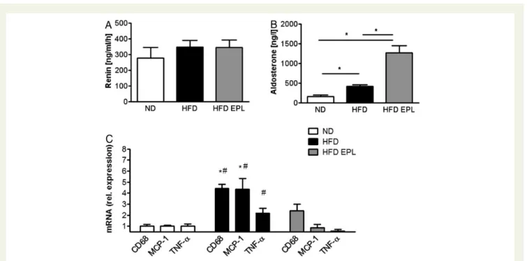
To assess a putative activation of the renin – angiotensin – aldos-terone system, aldosaldos-terone and renin plasma concentration were measured. Aldosterone levels were significantly higher, whereas renin concentration remained unaltered in mice fed an HFD
com-pared with lean mice (Figure1A and B). Compared with HFD-fed
obese mice, EPL treatment of obese mice did not change plasma
renin, whereas it increased plasma aldosterone levels (Figure 1A
and B). Feeding an HFD for 14 weeks resulted in a significant in-crease in body weight and epididymal WAT, it inin-creased fasting plasma glucose, and reduced glucose tolerance compared with mice fed an ND. Treatment with EPL administered with the HFD did not interfere with epididymal WAT and total body weight gain. However, EPL treatment did prevent worsening of glucose metabolism in these mice (Supplementary material online, Table S2).
mRNA levels of pro-inflammatory cytokines such as tumour ne-crosis factor-a (TNF-a) and monocyte chemoattractant protein-1 (MCP-1) tended to be increased (TNF-a) or were increased (MCP-1) in the epididymal WAT of obese mice compared with
lean control mice (Figure 1C). Chronic administration of
eplere-none prevented an increase in mRNA expression levels of these cytokines as well as macrophage-specific glycoprotein CD68 in the epididymal WAT. These data suggest that MR antagonism diminished the obesity-induced pro-inflammatory changes in the epididymal WAT.
Pharmacological mineralocorticoid
receptor antagonism prevents
obesity-induced endothelial dysfunction
To determine the effects of increased endogenous aldosterone levels in obesity on endothelial function, we performed ex vivo aortic ring contraction/relaxation experiments with aortae from C57BL/6 mice kept on ND, HFD, or HFD EPL, respectively(Figure 2). Endothelium-dependent relaxation to acetylcholine
was blunted in aortae of obese mice starting at a concentration
of 3× 1028M of acetylcholine and reached a maximal relaxation
obese mice prevented endothelial dysfunction (Figure 2A). Endothelium-independent relaxation to sodium nitroprusside
(SNP) was similar in all groups (Figure2B). Contractions to KCl
and norepinephrine (NE) were unaffected in mice fed HFD or HFD EPL, respectively (data not shown). These findings show that pharmacological MR antagonism prevents obesity-induced endothelial dysfunction.
Mineralocorticoid receptor antagonism
attenuates pro-inflammatory and
pro-oxidative changes in aortic
endothelial cells of obese mice
Obesity is known to promote a pro-inflammatory, pro-oxidative, and vasoconstricting state in the vascular endothelium, thereby Figure 1 Plasma aldosterone is increased in obesity and further elevated by mineralocorticoid receptor antagonism; obesity-induced expres-sion of pro-inflammatory cytokines in white adipose tissue is attenuated by mineralocorticoid receptor antagonism. C57BL/6 mice on ND, HFD, and HFD EPL were analysed concerning (A) plasma-renin and (B) plasma-aldosterone levels; n ¼ 8 – 10; *P , 0.05. (C ) mRNA levels of
epididy-mal fat pad in C57BL/6 mice on ND, HFD, and HFD EPL, norepididy-malized to ND levels; n ¼ 6 – 8; *P , 0.05 compared with ND levels,#P , 0.05
compared with HFD EPL. ND, normal chow diet; HFD, high-fat diet; HFD EPL, high-fat diet with eplerenone.
Figure 2 Mineralocorticoid receptor antagonism prevents obesity-induced worsening of endothelial function. Endothelial function studies in C57BL/6 mice on ND, HFD, and HFD EPL. Responses of aortic rings to increasing doses of (A) acetylcholine (Ach), mediating endothelial release of endogenous nitric oxide, and (B) sodium nitroprusside (SNP), a nitric oxide donor. See Supplementary material online, Table S4 for EC50 and
Emax values; n ¼ 8 – 10; *P , 0.05 for ND vs. HFD;#P , 0.05 for HFD vs. HFD EPL. ND, normal chow diet; HFD, high-fat diet; HFD EPL,
inducing endothelial dysfunction. To investigate the direct effects of obesity and eplerenone treatment on the genes expressed in aortic endothelial cells, we developed a method to isolate fresh endothe-lial cells from C57BL/6 mice exposed to ND, HFD, or HFD EPL (Supplementary material online, Figure S2A and B). We observed a slight increase in the MR mRNA expression under HFD and an even higher increase upon additional chronic eplerenone treat-ment (Suppletreat-mentary material online, Figure S3A). Mineralocortic-oid receptor mRNA was reduced in endothelial cells of mice in
which MR was deleted (EC MR2/2), indicating successful ablation
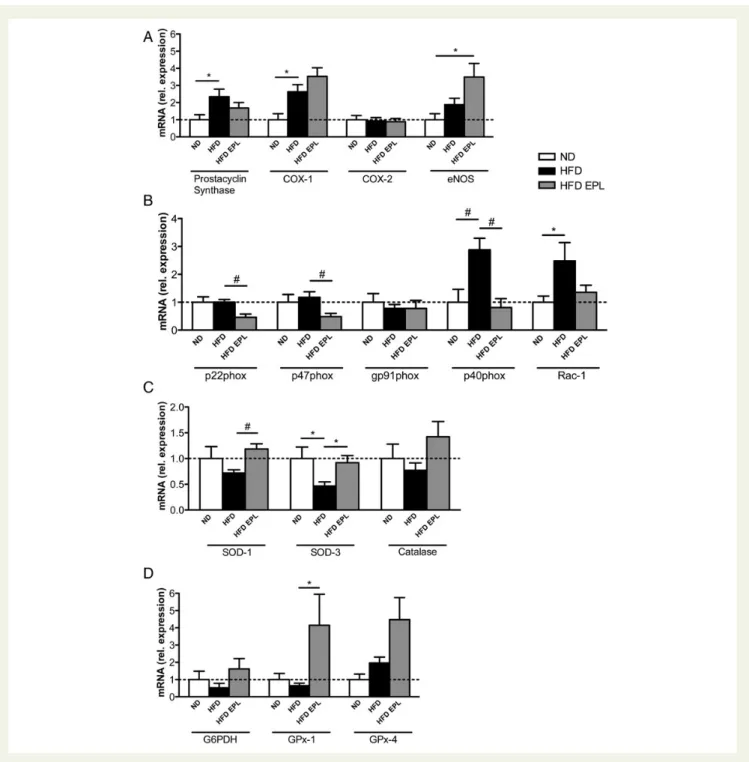
of endothelial MR (Supplementary material online, Figure S3B). Analyses of mediators involved in endothelial dysfunction revealed that prostacyclin synthase and COX-1 expression were
upregu-lated in obesity, whereas COX-2 remained unaltered (Figure3A).
Eplerenone treatment did neither prevent HFD-induced upregula-tion of prostacyclin synthase nor COX-1 expression. Furthermore, endothelial nitric oxide synthase (eNOS) expression increased in
HFD EPL compared with ND mice (Figure3A).
Figure 3 Obesity-induced pro-inflammatory and pro-oxidative changes are modulated by mineralocorticoid receptor antagonism in aortic endothelial cells. Aortic endothelial cell mRNA levels in C57BL/6 mice on ND, HFD, HFD EPL after 14 weeks (A) prostacyclin synthase, COX-1, COX-2, eNOS, (B) p22phox, 47phox, gp91phox, p40phox, Rac-1, (C ) SOD-1, SOD-3, catalase, (D) G6PDH, GPx-1, GPx-4,
Given the association of aldosterone with oxidative stress, we tested mRNA expression of NADPH oxidase subunits in isolated aortic endothelial cells. mRNA levels of the p22phox, p47phox, and gp91phox were not affected by diet-induced obesity. However, additional eplerenone treatment decreased the
expres-sion of p22phox and p47phox (Figure 3B). On the other hand,
obesity increased the expression levels of p40phox and Rac-1;
the increase in p40phox was attenuated by eplerenone (Figure3B).
mRNA expression levels of the antioxidant enzymes SOD-1 and SOD-3 were lower under HFD compared with ND (trend for SOD-1) and HFD EPL conditions; catalase expression remained
un-altered (Figure3C). The mRNA expression of the ROS-scavenging
enzyme GPx-1 was enhanced by eplerenone in obesity, whereas
GPx-4 and G6PDH remained unchanged (Figure3D).
Taken together, we observed the generation of an obesity-induced vasoconstrictive and pro-oxidative mRNA expression profile in aortic endothelial cells that was partially prevented by pharmacological MR antagonism.
Endothelium-specific mineralocorticoid
receptor ablation neither affects
hyperglycaemia nor inflammation in
white adipose tissue of obese or
aldosterone-infused lean mice
To investigate the role of the endothelial MR on obesity-induced
meta-bolic changes, we exposed EC MR2/2mice and their corresponding
MR+/+littermates to an ND or HFD for 14 weeks. After 14 weeks
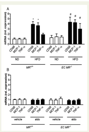
on a HFD, both obese MR+/+mice and EC MR2/2showed a similar
increase in plasma aldosterone levels (Supplementary material online, Figure S4A), weight gain, glucose intolerance (Supplementary material online, Table S3), as well as expression of pro-inflammatory markers MCP-1, and CD68; TNF-a was upregulated only in EC
MR2/2mice (Figure4A).
To test the effects of exogenous aldosterone on the endothelial MR, we implanted osmotic minipumps containing aldosterone or
vehicle in lean MR+/+ and EC MR2/2 mice. Aldosterone infusion
for 2 weeks increased plasma aldosterone to the same extent in
both EC MR2/2and MR+/+mice kept on ND (Supplementary
ma-terial online, Figure S4B) and did not induce the expression of
TNF-a, MCP-1, or CD68 in the epididymal WAT (Figure 4B).
These data indicate that endothelial MR deletion neither affects glucose tolerance nor the pro-inflammatory state of the WAT of obese or aldosterone-infused lean mice.
Deletion of the endothelial
mineralocorticoid receptor prevents
obesity- or exogenous aldosterone-induced
endothelial dysfunction to a similar extent
as does COX inhibition
To determine whether obesity-induced endothelial dysfunction
was mediated by the endothelial MR, we exposed EC MR2/2
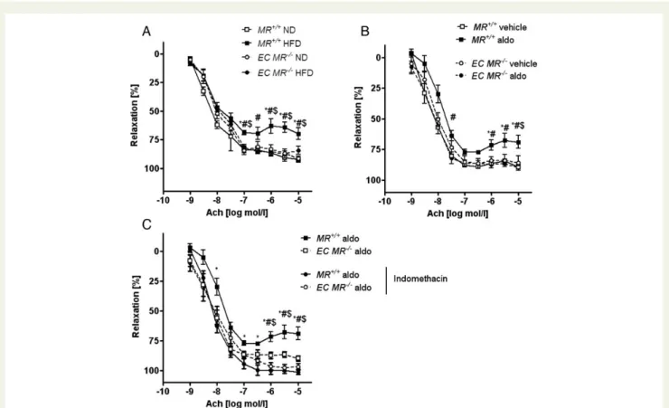
and MR+/+ mice to ND or HFD for 14 weeks. Compared with
lean controls, obese MR+/+ mice demonstrated a significant
im-pairment of endothelium-dependent vasodilation, whereas MR de-letion in endothelial cells prevented endothelial dysfunction
(Figure5A). Of note, aortic walls of obese MR+/+ mice contained
no macrophages (Supplementary material online, Figure S5). Infu-sion of aldosterone for 2 weeks using minipumps blunted endothe-lial function in lean mice. Genetic deletion of the endotheendothe-lial MR
prevented endothelial dysfunction (Figure5B). Thus, the
endothe-lial MR mediates both obesity-induced endogenous as well as ex-ogenous aldosterone-induced endothelial dysfunction.
COX inhibition using indomethacin normalized endothelial func-tion in aldosterone-infused lean mice to the same extent as
endo-thelial MR deletion (Figure 5C). Therefore, aldosterone induces
endothelial dysfunction by activating COX-dependent pathways.
Aldosterone-infused endothelium-specific
MR
2/2lean mice exhibit pro-inflammatory
changes in aortic endothelial cells
Next we evaluated the effects of aldosterone infusion or MR abla-tion on mediators of inflammaabla-tion, vasoconstricabla-tion, and oxidative
Figure 4 The obesity-induced increase in the expression of pro-inflammatory cytokines in white adipose tissue is independ-ent of endothelial mineralocorticoid receptor; aldosterone infu-sion in lean animals does not induce inflammation. mRNA
levels of the epididymal fat pad in wild-type (MR+/+) and
endothelial-specific MR knockout (EC MR2/2) mice (A) on ND
or HFD and (B) vehicle- or aldosterone-infused, standardized to S12 and normalized to ND levels; n ¼ 6 – 8; *P , 0.05 for
MR+/+ ND vs. MR+/+ HFD; #P , 0.05 for EC MR2/2 ND vs.
stress in aortic endothelial cells. For this purpose, we isolated fresh
aortic endothelial cells from lean MR+/+ and EC MR2/2 mice
infused with aldosterone for 2 weeks and determined mRNA ex-pression by qPCR. Our analyses showed that prostacyclin synthase expression was enhanced by exogenous aldosterone, independent
of the presence of endothelial MR (Figure 6A); in contrast,
aldosterone-induced COX-1 expression was decreased in EC
MR2/2 mice, whereas COX-2 and eNOS expression remained
unchanged.
Analyses of enzymes enhancing oxidative stress revealed that mRNA expression of the NADPH oxidase subunit p22phox was upregulated by aldosterone, whereas this effect was abolished in
EC MR2/2 mice; gp91phox remained unaltered in all conditions
(Figure 6B). Expression levels of the NADPH oxidase subunits
p47phox, p40phox, and Rac-1 were below the detection limit (data not shown). Regarding antioxidant enzymes, expression of SOD-1 mRNA was decreased by aldosterone, but did not reach statistical significance (P ¼ 0.077). SOD-3 mRNA was decreased
upon aldosterone infusion and remained low in EC MR2/2 mice,
both in the presence or absence of aldosterone (Figure 6C).
Catalase could not be detected (data not shown). ROS-scavenging enzymes GPx-1, GPx-4, and G6PDH remained unaltered in all
conditions (Figure6D). Thus, aldosterone enhances the expression
of mediators involved in the generation of ROS and prostanoids in an endothelial MR-dependent manner.
Discussion
Principle findings
This experimental study provides three main novel findings
(Figure7): (i) diet-induced obesity generates endothelial
dysfunc-tion by enhancing expression of NADPH oxidase and through ac-tivation of COX-1-dependent pathways in freshly isolated aortic endothelial cells; (ii) genetic deletion of the endothelial MR pre-vents obesity- as well as aldosterone-induced endothelial dysfunc-tion without affecting glucose tolerance or pro-inflammatory pathways in the WAT; (iii) activation of the endothelial MR is suf-ficient to induce endothelial dysfunction, in part via increased endothelial p22phox expression.
Figure 5 Loss of endothelial MR prevents obesity-induced endothelial dysfunction and COX inhibition restores aldosterone-induced endo-thelial dysfunction. The vascular response of aortic rings pre-constricted with norepinephrine to increasing doses of the
endothelium-dependent vasodilator acetylcholine (Ach) was measured in wild-type (MR+/+) and endothelial-specific MR knockout (EC MR2/2) mice;
n ¼ 6 – 8. (A) MR+/+and EC MR2/2 mice on ND and HFD; *P , 0.05 for MR+/+ ND vs. MR+/+ HFD;#P , 0.05 for MR+/+HFD vs. EC
MR2/2 HFD; $P , 0.05 for MR+/+ HFD vs. EC MR2/2
ND. (B) MR+/+ and EC MR2/2 mice after aldosterone or vehicle infusion;
*P , 0.05 for MR+/+ vehicle vs. MR+/+ aldo; #P , 0.05 for MR+/+ aldo vs. EC MR2/2 aldo; $P , 0.05 for MR+/+ aldo vs. EC MR2/2
vehicle. (C ) MR+/+on ND and HFD or after aldosterone or vehicle infusion; rings were pre-treated for 30 min with indomethacin. See
Sup-plementary material online, Table S4 for EC50 and Emax values. *P , 0.05 for MR+/+aldo vs. MR+/+aldo (indomethacin-treated);#P , 0.05 for
MR+/+aldo vs. EC MR2/2aldo;$P , 0.05 for MR+/+aldo vs. EC MR2/2
Added value of this study
Our study provides added value in the following contexts: In mice fed an HFD, we observed an increase in plasma aldoster-one. This effect was more pronounced if mice were pre-treated with eplerenone; renin levels remained unaltered. This is in line with publications showing a positive correlation between obesity and plasma aldosterone levels independent of plasma renin
activ-ity.25,26Since adipocytes induce the release of aldosterone by
adre-nocortical cells,27we propose a renin-independent mechanism in
obesity that stimulates aldosterone secretion. We observed a further increase in plasma aldosterone levels upon eplerenone
therapy corroborating previous findings.28,29This phenomenon is
likely related to a displacement of the mineralocorticoid from its receptor and/or its enhanced production during MR blockade due to a positive physiological feedback loop. We show for the first time that deletion of endothelial MR does not increase plasma aldosterone levels. Thus, we propose that the endothelial MR is not involved in the regulatory feedback loop that induces eplerenone-induced aldosterone production.
Our data reveal that pharmacological MR blockade in diet-induced obesity improved glucose tolerance. In line with our observation, MR antagonism has been reported to reduce insulin
resistance in genetically obese mice.23 Obesity-associated
pro-inflammatory changes in WAT can contribute to the Figure 6 Aldosterone-induced expression of COX-1 and reactive oxygen species-generating enzymes in endothelial aortic cells depend on
endothelial mineralocorticoid receptor. Aortic endothelial cell mRNA levels in wild-type (MR+/+) and endothelial-specific MR knockout
(EC MR2/2) mice after aldosterone or vehicle infusion (A) prostacyclin synthase, COX-1, COX-2, eNOS, (B) p22phox, gp91phox, (C )
development of insulin resistance.30 We and others22 demon-strated that eplerenone decreases mRNA of pro-inflammatory cytokines in WAT. Moreover, we extend these findings by showing for the first time that endothelial MR ablation neither pre-vents impaired glucose tolerance nor increases the expression of pro-inflammatory mediators in the obese WAT. Thus, endothelial MR signalling is neither involved in these obesity-induced diabetic nor pro-inflammatory changes.
Genetically obese mice develop endothelial dysfunction.31In the
current study, pharmacological MR antagonism prevented endo-thelial dysfunction in diet-induced obesity, suggesting that activa-tion of MR with a concomitant release of inflammatory molecules impaired endothelium-dependent relaxation. Notably, endothelium-specific MR deletion completely prevented both diet- and aldosterone-induced endothelial dysfunction. These novel findings demonstrate that activation of the endothelial MR is sufficient to mediate endothelial dysfunction in diet-induced obesity.
Aldosterone increases expression of COX-2, prostacyclin
pro-duction,32 and vascular inflammation.33 In the context of
diet-induced obesity with increased endogenous aldosterone levels, we observed that COX-1 was upregulated in freshly isolated aortic endothelial cells. Mineralocorticoid receptor antagonism did not attenuate this increased COX-1 expression. Our endothelial function studies revealed that pre-treatment of aortic rings with
the COX inhibitor indomethacin completely normalized
endothelium-dependent relaxations in response to acetylcholine
in obese mice. Similar effects were observed in spontaneously
hypertensive rats.11Our results corroborate another study
report-ing that obesity induces endothelial dysfunction in a
COX-1-dependent manner.34 Analyses of the prostacyclin synthase
revealed that both diet-induced obesity as well as exogenous al-dosterone administration enhanced expression of prostacyclin syn-thase in aortic endothelial cells. In line with our observation, it has been shown that prostacyclin increased endothelial dysfunction in
hypertensive rats.32Furthermore, inhibition of COX that produces
prostacyclin synthase substrates, restores aldosterone-induced
endothelial dysfunction.35Of note, neither pharmacological MR
an-tagonism in diet-induced obesity, nor genetic endothelial-specific MR deletion during aldosterone infusion attenuated prostacyclin synthase expression. These findings imply that increased endogen-ous aldosterone in obesity induces vasoconstricting prostanoids in-dependent of the endothelial MR. It is likely that aldosterone acts
in this context by non-genomic effects.36A more detailed
mechan-istic insight would be of interest. However, we consider the corre-sponding analyses beyond the scope of the current study.
We demonstrate that diet-induced obesity was associated with an increased expression of the pro-oxidant NADPH subunits (p40phox and rac-1) as well as a reduced expression of the anti-oxidant proteins SOD-1 and -3. Thus, increased ROS production may contribute to endothelial dysfunction. Indeed, the endothelial
MR mediates superoxide generation via Rac-1 activation.17This is
in line with our observation that MR antagonism in obesity con-ferred beneficial effects on the expression of the NADPH Figure 7 Mechanisms of aldosterone-induced endothelial dysfunction in obesity. The expression of NADPH oxidase subunit p22phox can be blocked by both eplerenone and endothelial mineralocorticoid receptor ablation. Therefore, p22phox seems to be the crucial mediator in in-ducing aldosterone-induced endothelial dysfunction in aortic endothelial cells of obese mice through genomic (solid arrow) or non-genomic (dashed arrow) effects.
oxidase subunits (p22phox, p47phox, p40phox, and rac-1), anti-oxidant enzymes SOD-1 and -3, and the ROS scavenging enzyme GPx-1. Exogenous aldosterone administration modulated exclusively the NADPH oxidase subunit p22phox mRNA expres-sion in aortic endothelial cells in an MR-dependent manner. Since endothelial MR ablation is sufficient to abolish both obesity- and exogenous aldosterone-induced endothelial dysfunction, we pos-tulate that the endothelial MR-mediated p22phox expression is sufficient to increase a pro-oxidative pathway leading to endothe-lial dysfunction.
Overexpression of endothelial MR in mice fed a normal chow induces an enhanced vasoconstrictive response in resistance arter-ies with a mild hypertension. However, these vessels do not exhibit signs of endothelial dysfunction nor increased oxidative
stress.37 Differences in experimental settings (our study:
diet-induced obesity, high cholesterol diet, and constitutive EC MR expression) are likely to account for the diverse findings.
Potential limitations
We provide evidence of increased endothelial MR expression in obese compared with lean mice and of a lack of MR expression
in endothelial cells of EC MR2/2 mice—all at the DNA and
RNA level. Despite extensive efforts, we did not succeed in visu-alizing MR expression at the protein level using various lots of
pre-viously described antibodies.38 Since the knockout procedure
consistently deletes the gene, we consider that our data provide solid evidence for MR deletion in endothelial cells. As the Tie2 promotor used to drive MR deletion in endothelial cells is also active in myeloid cells, we cannot conclude that endothelial
pro-tection seen in EC MR2/2 mice is due exclusively to endothelial
MR ablation.39Indeed, we observed MR ablation in macrophages
of EC MR2/2 mice. Moreover, MR ablation in macrophages
reduces MR-mediated cardiac pro-inflammatory responses in a
mouse model of cardiac fibrosis.40Since we found no macrophages
in the aortae of obese mice with intact MR and we observed similar pro-inflammatory expression patterns in the WAT of
obese EC MR2/2 and MR+/+ mice, we propose that Tie2-driven
ablation of MR in adipose tissue macrophages is unlikely to
contrib-ute to endothelial protection in diet-induced obesity in EC MR2/2
mice.
Blood pressure in the different animal groups was not assessed, although a minor increase in blood pressure of obese animals and a decrease in mice lacking the endothelial MR are possible. Indeed, increased blood pressure has been observed upon exogenous
al-dosterone administration19and in mice with endothelium-specific
MR overexpression.37 Thus, changes in blood pressure could
have taken place and may have modified aortic endothelial gene expression and participated in the observed alterations of endo-thelial function.
Conclusions and possible
implications
We demonstrate that both endogenous aldosterone in obesity as well as exogenous aldosterone cause endothelial dysfunction by
inducing expression of ROS-generating enzymes in an endothelial MR-dependent fashion and by activating the COX-1 pathway inde-pendent of the endothelial MR. Pharmacological MR blockade abolishes obesity-induced glucose intolerance, adipose tissue in-flammation, and endothelial dysfunction. Genetic ablation of the MR in endothelial cells identifies the endothelial MR as a crucial
mediator of obesity-induced endothelial dysfunction (Figure7). In
addition, changes in blood pressure secondary to endothelial MR ablation and/or the deletion of the MR in macrophages may have contributed to the observed prevention of obesity-induced effects by Tie2-driven MR deletion.
Our experimental findings have the following potential clinical implications: MR-antagonizing drugs appear worth testing for endothelial function in the rapidly growing population of obese patients that are particularly prone to atherosclerosis, hyperten-sion, and insulin resistance.
Supplementary material
Supplementary material is available at European Heart Journal online.
Acknowledgements
We thank the members of the cardiovascular research and epithe-lial transport laboratories for their help. Eplerenone was provided by Pfizer (Groton, CT, USA).
Funding
This work was supported by grants from the Swiss National Science Foundation 310030-130626/1 (C.M.M.), and 3100-068118 (T.F.L.) the University Research Priority Program Integrative Human Physiology at the University of Zurich (N.S., F.R, T.F.L., F.V, and C.M.M.). Further support was provided by an unrestricted grant from Pfizer (NYC, USA).
Conflict of interest: C.M.M. received grant from ZIHP, University of Zurich; MSD; AstraZeneca; EliLilly; Sirtris; Bayer; Swiss National Science Foundation; MSD; AstraZeneca; Roche; has expert testimony to MSD; has patent rights to Mabimmune, CH.
References
1. James PT, Rigby N, Leach R. The obesity epidemic, metabolic syndrome and future prevention strategies. Eur J Cardiovasc Prev Rehab 2004;355:763 – 778. 2. Spiegelman BM, Flier JS. Obesity and the regulation of energy balance. Cell 2001;
104:531 – 543.
3. Ahima RS. Adipose tissue as an endocrin organ. Obesity 2006;14:242S – 249S. 4. Wellen KE, Hotamisligil GS. Inflammation, stress, and diabetes. J Clin Invest 2005;
115:1111 – 1119.
5. Erhardt-Bornstein M, Lamounier-Zepter V, Schraven A, Langenbach J, Willenberg HS. Human adipocytes secrete mineralocorticoid-releasing factors. Proc Natl Acad Sci USA 2003;100:14211 – 14216.
6. Goodfriend TL, Egan BM, Kelley DE. Aldosterone in obesity. Endocr Res 1998;24: 789 – 796.
7. Pitt B, Remme W, Zannad F. Eplerenone, a selective aldosterone blocker, in patients with left ventricular dysfunction after myocardial infarction. N Engl J Med 2003;348:1309 – 1321.
8. Pitt B, Zannad F, Remme WJ. The effect of spironolactone on morbidity and mor-tality in patients with severe heart failure. N Engl J Med 1999;341:709 – 717. 9. Garnier A, Bendall JK, Fuchs S, Escoubet B, Rochais F, Hoerter J, Nehme J,
Ambroisine ML, Angelis ND, Morineau G, d‘Estienne P, Fischmeister R, Heymes C, Pinet F, Declayre C. Cardiac specific increase in aldosterone produc-tion induces coronary dysfuncproduc-tion in aldosterone synthase-transgenic mice. Circu-lation 2004;110:1819 – 1825.
10. Tang EH, Ku D, Tipoe GL, Feletou M, Man RY, Vanhoutte PM. Endothelium-dependent contractions occur in the aorta of wild-type and COX2-/- knockout but not COX1-/- knockout mice. J Cardiovasc Pharmacol 2005;46:761 – 765. 11. Luscher TF, Vanhoutte PM. Endothelium-dependent contractions to acetylcholine
in the aorta of the spontaneously hypertensive rat. Hypertension 1986;344: 344 – 348.
12. Leopold JA, Cap A, Scribner AW, Stanton RC, Loscalzo L. Glucose-6-phosphate dehydrogenase deficiency promotes endothelial oxidative stress and decreases endothelial nitric oxide bioviability. FASEB J 2001;15:1771 – 1773.
13. Hoen PAC, Lans CACVd, Eck MV, Bijsterbosch MK, Berkel TJCV, Twisk J. Aorta of ApoE-deficient mice responds to atherogenic stimuli by a prelesional increase and susequent decrease in the expression of antioxidant enzymes. Circ Res 2003; 93:262 – 269.
14. Sorescu D, Weiss D, Lassegue B, Clempus RE, Szo¨cs K, Sorescu GP, Valppu L, Quinn MT, Lambeth JD, Vega JD, Taylor R, Griendling KK. Superoxide production and expression of Nox family proteins in human atherosclerosis. Circulation 2002; 105:1429 – 1435.
15. Suzuki J, Iwai M, Mogi M, Oshita A, Yoshii T, Higaki J, Horiuchi M. Eplerenone with valsartan effectively reduces atherosclerotic lesion by attenuation of oxidative stress and inflammation. Arterioscler Thromb Vasc Biol 2006;26:917 – 921. 16. Sartorio CL, Fraccarollo D, Galuppo P, Leutke M, Ertl G, Stefanon I, Bauersachs J.
Mineralocorticoid receptor blockade improves vasomotor dysfunction and vascu-lar oxidative stress early after myocardial infarction. Hypertension 2007;50: 919 – 925.
17. Iwashima F, Yoshimoto T, Minami I, Sakurada M, Hirono Y, Hirata Y. Aldosterone induces superoxide generation via rac1 activation in endothelial cells. Endocrin-ology 2008;149:1009 – 1014.
18. Blanco-Rivero J, Cachofeiro V, Lahera V, Aras-Lopez R, Ma´rquez-Rodas I, Salaices M, Xavier FE, Ferrer M, Balfago´n G. Participation of prostacyclin in endo-thelial dysfunction induced by aldosterone in normotensive and hypertensive rats. Hypertension 2005;46:107 – 112.
19. Leopold JA, Dam A, Maron BA, Scribner AW, Liao R, Handy DE, Stanton RC, Pitt B, Loscalzo J. Aldosterone impairs vascular reactivity by decreasing glucose-6-phosphate dehydrogenase activity. Nat Med 2007;13:189 – 197.
20. Caprio M, Newfell B, Sala Al, Baur W, Fabbri A, Rosano G, Mendelsohn ME, Jaffe IZ. Functional mineralocorticoid receptor in human vascular endothelial cells regulate intercellular adhesion molecule-1 expression and promote leuko-cyte adhesion. Circ Res 2008;102:1359 – 1367.
21. Caprio M, Feve B, Claes A, Viengchareun S, Lombes M. Pivotal role of the min-eralocorticoid receptor in corticoid-induced adipogenesis. FASEB J 2007;21:1 – 10. 22. Guo C, Ricchiuti V, Lian BQ, Yao TM, Couthino P, Romero JR, Li J, Wiliams GH, Adler G. Mineralocorticoid receptor blockade reserves obesity-related changes in expression of adiponectin, peroxisome proliferator-activated receptor-gamma, and pro-inflammatory adipocytokines. Circulation 2008;117:2253 – 2261. 23. Hirata A, Maeda N, Hiuge A, Hibuse T, Fujita K, Okada T, Kihara S, Funahashi T,
Shimomura I. Blockade of mineralocorticoid receptor reverses adipocyte dysfunc-tion and insulin resistance in obese mice. Cardiovasc Res 2009;84:164 – 172. 24. Yamaji M, Tsutamoto T, Kawahara C, Nishiyama K, Yamamoto T, Fujii M, Horie M.
Effect of eplerenone versus spironolactone on cortisol and hemoglobin A1c levels in patients with chronic heart failure. Am Heart J 2012;160:915 – 921.
25. Goodfriend TL, Kelley DE, Goodpaster BH, Winters SJ. Visceral obesity and insulin resistance are associated with plasma aldosterone levels in women. Obes Res 1999;7:355 – 361.
26. Rocchini AP, Key J, Bondie D, Chico R, Moorehead C, Katch V, Martin M. The effect of weight loss on the sensitivity of blood pressure to sodium in obese ado-lescents. N Engl J Med 1989;321:580 – 585.
27. Goodfriend TL, Ball DL, Egan BM, Campbell WB, Nithipatikom K. Epoxy-keto de-rivative of linoleic acid stimulates aldosterone secretion. Hypertension 2004;43: 358 – 363.
28. Hlavacova N, Jezova D. Effects of single treatment with the antihypertensive drug eplerenone on hormone levels and anxiety-like behaviour in rats. Endocr Regul 2008;42:147 – 153.
29. Krum H, Nolly H, Workman D, He W, Roniker B, Krause S, Fakouhi K. Efficacy of eplerenone added to renin-angiotensin blockade in hypertensive patients. Hyper-tension 2002;40:117 – 123.
30. Kanda H, Tateya S, Tamori Y, Kotani K, Hiasa K, Kitazawa R, Kitazawa S, Miyachi H, Maeda S, Egashira K, Kasuga M. MCP-1 contributes to macrophage in-filtration into adipose tissue, insulin resistance, and hepatic steatosis in obesity. J Clin Invest 2006;116:1494 – 1505.
31. Avogaro A, de Kreutzenberg SV. Mechanisms of endothelial dysfunction in obesity. Clin Chim Acta 2005;360:9 – 26.
32. Xavier FE, Aras-Lopez R, Arroyo-Villa I, Campo LD, Salaices M, Rossoni LV, Ferrer M, Balfagon G. Aldosterone induces endothelial dysfunction in resistance arteries from normotensive and hypertensive rats by increasing thromboxane A2 and prostacyclin. Br J Pharmacol 2008;154:1225 – 1235.
33. Rocha R, Rudolph AE, Frierdich GE, Nachowiak DA, Kekec BK, Blomme EA, McMahon EG, Delyani JA. Aldosterone induces a vascular inflammatory pheno-type in the rat heart. Am J Physiol Heart Circ Physiol 2002;283:H1802 – H1810. 34. Traupe T, Lang M, Goettsch W, Mu¨nter K, Morawietz H, Vetter W, Barton M.
Obesity increases prostanoid-mediated vasoconstriction and vascular throm-boxan receptor gene expression. J Hypertens 2002;20:2239 – 2245.
35. Fe´le´tou M, Huang Y, Vanhoutte P. Vasoconstrictor prostanoids. Pflu¨gers Archiv 2010;459:941 – 950.
36. Gros R, Ding Q, Sklar LA, Prossnitz EE, Arterburn JB, Chorazyczewski J, Feldman RD. GPR30 expression is required for the mineralocorticoid receptor-independent rapid vascular effects of aldosterone. Hypertension 2011;57:442 – 451. 37. Nguyen Dinh Cat A, Griol-Charhbili V, Loufrani L, Labat C, Benjamin L, Farman N, Lacolley P, Henrion D, Jaisser F. The endothelial mineralocorticoid re-ceptor regulates vasoconstrictor tone and blood pressure. FASEB J 2010;24: 2454 – 2463.
38. Gomez-Sanchez CE, de Rodriguez AF, Romero DG, Estess J, Warden MP, Gomez-Sanchez MT, Gomez-Sanchez EP. Development of a panel of monoclonal antibodies against the mineralocorticoid receptor. Endocrinology 2006;147: 1343 – 1348.
39. Alva JA, Zovein AC, Monvoisin A, Murphy T, Salazar A, Harvey NL, Carmeliet P, Iruela-Aispe ML. VE-Cadherin-Cre-recombinase transgenic mouse: A tool for lineage analysis and gene deletion in endothelial cells. Dev Dynam 2006;235: 759 – 767.
40. Bienvenu LA, Morgan J, Rickard AJ, Tesch GH, Cranston GA, Fletcher EK, Delbridge LM, Young MJ. Macrophage mineralocorticoid receptor signaling plays a key role in aldosterone-independent cardiac fibrosis. Endocrinology 2012; 153:3416 – 3425.