Trypanosoma brucei brucei: differences in the nuclear
chromatin of bloodstream forms and procyclic culture
forms
W. SCHLIMME, M. BURRI, K. BENDER, B. BETSCHART and H. HECKER*
Swiss Tropical Institute, Postfach CH-4402, Basel, Switzerland (Received 9 December 1992; revised 1 March 1993; accepted 1 March 1993)
SUMMARY
Nucleosome filaments of two stages of the life-cycle of Trypanosoma brucei brucei, namely bloodstream forms and procyclic culture forms, were investigated by electron microscopy. Chromatin of bloodstream forms showed a salt-dependent condensation. The level of condensation was higher than that shown by chromatin from procyclic culture forms, but 30 nm fibres as formed in rat liver chromatin preparations were not found. Analysis of histones provided new evidence for the existence of HI-like proteins, which comigrated in the region of the core histones in SDS-PAGE and in front of the core histones in Triton acid urea gels. Differences were found between the HI-like proteins of the two trypanosome stages as well as between the core histones in their amount, number of bands and banding pattern. It can be concluded that T. b. brucei contains a full set of histones, including HI-like proteins, and that the poor condensation of its chromatin is not due to the absence of H1, but most probably due to histone—DNA interaction being weak. It is obvious that structural and functional differences of the chromatin exist not only between T. b. brucei and higher eukaryotes, but also between various stages of the life-cycle of the parasite. It is therefore not adequate to investigate the chromatin only of the procyclic culture forms as a model for all stages of the life-cycle of T. b. brucei.
Key words: Trypanosoma brucei brucei, chromatin, histones, lower eukaryotes, bloodstream forms, procyclic culture forms.
INTRODUCTION
Trypanosomes, the protozoan parasites of man and animals, have various nuclear features different from those of higher eukaryotes. No condensed chromo-somes can be visualized during nuclear division in trypanosomes (Vickerman & Preston, 1970), and the compaction of the chromatin in the nucleus is distinctly less pronounced as compared to the chromatin of rat liver nuclei (Hecker & Gander, 1985).
The chromatin of T.b. brucei and of T. cruzi procyclic culture forms was shown to be organized in a nucleosome filament-like form. However, the nucleosomes were spaced irregularly and no con-densation into a typical 30 nm fibre took place under experimental conditions. The nuclear chromatin was digested rapidly by micrococcal nuclease, and the interactions between DNA and proteins were rela-tively weak and were easily destabilized under experimental conditions, which are normally used for the isolation of chromatin from higher eukaryotes (Hecker & Gander, 1985; Hecker et al. 1989; Bender, Betschart & Hecker, 1992c). In addition, no histone HI could be demonstrated (Hecker & Gander, 1985 ; Hecker et al. 1989). Other workers also postulated that HI was absent (Bender et al. 1992 a, b, c, d). However, these results are controversial. Recent * Reprint requests to Professor H. Hecker.
studies demonstrated HI-like proteins in T. cruzi and Crithidia fasciculata (Toro & Galanti, 1988, 1990; Duschak & Cazzulo, 1990). These histones showed biochemical properties similar to those of histones from Tetrahymena fasciculata (Johmann & Gorovsky, 1976).
On the basis of the migration pattern in various gel systems and amino acid sequence analysis, Bender et
al. (1992 a, b) were able to show that the four core
histones of T. b. brucei procyclic culture forms, namely a, b, c, d are the counterparts of H3, H2B, H2A and H4 of higher eukaryotes. The core histones of higher eukaryotes are among the most conserved proteins known. Among the histones, H4 and H3 are the best conserved, followed by H2A, H2B and H I (van Holde, 1989). Biochemical differences between the core histones of T. b. brucei procyclic culture forms and the core histones of higher eukaryotes were found by Bender et al. (1992a, b). Differences of about 35 % exist in the amino acid sequence of the N- and C-terminal regions of the histone H4 of calf thymus and trypanosomes (Bender et al. 19926; Toro et al. 1992).
All previous investigations have been carried out with T. b. brucei procyclic culture forms which can be cultivated to a density of 3 x 10⁷/ml (Brun & Schdnenberger, 1979). T h e in vitro cultivation of bloodstream forms, the parasitic stage affecting mammals, is possible only to a density of 2 x 10⁶/ml (Hamm et al. 1990). Therefore studies on the
chromatin of this stage depend on its propagation in laboratory animals. It was not known, whether the differences found between the chromatin of procyclic culture forms and that of higher eukaryotes would also be found in bloodstream forms.
In the present study, the chromatin of procyclic culture forms and bloodstream forms of T. b. brucei was compared with rat liver chromatin, using a variety of new separation techniques and staining methods. Also electron microscopy (EM) and enzymic digestion were used to find out whether there were differences between the chromatin of the two forms, and between that of bloodstream forms and higher eukaryotes. The validity of taking the chromatin of procyclic culture forms as a model for all the stages of the life-cycle of T. b. brucei was evaluated.
MATERIALS AND METHODS
Livers were from rats from the SIV IVANOVAS strain. Trypanosoma b. brucei STIB 345 AB procyclic culture forms, cultivated in S D M 79 medium containing 1 0 % heat-inactivated foetal bovine serum, 10 /ig/ml haemin and 10 /jg/ml gentamicin at 27 °C were used (Brun & Schonenberger, 1979). Bloodstream forms of STIB 345 AB were grown in SIV rats. Rats were inoculated intraperitoneally with 106 trypanosomes, and killed on day 5.
Investigation of the influence of the temperature on the chromatin of procyclic culture forms
To investigate the effect of temperature, procyclic culture forms were cultivated as described above, but incubated for 1 week at 32 CC. After that period they were incubated at the following temperatures: 34, 35, 35-5, 36, 37 °C for various periods of time.
Purification of nuclei
Approximately 10 g liver from rats was homogenized in the presence of 1 mM phenylmethylsulphonyl-fluoride (PMSF) and the nuclei were purified by centrifugation through sucrose cushions (Thoma, Roller & Klug, 1979; Hecker & Gander, 1985). T. b.
brucei STIB 345 AB were harvested, and 2-3 x 10¹⁰
exponentially growing procyclic cells resuspended in hypotonic buffer containing 0-5 M 2-methyl-2,4-pentandiol (hexylene glycol), 2-5 mM CaCl2, 1 mM P M S F . They were lysed by nitrogen cavitation at 25 bar (Shapiro & Doxsey, 1982), vortexed for 30 s and the isolated nuclei were washed in 90 mM suspension buffer, pH 7-4, containing 0 1 mM disodium ethylen-diaminetetraacetic acid (Na2EDTA) (Thoma et al. 1979; Hecker & Gander, 1985).
Rats were anaesthetized with methoxyfluorane. Bloodstream forms were collected by cardiac punc-ture and separated from the blood by a DEAE-52
cellulose column (Lanham & Godfrey, 1970). Trypanosomes were isolated by centrifugation at 1800^ for 20 min. Isolation of the nuclei was the same from this step on as for procyclic culture forms.
Preparation of soluble chromatin
Nuclei of T. b. brucei were digested with 0-2 units of micrococcal nuclease (Sigma, N-3755) per 20 A260 at 30 °C for 50 s. Nuclei of rat liver were digested with 0-4 units of micrococcal nuclease per 20 A260 at 37 °C for 50 s. T h e nuclei were centrifuged, and the chromatin solubilized by nuclear lysis in a hypotonic buffer containing 1 mM triethanolamine
hydro-chloride (TEAC1) and 0-2 mM Na2EDTA, pH 7-4.
Insoluble material was removed by centrifugation (Thoma et al. 1979; Hecker & Gander, 1985). The preparation of soluble chromatin was identical for procyclic culture forms and for bloodstream forms.
Gradient analysis of the chromatin digest
Gradient analysis was done in 5-5-285 % (w/v) 17 ml isokinetic sucrose gradients containing 5 mM
TEAC1, pH 7-4, 0-2 mM Na2EDTA and 10 mM NaCl
(Noll, 1969). Centrifugation was performed for 14 h at 25 000 g in a Kontron T S T 28/17 swing-out rotor. The gradients were monitored at 254 nm and the bottom fractions containing the larger fragments of soluble chromatin were used for analysis by electron microscopy (Thoma & Roller, 1981).
Electron microscopy
The fractions with the large chromatin fragments were divided into 4 aliquots and dialysed against
5 mM TEAC1 (pH 7-4), 0-2 mM Na2EDTA
con-taining 0, 10, 40 or 100 mM NaCl respectively for 4 h. Glutaraldehyde was then added to the dialysis buffer at a concentration of 0'2 % (v/v) for trypano-some chromatin or 0-1 % for rat liver chromatin, and samples were fixed for at least 15 h and prepared for EM observation (Thoma et al. 1979). The con-centration of 0-2 % glutaraldehyde for trypanosomes was chosen after an experimental investigation of the effect of different levels of glutaraldehyde.
Protein extraction from purified nuclei
Nuclei were resuspended in 0-25 M HC1 (Elpidina, Zaitseva & Rrasheninnikov, 1979) or in 5 % (v/v) perchloric acid (PCA) or 350 mM NaCl in 10 mM TEAC1, pH 7-4, (Sanders, 1977) and proteins ex-tracted for 60 min under constant agitation. In-soluble material was pelleted at 4000 £ for 5 min. The supernatant fractions, containing the histones or non-histones, were removed and dialysed against
1 mM TEAC1, 0-2 mM Na2EDTA, pH 7-4, and
All the preparations were carried out at 0-4 °C if not otherwise stated.
Triton acid urea poly aery lamide gel electrophoresis
Lyophilized proteins were dissolved in sample buffer containing 2-5 M urea, 3 % (v/v) 2-mercaptoethanol, 001 % (v/v) pyronine G, 0'9 M acetic acid and incubated at 100 CC for 5 min. T h e stacking gel contained 1 2 % , the resolution gel 1 5 % poly-acrylamide and both 2'5 M urea, 0-9 M acetic acid and 0-38% (v/v) Triton D F 16. After a first pre-run (25 mA, up to constant voltage), a second pre-run was performed with 80 fi\ of 1 M cysteamine in 0-9 M acetic acid per lane at 25 mA for 1 h, to scavenge free radicals and to prevent oxidation of methionine residues. Then the probes were loaded and separated (Alfageme et at. 1974).
SDS-tricine-PAGE
Samples of proteins from soluble chromatin were either lyophilized or precipitated in 25 % (v/v) trichloroacetic acid (TCA), pelleted at 15000^, washed with 100% acetone and vacuum-dried. Samples were solubilized in sample buffer containing 4 % SDS, 12% (w/v) glycerol, 50 mM Tris, 2 % (v/v) 2-mercaptoethanol, 0-01 % (w/v) Servablue G, pH 68, boiled and separated in a 176% S D S -tricine-polyacrylamide resolution gel, with a 5 % stacking gel (Schagger & von Jagow, 1987).
Staining and destaining procedures
Coomassie brilliant blue. Gels were stained with
0-25% (w/v) Coomassie brilliant blue R-250 in methanol:water:glacial acetic acid (5:5:1) for l h and destained with methanol: water: glacial acetic acid (4:5:1) for l h and (10:83:7) overnight, respectively.
Differential destaining with FeCl3 of
Coomassie-stained histones. Differential destaining with 0-1 M
FeCl3 in 20 % (v/v) ethanol was performed for 6 h (Spiker, Key & Wakim, 1976; Duschak & Cazzulo,
1990).
Bromophenol blue. Gels, fixed in methanol: water:
acetic acid as described above were stained in 0-01 % (w/v) bromophenol blue in water (pH 3'0) for 24 h at room temperature, and differentially destained in 40 % (v/v) w-propanol for 24 h at 55 °C (Duschak & Cazzulo, 1990).
Micrococcal nuclease digestion and DNA agarose gel electrophoresis
Isolated nuclei of T. b. brucei procyclic culture forms as well as of bloodstream forms were digested with micrococcal nuclease, at concentrations of (M, 0-2 and 0-4 units per 20 A260 of nuclear suspension at
37 °C (Hewish & Burgoyne, 1973). Aliquots were withdrawn after 1, 4 and 10 min, and the digestion
stopped with Na2EDTA (final concentration
2'5 mM). DNA was prepared for gel electrophoresis, and samples of about 5 fig, DNA were separated on a horizontal 1"5% (w/v) agarose gel or on a linear 5'5 % polyacrylamide gel and stained with ethidium bromide according to methods described by Sam-brook, Maniatis & Fritsch (1989).
RESULTS
Effect of gentamicin on procyclic culture forms
Procyclic culture forms are routinely cultivated in the presence of 10 jug gentamicin/ml of culture medium. In bacteria the antibiotic substance genta-micin interferes with mRNA translation and there-fore leads to the synthesis of defective proteins. No influence of the antibiotic on the composition, structure and compaction of the chromatin could be seen, either in the absence of gentamicin or with a 5-fold higher concentration of gentamicin than is commonly used (50 fig/ml) (results not shown).
Structure and compaction pattern of soluble chromatin
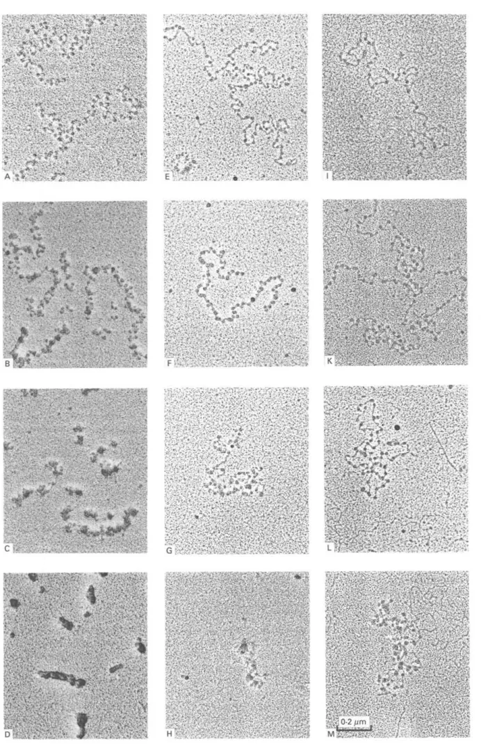
Soluble chromatin of T. b. brucei bloodstream forms, centrifuged through a sucrose gradient containing 10 mM NaCl, dialysed against concentrations of 0, 10, 40, or 100 mM NaCl, and prepared for EM, showed condensation at increasing ionic strength (Fig. 1E-H). The compaction was more pronounced as compared to procyclic culture forms (Fig. 1I-M). However, solenoids (30 nm fibres) which are typical for rat liver chromatin (Fig. 1A-D), were not formed. Free linker D N A could barely be seen in rat liver chromatin (Fig. 1A-C), but was clearly visible in chromatin from both trypanosome stages at low salt concentrations (Fig. IE—F and I—L). T h e nucleo-somes of the chromatin filaments of procyclic culture forms were irregularly spaced (Fig. I I ) . This was also seen in bloodstream forms but to a lesser extent (Fig. IE), while chromatin of rat liver never showed such an irregular arrangement (Fig. 1A).
Triton acid urea PAGE
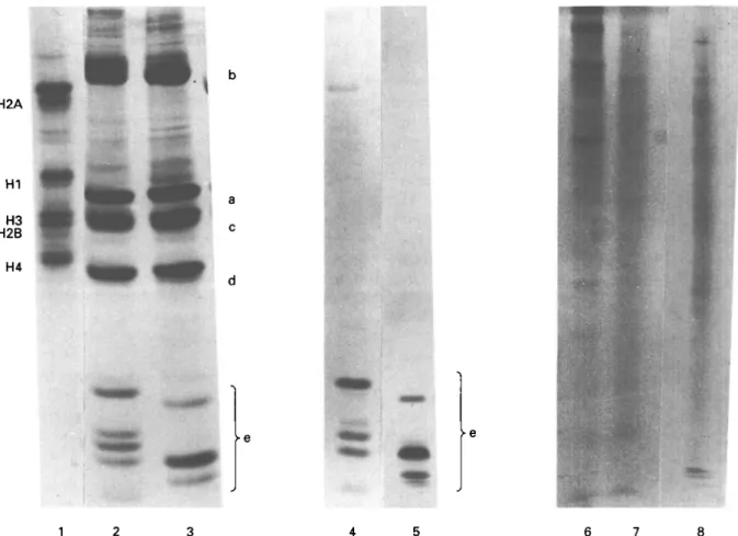
In triton acid urea gels, which separate proteins according to their hydrophobicity, core histones of trypanosomes migrated in four main complexes a, b, c, d and in a fast migrating, hydrophilic e-complex (Fig. 2, Lanes 2 and 3). Apart from the proteins of the d-regions, which had similar hydrophobic pro-perties to H4 of higher eukaryotes (Fig. 2, Lane 1), all the histones of trypanosomes differed from those of higher eukaryotes. No proteins of higher eukaryotes can be seen in the e-region.
5^/1'
* *
Chromatin of Trypanosoma brucei brucei
Differences in the number of bands, their position in the gel and their relative amount were seen between the histones of the two trypanosome stages. This was especially true for the e-region (Fig. 2, Lanes 2 and 3). All the proteins in the e-area could be selectively extracted with 5 % PCA (Fig. 2, Lanes 4 and 5). These proteins were metachromatically stained with Coomassie brilliant blue, which cannot be seen in black and white reproduction. Suspension of nuclei in 350 mM NaCl extracted non-histone proteins but not histone H I of rats (Fig. 2, Lane 8). Proteins of the e-region were not extracted from nuclei with 350 mM NaCl, neither from procyclic culture forms (Fig. 2, Lane 6) nor from bloodstream forms (Fig. 2, Lane 7).
Selective destaining of histones
Histones of bloodstream forms, separated in Triton acid urea gels, were stained with 0-01 % bromo-phenol blue, and the destaining of the proteins in 40 % n-propanol was densitometrically analysed (Fig. 3A). Selective destaining of the proteins in the e-region could be seen after 6 h (Fig. 3B). T h e proteins in the b- and c-regions were destained after 24 h, the staining intensity of the d-region was reduced, and no destaining took place in the a-region (Fig. 3C). Procyclic culture forms showed the same destaining behaviour (not shown). Differential de-staining of Coomassie brilliant blue-stained histones with 0-1 M FeCl3 revealed a selective destaining of the e-region after 2 h (not shown).
SDS-tricine-PAGE
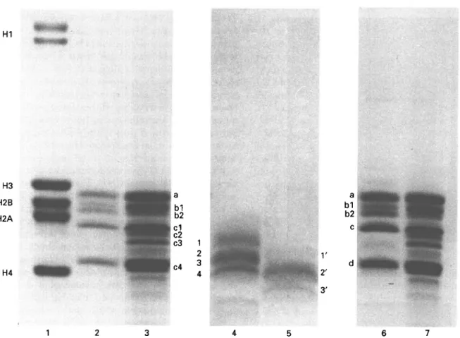
Histones of higher eukaryotes separated into a complex of 4 core histones and 2 H I variants in SDS-tricine-PAGE (Fig. 4, Lane 1). Core-histones of trypanosomes and higher eukaryotes could be seen to differ in their electrophoretic mobility (Fig. 4, Lanes 1, 2 and 3). T h e strongest differences existed between rat liver histones H3 and H4 and their trypanosome counterparts a and d. Histones of bloodstream forms showed a different banding pattern compared to that of procyclic culture forms. In this high-resolution gel system, the histone b was separated into two variants in both trypanosome stages (bl and b2) (Fig. 4, Lanes 2 and 3) and histone c shows three variants (cl, c2 and c3), but only in bloodstream forms (Fig. 4, Lane 3). Gels stained with Coomassie brilliant blue showed differences in the amounts of proteins bl and b2 in procyclic culture forms and bloodstream forms (Fig. 4, Lanes
2 and 3). Neither procyclic culture forms nor bloodstream forms had any protein migrating in the HI region of higher eukaryotes (Fig. 4, Lanes 1, 2 and 3). Proteins extracted with 5 % PCA migrated in the lower region of the core-histones and were metachromatically stained with Coomassie brilliant blue (Fig. 4, Lanes 4 and 5). These PCA-extractable proteins separated in four bands (1—4) in procyclic culture forms and in three bands (l'-3') in blood-stream forms.
Among the HCl-extractable proteins of procyclic culture forms, only band 1 was clearly separated from the others, while bands 2-4 were partly overlaid by histone d. Only faint bands above and below that of histone d indicate their presence (Fig. 4, Lane 6). Positioning of the bands by densitometric tracing revealed that the PCA-extractable bands 1', 2' and 3' of bloodstream forms were also visible in HC1 extracts.
No significant differences could be seen between the two stages in proteins extractable in 0-25 M HC1 or DNA-associated proteins derived from soluble chromatin (Fig. 4, Lanes 2, 3, 6 and 7).
Nucleosomal pattern after micrococcal nuclease digestion
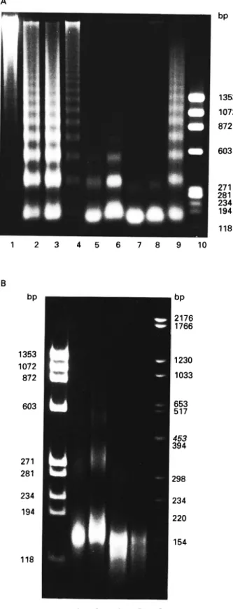
Ladders of D N A fragments ranging from oligo-nucleosomes to monooligo-nucleosomes were visible in agarose gels when chromatin of T. b. brucei procyclic culture forms were digested with micrococcal nuclease for 1 min (Fig. 5A, Lane 3). Chromatin of bloodstream forms was digested to a lesser extent, and that of rat liver still less (Fig. 5A, Lanes 4 and 1). After 10 min digestion, nucleosome ladders were still found for rat liver (Fig. 5A, Lane 9), whereas chromatin of both trypanosome stages was more extensively digested, mainly to mononucleosomes and dinucleosomes (Fig. 5A, Lanes 7 and 8). The strongest differences in the digestion patterns be-tween procyclic culture forms and bloodstream forms were seen after 4 min (Fig. 5A, Lanes 5 and 6). Mixed preparations of DNA fragments of procyclic culture forms and bloodstream forms after 1 min digestion (Fig. 5A, Lane 2) showed nucleosomal ladders identical to those of single preparations (Fig. 5A, Lanes 3 and 4), which demonstrates that the linker length is identical for both life-cycle stages of
T. b. brucei. T h e differences were not affected by
changes in the enzyme concentration (O'l, 0-2 and 0*4 units micrococcal nuclease per 20 A260 nuclei).
The same samples separated on a high-resolution polyacrylamide gel showed mainly
mononucleo-Fig. 1. Soluble chromatin of Trypanosoma brucei brucei and of rat liver from a sucrose gradient containing 10 mni NaCl, dialysed against concentrations of 0 mM NaCl (A, E, I), 10 mM NaCl (B, F, K), 40 mM NaCl (C, G, L) and 100 mM NaCl (D, H, M). Rat liver (A-D); bloodstream forms (E-H); procyclic culture forms (I-M).
H2A H1 H3 H2B H4 1 2 3 4 5 6 7 8
Fig. 2. Histone analysis in Triton acid urea PAGE. Lane 1: calf thymus histones. HCl-extracted histones of nuclei of
Trypanosoma brucei brucei procyclic culture forms (Lane 2) and of bloodstream forms (Lane 3); 5 % perchloric
acid-extracted histones of procyclic culture forms (Lane 4) and bloodstream forms (Lane 5). Non-histone proteins extracted with 350 mM NaCl from procyclic culture forms (Lane 6), bloodstream forms (Lane 7) and rat liver (Lane 8).
Fig. 3. Selective destaining of bromophenol blue stained histones. Histones of Trypanosoma brucei brucei
bloodstream forms, separated on a Triton DF-16 acid urea gel and stained with bromophenol blue, were treated with 40% n-propanol, which destains selectively histone HI, H2A and H2B. Densitometric Trace 1: before destaining; Trace 2: after 6 h; Trace 3 after 24 h of destaining. After 6 h the e-bands and after 24 h the b and c-bands, which are the counterparts of H2A and H2B in higher eukaryotes, are destained.
some-sized and traces of dinucleosome-sized DNA fragments in procyclic culture forms after digestion for lOmin (Fig. 5B, Lane 2). Bloodstream forms
showed fragments up to four nucleosomes at the same digestion time (Fig. 5B, Lane 3). The mean size of the mononucleosome fragments was larger for the bloodstream forms than for procyclic culture forms after 10 min digestion. The centre of the corresponding band in bloodstream forms was found above the 154 bp fragment of the DNA marker, while the centre of the band of the procyclic culture forms was situated in the region of this marker fragment. After 30 min digestion, DNA fragments were found in the region of 154 bp as well as below this marker for both trypanosome stages (Fig. 5B, Lanes 4 and 5).
DISCUSSION
The identification of histone HI can be difficult, since it is the most variable of all histones. In several cases, the original reports stated that HI was absent (van Holde, 1989) and only careful choice of experimental conditions, use of protease inhibitors and specially adapted gel systems allowed the demonstration of its presence. In T. b. brucei, a histone HI-like protein was postulated to be absent (Hecker & Gander, 1985; Hecker et al. 1989; Bender
H1 H3 H2B H2A H4 1 2 3 4 5 6 7
Fig. 4. Histone analysis in SDS-tricine—PAGE. Lane 1: calf thymus histones. HCl-extracted histones of Trypanosoma
brucei brucei procyclic culture forms (Lane 2) and bloodstream forms (Lane 3); 5 % perchloric acid-extracted histones
of bloodstream forms (Lane 4) and of procyclic culture forms (Lane 5); histones from soluble chromatin of procyclic culture forms (Lane 6) and bloodstream forms (Lane 7).
et al. 1992 a-d), although it was shown in other
trypanosomatids (Toro & Galanti, 1988, 1990; Duschak & Cazzulo, 1990).
In the present paper, proteins called e-region were seen in T. b. brucei for the first time. We postulate that the proteins of the e-region are histone HI-like proteins. This is supported by their extractability with 5 % PCA, a typical feature of histone HI of higher eukaryotes (Sanders, 1977). If these proteins are stained with Coomassie Brilliant blue, they show metachromasia; a reddish colour, which is a sign of a high lysine content (Duhamel, Meezan & Brendel, 1980). Additional evidence comes from destaining experiments with M-propanol (Duschak & Cazzulo, 1990) and FeCl3 (Spiker et al. 1976; Duschak & Cazzulo, 1990), which destains histone H I . Using both methods the e-region is destained first. Ad-ditionally, due to their non-extractability with 350 mM NaCl, it could be ruled out that these proteins were non-histones (Johns, 1982). Anti-bodies directed against HI of sea urchin showed cross-reactivity to the fast migrating proteins of T.
cruzi (Toro & Galanti, 1988), the counterparts of the
e-bands in T. b. brucei. It has been shown by means of amino acid analysis that these proteins of T. cruzi are histone HI-like proteins (G. C. Toro & N. Galanti, personal communication).
Triton binds to histones proportionally to their degree of hydrophobicity and thereby reduces the electrophoretic mobility (Hardison & Chalkley, 1978). The histone HI-like proteins of T. b. brucei are strongly hydrophilic and very fast migrating in Triton acid urea gels. The banding pattern in Triton DF-16 gels of histones of T. cruzi, T. b. brucei and
Crithidia fasciculata is similar and all species possess
fast migrating hydrophilic histone HI-like proteins. However, the banding pattern of the fast migrating bands in trypanosomatids differs. Differences in the number, amount and position of the HI-like proteins can also be seen, when the two stages of the life-cycle of T.b. brucei are compared.
Histone HI-like proteins of T. b. brucei run out of Triton and acid urea gels owing to their high mobility unless specially adapted systems are used. In SDS gels they can only be detected if the linear SDS-tricine-PAGE according to the method of Schagger & von Jagow (1987), optimized for highest resolution in the region of the core histones, is used. The resolution in this gel system is superior to that in gradient gels according to Laemmli (1970) which were originally used by Hecker & Gander (1985), and Bender et al. (1992a). The strong hydrophilic character and the small molecular weight of the H l -like proteins of T. b. brucei are the reasons why
A bp 118 10 bp 118
Hecker & Gander (1985), Hecker et al. (1989) and Bender et al. (I992a-d), failed to demonstrate their existence.
In higher eukaryotes, histone HI is composed of a central hydrophobic globular domain and two ad-jacent hydrophilic tails (Allan et al. 1980). We could show that the PCA extracted histone HI-like proteins of T. b. brucei run in the region of the core histones in S D S - P A G E and therefore they may be quite small. This finding is in line with a study of Hayashi, Hayashi & Iwai (1987) who described a histone HI with a small globular part in
Tetra-hymena.
If the N- and C-terminal hydrophilic tails of the histone HI-like proteins in T. b. brucei are of a similar length, compared to those of higher eukaryotes, then the hydrophobic globular region would occupy only a small proportion. The globular part of histone HI is the domain which is responsible for the binding to the nucleosome (van Holde, 1989). A small sized globular domain could indicate a weak interaction of the trypanosome HI-like proteins with the nucleosomes. The core histones of T. b. brucei procyclic culture forms and bloodstream forms, separated according to the molecular weight or to the hydrophobicity, are similar, but differ in the position and number of the bands as compared to higher eukaryotes. This result is in line with differences reported for the amino acid composition between
T. b. brucei procyclic culture forms and higher
eukaryotes (Bender et al. 1992 a). By selective destaining with w-propanol, we were able to show that the proteins of the bloodstream forms, which migrate in the positions of histones b and c in Triton gels are the counterparts of H2A and H2B (Duschak & Cazzulo, 1990). Life-cycle specific variants and/or modifications can be seen in histone b, which is resolved in two proteins bl and b2 in SDS-tricine-PAGE, as well as in histone c, which is resolved only in bloodstream forms into three bands cl, c2 and c3. Differences in the amount between the two bands of protein b occur between the two trypanosome stages and can be seen in SDS-tricine as well as in Triton acid urea gels.
Weak interaction and instability within the chro-matin enhances the accessibility of the linker DNA towards micrococcal nuclease and this leads to a
Fig. 5. (A) Separation of DNA fragments in a 1-5% agarose gel. Micrococcal nuclease digestion patterns of chromatin of rat liver, Trypanosoma brucei brucei procylic culture and bloodstream forms. Marker: HAE
III fragments of $ X 174 RF DNA (Lane 10). Lanes 1 and 9: rat liver; Lanes 3, 5 and 7: procyclic culture forms; Lanes 4, 6 and 8: bloodstream forms; Lane 2: mixture of procyclic culture and bloodstream forms. Digestion times: Lanes 1—4: 1 min; Lanes 5 and 6: 4 min; Lanes 7-9: 10 min. (B) Separation of DNA
fragments in a 5-5% polyacrylamide gel. Markers: HAE III fragments of 0 X 174 RF DNA (Lane 1) and DNA fragments of pBR328 cleaved with Bgl I and Hint I (Lane 6). Procyclic culture forms (Lanes 2 and 4) and bloodstream forms (Lanes 3 and 5) after 10 or 30 min digestion. The centre of the band of mononucleosome fragments of bloodstream forms (Lane 3) is above the 154 bp marker fragments (Lane 6), while one of the procyclic culture forms (Lane 2) is in the region of the latter.
faster digestion of the chromatin of the procyclic culture forms (Hecker et al. 1989) relative to higher eukaryotes. Bloodstream forms appear to be in-termediate. Digestion is slower than in procyclic culture forms, but still faster than for higher eukaryotes. There is a clear digestion barrier in higher eukaryotes in the presence of HI (Telford & Stewart, 1989), which cannot be seen in procyclic culture forms (Hecker et al. 1989). After digestion for lOmin, bloodstream forms yield larger mononucleosome-sized DNA fragments than the procyclic culture forms. These larger fragments are no longer seen after 30 min digestion. A weak digestion barrier in bloodstream forms may be due to a somewhat stronger interaction between histone HI-like proteins and the DNA, as compared to the procyclic culture form.
Genetically inactive chromatin of higher eukar-yotes forms 30 nm fibres (solenoids) at 100 mM NaCl. The formation of the 30 nm fibres depends on the presence of the lysine-rich histone H I . In the absence of histone H I , nucleosome filaments do not condense to solenoids, and form only loose aggre-gates (Thoma et al. 1979). The spacing of the nucleosomes on the DNA is irregular and nucleo-some sliding occurs (Stein & Bina, 1984).
Chromatin of T. b. brucei procyclic culture forms, prepared under identical conditions to rat liver chromatin, is organized in the form of nucleosome filaments. Previous studies on the ultrastructure of the chromatin of T. b. brucei procyclic culture forms revealed a bad conservation of the nucleosome filaments (Hecker & Gander, 1985). The nucleo-somes were irregularly spaced and at increasing ionic strength only a slight aggregation of the nucleosomes could be seen (Hecker & Gander, 1985). The interactions of histones and DNA are weak and are easily destabilized by experimental manipulation (Hecker & Gander, 1985; Hecker et al. 1989; Bender
et al. 1992c).
To try to improve the preservation of the chroma-tin filaments of trypanosomes, the effect of increasing the concentration of glutaraldehyde for the fixation of soluble chromatin for EM studies was investi-gated. The results obtained with 0-2 % glutaralde-hyde were superior to those with the 'conventional' concentration of 0-1 % (Thoma et al. 1979; Hecker &
Gander, 1985). Chromatin prepared with 0-5 %
glutaraldehyde showed an identical condensation behaviour to the one fixed with 0-2 %.
The stability of chromatin depends on the nature and strength of the DNA—protein interactions (Yager, McMurray & van Holde, 1989). In contrast to the chromatin of procyclic culture forms, the chromatin of bloodstream forms shows a stronger condensation at increasing ionic strength but it does not form 30 nm fibres. The compaction pattern of soluble chromatin of T. b. brucei bloodstream forms is structurally similar to that of T. cruzi epimastigote
culture forms (Hecker & Gander, 1985). Histone HI-like proteins were shown for T. cruzi by Toro & Galanti (1988, 1990). Hecker & Gander (1985) could induce a higher condensation level in T. cruzi with the addition of a histone HI-containing fraction from rat liver chromatin. This result suggested an HI binding site in T. cruzi chromatin. Additionally, Bender (1991) showed that a binding site for histone HI exists in the chromatin of T. b. brucei procyclic culture forms.
The finding of Hecker & Gander (1985), that no higher condensation level was achieved in procyclic culture forms of T. b. brucei by the addition of HI of rats, is most probably due to the damage of the chromatin having been introduced by the exper-imental procedure to make soluble chromatin and the concomitant dissociation of nucleosomes from the DNA. A subsequent reconstitution is no longer possible. The result is the formation of 'clumps on a string' with large stretches of free DNA.
The histone patterns of the e-region of procyclic culture forms and bloodstream forms are different in number and position of bands and in their relative amounts. Modifications of the histones may influence histone-histone interactions as shown by Simpson (1981) for higher eukaryotes, and may contribute to a higher stability of the core particle especially in bloodstream forms. Effects of histone modifications were shown on the condensation behaviour of nucleosome filaments into higher-order structures for higher eukaryotes (Marion et al. 1985).
The different histone patterns of the two compared trypanosome stages are most likely the reason for the different condensation behaviour of their chromatin. The HI-like proteins of procyclic culture forms seem to interact very weakly with the DNA and this could be the reason why no condensation takes place, since most of these proteins dissociate during the preparation of soluble chromatin. This would also explain the poor preservation of the nucleosome filaments of procyclic culture forms at 0 mM NaCl, which is in accordance with the results of Hecker et
al. (1989), who showed that chromatin of procyclic
culture forms is very unstable. As chromatin from both stages of the life-cycle was prepared under identical conditions, observed differences of the chromatin parameters must be intrinsic differences between the life-stages of T. b. brucei.
We wondered why trypanosomes undergo changes in their histones during their life-cycle and suggest that the stronger DNA-histone interaction in blood-stream forms might be a requirement for a stabil-ization of the chromatin at the higher temperatures of the mammalian host. When procyclic culture forms were cultivated above 35 °C, they stopped cell division and died after approximately 3 days. All chromatin parameters investigated, including the histone pattern in gels, were those usually found in procyclic culture forms. The fact that procyclic
culture forms cannot produce the histone HI pattern and core histone variants of modifications of blood-stream forms and die after several days when cultivated between 35 and 37 °C, supports this hypothesis. Since trypanosomes derive from free-living flagellates (Lumsden & Evans, 1976), the ability of bloodstream forms to support higher temperatures might be regarded as an adaptation to parasitism.
It can be concluded that T. b. brucei contains a full set of histones, including HI-like proteins, and that the poor condensation of its chromatin is not due to the absence of H I , but most probably to peculiar properties of HI and core histones. Despite common features, the histones and therefore various proper-ties of the nuclear chromatin are quite different between bloodstream and procyclic culture forms. As a consequence, the latter can no longer be regarded as the only model for all stages of the life-cycle of T.b. brucei.
We would like to thank Jennifer Jenkins and Patrick Lorenz for the careful reading of the manuscript, Ruth Nuesch for the preparation of the culture medium and Irene Herde for skilful technical assistance.
R E F E R E N C E S
ALFAGEME, C. R., ZWEIDLER, A., MAHOWALD, A. & COHEN,
L. H. (1974). Histones of Drosophila embryos. Journal
of Biological Chemistry 249, 3729-36. ALLAN, J., HARTMAN, P. G., CRANE-ROBINSON, C. &
AVILES, F. x. (1980). The structure of histone HI and its location in chromatin. Nature, London 288, 675—9.
BENDER, K. (1991). Biochemical and structural aspects of the nuclear chromatin of procyclic Trypanosoma
brucei brucei. Ph.D. thesis, University of Basel. BENDER, K., BETSCHART, B., SCHALLER, J., KAMPFER, U. &
HECKER, H. (1992a). Biochemical properties of histone-like proteins of procyclic Trypanosoma brucei brucei.
Ada Tropica 50, 169-84.
BENDER, K., BETSCHART, B., SCHALLER, J., KAMPFER, U. & HECKER, H. (19926). Sequence differences between histones of procyclic Trypanosoma brucei brucei and higher eukaryotes. Parasitology 105, 97-104.
BENDER, K., BETSCHART, B. & HECKER, H. (1992c).
Histone-DNA interactions in the chromatin of procyclic Trypanosoma brucei brucei. Parasitology
Research 87, 495-500.
BENDER, K., BETSCHART, B., MARION, C , MICHALON, P. & HECKER, H. (1992<f). Structural differences between the chromatin of procyclic Trypanosoma brucei brucei and higher eukaryotes as probed by immobilized trypsin. Ada Tropica 52, 69-78.
BRUN, R. & SCHONENBERGER, M. (1979). Cultivation and in
vitro cloning of Trypanosoma brucei in a semi-defined
medium. Ada Tropica 36, 289-92.
DUHAMEL, R. C , MEEZAN, E. & BRENDEL, K. (1980).
Metachromatic staining with Coomassie-Brilliant-Blue R-250 of the proline-rich calf thymus histone HI.
Biochimica et Biophysica Ada 626, 432^-2.
DUSCHAK, v. G. & CAZZULO, j . j . (1990). The histones of the insect trypanosomatid, Crithidia fasciculata.
Biochimica et Biophysica Acta 1040, 159-66. ELPIDINA, E. N., ZAITSEVA, G. N. & KRASHENINNIKOV, J. A.
(1979). Histones from Trypanosoma lewisi nuclei.
Biokhimiya 44, 1830-41.
HAMM, B., SCHINDLER, A., MECKE, D. & DUSZENKO, D.
(1990). Differentiation of Trypanosoma brucei bloodstream trypomastigotes from long slender to short stumpy-like forms in axenic culture. Molecular
and Biochemical Parasitology 40, 13—22.
HARDISON, R. & CHALKLEY, R. (1978). Polyacrylamide gel electrophoretic fractionation of histones. Methods in
Cell Biology 17, 235-51.
HAYASHI, T., HAYASHI, H. & IWAI, K. (1987). Tetrahymena histone HI. Isolation and amino acid sequence lacking the central hydrophobic domain conserved in other HI histones. Journal of Biochemistry 102, 369—76.
HECKER, H. & GANDER, E. s. (1985). The compaction pattern of the chromatin of trypanosomes. Biology of
the Cell 53, 199-208.
HECKER, H., BENDER, K., BETSCHART, B. & MODESPACHER,
u. P. (1989). Instability of the nuclear chromatin of procyclic Trypanosoma brucei brucei. Molecular and
Biochemical Parasitology 37, 225-34.
HEWISH, D. R. & BURGOYNE, L. A. (1973). Chromatin sub-structure. The digest of chromatin DNA at regularly spaced sites by a nuclear deoxyribonuclease.
Biochemical and Biophysical Research Communication 52, 504-10.
JOHMANN, c. A. & GOROVSKY, M. A. (1976). Purification and characterisation of histones associated with the macronucleus of Tetrahymena. Biochemistry 15, 1249-56.
JOHNS, E. w. (1982). Chapter 1. In The HMG
Chromosomal Proteins (ed. Johns, E. W.), pp. 1-7.
New York: Academic Press.
LAEMMLI, u. K. (1970). Cleavage of structural proteins during the assembly of the head of bacteriophage T4.
Nature, London 227, 680-5.
LANHAM, s. M. & GODFREY, D. G. (1970). Isolation of salivarian trypanosomes from man and other mammals using DEAE-Cellulose. Experimental
Parasitology 28, 521-34.
LUMSDEN, w. H. R. & EVANS, D. A. (1976). Biology of the
Kinetoplastida, Vol. I. New York: Academic Press. MARION, C , MARTINAGE, A., TIRARD, A., ROUX, B., DAUNE,
M. & MAZEN, A. (1985). Histone phosphorylation in native chromatin induces local structural changes as probed by electric birefringence. Journal of Molecular
Biology 186, 367-79.
NOLL, H. (1969). An automatic high-resolution gradient analyzing system. Analytical Biochemistry 27, 130-49.
SAMBROOK, J., MANIATIS, P. T. & FRITSCH, E. F. (1989). Molecular Cloning. A Laboratory Manual. Cold Spring
Harbor, New York: Cold Spring Harbor Laboratory. SANDERS, C. (1977). A method for the fractionation of the
high-mobility group non-histone proteins. Biochemical
and Biophysical Research Communications 78, 1034—42. SCHAGGER, H. & VON JAGOW, G. (1987).
Tricine-sodium-dodecyl sulfate—polyacrylamide gel electrophoresis for the separation of proteins in the range from 1 to 100 kDa. Analytical Biochemistry 166, 368-79.
SHAPIRO, s. z. & DOXSEY, s. j . (1982). Purification of
nuclei from a flagellate protozoan, Trypanosoma brucei. Analytical Biochemistry 127, 112-15.
SIMPSON, R. T. (1981). Modulation of nucleosome structure by histone subtypes in sea urchin embryos. Proceedings of the National Academy of Sciences, USA 78, 6803-7.
SPIKER, s., KEY, j . L. & WAKIM, B. (1976). Identification
and fractionation of plant histones. Archives of Biochemistry and Biophysics 176, 510-18.
STEIN, A. & BINA, M. (1984). A model chromatin
assembly system: factors affecting nucleosome spacing. Journal of Molecular Biology 178, 341-63.
TELFORD, D. J. & STEWART, B. W. (1989). MicrOCOCCal
nuclease: its specificity and use for chromatin analysis. International Journal of Biochemistry 21, 127-37.
THOMA, F., ROLLER, TH. & KLUG, A. (1979). Involvement
of histone HI in the organization of the nucleosome and the salt-dependent superstructures of chromatin. Journal of Cell Biology 83, 403-24.
THOMA, F. & ROLLER, TH. (1981). Unravelled
nucleosomes, nucleosome beads and higher-order structures of chromatin: influence of non-histone
components and histone H I . Journal of Molecular Biology 149, 709-33.
TORO, G. c. & GALANTI, N. (1988). H I histone and histone
variants in Trypanosoma cruzi. Experimental Cell Research 174, 16-24.
TORO, G. c. & GALANTI, N. (1990). Trypanosoma cruzi
histones. Further characterization and comparison with higher eukaryotes. Biochemistry International 21, 481-90.
TORO, G. C , WERNSTEDT, C , MEDINA, C , JARAMILLO, N.,
HELLMANN, u. & GALANTI, N. (1992). Extremely
divergent histone-H4 sequence from Trypanosoma cruzi — evolutionary implications. Journal of Cellular Biochemistry 49, 266-71.
VAN HOLDE, K. E. (1989). Chromatin. In Springer Series in Molecular Biology (ed. Rich, A.), pp. 168-80 and pp. 317-43. New York: Springer Verlag.
VICKERMAN, K. & PRESTON, T. M. (1970). S p i n d l e
microtubules in the dividing nuclei of trypanosomes. Journal of Cell Science 6, 365-83.
YAGER, T. D., McMURRAY, T. & VAN HOLDE, K. E. (1989). Salt-induced release of D N A from nucleosome core particles. Biochemistry 28, 2271-81.