Mutations in the fukutin-related protein gene (FKRP) identify limb girdle muscular dystrophy 2I as a milder allelic variant of congenital muscular dystrophy MDC1C
10
0
0
Texte intégral
(2) 2852 Human Molecular Genetics, 2001, Vol. 10, No. 25. Table 1. Summary of the clinical features of LGMD2I patients Family Sex. Age Age First symptom Serum CK (years) onset (years). Muscle hypertrophy. Weakest muscle. Cardiomyopathy Wheelchair Laminin α2 bound. α-Dystroglycan. Normal (I). ND. 1. M. 6. <0.5. Hypotonia, DMM. >1000. Calf, legs. Hip girdle. No. 2. F. 10. 1. Hypotonia, DMM. 3160. Tongue. Neck, shoulder. Borderline LVF No. Reduced (I). Severely reduced. 3. M. 18. 1.5. Waddle. 2850. Leg, calves, tongue. Hip = shoulder girdle. Severe LV, hypokinesia. 14. ND. ND. M. 11. 1.5. Waddle. 2328. Leg, calves, tongue. Hip = shoulder girdle. ND. No. ND. ND. 4. M. 19. 1.5. Waddle. 1700–3300 Leg, tongue. Hip = shoulder girdle. Moderate LV, hypokinesia. 12. ND. Severely reduced. 5. F. 20. 2. Waddle. 3160. Leg, calves. Deltoid. LVD. 13. Reduced (I). Severely reduced. 6. F. 10. 2.5. Toe walking, fatigue. 1380–7795 Leg, calves. Deltoid. ND. No. Reduced (I). Reduced. 7. F. 10. 2.5. DMM. 1278. No. Hip girdle. No. No. Reduced (I). ND. 8. M. 28. 4. Toe walking, no running. 4105. No. Hip girdle. Moderate LV, hypokinisia. No. ND. ND. M. 22. 4. No running. ND. No. Hip girdle. ND. No. ND. ND. 9. F. 37. 7. Waddle, arm weakness. 1150. Tongue. Shoulder girdle. No. 12. ND. ND. M. NA. NA. NA. NA. NA. NA. NA. NA. 10. F. 17. 9. Gowers. 2910–8200 Calf, legs. Neck, deltoid. Borderline LVF No. 11. F. 18. 9. Stairs. >1000. No. Tibialis anterior. No. No. ND. ND. M. NA. NA. NA. NA. NA. NA. NA. NA. ND. ND. 12. M. 16. 13. Gowers. 1305–6820 Leg, calves. Deltoid. ND. No. Reduced (I). Severely reduced. 13. F. 37. 27. Stairs. 2870. Calf. Hip girdle. No. Part-time, 35. Reduced (WB) Reduced. 14. F. 39. 28. Stairs. 2300. Calf, Triceps, hip girdle ND brachioradialis. No. Reduced (WB) ND. 15. F. 46. 40. Stairs, running 2230. Calf, Hip girdle brachioradialis. No. No. Reduced (WB) Reduced. M. 48. 35. Stairs. 1123. Brachioradialis Hip girdle. No. No. Reduced, (WB) ND. 16. M. 37. 29. Stairs. >1000. Calf, Hip girdle brachioradialis. No. Part-time, 36. Reduced (WB) Reduced. 17. F. 27. 23. Stairs. 4210. Leg, calves. No. No. Reduced (I). Hip = shoulder girdle. No. ND. ND. Reduced (I). Severely reduced. Reduced. WB, westerm blot; ND, no data; NA, not available; LVF, left ventricular function; LV, left ventricular.. adolescence or even adult life (2,3). Both CMD and LGMD are highly heterogeneous diseases. Inheritance in LGMD can be either autosomal dominant (LGMD type 1) or autosomal recessive (LGMD type 2), whereas CMD is always recessively inherited. To date, at least 14 types of LGMD have been reported and though the majority of causative genes have been found to encode proteins associated with the sarcolemma, genes encoding cytoskeletal proteins and muscle-specific enzymes have also been implicated (2). Fewer disease genes have been identified in CMD (reviewed in 4). Mutations in the LAMA2 gene on chromosome 6q22–23, encoding the laminin α2 chain of merosin (laminin-2), were identified in 1994 and account for ∼40% of CMD cases (5–7). Merosin is an extra-. cellular matrix protein that consists of three laminin chains, α2β1γ1, with α2 forming a link between the peripheral membrane protein α-dystroglycan and the basal lamina. Children with mutations in the LAMA2 gene usually have absent protein expression and a severe form of CMD, commonly referred to as merosin-deficient CMD or MDC1A. It is now clear, both clinically and genetically, that CMD and LGMD can overlap, suggesting that the underlying pathology in these diseases may follow a similar pathway. We have previously described a large Turkish kindred, with a clear LGMD phenotype, linked to the LAMA2 locus (8). Patients had a reduction in the expression of laminin α2 in their skeletal muscle biopsy. More recently, mutations in the LAMA2 gene in.
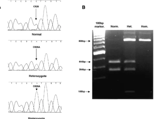
(3) Human Molecular Genetics, 2001, Vol. 10, No. 25 2853. Figure 1. (A) Sequencing chromatograms showing the C826A mutation. (B) Restriction enzyme analysis of the C826A mutation. A 996 bp PCR fragment was digested with BfaI. Wild-type DNA is cut into three fragments of 445, 364 and 189 bp, whereas products containing the mutation, which introduces the loss of a BfaI site, are cleaved into two fragments of 808 and 189 bp. Norm., wild-type DNA; Het., heterozygote; Hom., homozygote.. Table 2. Summary of FKRP mutations in LGMDI Family. Change. Protein effect. Mutational status. 6, 8, 13, 14, 15. C826A. Leu276Ile. Homozygous. 1. G1016T. Arg339Leu. Homozygous. C826A. Leu276Ile. 3, 4, 5, 12, 16, 17. ? 2. 9. 7. 10. 11. Compound Heterozygote. C826A. Leu276Ile. Compound. 390insTACC. Gly132Stop. Heterozygote. C826A. Leu276Ile. Compound. C934T. Arg312Cys. Heterozygote. C826A. Leu276Ile. Compound. C947G. Pro316Arg. Heterozygote. C826A. Leu276Ile. Compound. 426del12nt. 143delRMVE. Heterozygote. C427A. Arg143Ser. Compound. ?. Heterozygote. ?, second allelic mutation unidentified.. several patients with a LGMD phenotype and a partial laminin α2 deficiency have been identified (9–11), confirming that. mutations in this gene can result in either a severe disease (MDC1A) or a mild LGMD-like disorder, depending on the type and location of the mutation within the gene. A number of CMD forms have been described that have a reduction in laminin α2 expression not due to mutations in the LAMA2 gene (secondary laminin α2 deficiency). One of these is Fukuyama CMD (FCMD; MIM 253800), a multi-system disease in which brain, skeletal and cardiac muscle are affected (12). The FCMD gene encodes fukutin, a protein of unknown function (13). A profound depletion of skeletal and cardiac muscle α-dystroglycan has recently been reported in FCMD (14). We have recently identified a new member of the fukutin protein family, FKRP (15) (fukutin-related protein). Analysis of both FKRP and fukutin demonstrates sequence similarities to a family of proteins involved in modifying cell surface molecules, such as glycoproteins and glycolipids (16). All members of this family of proteins contain a conserved DxD motif found in many glycosyltransferases (17), though formal proof that FKRP is itself a glycosyltransferase remains to be demonstrated. Both proteins also have a putative type II membrane spanning a region consistent with localization to the Golgi apparatus, though it has been suggested that fukutin is also secreted (13). The FKRP gene is mutated in a severe form of CMD (MDC1C), characterized by early onset, inability to achieve independent ambulation, muscle hypertrophy, marked elevation of serum creatine kinases (CK), no brain involvement.
(4) 2854 Human Molecular Genetics, 2001, Vol. 10, No. 25.
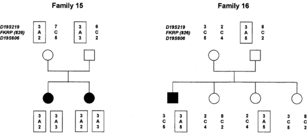
(5) Human Molecular Genetics, 2001, Vol. 10, No. 25 2855. Figure 2 (above and opposite). The pedigrees of 10 LGMD2I families showing the inheritance of the C826A mutation. The haplotypes containing the mutation are boxed. Markers D19S219 and D19S606 are based 3 cM apart on the Genethon map. D19S606 is <500 kb away from FKRP according to the latest sequence maps.. and a secondary deficiency of laminin α2 (15). In addition, affected individuals had a marked decrease in immunostaining of muscle α-dystroglycan and a reduction in its molecular weight on western blot analysis. We have suggested the abnormal expression of α-dystroglycan is most likely a result of its altered glycosylation. The FKRP gene maps to on an identical region on chromosome 19q13.3 as LGMD2I (17), between markers D19S219 and D19S606, suggesting that MDC1C and LGMD2I may be allelic disorders. To test this hypothesis we collected 25 LGMD families, including 14 that were consistent with links to the LGMD2I locus. Mutation analysis of the FKRP gene identified mutations in 17 families. In patients where muscle was available, a secondary laminin α2 deficiency and a variable reduction in α-dystroglycan expression were observed. RESULTS Mutation screening of the FKRP gene in LGMD2I Sequence analysis of the FKRP coding region identified mutations in individuals from 17 families, whose clinical features are summarized in Table 1 and mutations in Table 2. Unexpectedly, 15 families had an identical C826A (Leu276Ileu) mutation, of which five were homozygous for the mutation and the remainder were compound heterozygotes. None of the homozygous individuals was the offspring of consanguineous marriages. In the 10 families heterozygous for the C826A mutation we were able to identify the second allelic mutation in four cases. These were two missense mutations in families 7 and 9 (C934T and C947G), one nonsense mutation due to a 4 nt duplication in family 2 (390insTACC) and a 12 nt in-frame deletion (426del12) in family 10. In two families without the C826A mutation, we identified a homozygous G1016A missense mutation in family 1, a consanguineous family, and a C427A heterozygous missense mutation in family 11. Since the C826A mutation was present in a large number of families, we took advantage of the loss of a BfaI restriction site induced by the change (Materials and Methods; Fig. 1) to rule out this to be a polymorphism. On screening. controls, only one heterozygous individual was identified out of a total of 200 control chromosomes. We considered these mutations to be pathogenic since they were not present in at least 100 control chromosomes and segregated with the disease in an autosomal recessive fashion. In eight cases we did not observe any changes in the FKRP coding sequence. Four of these individuals came from genetically informative families that were compatible with linkage to the FKRP locus. The other four were isolated cases. Haplotype analysis of families with the C826A mutation We used markers D19S219 and D19S606 to haplotype families harbouring the C826A mutation, since radiation hybrid mapping suggested these were the closest markers flanking FKRP in the Genethon database. The order of markers is centromere–D19S219–FKRP–D19S606–telomere. The interval between the markers is 3 cM according to the Genethon map. Sequence data for this region of the genome puts D19S606 as the closest marker, no more the 500 kb downstream of the FKRP gene. In 10 families haplotyping was possible to check for linkage disequilibrium with the C826A mutation. Amongst the families tested, there was not a conserved haplotype (Fig. 2). However, marker D19S606, the most closely linked marker to FKRP, identified two alleles at a higher than expected frequency. Allele 2 was found in five haplotypes and allele 5 in four haplotypes (compared to a frequency of 0.036 for allele 2 and 0.018 for allele 5 in the CEPH database). Immunohistochemistry and western blot analysis The histological changes were characteristic of a muscular dystrophy in all patients. The expression of all proteins regularly screened for in the biopsies of patients with muscular dystrophy were normal. These included dystrophin, α-, β-, γsarcoglycans, caveolin, dysferlin, emerin, lamin A/C and calpain 3 (data not shown). Immunocytochemical analysis of laminin α2 using antibodies against both the 80 and 300 kDa fragments was reduced in several patients with a more severe phenotype (Fig. 3; Table 1), whereas it was normal in the remaining patients with the exception of few scattered fibres.
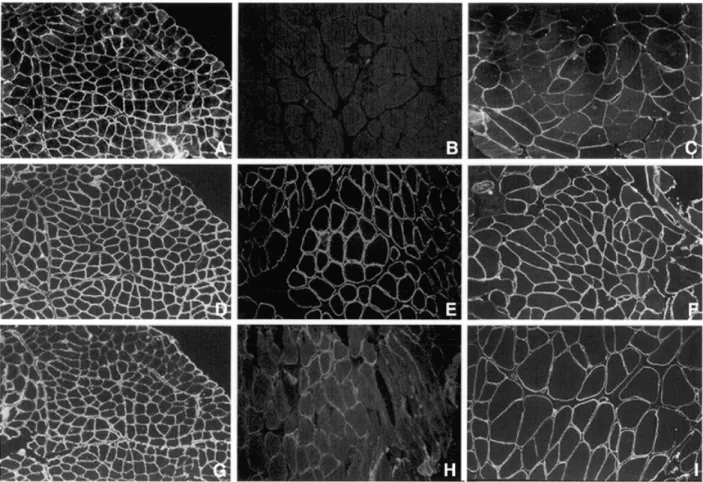
(6) 2856 Human Molecular Genetics, 2001, Vol. 10, No. 25. Figure 3. Immunocytochemical localization of α-dystroglycan (A–C), β-dystroglycan (D–F) and laminin α2 (300 kDa) (G–I) in control muscle (A, D and G), family 5 (B, E and H) and family 15 (C, F and I).. (data not shown). Since α-dystroglycan is a heavily glycosylated protein, we used two antibodies, V1A4-1 (Upstate Biotechnology) raised against an unknown epitope and a sheep polyclonal antibody, directed towards the core protein. Both antibodies clearly delineate individual muscle fibres with no apparent differences between fibre types in control muscle. In 10 patients with LGMD2I whose muscle was available to study, α-dystroglycan expression was found to be abnormal (Table 1). This was more marked using the V1A4-1 antibody than with the antibody to the core protein in (Fig. 3A). The apparent reduction in α-dystroglycan was more marked than laminin α2 (Fig. 3B). The pattern of β-dystroglycan labelling was normal, including those fibres in which α-dystroglycan was reduced. Western blot analysis using an antibody that recognized the 80 kDa merosin fragment revealed an absence or near-absence of labelling in family 15 (Fig. 4). This is identical to previous observations in this family, who along with families 13, 14 and 16, were reported as being affected by an ‘adult merosinopathy’ (18). The molecular mass of residual laminin α2 was normal, as was the expression of other proteins studied (Fig. 4). The virtual absence of laminin α2 on western blot was in marked contrast with the immunocytochemical analyses, where no reduction in its expression was evident.. DISCUSSION In this work mutations in the FKRP gene were identified in affected individuals from 17 families with LGMD2I. We originally demonstrated that mutations in this gene cause a severe form of CMD, MDC1C (15). The patients in this work had a variable phenotype, though all were less severe than MDC1C where patients never acquired independent ambulation. Patients at the severe end of the spectrum presented within the first 2 years of life with hypotonia, delayed motor milestones and followed by muscle hypertrophy (mostly in the legs, but also involving the tongue in several cases). These patients never acquired the ability to run or hop and followed a Duchenne-like disease course, with loss of independent ambulation in the early teens, followed by the development of cardiomyopathy. The remaining patients had milder phenotypes and are similar to those described in a large Tunisian family featured in the original description of LGMD2I (18). These patients were characterized by a later disease onset and a relatively benign course. Calf and to a lesser extent, thigh, brachioradialis and tongue muscle hypertrophy were present in most patients. Muscle cramps following exercise were also relatively common; one case had spontaneous myoglobinuria. Intelligence and brain MRI were normal. Serum CK was grossly elevated in all patients (between 10 and 30 times normal values) and the heart was variably affected, a feature not previ-.
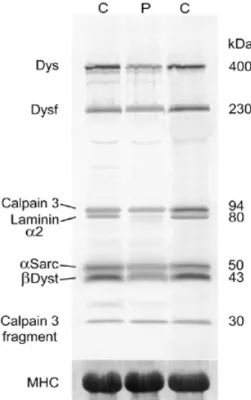
(7) Human Molecular Genetics, 2001, Vol. 10, No. 25 2857. Figure 4. Multiplex western blot showing the marked reduction of the 80 kDa fragment of the laminin α2 chain in family 15 (P) relative to controls (C). Protein loading of all samples was equivalent as shown by the staining of MHC on the gel after blotting.. ously reported in LGMD2I. The range of phenotypic severity due to FKRP mutations is surprisingly large and so far not observed in other forms of muscular dystrophy. The FKRP mutations identified in the LGMD2I families were different from those seen in MDC1C. The most severe patient in this series was a consanguineous child from family 1 who was symptomatic in the first year of life and whose motor milestones were delayed. His phenotype was clearly intermediate between MDC1C and LGMD2I. Fifteen families had an identical C826A missense mutation. The five patients homozygous for this mutation had a relatively mild phenotype, whereas compound heterozygotes for this mutation on the whole had a more severe phenotype. Linkage analysis failed to identify a single founder haplotype associated with this mutation. However, we did observe two rare alleles of D19S606 that appeared to be in linkage disequilibrium with the mutation, suggesting it may have arisen on a limited number of occasions. Although we identified FKRP mutations in 17 families, we were unable to find mutations in four families that were consistent with linkage to this locus. In addition, we have failed to identify the second mutation in six of the 10 compound heterozygotes for the C826A mutation. This strongly suggests that mutations exist outside the FKRP coding region in at least some of these families and analysis of these regions is presently under way. Patients at the severe end of the LGMD2I spectrum showed a mild deficiency of laminin α2 at the immunohistochemical level. Patients with a milder phenotype had normal expression of laminin α2 on immunohistochemistry, but in four patients. western blot analysis identified a severe deficiency or total absence of this protein. These four families have been described previously by Bushby et al. (19). The discrepancy between the immunocytochemical and western blot analysis suggests that mutations in FKRP result in the altered processing, epitope masking or abnormal folding of the laminin α2 polypeptide chain. The expression of α-dystroglycan, but not β-dystroglycan, was abnormal in all muscle biopsies of LGMD2I patients available for study, an identical finding to MDC1C. α- and β-dystroglycans are derived from post-translational cleavage of a single precursor peptide (20). α-Dystroglycan is a heavily glycosylated peripheral membrane protein that forms a link between the actin associated cytoskeleton and, via laminin α2, the extracellular matrix (21,22). Laminin α2 binds binds to a carbohydrate moiety of α-dystroglycan (23). The pattern of α-dystroglycan glycosylation differs not only between tissues but also within them, and different glycoforms may have different binding partners that mediates the function of α-dystroglycan (23,24). Mutations in the FKRP gene appear to result in a selective deficiency of αdystroglycan in skeletal muscle, since the expression of other muscle proteins was normal. This is most likely due to an altered pattern of glycosylation, which would be expected to affect the function of α-dystroglycan and be integral to the pathology in these patients. Recent evidence to support this comes from the abnormal expression of α-dystroglycan in both FCMD (14) and the myd mouse, in which the LARGE glycosyltransferase gene is mutated (25). The deficiency of αdystroglycan represents an important biochemical marker in these conditions and in our experience is more sensitive than a deficiency of laminin α2 expression. Mutations in the FKRP gene account for a significant proportion of our patients with muscular dystrophy whose severity varied from a severe, congenital form characterized by inability to walk and early death (MDC1C), to a much milder form where patients are still ambulant in the fifth decade of life (LGMD2I). In our experience LGMD2I accounts for a large proportion of patients who have been previously excluded from other LGMD disease loci. Most patients with a relatively mild disease course had a C826A mutation. As this mutation was mostly observed within two haplotypes, it is unlikely that it has arisen independently on multiple independent events. It may be there is some unknown selection pressure for the maintenance of this mutation within the population. MATERIALS AND METHODS Patients Twenty-five families were studied after informed consent and approval of the local ethic committees. We have divided the case histories in two groups, depending on disease severity. Clinical information is summarized in Table 1. Patients with a severe phenotype (families 1–5) In five families the clinical features were clearly more severe than in the remaining cases with a typical LGMD2I phenotype, with onset of symptoms before the age of 2 years. Despite the overlap with MDC1C, the disease course was significantly.
(8) 2858 Human Molecular Genetics, 2001, Vol. 10, No. 25. milder when compared to this form of CMD, and resembled Duchenne muscular dystrophy in several aspects. The clinical features in these five families were characterized by delay in early motor milestones, waddling gait, difficulties in running, mild facial weakness, calf and, in a few cases, tongue hypertrophy and Achilles tendon contractures. Intelligence and brain MRI were invariably normal whereas serum CK was markedly elevated (ranging from 1000 to 3160 IU/l, normal value < 190). Spontaneous bouts of myoglobinuria were observed in one case. Three cases lost the ability to walk before the age of 14; evidence of cardiac involvement was present in four of these children. Additional features noticed in some families were soft and hyperextensible skin. Families with a mild phenotype (6–17) The remaining 12 families belonged to this group. Four of these families have been described in the past (19). There were a total of 16 affected individuals; all families were non-consanguineous. Early motor milestones were normal in each case. Onset of symptoms was in the first decade of life in one patient who complained of calf cramps aged 3 and enlargement of the calves. In the remaining patients onset of symptoms was in the second or third decade of life. Weakness affected the lower girdle causing waddling gait. With the exception of one patient, who deteriorated following an accident, making her wheelchair dependent, progression was not severe with all patients remaining ambulant. Mild facial and neck flexion weakness was present in most patients. Calf and, less commonly, brachioradialis hypertrophy were observed. Proximal muscles in the lower and upper limbs were weaker than distal muscles. There was a mild reduction in lateral flexion and rotation of the neck in several patients but otherwise no significant contractures. Serum CK was markedly elevated in all patients (1545–3696 IU/l, normal value < 140 IU/l). Cardiac involvement, in the form of reduction of left ventricular function, was noticed in three families. Cardiac involvement was also noticed in patients with MDC1C (15). Linkage and haplotype analysis DNA was extracted from whole blood using standard procedures after obtaining informed consent. Markers D19S606 and D19S219, separated by 3 cM on the Genethon map and spanning the FKRP locus, were used in initial linkage analysis. Primers amplifying these markers were purchased from Invitrogen, with the forward primer modified at the 5′ end by the addition of either a FAM or NED fluorescent label. PCR products were separated on a 5% denaturing gel (Amresco) in a ABI 377A automated DNA sequencer (ABI) and analysed using Genescan v3.01 and Genotyper v2.01 software. FKRP mutational analysis A 1.7 kb fragment containing FKRP exon 4 and the entire coding sequence was amplified from genomic DNA using Advantage-GC Genomic Polymerase Mix (Clontech) and primers FKRP-1F (AAAGGGAATTGAGAAAGAGC) and FKRP-5 (GCTCACACAGAGCTTCTCC). PCR products. were separated by agarose gel electrophoresis, purified (Qiagen) and used for direct sequencing. Sequencing reactions were carried out using an ABI Prism BigDye Terminator Cycle Sequencing kit (Applied Biosystems) and reverse primers FKRP-1R (GCAGGAAGGAGTCTACCAG), FKRP-2R (CCGAGAGGTTGAAGAGGT), FKRP-3R (CTCCTCGTAGAGGTAGGC), FKRP-4R (CCTTCTCCCATACGAAGC) and FKRP-5R. The C826A mutation was further analysed by restriction enzyme analysis. A 900 bp DNA fragment was amplified using pimers FKRP 2F (CCGAGAGGTTGAAGAGGT) and FKRP 4R and digested under the manufacturer’s conditions with BfaI (New England Biolabs). PCR products were separated on 2% agarose gels. Wild-type DNA was cut into two fragments of 500 and 400 bp, whereas products harbouring the mutation, which introduced the loss of this BfaI site, remained uncut. Immunocytochemistry Unfixed frozen 8 µm sections were incubated with monoclonal antibodies to β-spectrin, laminin α2, α5, β1, γ1 (Chemicon), V1A4-1 (a monoclonal antibody raised against α-dystroglycan; Upstate Biotechnology), sheep polyclonal raised against α-dystroglycan. All primary antibodies were applied for 1 h and revealed with an appropriate biotinylated secondary antibody (Jackson ImmunoResearch Laboratories) for 30 min, followed by streptavidin congugated to Alexa 594 (Jackson ImmunoResearch Laboratories) for 15 min. All dilutions and washings were made in phosphate buffered saline. Sections were mounted in aqueous mountant and viewed with epifluorescence using a Leica Aristoplan microscope. Control sections were labelled without primary antibodies, and all sections were compared with control samples from other neuromuscular disorders, and with normal muscle. Western blotting Muscle proteins were extracted in treatment buffer containing 0.125 mil/l Tris–HCL buffer pH 6.4, 10% glycerol, 4% SDS, 4 mol/l urea, 10% mercaptoethanol and 0.001% bromophenol blue (final pH of the treatment buffer was 6.8). Soluble proteins were separated using the Mutiplex System of western blotting and then electrophoretically transferred onto nitrocellulose for 7 h as described previously (18). After blotting, the gels were fixed in 20% trichloroacetic acid and stained in 0.115% Coomassie brilliant blue R250 in 25% ethanol/10% acetic acid and destained in 10% ethanol/10% acetic acid. The density of the myosin heavy-chain band on the dried, postblotted gel was used to indicate how much muscle protein had been loaded. For visualization of blotted proteins nitrocellulose strips were blocked in 5% milk powder in a pH 8 buffer containing 10 mmol/l Tris–HCL, 0.15mol/l NaCl and 0.05% Tween 20 (TBST). Blots were probed with antibodies to dystrophin [Dy8/6C5, calpain 3, α-sarcoglycan, β-dystroglycan (all available from Novocastra Laboratories) and laminin α2 chain (80 kDa fragment; Chemicon)] and visualized using peroxidase-conjugated anti-mouse secondary antibody followed by exposure to freshly prepared 0.05% diaminobenzidine..
(9) Human Molecular Genetics, 2001, Vol. 10, No. 25 2859. ACKNOWLEDGEMENTS The authors wish to thank Stephan Kroger for the gift of the sheep polyclonal antibodies against α-dystroglycan. The authors also wish to thank Dr Lucy Feng for technical assistance. We wish to acknowledge the Muscular Dystrophy Campaign grant to F.M. and the European Community grant (QLG1 CT 1999 00870) Myo-Cluster GENRE (Genetic Resolution of Congenital Muscular Dystrophy). The Wellcome grant to D.J.B. and the DFG grant (STR498/3-1) to T.V. and V.S. are also gratefully acknowledged. D.J.B. is a Wellcome Trust Senior Fellow and P.P. was supported by a University of Padua Fellowship. REFERENCES 1. Dubowitz, V. (1995) Muscle Disorders in Childhood, 2nd edn. WB Saunders, London. 2. Bushby, K.M. (1999) The limb-girdle muscular dystrophies—multiple genes, multiple mechanisms. Hum. Mol. Genet., 8, 1875–1882. 3. Brown, S.C., Muntoni, F. and Sewry, C.A. (2001) Non-sarcolemmal muscular dystrophies. Brain Pathol., 11, 193–205. 4. Mercuri, E. and Muntoni, F. (2001) Congenital muscular dystrophy. In Emery, A. (ed.), The Muscular Dystrophies. Oxford University Press, Oxford, UK. 5. Tome, F.M., Evangelista, T., Leclerc, A., Sunada, Y., Manole, E., Estournet, B., Barois, A., Campbell, K.P. and Fardeau, M. (1994) Congenital muscular dystrophy with merosin deficiency. C. R. Acad. Sci. III, 317, 351–357. 6. Helbling-Leclerc, A., Zhang, X., Topaloglu, H., Cruaud, C., Tesson, F., Weissenbach, J., Tome, F.M., Schwartz, K., Fardeau, M., Tryggvason, K. et al. (1995) Mutations in the laminin α2-chain gene (LAMA2) cause merosin-deficient congenital muscular dystrophy. Nat. Genet., 11, 216– 218. 7. Pegoraro, E., Marks, H., Garcia, C.A., Crawford, T., Mancias, P., Connolly, A.M., Fanin, M., Martinello, F., Trevisan, C.P., Angelini, C. et al. (1998) Laminin α2 muscular dystrophy: genotype/phenotype studies of 22 patients. Neurology, 51, 101–110. 8. Tan, E., Topaloglu, H., Sewry, C., Zorlu, Y., Naom, I., Erdem, S., D’alessandro, M., Muntoni, F. and Dubowitz, V. (1997) Late onset muscular dystrophy with cerebral white matter changes due to partial merosin deficiency. Neuromuscul. Disord., 7, 85–89. 9. Allamand, V., Sunada, Y., Salih, M.A., Straub, V., Ozo, C.O., Al Turaiki, M.H., Akbar, M., Kolo, T., Colognato, H., Zhang, X. et al. (1997) Mild congenital muscular dystrophy in two patients with an internally deleted laminin α2-chain. Hum. Mol. Genet., 6, 747–752. 10. Naom, I., D’alessandro, M., Sewry, C.A., Philpot, J., Manzur, A.Y., Dubowitz, V. and Muntoni, F. (1998) Laminin α2-chain gene mutations in two siblings presenting with limb-girdle muscular dystrophy. Neuromuscul. Disord., 8, 495–501.. 11. Naom, I., D’alessandro, M., Sewry, C.A., Jardine, P., Ferlini, A., Moss, T., Dubowitz, V., and Muntoni, F. (2000) Mutations in the laminin α2-chain gene in two children with early-onset muscular dystrophy. Brain, 123, 31–41. 12. Fukuyama, Y., Osawa, M., and Suzuki, H. (1981) Congenital progressive muscular dystrophy of the Fukuyama type—clinical, genetic and pathological considerations. Brain Dev., 3, 1-29. 13. Kobayashi, K., Nakahori, Y., Miyake, M., Matsumura, K., Kondo-Iida, E., Nomura, Y., Segawa, M., Yoshioka, M., Saito, K., Osawa, M. et al. (1998) An ancient retrotransposal insertion causes Fukuyama-type congenital muscular dystrophy. Nature, 394, 388–392. 14. Hayashi, Y.K., Ogawa, M., Tagawa, K., Noguchi, S., Ishihara, T., Nonaka, I., and Arahata, K. (2001) Selective deficiency of α-dystroglycan in Fukuyama-type congenital muscular dystrophy. Neurology, 57, 115– 121. 15. Brockington, M., Blake, D.J., Prandini, P., Brown, S.C., Torelli, S., Benson, M.A., Ponting, C., Estournet, B., Romero, N.B., Mercuri, E. et al. (2001) Mutations in the fukutin-related protein gene (FKRP) cause a form of congenital muscular dystrophy with secondary laminin α2 deficiency and abnormal glycosylation of α-dystroglycan. Am. J. Hum. Genet., 69, 1198–1209. 16. Aravind, L. and Koonin, E.V. (1999) The fukutin protein family–predicted enzymes modifying cell-surface molecules. Curr. Biol., 9, R836– R837. 17. Breton, C. and Imberty, A. (1999) Structure/function studies of glycosyltransferases. Curr. Opin. Struct. Biol., 9, 563–571. 18. Driss, A., Amouri, R., Ben Hamida, C., Souilem, S., Gouidier-Khouja, N., and Hentati, F. (2000) A new locus for autosomal recessive limb-girdle muscular dystrophy in a large consanguineous Tunisian family maps to chromosome 19q13.3. Neuromuscul. Disord., 10, 240– 246. 19. Bushby, K., Anderson, L.V., Pollitt, C., Naom, I., Muntoni, F. and Bindoff, L. (1998) Abnormal merosin in adults. A new form of late onset muscular dystrophy not linked to chromosome 6q2. Brain, 121, 581–588. 20. Holt, K.H., Crosbie, R.H., Venzke, D.P. and Campbell, K.P. (2000) Biosynthesis of dystroglycan: processing of a precursor propeptide. FEBS Lett., 18, 79–83. 21. Ibraghimov-Beskrovnaya, O., Ervasti, J.M., Leveille, C.J., Slaughter, C.A., Sernett, S.W. and Campbell, K.P. (1992) Primary structure of dystrophin-associated glycoproteins linking dystrophin to the extracellular matrix. Nature, 355, 696–702. 22. Ervasti, J.M. and Campbell, K.P. (1993) A role for the dystrophin–glycoprotein complex as a transmembrane linker between laminin and actin. J. Cell Biol., 122, 809–823. 23. Henry, M.D. and Campbell, K.P. (1999) Dystroglycan inside and out. Curr. Opin. Cell Biol., 11, 602–607. 24. Leschziner, A., Moukhles, H., Lindenbaum, M., Gee, S.H., Butterworth, J., Campbell, K.P. and Carbonetto, S. (2000) Neural regulation of α-dystroglycan biosynthesis and glycosylation in skeletal muscle. J. Neurochem., 74, 70–80. 25. Grewal, P.K., Holzfeind, P.J., Bittner, R.E. and Hewitt, J.E. (2001) Mutant glycosyltransferase and altered glycosylation of α-dystroglycan in the myodystrophy mouse. Nat. Genet., 28, 151–154..
(10) 2860 Human Molecular Genetics, 2001, Vol. 10, No. 25.
(11)
Figure
Documents relatifs
We also identify a specific region in FKRP protein localized between residues 300 and 321 in which genetic variants found in patients lead to correctly localized proteins but
To summarize, for each child the Ker-EGI, the Global EMG-PS (global index of muscle activity), the EMG-PS (index of muscle activity for each muscle for the whole of gait cycle,
gingivalis induces RANKL expression in gingival and periodontal ligament fibroblasts [ 18 ], as well as bone marrow stromal cells [ 19 ].. Therefore, the present finding confirms
construction of a new building, or struc- ture, and the moving, reconstruction, al- teration, major maintenance or repair involving a color change materially
Condo Bundles can be defined as any form of residential development that features several apartment buildings and single family houses, private amenities -- a
The objective consists of choosing, at each time step, a vertex which will be protected by a firefighter such that a maximum number of vertices in the graph is saved at the end of
Since the computation cost of the Shapley value can be extremely high in network scenarios with many players, in this paper we consider another approach to cooperative game:
Votre dédicace est la plus difficile à faire pour nous, car les mots nous manquent pour vous traduire notre reconnaissance et dire tout ce que nous pensons de vous. Ce travail
