The effects of arterial flow on platelet activation,
thrombus growth, and stabilization
Judith M.E.M. Cosemans
1, Anne Angelillo-Scherrer
2, Nadine J.A. Mattheij
1,
and Johan W.M. Heemskerk
1*
1
Department of Biochemistry, Cardiovascular Research Institute Maastricht (CARIM), Maastricht University, PO Box 616, Maastricht 6200 MD, The Netherlands; and2
Service and Central Laboratory of Hematology, Lausanne University Hospital, Lausanne, Switzerland
Received 14 February 2013; revised 30 April 2013; accepted 2 May 2013; online publish-ahead-of-print 10 May 2013 Time for primary review: 31 days
Abstract Injury of an arterial vessel wall acutely triggers a multifaceted process of thrombus formation, which is dictated by the high-shear flow conditions in the artery. In this overview, we describe how the classical concept of arterial thrombus formation and vascular occlusion, driven by platelet activation and fibrin formation, can be extended and fine-tuned. This has become possible because of recent insight into the mechanisms of: (i) platelet-vessel wall and platelet – platelet commu-nication, (ii) autocrine platelet activation, and (iii) platelet – coagulation interactions, in relation to blood flow dynamics. We list over 40 studies with genetically modified mice showing a role of platelet and plasma proteins in the control of thrombus stability after vascular injury. These include multiple platelet adhesive receptors and other junctional mole-cules, components of the ADP receptor signalling cascade to integrin activation, proteins controlling platelet shape, and autocrine activation processes, as well as multiple plasma proteins binding to platelets and proteins of the intrinsic coagulation cascade. Regulatory roles herein of the endothelium and other blood cells are recapitulated as well. Patient studies support the contribution of platelet- and coagulation activation in the regulation of thrombus stability. Analysis of the factors determining flow-dependent thrombus stabilization and embolus formation in mice will help to understand the regulation of this process in human arterial disease.
-Keywords Platelets † Coagulation † Thrombus † Shear rate † Stabilization
-This article is part of the Spotlight Issue on: Biochemical factors in cardiovascular disease.
1. Introduction
At prevalent flow conditions, platelets interact only limitedly with the healthy vessel wall, e.g. to maintain vascular integrity. However, this changes dramatically at conditions of vascular inflammation, damage, or disruption (situations leading to atherogenesis, haemostasis, or arter-ial thrombosis, respectively), where platelets massively adhere to the activated endothelium or the underlying endothelial matrix.1–3In this overview, we discuss shear-dependent mechanisms by which platelets are capable to adhere to activated, damaged or disrupted arterial vessels, and subsequently assemble into a stable or unstable throm-bus.4,5We describe how the classical concept of arterial thrombus for-mation, developed almost 20 years ago, is currently extended and fine-tuned due to better insights into the underlying molecular mechan-isms in relation to blood flow dynamics. We illustrate how local changes in fluid shear stress can control both platelet and coagulation activation and, thereby, the growth and stabilization of a thrombus. Furthermore,
we recapitulate key roles of the vessel wall and leucocytes in the control of thrombus formation. By comparing recently published studies, where the effects of gene knockout have been determined on thrombus stabil-ity and embolus formation in mouse, we provide a first encompassing overview of the activation pathways in platelets and blood plasma that may control these processes. Subsequently, we discuss the possible clin-ical relevance of these findings on flow-dependent thrombus stabiliza-tion and embolizastabiliza-tion. For reasons of space, we only briefly touch the processes of venous thromboembolism and fibrinolysis.
2. Classical concept of
flow-dependent platelet adhesion
and thrombus formation
How platelets adhere at a site of vascular activation or injury greatly depends on the local blood flow and shear conditions. In the arterial
*Corresponding author. Tel:+31 433881671; fax: +31 433881674, Email: [email protected]
system, the mechanical force that is most relevant for platelet adhesion is the shear stress of the blood.6Since the velocity of flowing blood is greater in the centre of the artery than near the vessel wall, blood consists of con-centric layers through the artery lumen that markedly differ in flow rate. Shear stress is defined as the force per unit area between such layers, and wall shear stress is the force per unit area applied onto the vessel wall. Accordingly, also the shear rate (expressed as shear stress times the viscosity of the blood) varies through the artery lumen from minimal at the centre-line to maximal near the wall. Typical wall shear rates are in the range of 300 –800 s21in large arteries, and of 500 – 1600 s21 in arterioles of the microcirculation.6 Especially in stenotic vessels, the wall shear rates can increase up to 10 000 s21and even higher.7Wall shear rates in the venous part of the circulation are in general low.
At a shear rate of .500 s21, initial tethering of platelets to the vessel wall is primarily mediated by interaction of the receptor complex glyco-protein (GP)Ib-V-IX to the von Willebrand factor (vWF). This multi-meric adhesive protein is abundantly present in the plasma and secreted by endothelial cells. It is also bound to locally activated endo-thelium and deposits at the exposed extracellular matrix, particularly binding to collagen fibres.8At high wall shear rates (.5000 s21) or at sharp gradients of shear rate, the interaction of GPIb-V-IX with the vWF can be sufficient for unstable thrombus formation, albeit it results in no more than weak intracellular signalling in platelets.9,10 The marked shear gradients around stenotic sites, such as present in ar-teries with advanced atherosclerosis, stimulate the endothelial release of the vWF and trigger GPIb-V-IX-dependent thrombus formation.11
The initial, shear-dependent adhesion of platelets is possible due to unique biomechanical properties of the vWF bond with GPIb-V-IX, as this is characterized by a very rapid on-rate and facilitated by unfolding of vessel wall-adhered vWF multimers.2However, the bond between GPIb-V-IX and vWF also has a rapid off-rate, implicating that by itself it is insufficient for stable platelet adhesion, except in situations of quite high-shear rate. The adhesion of platelets to vWF is stabilized by weak activation of integrin aIIbb3, which mediates the integrin-dependent
binding of platelets to vWF, and also facilitates the binding to platelets of plasma components such as fibrinogen and fibronectin.12,13 Interest-ingly, GPIb-V-IX-dependent activation of aIIbb3appears to be impaired
in platelets from mice lacking phospholipase D1 by a mechanism that relies on reduced phosphatidic acid production.14,15
In both the human and mouse systems, platelet interaction with col-lagen/vWF provides one of the most potent ways to attain stable adhe-sion and to trigger platelet activation processes to thrombus formation.3,12,16The signalling in platelets occurs by way of interplay between multiple receptors with, next to GPIb-V-IX and aIIbb3, the
immunoglobulin-family collagen receptor, GPVI, and the adhesive colla-gen receptor, integrin a2b1.17,18Activation of platelets via GPVI, in
complex with the Fc receptor g-chain (FcRg), is mediated by a ‘signalo-some’ of multiple proteins, including various adapter and scaffold proteins (e.g. LAT, Cbl-b), tyrosine protein kinases (e.g. Syk), phosphatidylinositol 3-kinases (PI 3-kinases), and small GTP-binding proteins and their regulators (like Rac, Rho, CalDAG-GEFI).19 The GPVI-induced signalling culminates in activation of phospholipase Cg2 (PLCg2), which produces second messengers causing an intracellular release of Ca2+, followed by store-regulated influx of extracellular Ca2+ via the Ca2+-sensor STIM1, and activation of downstream protein kinases.20Integrin a2b1, like other integrins interacting with
their substrates, strengthens and stabilizes the adhesion of platelets to collagen.16,18 Platelets dispose of signalling mechanisms to tightly
synchronize the activation state—and thus adhesiveness—of their various integrins.21Under shear conditions, platelets can also arrest at other extracellular matrix proteins, like thrombospondin-1, but the ensuing signalling pathways are less intensively studied than for adhesion to collagen/vWF.8,22
The shear-dependent adhesion and subsequent intracellular signalling leads to a range of biochemical and morphological platelet responses. Alterations in platelet shape occur via remodelling of the actin-myosin cytoskeleton (via Rac1 and Rho-kinase pathways) and polymerization of microtubules (with Ran-binding protein 10), resulting in the formation of pseudopods and lamellipods after adhesion.23,24Other prothrombo-tic responses include enhanced activation of the various integrins, the release of mediator molecules from platelet dense and alpha granules (such as ADP, ATP, and Gas6), the formation of thromboxane A2, and
the scrambling of membrane phospholipids.3,15,25 These processes achieve capturing of flowing platelets, thus leading to a growing platelet aggregate, where platelets primarily interact via activated aIIbb3integrins
that bridge fibrinogen or fibronectin molecules. At high-shear condi-tions, platelet – platelet interaction can also be achieved via GPIb-V-IX interacting with vWF, bound itself to the platelet aggregate.26Defects in these activation pathways may lead to impaired haemostasis and bleeding, as described in detail elsewhere.27
In the classical concept, the consolidation of a growing platelet throm-bus is achieved by activation of the coagulation process. Blood coagula-tion can be initiated by (extrinsic) tissue factor highly expressed at the surface of subendothelial cells and, alternatively, by collagen and platelet-derived polyphosphates, which trigger the (intrinsic) factor XII pathway of coagulation.28–30Subendothelial tissue factor, particular-ly on fibroblasts and smooth muscle cells, needs encryption before it becomes active in coagulation, possibly through disulfide bond changes by protein disulfide isomerase.3,31Also tissue factor on micro-particles can fulfil such a role.32These initial processes can only become effective if enforced in the propagation phase of coagulation, depending on the exposure of pro-coagulant phospholipids, formed at the mem-brane of platelets and other cells by a Ca2+-dependent phospholipid scramblase.3,33Thus, membranes with the surface expression of phos-phatidylserine provide an active site for coagulation factor complexes formation and thrombin generation.2,34The generated thrombin pro-duces fibrin fibres at the platelet surface, which are considered to stabil-ize and consolidate the thrombus, and to mediate platelet-dependent clot retraction.35,36This fibrin network can trap flowing erythrocytes and leucocytes, ultimately resulting in full-vessel occlusion.
This sequential scheme of arterial (occlusive) thrombus formation explains why such a wide variety of drugs in use or in study can suppress this process, i.e. blockers of: GPIb-V-IX (humanized antibodies), thromb-oxane A2formation (aspirin), P2Y12receptors (clopidogrel, prasugrel,
ticagrelor), aIIbb3integrins (abciximab, eptifibatide, tirofiban), thrombin
generation (anticoagulants), and protease-activated receptor (PAR)-family thrombin receptors.37However, as discussed extensively else-where, minor or major bleeding is a known undesired side effect of essen-tially all these drug types.15,27This explains the still continuous search for new targets of antithrombotic drugs, where the risk of bleeding is minimal.
3. Vascular control
Discussed extensively elsewhere are the molecular processes by which the healthy endothelium suppresses the coagulation process.3 The thrombo-protective potential of vascular endothelial cells also extends to preventing stable platelet adhesion and activation by a variety of
mechanisms (Figure1). The endothelial products nitric oxide and prosta-cyclin (prostaglandin I2) both act to relax vascular smooth muscle cells and
inhibit platelet activation.38Thus, in healthy endothelium, nitric oxide and prostacyclin maintain a low blood pressure, reduce blood shear forces, and suppress platelet activation. Mouse models confirm the antithrombo-tic roles of both products. Hence, mice lacking endothelial nitric oxide synthase show an impaired vasodilator response in conductance vessels. Mouse models, in which the COX2-dependent formation of pros-tacyclin or its action is disrupted, present with a predisposition to throm-botic events.39 Another platelet-inhibiting protein at the endothelial surface is the ectonucleotidase CD39, which degrades (endothelial-derived) ATP and ADP, and thus prevents platelet activation by these
nucleotides. Furthermore, the adenosine produced by CD39 has anti-platelet activity.40 Clearly, these thrombo-protective effects of the vessel wall are abolished upon local damage or disruption of the endothelium.
A recent topic of interest is the role of vessel wall-adherent leucocytes in thrombus formation. In certain mouse models, neutrophils can adhere even earlier than platelets upon vascular damage, with as a result increased tissue factor-dependent fibrin generation and platelet accumulation.41 Both neutrophils (forming neutrophil extracellular traps) and monocytes (exposing tissue factor) support the thrombotic process especially under conditions of venous thrombosis.42,43The relevance of these processes for the development of arterial thrombosis is still unclear.
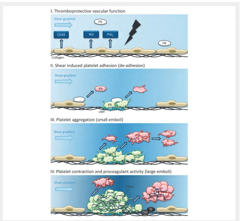
Figure 1 Thrombus growth and instability in a damaged artery. (I) Endothelial activity at the healthy vessel wall prevents platelet (Plt) adhesion. (II) Shear-induced adhesion of platelets at exposed subendothelial collagen/vWF allows platelets to aggregate, partly in a reversible way. (III) During the build-up phase of a thrombus, small emboli are shed. (IV) In a grown thrombus containing contracted and fibrin-anchored platelets, local heterogeneities and blood flow shear gradients allow the shedding of larger emboli.
4. New concepts: dynamic,
fine-tuned regulation of thrombus
growth and stabilization
The classical scheme described above considers arterial thrombus for-mation as a simple progressive process, starting with platelet adhesion and ending with occlusion of the locally activated or damaged vessel. However, many experimental studies, either in vivo with experimentally damaged arteries in mice, or in vitro with flow chambers perfused with whole-blood, point to a more complex organization of the thrombosis process in time. In the macro-circulation (carotid artery) and the micro-circulation (mesenteric and cremaster arterioles), it is often examined that single platelets can adhere and detach during the build-up phase of a thrombus. Furthermore, once a discernible thrombus has been formed, it contracts and tends to shed smaller or larger emboli for a certain period of time (Figure1). Accordingly, flow-dependent dynamics of platelet detachment, embolus shedding, and unstable occlusion seem to be common events during the process of thrombus formation. A large number of studies with genetically modified mice point to the involve-ment of many platelet-derived and coagulant proteins in the dynamic regulation of the stability of thrombi formed at arterial flow conditions (Table1). Hence, the classical concept of thrombus formation needs adjustments accommodating the flow-dynamic components. Refine-ments, particularly, explaining the dynamics and heterogeneities of thrombus build-up and fate, are described below. Participation of vascular- or leucocyte-derived proteins in the control of thrombus sta-bility has hardly been described in the literature.
4.1 Integrin a
IIbb
3activation and reversible
platelet aggregation
Impaired or diminished activation of platelet aIIbb3is known to cause
in-stability of thrombi, which are formed under flow in vivo or in vitro, and to stimulate the detachment of single platelets and small aggregates.44,45In vitro observations support the notion that aIIbb3activation is a reversible
process, and that persistent signalling in platelets is required to keep this integrin in an activated, pro-adhesive conformation.46Several autocrine and paracrine (between platelets) signalling processes appear to con-tribute to persistent integrin activation, and many of these have been shown to be involved in thrombus stabilization. A prominent factor is the release of ADP and its interaction with platelet P2Y12receptors,
which stimulates integrin activation via a pathway involving PI 3-kinase, Akt2, Rap1b, and filamin A (Figure2).46–49Enforced integrin activation is furthermore achieved by interaction of CD40L with its supposed ligand CD40, both of which are membrane proteins that regulate throm-bus stability.50Another mechanism for continued integrin activation is provided by interaction of the soluble molecule Gas6 (present in plasma and limitedly stored in platelets) with the platelet TAM recep-tors, Tyro, Axl, and Mer.51–53New data yet suggest that plasma Gas6 may also stimulate the coagulation process by regulating the expression of vascular tissue factor.54
Limited integrin activation also explains why, at gradients of shear stress, platelets tend to loosely adhere to a growing thrombus via GPIb-V-IX in an often instable way.10Another family of proteins that is considered to modulate platelet integrin function is provided by the tet-raspanins, of which TSSC6 and CD151 are abundantly expressed in the platelet membrane. Tetraspanin control of aIIbb3activation may explain
why the genetic ablation of TSSC6 or CD151 results in thrombus in-stability and increased embolus formation.55,56
Another mechanism controlling the activation of aIIbb3and other
integrins is by redox control of free-cysteine thiols in the extracellular chains, particularly by the protein disulfide isomerase.57 How the redox control affects thrombus stabilization is still unknown.
4.2 Contact-dependent signalling to tight
platelet interactions
Particularly, the work of Brass et al. has led to substantial insight into so-called contact-dependent activation pathways, by which platelets can tightly interact with each other in a thrombus.5,58
Contact-dependent signalling occurs by pairs of ligands and recep-tors, such as ephrin B1-EphA4 (which enforces aIIbb3activation and
pre-vents platelet disaggregation), and semaphorin 4D-plexin (which supports Syk-mediated platelet activation); as well as by tight plate-let – plateplate-let interactions through JAM- and SLAM-family members.5,59 Deficiency in several of these proteins has been found to impair the sta-bilization of mouse thrombi formed in vivo (Table1). Jointly, these inter-action establish close platelet – platelet contacts, which is considered to be a requirement for the stabilizing contraction of platelets in the throm-bus core.58Inside platelets, signalling via Rho-kinase to myosin and actin
appears to be a key mechanism transmitting the contractile forces from the cell surface to the cytoskeleton.60This may explain why arterial thrombi in mice lacking RhoA or myosin heavy chain-9 are characteris-tically unstable and show frequent embolization.23,61
A different set of contact receptors has been identified that negatively regulates platelet activation and thrombus stability. In mice, the absence of the CEACAM1 or ESAM receptors resulted in an increased thrombus growth and reduced embolus shedding.62,63 It is hence tentative to suggest that, within a thrombus, local balances of the platelet-activating and platelet-inhibiting contact signalling events determine which part of the thrombus can contract to form a stable plug, and which part of the thrombus does not contract and is susceptible for detachment of single of aggregated platelets. Intra-thrombus differences in contact-dependent signalling may also explain the reported heterogeneity within a thrombus with contracted, pro-coagulant, and loosely aggre-gated platelets.64However, as described above, also the partial penetra-tion of thrombin and fibrin into a thrombus may contribute to this heterogeneity.
A negative role in thrombus stability has also been observed for the contact protein, connexin 37.65. This is a gap junction protein expressed in platelets, as well as in endothelial cells, smooth muscle cells, mono-cytes, and macrophages.65–67Interestingly, the conclusions from the two publications regarding the role of connexin 37 in platelets are differ-ent: one research group concludes that it functions by limiting platelet activation and thrombus stabilization,65 while the other group finds that it promotes platelet activation.67An approach to take this further would be the generation of mice lacking connexin 37 only in platelets.
4.3 Multisided regulation by coagulation
The role of coagulation in thrombus formation and stabilization appears to be more complex than earlier thought. Thrombin that is formed at the thrombus surface contributes to platelet activation by interaction with PARs.68,69Recent evidence suggests that the contribution of PARs to platelet activation is dependent on the blood flow rate. Whereas PARs ac-tivate platelets at low-shear conditions, their role becomes diminished at pathologically high shear rates.70Yet, thrombin-induced signalling contri-butes to the generation of pro-coagulant PS exposing platelets, which are abundantly formed in arterial thrombi.71There is ongoing research to find
. . . . Table 1 Reported effects of genetic deficiency in mouse on embolization during arterial thrombus formation in vivo or in vitro
Gene defect MGI Protein defect Thrombosis Model Effect on arterial thrombus formation
Emboli Ref(s) Platelet receptors and membrane proteins
Axl 1 347 244 Axl (Gas6 receptor) Flow device Increased thrombus disaggregation + 53
Adra2a 87 934 a2 adrenergic receptor Mesentery/FeCl3 Increased formation of emboli + 95
Cd151 1 096 360 Tetraspanin CD151 Carotis/ligation, cremaster/ laser
Increased thrombus instability ++ 56
Cd40lg 88 337 CD40L Mesentery/FeCl3 Delayed occlusion, more unstable thrombi ++ 50
Fcer1g 95 496 FcR g-chain Carotis/ligation, cremaster/ laser
Smaller thrombi, more emboli formed + 76,96
Gp1ba 1 333 744 GPIba Carotis/ligation Smaller thrombi, reduced stable platelet adhesion ++ 97
Gp5 1 096 363 GPV Mesentery/FeCl3 Smaller thrombi, increased detachment ++ 98,99
Gp6 1 889 810 GPVI Carotis/FeCl3, ligation Smaller thrombi, reduced stable platelet adhesion ++ 100,101
Itga2 96 600 Integrin a2 Mesentery/FeCl3, flow device Smaller thrombi, more emboli formed + 75,102
Lat 1 342 293 LAT Cremaster/ laser Smaller thrombi, more emboli formed ++ 76
Mertk 96 965 Mer (Gas6 receptor) Flow device Increased thrombus disaggregation + 53
P2yr12 1 918 089 P2Y12receptor Mesentery/FlCl3, cremaster/
laser
Smaller thrombi, more emboli formed ++ 47,103
Flow device Increased thrombus embolization ++ 103,104
Slamf1 1 351 314 SLAM (CD84) Mesentery/FeCl3 Delayed occlusion, more emboli formed + 59
Tspan32 1 350 360 Tetraspanin TSSC6 Mesentery/FeCl3 Increased thrombus instability + 55
Tyro3 104 294 Sky (Gas6 receptor) Flow device Increased thrombus disaggregation + 53
Ceacam1 1 347 245 CEACAM1 Mesentery/FeCl3 Larger thrombi, less emboli formed 2 62
Esam 1 916 774 ESAM Cremaster/laser Larger thrombi, less detachment 2 63
Gja4 95 715 Connexin 37 Mesentery/FeCl3\ Increased thrombus formation, less emboli 2 65
Platelet intracellular signalling proteins
Akt2 104 874 Akt2 Carotis/FeCl3 Smaller thrombi, increased instability ++ 105
Cblb 2 146 430 Cbl-b Carotis/FeCl3 Delayed occlusion, unstable thrombi + 106
Flna 95 556 Filamin A Flow device Increased platelet detachment + 107
Myh9 107 717 Myosin heavy chain-9 Carotis/FeCl3 Reduced thrombus growth, more emboli formed + 23
Plcg2 97 616 Phospholipase Cg2 Cremaster/ laser, flow device (smaller) thrombi, increased instability ++ 76,108
Prkaa2 1 336 173 AMPK-a2 Carotis/FeCl3 Less compact thrombus, more emboli formed + 109
Pik3cb 1 922 019 PI 3-kinase-b Flow device Increased thrombus disaggregation ++ 46
Pik3cg 1 353 576 PI 3-kinase-g Flow device Unstable thrombi, increased disassembly ++ 46
Rac1 97 845 Rac1 Cremaster/laser, flow device Increased instability of thrombi ++ 110
Ranbp10 1 921 584 Ran-binding protein 10 Mesentery/FeCl3 Reduced occlusion, unstable thrombi + 24
Rhoa 1 096 342 RhoA Mesentery/FeCl3 Reduced occlusion, more emboli formed ++ 61
Stim1 107 476 STIM1 Mesentery/FeCl3 Delayed occlusion, increased platelet detachment + 111
Plasma proteins
C3 88 227 Complement factor 3 Cremaster/photochemical Delayed thrombus formation, more emboli + 112
F11 99 481 Factor XI Mesentery/FeCl3 Increased detachment of thrombi + 80
F12 1 891 012 Factor XII Carotis/FeCl3, mesentery/FeCl3 Increased detachment of thrombi ++ 80,81
Fgg 95 526 Fibrinogen g-chain Carotis/FeCl3, mesentery/FeCl3 Increased detachment of thrombi ++ 113,114
Fn 95 566 Fibronectin Mesentery/FeCl3 Delayed formation of unstable thrombi ++ 115
Gas6 95 660 Gas6 Flow device Increased thrombus disaggregation + 53
Klk4 1 861 379 Prekallikrein Mesentery/FeCl3 Reduced thrombus formation, more emboli + 81
Lep 104 663 Leptin Carotis/FeCl3 Delayed occlusion, unstable thrombi + 116
Serpine1 97 608 PAI-1 Carotis/FeCl3 Longer time to occlusion, unstable thrombi + 117
Thbs1 98 737 Thrombospondin-1 Mesentery/photochemical Prolonged occlusion, more emboli formed + 118
Vtn 98 940 Vitronectin Carotis/FeCl3, mesentery/FeCl3 Longer time to occlusion, unstable thrombi ++ 117,119
Wvf 98 941 vWF Mesentery/FeCl3(venules) Reduced thrombus formation, unstable + 120
Plg 97 620 Plasminogen Carotis/photochemical Shortened occlusion, less emboli 2 121
Indicated are the mouse genes, the corresponding proteins in blood platelets or plasma, the murine thrombosis model used (flow device in case of in vitro studies), the effect on embolus formation (+, increased; ++, highly increased; 2, decreased).
other strong agonists—besides thrombin—that can support collagen-induced platelet activation (via GPVI). Currently in the spot light are the CLEC-2 receptors, which via an unknown ligand, have been implicated in arterial thrombus formation in mice.72Such strong agonists other
than thrombin may also stimulate the contraction of platelets, making an aggregate stable.60On the other hand, fibrin clot retraction, mediated
via activated aIIbb3integrins, is still considered to be a main mechanism for
platelet contraction in a stabilising thrombus.34
Although it may be obvious that fibrin formation is needed for a stable thrombus, reports on the effects of thrombin inhibitors in arterial models in vivo primarily point to a reduced thrombus growth, rather than to thrombus instability.73–76A certain amount of fibrin formation
yet seems to be important, since in flow devices in vitro the inhibition of
fibrin polymerization resulted in shear-induced shedding of emboli.77
Mechanistically, these findings are not easy to explain. At the one hand, the formation rates of thrombin and fibrin decrease at a higher shear rate, as a consequence of thrombin dilution by blood flow, which suggests that thrombin generation is a limiting factor in arterial thrombus formation.69,78 At the other hand, neither thrombin nor
fibrin is uniformly distributed in an arterial thrombus,73,79which may
imply that a consolidating fibrin network is only present in parts of the thrombus.
Another relevant finding is that especially deficiencies in the intrinsic coagulation pathway (prekallikrein, factor XII, or factor XI) reduce thrombus stability and provoke embolus formation.80,81In agreement
with this, pharmacological inhibition of the factor XII pathway results Figure 2 Key platelet and plasma proteins contributing to thrombus stability. (I) Platelet receptors and ligands involved in initial integrin aIIbb3activation
and reversible platelet aggregation (seeTable 1). The absence of these molecules increases thrombus instability. Also indicated is a box with intracellular signalling proteins controlling this process. II. Contact-dependent signalling mechanisms implicated in platelet contraction and irreversible platelet aggre-gation. Fibrin formed by the coagulation process stabilizes the platelet aggregate. (III) Plasma coagulation factors, via the intrinsic (factor XII, FXII) and ex-trinsic (tissue factor, TF) pathways, mediating platelet-dependent thrombin and fibrin generation, stabilizing a growing thrombus. Also indicated is a primary mechanism of platelet-leucocyte interaction via P-selectin and PSGL-1. See further Versteeg et al.3.
in the formation of large emboli shed from arterial thrombi.82These data point to a thrombus-destabilizing effect upon partial—and likely non-uniform—suppression of the clotting process within a thrombus. More research is needed to understand the precise role of the intrinsic coagulation pathway.
Interestingly, reports on the roles of anticoagulant proteins carried by platelets do not describe effects on thrombus stability. For example, tissue-factor pathway inhibitor located in platelets plays a significant role in the control of thrombus growth, but was not reported to influ-ence thrombus stability.83The same is true for its cofactor, protein S (Calzavarini, Angelillo-Scherrer, unpublished observations, 2012). More thorough studies to the effect of genetic deficiency restricted to mouse platelet proteins are needed to advance this field.
4.4 Disturbances in blood rheology
An aspect that is discussed extensively elsewhere,2is the contribution of blood flow, and in particular of rheological disturbances, on the stabiliza-tion or embolizastabiliza-tion of near-occlusive thrombi. When a stenotic or otherwise vulnerable vessel tends to become occluded, high-shear gra-dients are generated around the growing thrombus. These flow distur-bances will not only accelerate platelet activation and fibrin formation, but also provide the force for embolization of smaller or larger platelet aggregates (Figure1). Flow pulsations by the heart rhythm and vascular distension may further aggravate the extent of embolization and perhaps the size of the emboli, but this has hardly been investigated. Another still poorly studied aspect is how red blood cells—either flowing or when bound to fibrin fibres—contribute to thrombus stabil-ity under arterial flow conditions.
5. Genetic mouse models and flow
chambers: key pathways of thrombus
(in)stability identified?
The above described mechanisms point to involvement of a surprisingly high number of proteins in the vessel wall, platelets, and the coagulating plasma, that contribute to the formation and stabilization of an arterial
thrombus. Table1provides a list of experimental thrombosis studies using mice, where effects have been measured of genetic modification on stable platelet adhesion or shedding of emboli, following damage of vessels of the macro-circulation (carotis artery) or microcirculation (mesenteric or cremaster artery). The evidence for thrombus instability comes from either intravital microscopic observations or rapid changes in blood flow, measured with Doppler probes. Table2gives a list of drug interventions that have been shown to influence thrombus stability in vitro during the perfusion of the human blood through a flow device at a high arterial shear rate.
As indicated in Table1, for 44 different mouse genes a notable change in stability of arterial thrombi has been reported. This list mostly con-cerns genes and proteins that also play a role in the overall process of thrombus growth. In short, referring to the mechanisms described above, this concerns genes implicated in: (i) GPIb-V-IX and GPVI-dependent platelet adhesion (also a2b1, FcR g-chain); (ii)
GPVI-mediated platelet signalling to Ca2+rises and beyond (PLCg2, LAT, Cbl-b, STIM1, Rac1); (iii) integrin activation (PI 3-kinases, Akt2, filamin A, tetraspanins); (iv) autocrine and paracrine regulatory mechanisms supporting integrin activation (P2Y12, CD40, CD40L, Axl, Mer, Sky,
SLAM); (v) and regulation of platelet contraction (RhoA, myosin). Further-more relevant are genes of plasma proteins involved in: (vi) adhesion to platelets (vWF, fibrinogen, fibronectin, vitronectin, thrombospondin-1); (vii) activation of platelets (Gas6, leptin); and (viii) activation of the intrinsic coagulation cascade (prekallikrein, factors XI, XII). Interestingly, hardly any reports are available on thrombus instability due to specific platelet secretion defects. A suppressive role is reported for negative regulators of the contact activation (CEACAM1, ESAM, connexin 37).
The studies with human blood and flow devices to a certain extent support involvement of the same platelet activation pathways in throm-bus stability and embolization in the human system (Table2). In particu-lar, this concerns a role of the aIIbb3activation pathway, in that inhibition
of P2Y receptors, PI 3-kinases, or the integrins themselves results in em-bolization. Thrombus instability is also examined upon inhibition of platelet contraction (EphA4, ephrinB1), Gas6 activity or fibrin forma-tion. More work is clearly needed to demonstrate the importance of the other proteins identified in mouse for the human system.
. . . . Table 2 Reported effects of pharmacological inhibitors on embolization of human thrombi under high-shear flow conditions in vitro
Target protein Inhibitor Effect on thrombus formation ex vivo Emboli Ref(s) Platelet proteins
EphA4/ephrinB1 Soluble fragments Increased platelet disaggregation + 122
IntegrinaIIbb3 Abciximab, eptifibatide Increased platelet disaggregation + 123,124
Myosin heave chain-II Blebbistatin Increased thrombus instability + 125
P2Y1/ P2Y12receptors MRS2179/ticagrelor/2-MeSADP Increased thrombus instability + 44,124,126
AR-C69931MX Increased platelet disaggregation + 46
PI 3-kinase-b TGX-221 Increased platelet disaggregation + 46
RhoA kinase Y-27632 Reduced thrombus formation, increased instability + 60,125
Plasma proteins
Fibrin polymer GPRP More unstable thrombi, releasing platelets + 77
Gas6 Depleted plasma Increased thrombus instability + 53
6. What can we learn more from
patient observations?
In man, thrombosis refers to the pathological condition where thrombi form inopportunely in the lumen of vulnerable vessels, leading to inter-ruption of blood flow, occlusion, and ensuing tissue damage. Antithrom-botic drugs, which comprise antiplatelet, anticoagulant, and anti-fibrinolytic drugs, are commonly used for the treatment and secondary prevention of such thromboses in arteries and veins.2,37The common cause is rupture or erosion of an atherosclerotic plaque or a local dis-turbance in haemodynamic shear forces in the flowing blood. Arterial thrombosis, causing heart attacks, stroke, or limb gangrene, is respon-sible for almost 50% of mortality in industrialized countries. Next to treatments stimulating vasodilatation, antiplatelet drugs are the first choice for treatment of (secondary) arterial thrombosis.84It is relevant to note here that non-steroidal anti-inflammatory drugs, particularly those which inhibit COX2-dependent formation of prostacyclin in the vessel wall, confer a cardiovascular hazard.39Such drugs antagonize the capacities of prostacyclin to suppress platelet activation and vaso-constriction. This can predispose to thrombosis, hypertension, and atherosclerosis.
Patients with a transient ischaemic attack/stroke or myocardial infarc-tion mostly suffer from thrombosis of the atherosclerotic carotid or cor-onary artery. Such patients may present with symptomatic emboli that are shed from the earlier formed thrombi. However, in case of acute stroke or post-operatively after carotid endarterectomy, patients may also develop clinically asymptomatic embolization.85Asymptomatic em-bolization has also been reported following carotid artery stenosis.86In such patients, shedding of platelet emboli from the thrombotic carotid artery can be detected using trans-cranial Doppler ultrasound, for the major part without pathological consequences.87 This indicates that embolization is a frequent phenomenon after a thrombotic event that, although often clinically silent, yet may form an increased risk of becom-ing symptomatic. More research is clearly needed to ascertain this.
Because of the reduced blood flow in the vein system, venous throm-bosis relies more on thrombin and fibrin generation. Patients with venous thrombosis or venous thromboembolism are treated with several types of anticoagulants.88,89 Vitamin K antagonists produce their anticoagulant effect by interfering with the g-carboxylation of vitamin K-dependant prothrombin and factors VII, IX, and X. Unfractio-nated heparins are indirect anticoagulants that bind to antithrombin, enhancing its ability to inhibit activated factor X, thrombin, and other coagulation factors. Low molecular-weight heparins and analogues (danaparoid, fondaparinux) bind to antithrombin, and selectively po-tentiate its anti-factor Xa activity. Drugs like lepirudin (also bivalirudin, argatroban, dabigatran) are used as direct, selective inhibitors of throm-bin, whereas the novel compounds rivaroxaban, apixaban, and edoxa-ban are direct inhibitors of factor Xa. Whereas all these drugs have proved to be clinically effective, there is hardly any knowledge on how their action is determined by the local flow conditions at the site of the thrombus.
An interesting case is provided by patients with specific coagulation defects in the absence of bleeding. Patients with afibrinogenaemia (com-plete fibrinogen deficiency) sometimes develop thrombosis. The thrombotic events can be located in either the arterial or venous terri-tories.90It is considered that in these patients thrombin that is formed is more active, since it cannot be inactivated by binding to fibrin.91One of the consequences is increased platelet activation.92The resulting,
fibrin-poor thrombi are described as large but loosely packed, confirm-ing that fibrin provides thrombus stability.93Interestingly, emboli are fre-quently observed in these patients. Treatment comprises concomitant infusion of fibrinogen and an anticoagulant, capable of binding fibrin-bound and free thrombin, e.g. a direct thrombin inhibitor.
Severe factor XII deficiency may also provoke pulmonary emboliza-tion, e.g. in John Hageman, the index patient with factor XII deficiency. Epidemiological studies show a complex relation between severe factor XII deficiency and increased thrombotic risk.94One of the expla-nations is that complete deficiency in factor XII restricts the formation of fibrin, and facilitates symptomatic embolization in a similar way as observed in murine studies. More translational research is required to link these clinical observations to those of the mouse models.
7. Conclusions: the good and the bad
of arterial thrombus stabilization and
embolus formation
As described above, mouse experimental thrombosis studies in general indicate that arterial thrombus growth and thrombus stability can be linked processes. Many platelet and plasma proteins that control throm-bus growth also appear to play a role in stabilization of the thromthrom-bus. This is directly evident from intravital microscopy observations showing that flow-mediated adhesion of platelets can be a reversible event, and that single platelets as well as small or large platelet aggregates regularly detach from a thrombus even in wild-type mice. Hence, some degree of instability may be considered as a natural phenomenon in arterial thrombus formation. This is illustrated in Figure1, schematizing that during thrombus growth smaller and larger emboli are shed. The limited clinical observations so far indicate that such shedding of emboli also occurs in thrombotic human arteries.
On the other hand, thrombus growth and stability do not seem to be controlled in exactly the same ways. For instance, there are surprisingly few reports on a role of platelet secretion products in thrombus stabil-ization (Table1), whereas platelet secretion is considered to be major determinant of thrombus growth. As schematized in Figure2, key pro-cesses controlling the stability of a thrombus are (i) initial platelet integrin activation controlling platelet aggregation, which can be reversible resulting in shedding of platelet emboli; (ii) contact-dependent signalling, stabilizing the platelet aggregates; and (iii) plasma thrombin and fibrin generation via the intrinsic and extrinsic coagulation pathways, which provides the thrombus with a fibrin network, but still allows shedding of platelet-fibrin emboli (microclots).
In the clinical situation, the shedding of relatively large (fibrin-containing) emboli may be most harmful, giving rise to (semi)occlusive thrombus formation downstream in the vasculature. Clinically silent likely are those emboli that are smaller and prone to disintegration. Pres-ently, we can only speculate on the mechanisms that favour the shedding of large emboli with pathological consequences. An interesting hypoth-esis is that these are due to local or temporal inhomogeneities in a thrombus, e.g. differences in platelet contraction or local incomplete-ness of fibrin formation. Thus, partial inhibition of ‘later’ pathways (irre-versibly contracted platelets, platelet-fibrin clots) may result in emboli that are not only larger, but also more stable themselves and clinically symptomatic. Another possibility is that such emboli are formed by partial thrombolysis due to restricted fibrinolytic activity. This clearly needs further study. More thorough investigation is also needed to
understand the roles of the natural platelet-inhibiting, coagulation-inhibiting and fibrinolytic pathways in the control of flow-dependent thrombus stability.
Conflict of interest: none declared.
Funding
Swiss National Foundation for Scientific Research 310030-135822/1; Center for Translational Molecular Medicine (INCOAG); Netherlands Heart Foun-dation (2011T6); ZonMW (MKMD 114021004).
References
1. Ruggeri ZM, Mendolicchio GL. Adhesion mechanisms in platelet function. Circ Res 2007; 100:1673 – 1685.
2. Jackson SP. Arterial thrombosis: insidious, unpredictable and deadly. Nat Med 2011;17: 1423 – 1436.
3. Versteeg HH, Heemskerk JW, Levi M, Reitsma PS. New fundamentals in hemostasis. Physiol Rev 2013;93:327 – 358.
4. Jackson SP. The growing complexity of platelet aggregation. Blood 2007;109: 5087 – 5095.
5. Brass LF, Wannemacher KM, Ma P, Stalker TJ. Regulating thrombus growth and stability to achieve an optimal response to injury. J Thromb Haemost 2011;9(Suppl. 1):66 – 75. 6. Kroll MH, Hellums JD, McIntire LV, Schafer AI, Moake JL. Platelets and shear stress.
Blood 1996;88:1525 – 1541.
7. Sakariassen KS. Thrombus formation on apex of arterial stenoses: the need for a fluid high shear stenosis diagnostic device. Future Cardiol 2007;3:193 – 201.
8. Ruggeri ZM. Platelet-vessel wall interactions in flowing blood. In: Colman RW et al., eds. Hemostasis and Thrombosis. Philadelphia, PA, USA: Lippincott; 2001. p683 – 698. 9. Ruggeri ZM, Orje JN, Habermann R, Federici AB, Reininger AJ. Activation-independent
platelet adhesion and aggregation under elevated shear stress. Blood 2006;108: 1903 – 1910.
10. Nesbitt WS, Westein E, Tovar-Lopez FJ, Tolouei E, Mitchell A, Fu J et al. A shear gradient-dependent platelet aggregation mechanism drives thrombus formation. Nat Med 2009;15:665 – 673.
11. Westein E, van der Meer AD, Kuijpers MJ, Frimat JP, van den Berg A, Heemskerk JW. Atherosclerotic geometries spatially confine and exacerbate pathological thrombus formation poststenosis in a von Willebrand factor-dependent manner. Proc Natl Acad Sci USA 2013;110:1357 – 1362.
12. Ruggeri ZM, Dent JA, Saldı´var E. Contribution of distinct adhesive interactions to plate-let aggregation in flowing blood. Blood 1999;94:172 – 178.
13. Wu YP, Vink T, Schiphorst M, van Zanten GH, IJsseldijk MJ, de Groot PG et al. Platelet thrombus formation on collagen at high shear rates is mediated by von Willebrand factor-glycoprotein Ib interaction and inhibited by von Willebrand factor-glycoprotein IIb/IIIa interaction. Arterioscler Thromb Vasc Biol 2000;20:1661 – 1667.
14. Elvers M, Stegner D, Hagedorn I, Kleinschnitz G, Braun A, Kuijpers MJ et al. Impaired integrin aIIbb3 activation and shear-dependent thrombus formation in mice lacking phospholipase D1. Sci Signal 2010;3:ra101.
15. Stegner D, Nieswandt B. Platelet receptor signaling in thrombus formation. J Mol Med 2011;89:109 – 121.
16. Farndale RW, Sixma JJ, Barnes MJ, de Groot PG. The role of collagen in thrombosis and haemostasis. J Thromb Haemost 2004;2:561 – 573.
17. Siljander PR, Munnix IC, Smethurst PA, Deckmyn H, Lindhout T, Ouwehand WH et al. Platelet receptor interplay regulates collagen-induced thrombus formation in flowing human blood. Blood 2004;103:1333 – 1341.
18. Auger JM, Kuijpers MJ, Senis YA, Watson SP, Heemskerk JW. Adhesion of human and mouse platelets to collagen under shear: a unifying model. FASEB J 2005;19:825 – 827. 19. Watson SP, Auger JM, McCarty OJ, Pearce AC. GPVI and integrin aIIbb3signaling in
pla-telets. J Thromb Haemost 2005;3:1752 – 1762.
20. Varga-Szabo D, Braun A, Nieswandt B. STIM1 and Orai1 in platelet function. Cell Calcium 2011;50:70 – 278.
21. Van de Walle G, Schoolmeester A, Iserbyt BF, Cosemans JM, Heemskerk JW, Hoylaerts MF et al. Activation of aIIbb3 is sufficient but also an imperative prerequisite to activate a2b1 on platelets. Blood 2007;109:595 – 602.
22. Jurk K, Clemetson KJ, de Groot PG, Brodde MF, Steiner M, Savion N et al. Thrombospondin-1 mediates platelet adhesion at high shear via glycoprotein Ib (GPIb): an alternative/backup mechanism to von Willebrand factor. FASEB J 2003;17: 1490 – 1492.
23. Le´on C, Eckly A, Hechler B, Aleil B, Freund M, Ravanat C et al. Megakaryocyte-restricted MYH9 inactivation dramatically affects hemostasis while preserving platelet aggrega-tion and secreaggrega-tion. Blood 2007;110:3183 – 3191.
24. Meyer I, Kunert S, Schwiebert S, Hagedorn I, Italiano JE, Dutting S et al. Altered micro-tubule equilibrium and impaired thrombus stability in mice lacking RanBP10. Blood 2012;120:3594 – 3602.
25. Gibbins JM. Platelet adhesion signalling and the regulation of thrombus formation. J Cell Sci 2004;117:3415 – 3425.
26. Nesbitt WS, Mangin P, Salem HH, Jackson SP. The impact of blood rheology on the mo-lecular and cellular events underlying arterial thrombosis. J Mol Med 2006;84:989 – 995. 27. Wei AH, Schoenwaelder SM, Andrews RK, Jackson SP. New insights into the
haemo-static function of platelets. Br J Haematol 2009;147:415 – 430.
28. Mackman N, Tilley RE, Key NS. Role of the extrinsic pathway of blood coagulation in hemostasis and thrombosis. Arterioscler Thromb Vasc Biol 2007;27:1687 – 1693. 29. Mu¨ller F, Mutch NJ, Schenk WA, Smith SA, Esterl L, Spronk HM et al. Platelet
polypho-sphates are proinflammatory and procoagulant mediators in vivo. Cell 2009;139: 1143 – 1156.
30. Van der Meijden PE, Munnix IC, Auger JM, Govers-Riemslag JW, Cosemans JM, Kuijpers MJ et al. Dual role of collagen in factor XII-dependent thrombus and clot for-mation. Blood 2009;114:881 – 890.
31. Chen VM, Ahamed J, Versteeg HH, Berndt MC, Ruf W, Hogg PJ. Evidence for activation of tissue factor by an allosteric disulfide bond. Biochemistry 2006;45:12020 – 12028. 32. Owens AP, Mackman N. Role of tissue factor in atherothrombosis. Curr Atheroscler Rep
2012;14:394 – 401.
33. Monroe DM, Hoffman M. What does it take to make the perfect clot? Arterioscler Thromb Vasc Biol 2006;26:41 – 48.
34. Heemskerk JW, Mattheij N, Cosemans JM. Platelet-based coagulation: different popula-tions, different functions. J Thromb Haemost 2013;11:2 – 16.
35. Reininger AJ, Bernlochner I, Penz SM, Ravanat C, Smethurst P, Farndale RW et al. A 2-step mechanism of arterial thrombus formation induced by human atherosclerotic plaques. J Am Coll Cardiol 2010;55:1147 – 1158.
36. Cosemans JM, Schols SE, Stefanini L, de Witt S, Feijge MA, Hamulyak K et al. Key role of glycoprotein Ib/V/IX and von Willebrand factor in platelet activation-dependent fibrin formation at low shear flow. Blood 2011;117:651 – 660.
37. Borissoff JI, Spronk HM, ten Cate H. The hemostatic system as a modulator of athero-sclerosis. N Engl J Med 2011;364:1746 – 1760.
38. Moncada S, Higgs EA. Nitric oxide and the vascular endothelium. Handb Exp Pharmacol 2006;176:213 – 254.
39. Funk CD, FitzGerald GA. COX-2 inhibitors and cardiovascular risk. J Cardiovasc Phar-macol 2007;50:470 – 479.
40. Johnston-Cox HA, Ravid K. Adenosine and blood platelets. Purinergic Signal 2011;7: 357 – 365.
41. Darbousset R, Thomas GM, Mezouar S, Fre`re C, Bonier R, Mackman N et al. Tissue factor-positive neutrophils bind to injured endothelial wall and initiate thrombus for-mation. Blood 2012;120:2133 – 2143.
42. Fuchs TA, Brill A, Duerschmied D, Schatzberg D, Monestier M, Myers DD et al. Extra-cellular DNA traps promote thrombosis. Proc Natl Acad Sci USA 2010;107: 15880 – 15885.
43. Von Bru¨hl ML, Stark K, Steinhart A, Chandraratne S, Konrad I, Lorentz M et al. Mono-cytes, neutrophils and platelets cooperate to initiate and propagate venous thrombosis in mice in vivo. J Exp Med 2012;209:819 – 835.
44. Goto S, Tamura N, Ishida H, Ruggeri ZM. Dependence of platelet thrombus stability on sustained glycoprotein IIb/IIIa activation through adenosine 5-diphosphate receptor stimulation and cyclic calcium signaling. J Am Coll Cardiol 2006;47:155 – 162. 45. Cosemans JM, Iserbyt BF, Deckmyn H, Heemskerk JW. Multiple pathways to switch
platelet integrins on and off. J Thromb Haemost 2008;6:1253 – 1261.
46. Cosemans JM, Munnix IC, Wetzker R, Heller R, Jackson SP, Heemskerk JW. Continuous signaling via phosphoinositide 3-kinase isoforms b and g is required for platelet ADP receptor function in dynamic thrombus stabilization. Blood 2006;108:3045 – 3052. 47. Andre´ P, Delaney SM, LaRocca T, Vincent D, DeGuzman F, Jurek M et al. P2Y12regulates
platelet adhesion/activation, thrombus growth, and thrombus stability in injured arter-ies. J Clin Invest 2003;112:398 – 406.
48. Canobbio I, Stefanini L, Cipolla L, Ciraolo E, Gruppi C, Balduini C et al. Genetic evidence for a predominant role of PI3Kb catalytic activity in ITAM- and integrin-mediated signal-ing in platelets. Blood 2009;114:2193 – 2196.
49. Nieswandt B, Varga-Szabo D, Elvers M. Integrins in platelets. J Thromb Haemost 2009; 7(Supp. 1):206 – 209.
50. Andre´ P, Srinivasa Prasad KS, Denis CV, He M, Papalia JM, Hynes RO et al. CD40L sta-bilizes arterial thrombi by a b3integrin-dependent mechanism. Nat Med 2002;8: 247 – 252.
51. Angelillo-Scherrer A, Garcia de Frutos P, Aparicio C, Melis E, Savi P, Lupu F et al. Defi-ciency or inhibition of Gas6 causes platelet dysfunction and protects mice against thrombosis. Nat Med 2001;7:215 – 221.
52. Angelillo-Scherrer A, Burnier L, Flores N, Savi P, DeMol M, Schaeffer P et al. Role of Gas6 receptors in platelet signaling during thrombus stabilization and implications for antithrombotic therapy. J Clin Invest 2005;115:237 – 246.
53. Cosemans JM, van Kruchten R, Olieslagers S, Schurgers LJ, Verheyen FK, Munnix ICA et al. Potentiating roles for Gas6 and Tyro, Axl and Mer (TAM) receptors in human and murine platelet activation and thrombus stabilization. J Thromb Haemost 2010;8: 1797 – 1808.
54. Foley JH, Conway EM. Gas6 gains entry into the coagulation cascade. Blood 2013;121: 570 – 571.
55. Goschnick MW, Lau LM, Wee JL, Liu YS, Hogarth PM, Robb LM et al. Impaired outside-in integrin aIIbb3signaling and thrombus stability in TSSC6-deficient mice. Blood 2006;108:1911 – 1918.
56. Orlowski E, Chand R, Yip J, Wong C, Goschnick MW, Wright MD et al. A platelet tetra-spannin superfamily member, CD515, is required for regulation of thrombus growth and stability in vivo. J Thromb Haemost 2009;7:2074 – 2084.
57. Essex DW. Redox control of platelet function. Antioxid Redox Signal 2009;11: 1191 – 1225.
58. Brass LF, Zhu L, Stalker TJ. Minding the gaps to promote thrombus growth and stability. J Clin Invest 2005;115:3385 – 3392.
59. Nanda N, Andre P, Bao M, Clauser K, Deguzman F, Howie D et al. Platelet aggregation induces platelet aggregate state stability via SLAM family receptor signalling. Blood 2005; 106:3028 – 3034.
60. Ono A, Westein E, Hsiao S, Nesbitt WS, Hamilton JR, Schoenwaelder SM et al. Identi-fication of a fibrin-independent platelet contractile mechanism regulating primary hemostasis and thrombus growth. Blood 2008;112:90 – 99.
61. Pleines I, Hagedorn I, Gupta S, May F, Chakarova L, van Hengel J et al. Megakaroycyte-specific RhoA deficiency causes macrothrombocytopenia and defective platelet activa-tion in hemostasis and thrombosis. Blood 2012;119:1054 – 1063.
62. Wong C, Liu Y, Chand R, Wee JL, Oates L, Nieswandt B et al. CEACAM1 negatively reg-ulates platelet-collagen interactions and thrombus growth in vitro and in vivo. Blood 2009; 113:1818 – 1828.
63. Stalker TJ, Wu J, Morgans A, Traxler EA, Wang L, Chatterjee MS et al. Endothelial cell specific adhesion molecule (ESAM) localizes to platelet-platelet contacts and regulates thrombus formation in vivo. J Thromb Haemost 2009;7:1886 – 1896.
64. Munnix IC, Kuijpers MJ, Auger JM, Thomassen CM, Panizzi P, van Zandvoort MA et al. Segregation of platelet aggregatory and procoagulant microdomains in thrombus for-mation. Regulation by transient integrin activation. Arterioscler Thromb Vasc Biol 2007; 27:2484 – 2490.
65. Angelillo-Scherrer A, Fontana P, Burnier L, Roth I, Sugamele R, Brisset A et al. Connexin 37 limits thrombus propensity by downregulating platelet reactivity. Circulation 2011; 124:930 – 939.
66. Chanson M, Kwak BR. Connexin 37: a potential modifier gene of inflammatory disease. J Mol Med 2007;85:787 – 795.
67. Vaiyapuri S, Jones CI, Sasikumar P, Moraes LA, Munger SJ, Wright JR et al. Gap junctions and connexin hemichannels underpin hemostasis and thrombosis. Circulation 2012;125: 2479 – 2491.
68. Siljander P, Farndale RW, Feijge MA, Comfurius P, Kos S, Bevers EM et al. Platelet adhe-sion enhances the glycoprotein VI-dependent procoagulant response: involvement of p38 MAP kinase and calpain. Arterioscler Thromb Vasc Biol 2001;21:618 – 627. 69. Berny MA, Munnix IC, Auger JM, Schols SE, Cosemans JM, Panizzi P et al. Spatial
distri-bution of factor Xa, thrombin, and fibrin(ogen) on thrombi at venous shear. Plos One 2010;5:e10415.
70. Lee H, Sturgeon SA, Jackson SP, Hamilton JR. The contribution of thrombin-induced platelet activation to thrombus growth is diminished under pathological blood shear conditions. Thromb Haemost 2012;107:328 – 337.
71. Munnix IC, Strehl A, Kuijpers MJ, Auger JM, van der Meijden PE, van Zandvoort MA et al. The glycoprotein VI-phospholipase Cg2 signaling pathway controls thrombus forma-tion induced by collagen and tissue factor in vitro and in vivo. Arterioscler Thromb Vasc Biol 2005;25:2673 – 2678.
72. May F, Hagedorn I, Pleines I, Bender M, Vo¨gtle T, Eble J et al. CLEC-2 is an essential platelet-activating receptor in hemostasis and thrombosis. Blood 2009;114: 3464 – 3472.
73. Furie B, Furie BC. Thrombus formation in vivo. J Clin Invest 2005;115:3355 – 3362. 74. Mangin P, Yap CL, Nonne C, Sturgeon SA, Goncalves I, Yuan Y et al. Thrombin
over-comes the thrombosis defect associated with platelet GPVI/FcRg deficiency. Blood 2006;107:4346 – 4353.
75. Kuijpers MJ, Pozgajova M, Cosemans JM, Munnix IC, Eckes B, Nieswandt B et al. Role of murine integrin a2b1in thrombus stabilization and embolization: contribution of thromboxane A2. Thromb Haemost 2007;98:1072 – 1080.
76. Kalia N, Auger JM, Atkinson B, Watson SP. Critical role of FcR g-chain, LAT, PLCg2 and thrombin in arteriolar thrombus formation upon mild, laser-induced endothelial injury in vivo. Microcirculation 2008;15:325 – 335.
77. Colace TV, Muthard RW, Diamond SL. Thrombus growth and embolism on tissue factor-bearing collagen surfaces under flow. Role of thrombin with and without fibrin. Arterioscler Thromb Vasc Biol 2012;32:1466 – 1476.
78. Okorie UM, Denney WS, Chatterjee MS, Neeves KB, Diamond SL. Determination of tissue factor thresholds that trigger coagulation versus venous and arterial shear rates: amplification of 100 fM circulating tissue factor requires flow. Blood 2008;111: 3507 – 3513.
79. Welsh JD, Colace TV, Muthard RW, Stalker TJ, Brass LF, Diamond SL. Platelet-targeting sensor reveals thrombin gradient within blood clots forming in microfluidic assay in mouse. J Thromb Haemost 2012;10:2344 – 2353.
80. Renne´ T, Pozgajova M, Gru¨ner S, Schuh K, Pauer HU, Burfeind P et al. Defective throm-bus formation in mice lacking coagulation factor XII. J Exp Med 2005;202:271 – 281. 81. Revenko AS, Gao D, Crosby JR, Bhattacharjee G, Zhao C, May C et al. Selective
deple-tion of plasma prekallikrein or coaguladeple-tion factor XII inhibits thrombosis in mice without increased risk of bleeding. Blood 2011;118:5302 – 5311.
82. Hagedorn I, Schmidbauer S, Pleines I, Kleinschnitz C, Kronthaler U, Stoll G et al. Factor XIIa inhibitor recombinant human albumin infestin-4 abolishes occlusive arterial thrombus formation without affecting bleeding. Circulation 2010;121:1510 – 1517.
83. Maroney SA, Cooley BC, Ferrel JP, Bonesho CE, Mast AE. Murine hematopoietic cell tissue factor pathway inhibitor limits thrombus growth. Arterioscler Thromb Vasc Biol 2011;31:821 – 826.
84. Ruggeri ZM. Platelets in atherothrombosis. Nat Med 2002;8:1227 – 1234.
85. King A, Markus HS. Doppler embolic signals in cerebrovascular disease and prediction of stroke risk: a systematic review and meta-analysis. Stroke 2009;40:3711 – 3717. 86. Markus HS, King A, Shipley M, Topakian R, Cullinane M, Reihill S et al. Asymptomatic
embolisation for prediction of stroke in the Asymptomatic Carotid Emboli Study (ACES): a prospective observational study. Lancet Neurol 2010;9:663 – 671. 87. Kruis RW, Vlasveld FA, van Dijk D. The (un)importance of cerebral microemboli. Semin
Cardiothorac Vasc Anesth 2010;14:111 – 118.
88. Maan A, Padmanabhan R, Shaikh AY, Mansour M, Ruskin JN, Heist EK. Newer anticoa-gulants in cardiovascular disease: a systematic review of the literature. Cardiol Rev 2012; 20:209 – 221.
89. Schulman S. Advances in the management of venous thromboembolism. Best Pract Res Clin Haematol 2012;25:361 – 377.
90. Girolami A, de Marinis GB, Bonamigo E, Lombardi AM. Recombinant FVIIa concentrate-associated thrombotic events in congenital bleeding disorders other than hemophilias. Hematology 2012;17:346 – 349.
91. De Bosch NB, Mosesson MW, Ruiz-Sa´ez A, Echenagucia M, Rodriguez-Lemoin A. In-hibition of thrombin generation in plasma by fibrin formation (antithrombin I). Thromb Haemost 2002;88:253 – 258.
92. Korte W, Feldges A. Increased prothrombin activation in a patient with congenital afi-brinogenemia is reversible by fibrinogen substitution. Clin Invest 1994;72:396 – 398. 93. Remijn JA, Wu YP, IJsseldijk MJ, Zwaginga JJ, Sixma JJ, de Groot PG. Absence of
fibrino-gen in afibrinofibrino-genemia results in large but loosely packed thrombi under flow condi-tions. Thromb Haemost 2001;85:736 – 742.
94. Zeerleder S, Schloesser M, Redondo M, Wuillemin WA, Engel W, Furlan M et al. Re-evaluation of the incidence of thromboembolic complications in congenital factor XII deficiency: a study on 73 subjects from 14 Swiss families. Thromb Haemost 1999;82: 1240 – 1246.
95. Pozgajova M, Sachs UJ, Hein L, Nieswandt B. Reduced thrombus stability in mice lacking the a2A-adrenergic receptor. Blood 2006;108:510 – 514.
96. Dubois C, Panicot-Dubois L, Merrill-Skoloff G, Furie B, Furie BC. Glycoprotein VI-dependent and -independent pathways of thrombus formation in vivo. Blood 2006; 107:3902 – 3906.
97. Massberg S, Gawaz M, Gru¨ner S, Schulte V, Konrad I, Zohlho¨fer D et al. A crucial role of glycoprotein VI for platelet recruitment to the injured arterial wall in vivo. J Exp Med 2003;197:41 – 49.
98. Moog S, Mangin P, Lenain N, Strassel C, Ravanat C, Schuhler S et al. Platelet glycoprotein V binds to collagen and participates in platelet adhesion and aggregation. Blood 2001;98: 1038 – 1046.
99. Ni H, Ramakrishnan V, Ruggeri ZM, Papalia JM, Phillips DR, Wagner DD. Increased thrombogenesis and embolus formation in mice lacking glycoprotein V. Blood 2001; 98:368 – 373.
100. Bender M, Hagedorn I, Nieswandt B. Genetic and antibody-induced glycoprotein VI de-ficiency equally protects mice from mechanically and FeCl3-induced thrombosis. J Thromb Haemost 2011;9:1423 – 1426.
101. Gru¨ner S, Prostredna M, Koch M, Miura Y, Schulte V, Jung SM et al. Relative antithrom-botic effect of soluble GPVI dimer compared with anti-GPVI antibodies in mice. Blood 2005;105:1492 – 1499.
102. Neeves KB, Maloney SF, Fong KP, Schmaier AA, Kahn ML, Brass LF et al. Microfluidic focal thrombosis model for measuring murine platelet deposition and stability: PAR4 signaling enhances shear-resistance of platelet aggregates. J Thromb Haemost 2008;6: 2193 – 2201.
103. Stolla M, Stefanini L, Roden RC, Chavez M, Hirsch J, Greene T et al. The kinetics of aIIbb3 activation determines the size and stability of thrombi in mice: implications for antiplate-let therapy. Blood 2011;117:1005 – 1013.
104. Nergiz-Unal R, Cosemans JM, Feijge MA, van der Meijden PE, Storey RF, van Giezen JJ et al. Stabilizing role of platelet P2Y12receptors in shear-dependent thrombus forma-tion on ruptured plaques. Plos One 2010;5:e10130.
105. Woulfe D, Jiang H, Morgans A, Monks R, Birnbaum M, Brass LF. Defects in secretion, aggregation and thrombus formation in platelets from mice lacking Akt2. J Clin Invest 2004;113:441 – 450.
106. Daniel JL, Dangelmaier CA, Mada S, Buitrago L, Jin J, Langdon WY et al. Cbl-b is a novel physiological regulator of glycoprotein VI-dependent platelet activation. J Biol Chem 2010;285:17282 – 17291.
107. Falet H, Pollitt AY, Begonja AJ, Weber SE, Duerschmied D, Wagner DD et al. A novel interaction between FlnA and Syk regulates platelet ITAM-mediated receptor signaling and function. J Exp Med 2011;207:1967 – 1979.
108. Rathore V, Wang D, Newman DK, Newman PJ. Phospholipase Cg2 contributes to stable thrombus formation on VWF. FEBS Lett 2004;27:26 – 30.
109. Randriamboavonjy V, Isaak J, Fromel T, Viollet B, Fisslthaler B, Preissner KT et al. AMPK a2subunit is involved in platelet signaling, clot retraction and thrombus stability. Blood 2010;116:2134 – 2140.
110. McCarty OJ, Larson MK, Auger JM, Kalia N, Atkinson BT, Pearce AC et al. Rac1 Is essen-tial for platelet lamellipodia formation and aggregate stability under flow. J Biol Chem 2005;280:39474 – 39484.
111. Varga-Szabo D, Braun A, Kleinschnitz C, Bender M, Pleines I, Pham M et al. The calcium sensor STIM1 is an essential mediator of arterial thrombosis and ischemic brain infarc-tion. J Exp Med 2008;205:1583 – 1591.
112. Gushiken FC, Ha H, Li J, Rumbaut RE, Afshar-Kharghan V. Abnormal platelet function in C3-deficient mice. J Thromb Haemost 2009;7:865 – 870.
113. Ni H, Denis CV, Subbarao S, Degen JL, Sato TN, Hynes RO et al. Persistence of platelet thrombus formation in arterioles of mice lacking both von Willebrand factor and fi-brinogen. J Clin Invest 2000;106:385 – 392.
114. Jirouskova M, Chereshnev I, Vaananan H, Degen JL, Coller BS. Antibody blockade or mutation of the fibrinogen g-chain C-terminus is more effective in inhibiting murine ar-terial thrombus formation than complete absence of fibrinogen. Blood 2004;103: 1995 – 2002.
115. Ni H, Yuen PTS, Papalia JM, Trevithick JE, Sakai T, Fa¨ssler R et al. Plasma fibronectin pro-motes thrombus growth and stability in injured arterioles. Proc Natl Acad Sci USA 2003; 100:2415 – 2419.
116. Konstantinides S, Schafer K, Neels JG, Dellas C, Loskutoff DJ. Inhibition of endogenous leptin protects mice from arterial and venous thrombosis. Arterioscler Thromb Vasc Biol 2004;24:2196 – 2201.
117. Koschnick S, Konstantinides S, Schafer K, Crain K, Loskutoff DJ. Thrombotic phenotype of mice with a combined deficiency in plasminogen activator inhibitor 1 and vitronectin. J Thromb Haemost 2005;3:2290 – 2295.
118. Bonnefoy A, Daenens K, Feys HB, De Vos R, Vandervoort P, Vermylen J et al. Thrombospondin-1 controls vascular platelet recruitment and thrombus adherence in mice by protecting (sub)endothelial vWF from cleavage by ADAMTS-13. Blood 2006;107:955 – 964.
119. Reheman A, Gross P, Yang H, Chen P, Allen D, Leytin V et al. Vitronectin stabilizes thrombi and vessel occlusion but plays a dual role in platelet aggregation. J Thromb Haemost 2005;3:875 – 883.
120. Chauhan AK, Kisucka J, Lamb CB, Bergmeier W, Wagner DD. Von Willebrand factor and factor VIII are independently required to form stable occlusive thrombi in injured veins. Blood 2007;109:2424 – 2429.
121. Matsuno H, Kozawa O, Okada K, Ueshima S, Matsuo O, Uematsu T. Plasmin generation plays different roles in the formation and removal of arterial and venous thrombus in mice. Thromb Haemost 2002;87:98 – 104.
122. Pre´vost N, Woulfe DS, Jiang H, Stalker TJ, Marchese P, Ruggeri ZM et al. Eph kinases and ephrins support thrombus growth and stability by regulating integrin outside-in signal-ing in platelets. Proc Natl Acad Sci USA 2005;102:9820 – 9825.
123. Speich HE, Furman RR, Lands LT, Moodie GD, Jennings LK. Elevating local concentra-tions of GPIIb-IIIa antagonists counteracts platelet thrombus stability. J Thromb Thromb-olysis 2012 (in press).
124. Hosokawa K, Ohnishi T, Fukasawa M, Kondo T, Sameshima H, Koide T et al. A microchip flow-chamber system for quantitative assessment of the platelet thrombus formation process. Microvasc Res 2012;83:154 – 161.
125. Calaminus SD, Auger JM, McCarty OJ, Wakelam MJ, Mecheskys LM, Watson SP. Myo-sinIIa contractility is required for maintenance of platelet structure during spreading on collagen and contributes to thrombus stability. J Thromb Haemost 2007;5:2136 – 2145. 126. Stephens G, He M, Wong C, Jurek M, Luedemann HC, Shapurian G et al. Development of a perfusion chamber assay to study in real time the kinetics of thrombosis and the antithrombotic characteristics of antiplatelet drugs. Thromb J 2012;10:11.