Original article
Sequential administration of temozolomide and fotemustine: Depletion of
O
6-alkyl guanine-DNA transferase in blood lymphocytes and in tumours
M. Gander,
1S. Leyvraz,
1L. Decosterd,
2M. Bonfanti,
3C. Marzolini,
2F. Shen,
2D. Lienard,
1L. Perey,
1G. Colella,
3J. Biollaz,
2F. Lejeune,
1D. Yarosh,
4M. Belanich
4& M. D'lncalci
3x
Centre Pluridisciplinaire d'Oncologie, 2Division de Pharmacologie Clinique, Departement de Medecine, Centre Hospitalier Universitaire Vaudois, Lausanne, Switzerland; 3Istitutodi Ricerche Farmacologiche Mario Negri, Milano, Italy; 4Applied Genetics Inc., Freeport, NY, USA
Summary
Background: The DNA repair protein O6-alkylguanine-DNA alkyl transferase (AT) mediates resistance to chloroethylnitro-soureas. Agents depleting AT such as DTIC and its new ana-logue temozolomide (TMZ) can reverse resistance to chloro-ethylnitrosoureas. We report the results of a dose finding study of TMZ in association with fotemustine.
Patients and methods: Twenty-four patients with metastatic
melanoma or recurrent glioma were treated with escalating dose of oral or intravenous TMZ ranging from 300 to 700 mg/m2, divided over two days. Fotemustine 100 mg/m2 was given intravenously on day 2, 4 hours after TMZ. AT depletion was measured in peripheral blood mononuclear cells (PBMCs) and in selected cases in melanoma metastases and was com-pared to TMZ pharmacokinetics.
Results: The maximum tolerated dose (MTD) of TMZ was
400 mg/m2 (200 mg/m2/d) when associated with fotemustine the 2nd day with myelosuppression as dose limiting toxicity. The decrease of AT level in PBMCs was progressive and reached 34% of pretreatment values on day 2. There was how-ever wide interindividual variability. AT reduction was neither dose nor route dependent and did not appear to be related to TMZ systemic exposure (AUC). In the same patients, AT depletion in tumour did not correlate with the decrease of AT observed in PBMCs.
Conclusions: PBMCs may not be used as a surrogate of
tumour for AT depletion. Further study should concentrate on the pharmacokinetic pharmacodynamic relationship in tumour to provide the basis for individually tailored therapy.
Key words: melanoma, pharmacodynamics, pharmacokinetics,
temozolomide
Introduction
Chloroethylnitrosoureas are widely used to treat solid tumours including brain tumours and metastatic mela-nomas [1]- They act through the formation of DNA interstrand crosslinks starting with a carbonium ion attack at the Opposition of the guanine and 6 to 12 hours later binding to the opposite cytidine [2, 3]. There is a good correlation between the number of interstrand crosslinks and the cytotoxicity induced by nitrosoureas [4]. The number of crosslinks depends on the ability of the cell to remove the chloroethyl group from the gua-nine before the crosslink is established. This removal is due to the O6-alkylguanine DNA alkyltransferase (AT) protein [4, 5]. AT transfers the alkyl group from the DNA to an AT internal cysteine residue in an irreversible and self-inactivating reaction [6]. Restoration of activity requires new protein synthesis [7]. AT level in human tumour cells correlates inversely with cytotoxicity of the chloroethylnitrosoureas [8-11]. In vitro resistance to nitrosourea can be overcome by the use of methylating agents [12] or O6-benzylguanine [13] to deplete AT levels in tumour cells. In the rat, dacarbazine (DTIC) has been shown to be able to deplete AT [14].
In the clinic, a sequential administration of DTIC and nitrosourea produced a 20% response rate in meta-static melanoma [15]. Lee et al. have shown that AT depletion of peripheral blood mononuclear cells was dependent on the dose of DTIC. However, the inactiva-tion of AT reached a plateau at the highest dose level of DTIC, suggesting a saturation of its hepatic metabolism into 5-(3 methyl-l-triazeno)imidazole-4-carboxamide (MTIC), its active metabolite [16].
Temozolomide (TMZ) is a recently synthetised imi-dazotetrazinone derivative. Its activation is independent of the hepatic microsomal system. At physiological pH, TMZ undergoes spontaneous degradation to generate the methylating compound MTIC [17]. TMZ is able to reverse the resistance to nitrosourea of a L1210 cell subline in vitro [18]. In phase I studies, leucopenia and thrombocytopenia were the dose limiting toxicities at 1000 mg/m2 [19]. Antitumour activity was observed in the treatment of malignant glioma and metastatic mela-noma [19-21].
Interaction between TMZ and nitrosoureas has never been studied in the clinic. We initiated a study of a sequential administration of TMZ and fotemustine (diethyl-l-3-(2-chloroethyl)-3-nitrosoureido ethyl
phos-Table 1. Patient characteristics. Characteristics Total Sex Male Female
Mean performance status Mean age (in years) Primary Malignant melanoma High-grade glioma Previous treatment None Chemotherapy Without DTIC With DTIC Immunotherapy Radiotherapy n (range) 24 14 10 1 (0-2) 51(31-71) 22 2 12 2 4 4 2 Route and dose of temozolomide (mg/m2, divided over two days) 300 i.v. 4 500 i.v. 4 700 i.v. 1 300 p.o. 3 500 p.o. 6 400 i.v./p.o. 4 400 p.o./i.v. 2
phonate), a third generation nitrosourea which showed high activity against metastatic melanoma and brain metastases from melanoma [22, 23]. The aims of the study were the following: a) to define the maximal tolerated dose (MTD) of TMZ administered four hours before a fixed dose of fotemustine b) to study the AT depletion in peripheral blood mononuclear cells and in selected tumour biopsies c) to measure the pharmaco-kinetics of TMZ in order to relate them to AT depletion.
Vaudois (Lausanne) in 20 mg and 100 mg gelatine capsules, accord-ing to the recommendations of the CRC. Capsules were accord-ingested during a light meal. The total dose was also divided over two days. Ondansetron (8 mg i.v.) was given prophylactically as antiemetic to all patients.
Dose escalation of TMZ was performed in two groups of patients: one receiving intravenous infusion, the other oral administration. The starting dose level of TMZ was 300 mg/m2 (150 mg/m2 x 2 days). Depending on the encountered toxicity, dose escalation was planned to 500 mg/m2 (250 mg/m2/d) and then to 700 mg/m2 (350 mg/m2/d). Toxicity was defined as dose limiting if any grade III or IV toxicity according to WHO criteria was observed [24]. Three consecutive patients were treated at each dose level. If one patient experienced a dose limiting toxicity (DLT), three additional patients were enrolled at the same dose level. If two patients reached DLT, this dose level was considered as the maximal tolerated dose (MTD). After two patients experienced DLT at the 500 mg/m2 dose level, the dose of TMZ was then decreased to 400 mg/m2. In order to study its bioavailability, it was administered to four patients intravenously on day 1 and orally on day 2, and to two patients orally on day 1 and intravenously on day 2.
Fotemustine was supplied by IRIS, Courbevoie, France as 4 ml vials containing 200 mg freezed dried powder that was dissolved in ethanol. The solution was diluted in a further 250 ml isotonic glucose and administered as a one hour intravenous infusion. Flask and tubing were protected from light. Fotemustine was administered at the dose of 100 mg/m2 intravenously on day 2, four hours after the beginning of TMZ administration, time interval after which the maximum AT depletion by DTIC has been reported [25],
Due to the limited amount of TMZ available at the time of the study, TMZ was replaced by DTIC 500 mg/m2 i.v. for the second and subsequent cycles. The treatment was repeated at a four-week interval until disease progression.
Toxicity and response evaluation
Follow-up consisted of a weekly blood cell count. Liver and lung function tests were done before the second cycle of treatment, then every two cycles. Toxicity was graded according to WHO [24]. Anti-tumour response was evaluated before the second cycle of treatment, then every two cycles according to WHO response criteria.
Pharmacokinetics Patients and methods
Patients with histologically proven metastatic disseminated melanoma or recurrent high-grade glioma were eligible for the study. The inclu-sion criteria were ^ 1 8 years, performance status $ 2 according to Eastern Cooperative Oncology Group (ECOG), normal hematological, hepatic, renal and pulmonary function tests. Each patient had to give a written informed consent prior to enrolment in the study. Patients having received chemotherapy within eight weeks or having a history of lung irradiation were not included. The study was approved by the local ethics committee.
Drug formulation and treatment schedule
TMZ was supplied by the Cancer Research Campaign (CRC, London, UK) in 5 ml vials containing 150 mg TMZ in dimethylsulfoxide (DMSO). The total dose was diluted in 500 ml normal saline and was administered as a one-hour constant rate infusion. In order to achieve a substantial decrease of AT at the time of fotemustine administration, TMZ treatment was divided over two days. TMZ was planned to be administered by intravenous infusion, but, during the course of the study, TMZ became available only as a bulk powder. It was then formulated at the Pharmacy of the Centre Hospitalier Universitaire
Pharmacokinetics of TMZ were studied in plasma on day 1 and 2. Two ml blood samples were collected into heparinised tubes prior to TMZ administration and at 30', 1-hour, 1-hour 30', 2-, 3-, 4-, 6-, 12- and 24-hour post administration on days 1 and 2. Blood samples were immediately centrifuged at 2000 rpm for 10' at 4 °C. An aliquot of I ml plasma was acidified with 0.1 ml 1 M HCI and stored at - 2 0 °C for later analysis.
TMZ assay was performed as previously described [26, 27]. In brief, matrix components were first eliminated by solid phase extrac-tion on a 100 mg C\% cartridge, with subsequent desorpextrac-tion of TMZ with methanol. The resulting eluate was evaporated under nitrogen at room temperature and reconstituted in 200 ul of 0.5% acetic acid before HPLC analysis on an ODS-column with MeOH 0.5% AcOH, 10 : 90 as solvent system. The lower limit of quantitation was 0.2 ug/ml. The specifications of the analytical methods were in accordance with the recommendations promulgated by the Conference Report on bio-analytical validation [28]. Pharmacokinetic analysis was carried out using the computer program SIPHAR (4.0, SIMED, Creteil, France). Pharmacokinetic parameters were calculated by non compartmental approach. The elimination rate constant was determined as the slope of monoexponential curve-fit of logarithmic plasma concentrations
versus time. The area under plasma concentration-time curve (AUC)
was obtained by the linear trapezoidal method and the residual area from the last data point to infinity was estimated as the plasma concentration at this time divided by the elimination rate constant.
O*-Alkylguanine DNA alkyltransferase measurement
Heparinized blood samples of 10 ml were collected beforeTMZ and 2, 4, 8 and 24 hours after TMZ administration in prechilled syringes. Peripheral blood mononuclear cells (PBMCs) were separated from heparinized blood using Ficoll-Hypaque, washed with phosphate buffered saline and stored at - 8 0 °C. In selected patients with skin metastases of malignant melanoma, biopsies were taken one to three days before and four hours after the beginning of the administration of TMZ, time interval after which AT inactivation by DTIC reaches its nadir [25]. Biopsy samples were immediately frozen in liquid nitrogen. AT assay of PBMCs and of tissue samples was performed as reported elsewhere [25]. Samples (1-3 x 10⁶ PBMCs or 30-90 mg tumor tissue) were sonicated, the supernatants were assayed using 10 ug (3H)-methyl DNA that had been reacted with N-(3 H)-methyl-N-nitrosourea 20 Ci/mmol. Activity was expressed as fmol methyl trans-ferred to protein per mg of protein. AT depletion was expressed as absolute decline (in fmol/mg) and as percentage of pretreatment value. The limit of quantitation of the assay was 10 fmol/mg of protein. The coefficient of variation of the same sample determined in the same day or in different days of the same week, was within 10% for PBMCs and 15% for tumour samples.
Quantitative immunofluorescence microscopy
Quantitative immunofluorescence microscopy was performed as de-scribed [29, 30]. Briefly, two paraffin slides from each tumour sample were stained in parallel, one with a monoclonal antibody against human alkyltransferase and another without antibody as a negative control. A positive control of HT29 tumour cells was also included. The slides were double-labeled with DAPI to delineate the nuclei and secondary anti-mouse FITC-labeled antibody to visualize AT-specific staining. The level of fluorescence in nuclei was calculated from the digitized epifluorescence image, and converted to molecules per nucleus using bead standards that related fluorescence intensity to molecules per unit area.
Statistical analysis
The relationship between parameters was evaluated by determination of the standard linear correlation coefficient (Pearson). The paired /-test was used to test whether differences between PBMCs and tumour AT pretreatment values and those at two time intervals were signifi-cantly different from zero [31, 32].
Results
Patients characteristics
Characteristics of the 24 patients are presented in Table 1. Six patients with metastatic malignant melano-ma had received prior chemotherapy and four of them a regimen containing DTIC. Two patients with glioma had a recurrent disease after surgery and radiotherapy. Nine patients received TMZ as an i.v. infusion, nine patients as p.o. capsules and six patients p.o. and i.v., or the reverse sequence. Among the 24 patients, one was not assessable for toxicity due to rapid disease progression.
Dose escalation
Dose escalation in relation to toxicity is presented in Table 2. Myelosuppression was the dose limiting toxicity
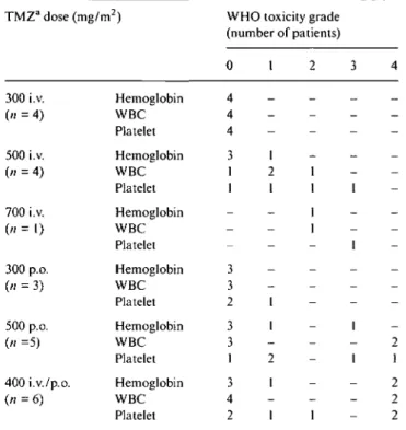
Table 2. Hematological toxicity.
TMZa dose (mg/m ) 300 i.v. (" = 4) 500 i.v. (n = 4) 700 i.v. (« = 1) 300 p.o. (« = 3) 500 p.o. (« =5) 400i.v./p.o. (n=6) Hemoglobin WBC Platelet Hemoglobin WBC Platelet Hemoglobin WBC Platelet Hemoglobin WBC Platelet Hemoglobin WBC Platelet Hemoglobin WBC Platelet
WHO toxicity grade (number of patients) 0 4 4 4 3 1 1 ; 3 3 2 3 3 1 3 4 2 1 -1 2 1 1 1 2 1 1 2 -1 1 1 1 : i 3 -1 1 1 1 -4 -— 2 1 2 2 2 " TMZ - indicated temozolomide.
of the sequential administration of TMZ and fotemus-tine. At the 500 mg/m2 dose level (250 mg/m2/d) two among the five evaluable in the p.o. groiup had leuco-cytopenia and thromboleuco-cytopenia grade 3-4. In the i.v. infusion group, one among four evaluable patients had thrombocytopenia grade 3. All the patients who experi-enced DLT at this dose level were non pretreated. The patient at 700 mg/m2 i.v. (350 mg/m2/d) also developed grade 3 thrombocytopenia. Among the six patients who received 400 mg/m2 (200mg/m2/d) orally and intrave-nously, two reached a DLT consisting in WHO grade 4 leucopenia, thrombopenia and anemia. One of these two patients had received nitrosourea as previous che-motherapy. The dose of 400 mg/m2 (200 mg/m2/d) should be considered as the MTD of TMZ when asso-ciated with fotemustine 100 mg/m2 on day 2, four hours after TMZ. Duration of myelosuppression in patients who experienced WHO grade 3-4 toxicity was a median of 35 days (range 26-41 days) and was similar for leucocytes and platelets. WHO grade 1 and 2 nausea occured in 28% of the patients despite the antiemetic prophylaxis by ondansetron. No respiratory symptoms were observed after one cycle of treatment. Lung func-tion tests did not show any modificafunc-tion.
We cannot present data on the cumulative toxicity of this regimen, since only one cycle of TMZ and fotemus-tine was administered. Among the 19 patients who received 39 further cycles of DTIC and Fotemustine, 5 experienced WHO grade 3-4 leuco- and thrombocyto-penia. The toxic events occurred after three cycles in three patients without toxicity after TMZ and fotemus-tine but after the first cycle in two patients who had had
a grade 4 toxicity after TMZ and fotemustine. Three patients experienced transitory lung toxicity (two WHO grade 2, one grade 4), after three (one patient) and four cycles (two patients) of DTIC-Fotemustine (i.e., after a cumulative dose of DTIC of 1500 and 2000 mg/m2).
Pharmacokinetics
Pertinent pharmacokinetics parameters are reported in Table 3. A more comprehensive detailed pharmaco-kinetic evaluation of TMZ is reported elsewhere [28]. Four out of six patients received the MTD (400 mg/m2 divided over two days) i.v. on day 1 and p.o. on day 2 while the last two were on the opposite regimen. Under controlled conditions (standardized food intake and posture) the mean bioavailabilty in six patients was 0.96 ± 0.1 without any noticeable influence of the adminis-tration sequence. Pharmacokinetics of TMZ was linear after i.v. administration with AUC increasing proportion-nally to the dose in mg/m2 (r - 0.86 and 0.91 for day 1
and day 2, respectively). When considering all patients having received TMZ by the oral route, the observed AUC were lower and more variable indicating a marked inter-patient variability, possibly influenced by the con-ditions of capsule administration.
As shown in Figure 1, there was some correlation
Table 3. Pharmacokinetic parameters of temozolomide.
TMZ dose (mg/m2) 150 i.v. 200 i.v. 250 i.v. 350 i.v. 150 p.o. 200 p.o. 250 p.o. No. of pts AUC (mg min/1) mean ± SD mean ± SDg
Day 1 Day 2 Day 1 Day 2 1187 ±237 1704 ± 149 2238 ± 396 2819 1206 ±200 1229 ± 14 1383 ±298 1153 ± 122 1376 ± 24 1888 ±289 2367 1108 ±306 1677 ±201 1447 ± 388 7.2 ± 2 . 4 7.1 ±1.8 9.1 ± 1 . 8 7 . 5 ± 0 12.7 ±1.3 10.1 ±2.0 14.5 11.8 5.3 ± 2 . 0 7.1 ± 1.1 7.2 ± 2 . 3 4.2 ± 1.7 8.9 ± 1.5 7.7 ±5.7 a AT 200 mg/m2, the four patients on i.v. day 1 were the same as on p.o. day 2 and the two patients on p.o. day 1 the same as on i.v. day 2. Time range of Cmax was after i.v. 60-98 minutes on day 1, 50-72 minutes on day 2, after p.o. 40-180 minutes on day 1 and 42-240 minutes on day 2.
(Pearson correlation test) between the degree of leuco-cytopenia and thromboleuco-cytopenia (percent decrease count at the nadir) and TMZ AUC after i.v. administra-tion (r = 0.68, P = 0.04 and r = 0.72, P = 0.03, respec-tively). After oral administration however, TMZ AUC was not a predictive index of leucocyte and thrombocyte
(a) 100 "I
I
re ; 8 • o 3 o o 80 60 40 -20 " o --20 I.V. Direct correlation coefficient (Pearson) r = 0.68 p = 0.04 1000 3000 5000 TMZ AUC (mg.min/L) p.o. (c) 100 -\ 50 " •o S o --50 o o 1000 3000 5000 T M Z A U C (mg.min/L) i.v./p.o. p.o./i.v. (e) 100 £ 80 -o w n £ 60 " £ 40 " 20 " o o • o iv/po po/iv 1000 3000 5000 T M Z A U C (mg.min/L) (b) 100 "I £ o (0 re a u o 73 yt e U 2 80 " 60 40 -20 " Direct correlation coefficient (Pearson) r = 0.72 p = 0.03 1000 3000 5000 TMZ AUC (mg.min/L) (d) oo -80 " 60 " 40 " 20 " o -o -o o o %o o 1 * 1 1000 3000 5000 TMZ AUC (mg.min/L) (f) 100 "I £ o (0 n s o o • D 2 >< 3s
80 " 60 " 40 " 20 " • • iv/po po/iv 1000 3000 5000 TMZ AUC (mg.min/L)Figure I. Relationship between temozolomide area-under-the-curve on day 1 and 2 (TMZ AUC) and the degree of leuco- and thrombocytopenia
(pretreatment count minus count at the nadir/ pretreatment count) in patients who received TMZ intravenously (i.v.), orally (p.o.), intravenously on day 1 and orally on day 2 (i.v./p.o.) and orally on day 1 and intravenously on day 2 (p.o./i.v.) in association with fotemustine. TMZ was given on day 1 and 24 hours later on day 2. Fotemustine was given four hours after TMZ on day 2.
120
300-20
Hours
Figure 2. Mean alkyltransferase (AT) decrease in peripheral blood
mononuclear cells (PBMC) of all patients ± standard errors of the means (SEM).
toxicity. Substantial decrease in thrombocyte and in leucocyte counts was observed at lower TMZ AUC when the patients received TMZ by oral route, in com-parison with those who received TMZ by i.v. infusion.
Pharmacodynamic assessment of AT depletion
AT level in PBMCs was measured in 20 patients on day 1, in 19 patients on day 2 and in 6 patients also after DTIC, on day 29. Pretreatment values were scattered ranging from 121 to 1147 fmol/mg protein (mean 376 fmol/mg). In the whole group, mean AT levels after treatment showed a progressive and cumulative decrease corresponding to (± SEM) 62% ± 8% of pretreatment value after eight hours, 52% ± 14% at the time of fote-mustine administration and reaching its lowest value at 48 hours with 34% ± 7% (Figure 2). AT levels at eight hours on day 1 and at four hours on day 2 were statisti-cally different from pretreatment values values (P < 0.0004). The absolute AT decline (pretreatment minus AT level at the nadir) was directly related to the pretreat-ment AT level with a correlation coefficient of 0.97. The most prominent feature was, however, a wide interindi-vidual variation in the extent of the decline of AT, even in patients treated at the same dose level. Figure 3, for example, shows the AT levels in the group of patients who were given TMZ 250 mg/m2/day by i.v. infusion. Due to this high interpatient variability, we were unable to assess differences between doses and routes of admin-istration. Similarly AT decrease could not be related to TMZ systemic exposure (TMZ plasma AUC, Figure 4). Complete AT depletion, was observed at each dose level, in five patients. AT decrease was observed in patients who had received previous treatment as well as in non pretreated patients. However, two out of three patients who had received DTIC as previous chemotherapy had no or minimum AT depletion (nadir at 108 and and 77%
0 8 16 24 32 40 48 56 Hours
I tt
T T F
Figure 3. Alkyltransferase (AT) levels (fmol/mg) in peripheral blood
mononuclear cells in four patients who received Temozolomide (T) 250 mg/m2 i.v. T was given on day I and 24 hours later on day 2. Fotemustine (F) was administered four hours after temozolomide on day 2. V (0 (0 £ o o • o < IUU -80 " 60 " 40 " 20 o --20 " • • I* • • • • 1000 2000 3000 4000 5000
TMZ A U C (mg.min/L)
6000Figure 4. Relationship between the temozolomide
area-under-the-curve on day I and 2 and the degree of alkyltransferase (AT) depletion (percent decrease at the AT nadir) in peripheral blood mononuclear cells (PBMCs).
of pretreatment level, respectively). Ten patients had partial or complete recovery of their initial AT level on day I and five on day 2. PBMCs AT depletion was not related to myelosuppression (data not shown).
On day 29, AT level reached the pretreatment values in five among six assessed patients. One patient retained a low AT level at 22% of its pretreatment value. AT depletion achieved by 500 mg/m2 DTIC i.v. on day 29
was more rapid than that observed on day 1 and 2 after TMZ at 250 mg/m2/d i.v., but the nadir of AT depletion was similar.
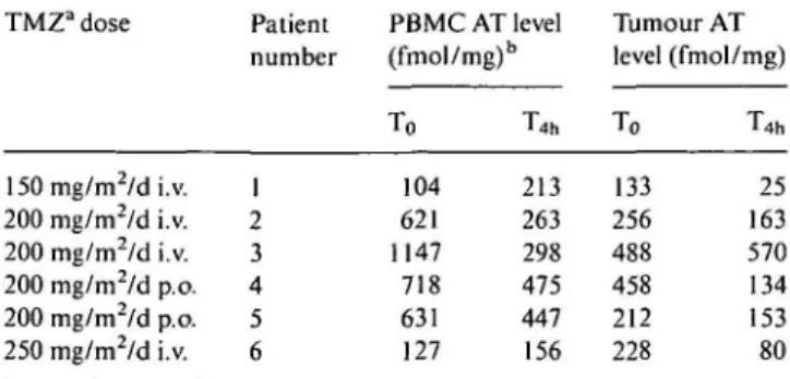
Six patients had biopsies of melanoma skin metas-tases before and four hours after TMZ administration. There was also wide interindividual variations in pre-treatment levels of AT in tumour (mean value 296 fmol/mg, range 133-488 fmol/mg). After TMZ, mean AT level was 56% ± 15% of pretreatment level in tumour. It decreased from 19% to 72% in all but one patient in whom AT level increased by 16%. As shown in Table 4, there was, in three patients, a discrepancy between AT inactivation in peripheral blood lympho-cytes and in tumours. Two patients (patients no. 1 and no. 6, Table 4) had a decrease in their tumour AT activity and an increase in their PBMCs AT, one patient (patient no. 3, Table 4) had an increase in tumour AT and a decrease in PBMCs AT. The extent of AT depletion in tumor could not be related to TMZ dose in this small group of patients. The maximum AT decrease was even observed in a patient treated at the TMZ lowest dose level (patient no. 1, Table 4).
Quantitative immunohistochemistry of AT gave results similar to those obtained with the biochemical method. For example, a section from a melanoma skin metas-tasis biopsy taken prior to TMZ treatment showed much greater AT-specific immunofluorescence than a section taken 4 hours after treatment (Figure 5). Quantitation of the nuclear AT-specific staining in this patient showed a reduction from an average of 44,000 AT molecules per nucleus before to 16,000 molecules per nucleus after treatment. The AT appeared to be localized in the cyto-plasm prior to treatment (panel a), as observed previ-ously [30], and was depleted from both the nucleus and cytoplasm after TMZ treatment (panel b).
Clinical assessment and relationship with AT depletion
The antitumour response was not a main goal of this study. Tumour evaluation on day 28 revealed tumour progression in five patients and stable disease in 19 patients. Three patients with stable disease had minor tumour regression: one had the disappearance of multi-ple skin metastases with the persistance of visceral, pulmonary and hepatic lesions, another had a radio-logical improvement of a lung infiltrate and the third had a 25% radiological decrease of an iliac lymph node. The 19 patients with stable disease received the treat-ment as planned with DTIC and fotemustine. Objective responses were observed in three patients (16%): one complete response in the patient with the improvement of a lung infiltrate and 2 partial remissions (in the patient with the disappearance of skin metastases and in another one with lung metastases).
AT levels in melanoma were measured in 6 out of 24 patients. This small number did not allow to draw definite conclusion regarding the relationship between the AT decrease in the tumour and the clinical response. Noticeably however, the patient with melanoma skin metastasis experiencing a complete response had a 70%
Table 4. AT level in peripheral blood mononuclear cells and in tumour
(six patients). TMZa dose 150 mg/m2/d i.v. 200 mg/m2/d i.v. 200 mg/m2/d i.v. 200 mg/m2/d p.o. 200 mg/m /d p.o. 250 mg/m2/d i.v. Patient number 1 2 3 4 5 6 PBMC AT level (fmol/mg)b To 104 621 1147 718 631 127 T4h 213 263 298 475 447 156 Tumour AT level To 133 256 488 458 212 228 (fmol/mg) T4h 25 163 570 134 153 80 a TMZ - indicates temozolomide.
b PBMC AT level for alkyl-transferase level in peripheral blood mono-nuclear cells.
(a)
* •
Figure 5. Alkyltransferase (AT) immunofluorescence in sections from
biopsies taken before (a) and four hours after (b) Temozolomide. The AT-specific antibody staining in the sections was visualized with FITC-secondary antibodies. In the sample taken before (a) note the cyto-plasmic localization with relatively low nuclear fluorescence (arrow).
decrease of AT level in the tumour biopsy (patient no. 4), while another patient (no. 3) with rapid tumour progres-sion had a 17% increase of AT level in the excised tumour.
Discussion
Part of the resistance to nitrosourea is mediated through the O6-alkylguanine-DNA alkyltransferase protein (AT), which is depleted by TMZ in vitro and in vivo [33, 34]. To our knowledge, this is the first report of the clinical application of the sequential administration of TMZ and nitrosourea. TMZ administration was divided over two days to achieve a substantial decrease of AT at the time of fotemustine administration. The main toxicity was myelosuppression, including thrombocytopenia and anemia. The MTD of TMZ was 400 mg/m2 divided over two days when given in combination with fotemustine
100 mg/m2 the second day, four hours after TMZ. When the study was initiated, TMZ was available as injection form only, it then became available as oral formulation. This form is currently approved for general use. Thrombocytopenia and leucocytopenia were more pronounced when TMZ was administered by the oral route as seen in Figure 1. The toxicity was however not predictable but occurred generally at lower AUC as
compared to i.v. administration. Since we confirmed that the bioavailability of TMZ was close to 1 (i.e. AUC p.o./AUC i.v. = 1), the difference, observed in this small group of patients, reflects interpatient variability and susceptibility to TMZ rather than route dependency. However, some unrecognized effect of TMZ adminis-tered by oral route cannot be fully excluded.
For further phase II trials of TMZ in association with a nitrosourea, a starting dose of 300 mg/m" (150 mg/m2/day) divided over two days should be recom-mended. This dose should be escalated in patients with-out toxicity.
However, the cumulative toxicity of this regimen, and in particuliar the risk of lung toxicity, is not known, since only one cycle of TMZ and fotemustine was administered in this study. The pulmonary toxicity that occurred in three patients after further treatment with DTIC and fotemustine has been previously reported after this regimen, and a cumulative dose of DTIC of 1500 mg/m" [15]. Signs of such a toxicity should be carefully looked for in future phase II trials of TMZ in association with a nitrosourea.
TMZ clearly showed a linear pharmacokinetics with AUC increasing proportionally with dose after i.v. ad-ministration. Pharmacokinetic parameters and bioavail-ability values found in this study [28] are in good accordance with those previously published [19].
The progressive and cumulative AT depletion in PBMCs with wide interindividual variation has been previously reported after DTIC [16, 25] and after TMZ [34]. It has been hypothesized that interindividual varia-bility in AT depletion was due to differences in pharma-cokinetics of TMZ. However, in our study, the variabil-ity of TMZ pharmacokinetics did not influence the absolute, relative or cumulative AT decrease. AT deple-tion by DTIC was dose dependent [16] but we could not demonstrate the same dose-response effect after TMZ. Indeed, there was no clear relationship between the TMZ dose administered and the extent of AT depletion.
Previous reports have mentioned the heterogeneity of AT level in liver metastases from colon cancer [32] and in skin metastases from melanoma [35]. In the same patient a variation of up to 400% among lesions was also observed [35]. Although we cannot rule out that the lower AT levels we observed in melanoma skin metas-tases after treatment might be due to interlesional heter-ogeneity, the consistency of the decrease strongly suggests that it is an effect of the treatment. Interestingly enough, AT depletion in tumour and in PBMCs were not related. An analogue observation has been reported in seven patients with liver metastases from colon cancer treated with streptozotocin and BCNU [32]. These observations indicate that depletion of AT in PBMC is a marker for production of O6-alkylguanine in DNA, but the extent of depletion in PBMCs may not accurately reflect the extent of AT depletion in metastases from melanoma and colon cancer. Inaccessibility prevented AT decrease assessment in patients with glioma. A direct relationship between AT depletion in glioma and in PBMCs can therefore not be excluded.
In our small group of patients maximum AT deple-tion was observed at all TMZ dose levels in PBMCs and at the lowest dose level in melanoma. Moreover, AT depletion in tumour and in PBMCs could not be directly related to TMZ exposure. If the aim of the TMZ treat-ment is to sensitize tumour to the action of chloroethyl-nitrosoureas, it may not be necessary to use the MTD. Modulation of AT depletion by TMZ might be better achieved by individualizing the dose of TMZ. Most of dose adjustment strategies described to date rely on pharmacokinetic measurements and on dose adjuste-ment based on pharmacokinetic-pharmacodynamic models [36]. In clinical practice, it has been successfully applied to define the doses of methotrexate, carboplatin, etoposide, teniposide, 5-fluorouracil and suramin [37]. Modulation of AT could be a future approach and should concentrate on AT measurement in tumour. The ultimate aim is to provide a model, based on the meas-urement of AT and its depletion in tumour, to tailor the administration of AT depleting agents to the individual patient. Among the agents designed to inactivate AT, O6-benzylguanine has recently received considerable at-tention. Unlike TMZ, O6-benzylguanine acts as a direct inactivator of AT without interaction with DNA and has no intrinsic tumour activity [38, 39]. In a phase I study in patients with glioma, it depleted completely AT levels in glioma without occurrence of toxicity [39]. Phase I trials defining the optimal dose of the chloroethylnitrosourea carmustine to be combined with O6-benzylguanine are ongoing. Preliminary reports seem however to show a substantial enhancement of the hematologic toxicity of carmustine by O6-benzylguanine requiring the dose of carmustine to be decreased [40]. It is however still not clear which approach offers the best therapeutic window: either a complete depletion by agents such a O6 -benzyl-guanine but resulting in increased end organ toxicities necessitating a decrease in the dosage of the alkylating agent or only a partial depletion by methylating agents such as TMZ which have intrinsic cytotoxic activities.
Acknowledgements
We wish to thank J. Kibitel and Y. Garcia for their technical assistance in immunostaining, and Ms M. Gonin for secretarial help, as well as MEDIC founda-tion for financial support. Gordon McVie and David Secher from the Cancer Research Campaign are grate-fully acknowledged for the gift of temozolomide and Bruno Giroux from Servier Laboratories for the gift of fotemustine.
References
1. Berger NA. Alkylating agents. In De Vita VT Jr, Hellman S, Rosenberg SA (eds): Cancer Principles and Practice of Oncology. Philadelphia: JB Lippincott 1993; 400-9.
2. Kohn K.W. Interstrand cross-linking of DNA by 1,3-bis (2-chloro-ethyl)-l-nitrosourea and other l-(2-halo(2-chloro-ethyl)-l-nitrosoureas. Cancer Res 1977; 37: 1450-4.
3. Tong WP, Kirk MC, Ludlum DB. Formation of the cross-link l-(Afi-deoxycytidyl),2-(A'/-deoxyguanosinyl)ethane in DNA treated with N.N'-bis (2-chloroethyl)-A'-nitrosourea. Cancer Res 1982; 42: 3102-5.
4. Erickson LC, Laurent G, Sharkey NA, Kohn K.W. DNA cross-linking and monoadduct repair in nitrosourea-treated human tumour cells. Nature 1980; 288: 727-9.
5. D'lncalci M, Citti L, Taverna P, Catapano CV. Importance of the DNA repair enzyme O6"alkyl guanine alkyltransferase (AT) in cancer chemotherapy. Cancer Treat Rev 1988; 15: 279-92. 6. Olsson M, Lindahl T. Repair of alkylated DNA in Escherichia
coli. Methyl group transfer from O6-methylguanine to a protein cysteine residue. J Biol Chem 1980; 255: 10569-71.
7. Yarosh DB, Rice M, Day RS et al. O6-methylguanine-DNA methyltransferase in human cells. Mutat Res 1984; 131: 27-36. 8. Domoradzki J, Pegg AE, Dolan ME et al. Correlation between
O6-methylguanine-DNA-methyltransferase activity and resistance of human cells to the cytotoxic and mutagenic effect of N-methyl-N'-nitro-N-nitrosoguanidine. Carcinogenesis 1984; 5: 1641-7. 9. Brent TP, Houghton PJ, Houghton JA. O6-alkylguanine-DNA
alkyl transferase activity correlates with the therapeutic response of human rhabdomyosarcoma xenografts to l-(2-chloroethyl)-3-(trans-4-methylcyclohexyl)-l-nitrosourea. Proc Natl Acad Sci USA 1985; 82: 2985-9.
10. Dolan ME, Norbeck L, Clyde C et al. Expression of mammalian O6-alkylguanine-DNA alkyltransferase in a cell line sensitive to alkylating agents. Carcinogenesis 1989; 10:1613-9.
11. Schold SC, Brent TP, von Hofe E et al. O6-Alkylguanine-DNA alkyl transferase and sensitivity to procarbazine in human brain-tumor xenografts. J Neurosurg 1989; 70: 573-7.
12. Zlotogorski C, Erickson LC. Pretreatment of human colon tumor cells with DNA methylating agents inhibits their ability to repair chloroethyl monoadducts. Carcinogenesis (London) 1984; 5: 83-7. 13. Dolan ME, Moschel RC, Pegg AE. Depletion of mammalian O6 -alkylguanine-DNA alkyltransferase activity by O6-benzylguanine provides a means to evaluate the role of this protein in protection against carcinogenic and therapeutic alkylating agents. Proc Natl Acad Sci USA 1990; 87: 5368-72.
14. Meer L, Schold SC, Kleihues P. Inhibition of the hepatic O6-alkylguanine-DNA alkyltransferase in vivo by pretreatment with antineoplastic agents. Biochem Pharmacol 1989; 38: 929-34. 15. Gerard B, Aamdal S, Lee SM et al. Activity and unexpected lung toxicity of the sequential administration of two alkylating agents - dacarbazine and fotemustine - in patients with melanoma. Eur J Cancer 1993; 29A: 711-9.
16. Lee SM, Thatcher N, Dougal M, Margison GP. Dosage and cycle effects of dacarbazine (DTIC) and fotemustine on O6 -alkylgua-nine-DNA alkyltransferase in human peripheral blood mono-nuclear cells. Br J Cancer 1993; 67: 216-21.
17. Stevens MFG, Hickman JA, Langdon SP et al. Antitumor activity and pharmacokinetics in mice of 8-carbamoyl-3-methyl-imidazo(5,l-d)-l,2,3,5-tetrazin-4(3H)-one (CCRG 81045; M&B 39831), a novel drug with potential as an alternative to dacarba-zine. Cancer Res 1987; 47: 5846-52.
18. D'lncalci M, Taverna P, Erba E et al. O6-methylguanine and temozolomide can reverse the resistance to chloroethylnitrosour-eas of a mouse L12I0 leukemia. Anticancer Res 1991; 11: 115-22. 19. Newlands ES, Blackledge GRP, Slack JA et al. Phase I trial of
temozolomide. (CCRG 81045: M&B 39831: NSC 362856). Br J Cancer 1992; 65: 287 91.
20. O'Reilly SM, Newlands ES, Glaser MG et al. Temozolomide: A new oral cytotoxic chemotherapeutic agent with promising activity against primary brain tumours. Eur J Cancer 1993; 29A: 940 2.
21. Bleehen NM, Newlands ES. Lee SM et al. Cancer Research Campaign phase II trial of temozolomide in metastatic melano-ma. J Clin Oncol 1995: 13: 910-3.
22. Jacquillat C, Khayat D. Banzet P et al. Final report on the french multicenter phase II study of the nitrosourea fotemustine in 153 evaluable patients with disseminated melanoma, including pa-tients with cerebral metastases. Cancer 1990; 66: 1873-8.
23. Jacquillat C, Khayat D, Banzet P et al. Chemotherapy by fote-mustine in cerebral metastases of disseminated melanoma. Cancer Chemother Pharmacol 1990; 25: 263-9.
24. Wolrd Health Organization. Handbook for Reporting Results of Cancer Treatment. WHO Offset Publication No. 48. Geneva: WHO 1979.
25. Lee SM, Thatcher N, Margison GP. O6-alkylguanine-DNA alkyl-transferase depletion and regeneration in human peripheral lym-phocytes following dacarbazine and fotemustine. Cancer Res 1991; 51: 619-23.
26. Shen F, Decosterd LA, Gander M et al. Determination of temo-zolomide in human plasma and urine by high-performance liquid chromatography after solid-phase extraction. J Chromatogr Bi-omedAppl 1995; 667: 291-300.
27. Marzolini C, Decosterd LA, Shen F et al. Pharmacokinetics of temozolomide in association with fotemustine in malignant melanoma and malignant glioma patients: Comparison of oral, intravenous and hepatic intraarterial administration. Cancer Chemother Pharmacol 1998; 42: 433-40.
28. Shah VP, Midha KK, Dighe S et al. Analytical methods valida-tion: Bioavailability, bioequivalence, and pharmacokinetic studies. Conference report. Eur J Drug Metab Pharmacokinet 1991; 16: 249-55.
29. Belanich M, Ayi TC, Li BF et al. Analysis of O6 -methylguanine-DNA methyltransferase in individual human cells by quantitative immunofluorescence microscopy. Oncol Res 1994; 6: 129-37. 30. Belanich M, Randall T, Pastor MA et al. Intracellular
localiza-tion and intercellular heterogeneity of the human DNA repair protein O6-methylguanine-DNA methyltransferase. Cancer Chemother Pharmacol 1996; 37: 547-55.
31. Fisher RA, Yates F. Statistical Tables for Biological, Agricultural and Medical Research. Edinburgh: Oliver and Boyd 1974. 32. Willson JKV, Haaga JR, Trey JE et al. Modulation of O6
-alkyl-guanine alkyltransferase-directed DNA repair in metastatic colon cancers. J Clin Oncol 1995; 13: 2301-8.
33. Mitchell RB, Dolan ME. Effect of temozolomide and dacar-bazine on O6-alkylguanine-DNA alkyltransferase activity and sensitivity of human tumor cells and xenografts to l,3-bis(2-chloro-ethyl)-l-nitrosourea. Cancer Chemother Pharmacol 1993; 32: 59-63.
34. Lee SM, Thatcher N, Crowther D. Margison GP. Inactivation of O6-alkylguanine-DNA alkyltransferase in human peripheral blood mononuclear cells by temozolomide. Br J Cancer 1994; 69: 452-6.
35. Egyhazi S, Hansson J, Ringborg U. O6-Methylguanine-DNA methyltransferase activities in biopsies of human melanoma tu-mours. Br J Cancer 1995; 71: 37-9.
36. Kobayashi K, Jodrell DI, Ratain MJ. Pharmacodynamic-pharmacokinetic relationships and therapeutic drug monitoring. Cancer Surv 1993; 17:51-78.
37. Kobayashi K, Ratain MJ. Individualizing dosing of cancer che-motherapy. Semin Oncol 1993; 20: 30-42.
38. Dolan ME, Roy SK, Fasanmade AA et al. 06-benzylguanine in humans: Metabolic, phamacokinetic and pharmacodynamic find-ings. J Clin Oncol 1998; 16: 1803-7.
39. Friedman HS. Kokkinakis DM, Pluda J et al. Phase I trial of O6-benzylguanine for patients undergoing surgery for malignant glioma. J Clin Oncol 1998; 16: 3570-5.
40. Schilsky RL, Dolan ME, Vogelzang NJ et al. Phase I clinical and pharmacologic study of O6-benzylguanine (BG) plus carmustine (BCNU) in patients with advanced cancer. Proc Am Soc Clin Oncol 1998; 17: 198a (Abstr).
Received 1 March 1999: accepted 11 May 1999.
Correspondence to:
S. Leyvraz, MD
Centre Pluridisciplinaire d'Oncologie CHUV BH 10, Rue du Bugnon 46 CH-1011 Lausanne
Switzerland