Case report
Langerhans cell histiocytosis as differential diagnosis of
a mediastinal tumor
Rene
´ Fahrner, Beatrix Hoksch, Mathias Gugger, Ralph Alexander Schmid
*
Division of Thoracic Surgery, University Hospital Bern, Switzerland
Received 17 September 2007; received in revised form 7 December 2007; accepted 10 December 2007
Abstract
We describe the case of a 55-year-old man who presented with parasternal swelling. The chest CT scan showed a large tumor of the chest wall infiltrating the subcutaneous tissue. To assume histologic diagnosis an open biopsy was performed. Between the myofibrils a coarse, white tumor with infiltrative growth was noted. Histopathologic examination revealed expanded atrophic skeletal muscle that was infiltrated by histiocytic cells. Numerous eosinophilic granulocytes and lymphocytes CD20 and CD3 positive could be detected and immunohistochemical staining was also positive for S-100 proteins and CD1a. Histologic findings were characteristic of Langerhans cell histiocytosis (LCH). To the best of our knowledge a LCH originating from the mediastinum in an adult as presented has not been previously described.
#2007 European Association for Cardio-Thoracic Surgery. Published by Elsevier B.V. All rights reserved.
Keywords: Langerhans cell histiocytosis; Mediastinum; Tumor
1. Introduction
The Langerhans cell is an important antigen-presenting cell and belongs to the bone marrow-derived dendritic cells. Historic studies showed that in Langerhans cell histiocytosis (LCH) the immature dendritic cells have poor antigen-presentation. LCH is rare and is characterized by infiltration and accumulation of monocytes and large histiocytes[1]with local aggressive expansion and positive immunohistological staining for S-100 protein, and CD1a[2,3]. LCH can involve different types of tissue, even multiple organs. In this case report an unusual presentation of LCH originating from the mediastinum is discussed as differential diagnosis of mediastinal tumors.
2. Case report
A 55-year-old man presents with parasternal swelling since 6 months and a slight increase in size. He noted significant weight loss of approximately 6 kg over a 4-months period, night sweats, short periods of double vision, and sensation of thoracic compression. On clinical examination a parasternal mass was noted with marked venous rete. Examination of the cardiopulmonal system and abdomen
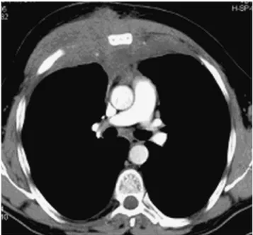
was unremarkable. Extensive laboratory tests showed with the exception of low hemoglobin (121 g/l), mild reduction of lymphocytes (0.70 G/l), elevated C-reactive protein (53 mg/ l), protein (87 g/l), and alkaline phosphatase (139 U/l), normal results. In a conventional chest X-ray, diffuse compression of the soft tissue and enlargement of the retrosternal space was seen. No prominent alteration of the heart configuration and lung perfusion was seen. In the CT scan of the thorax a large tumor was detected in the chest wall with infiltration of the subcutaneous soft tissue and anterior mediastinum. No osteolysis was observed, but mild alteration of the spongiosal structure of the sternum was seen (Fig. 1). For diagnosis an open biopsy was performed which showed a coarse, white tumor growing infiltratively and expansively into the chest wall. Histopathologic examination revealed intact skeletal muscle but also an expanded atrophic skeletal muscle that was infiltrated by a tight cluster of histiocytes. Likewise numerous eosinophilic granulocytes and lymphocytes CD20 and CD3 positive were seen. Immunohistochemical staining for S-100 proteins, and CD1a was positive (Fig. 2). Subsequently the patient underwent chemotherapy with two cycles of cladribine (2-CdA, LitakW). A combination of vinblastine (VelbeW) and prednisone (PrednisoneW
) was administered weekly for 2 months. As maintenance therapy seven cycles of vinblastine (VelbeW
), 6-mercaptopurine (Puri-NetholW
) and prednisone were administered. After 6 months, the chest CTscan showed a discrete decrease of tumor mass in the chest wall but no new lesions. At a recent clinical examination 10 months after diagnosis the patient was weak but in good general condition.
www.elsevier.com/locate/ejcts European Journal of Cardio-thoracic Surgery 33 (2008) 515—516
* Corresponding author. Address: Division of Thoracic Surgery, Inselspital Bern, 3010 Bern, Switzerland. Tel.: +41 31 6322330; fax: +41 31 6322327.
E-mail address:ralph.schmid@insel.ch(R.A. Schmid).
1010-7940/$ — see front matter # 2007 European Association for Cardio-Thoracic Surgery. Published by Elsevier B.V. All rights reserved. doi:10.1016/j.ejcts.2007.12.026
3. Discussion
Mediastinal masses are a very heterogeneous group of tumors. Lymphoma, thymoma, bronchial or esophageal cysts, and struma endothoracica are the most common tumors[4].
For planning adequate therapy, surgical biopsy and histolo-gical analysis are in most cases mandatory. In the presented patient the chest CT scan showed a large tumor of the chest wall with involvement of the anterior mediastinum. Lym-phoma, sarcoma, carcinoma of the lung or of the thymus were the differential diagnosis. The histological analysis, however, demonstrated a LCH originating from the medias-tinum.
The antigen-presenting Langerhans cell belongs to the bone marrow-derived dendritic cells and is typically CD1a-positive[5]. In case of LCH the tumor Langerhans cells have lost the ability to present the antigen, as they remain in an immature state [6]. The etiology of this tumor is still unknown; a viral cause has often been discussed but has not been verified. LCH occurs in childhood and in adults. Initial complaints independent of the location include weight loss, fever, lymphadenopathy and unspecific symptoms such as skin rash, dyspnea or bone pain[1,7,8]. LCH can present as single site (skin, bone, lymph node) or systemic disease (liver, spleen, lungs, bone marrow, endocrine system, gastrointest-inal or central nervous system). The therapy of choice is chemotherapy with the purine analogue 2-chlorodeoxyade-nosine (2-CdA, cladribine, LitakW
), which has shown good response rates in pediatric and adult patients[7].
To the best of our knowledge a LCH originating from the mediastinum in an adult has not been previously described. This case shows that in patients with unclear mediastinal tumors, a histological diagnosis by excision or biopsy is always necessary to plan further adequate therapy. The presented patient responded well to the treatment with 2-chlorodeox-yadenosine and without severe complications or side effects.
References
[1] Smets A, Mortele´ K, De Praeter G, Francois O, Benoit Y, Kunnen M. Pulmonary and mediastinal lesions in children with Langerhans cell his-tiocytosis. Pediatr Radiol 1997;27:873—6.
[2] Nakhla H, Jumbelic MI. Sudden death of a patient with pulmonary Lan-gerhans cell histiocytosis. Arch Pathol Lab Med 2005;129:798—9. [3] Terraciano L, Kocher T, Cathomas G, Bubendorf L, Lehmann FS. Langerhans
cell histiocytosis of the stomach with atypical morphological features. Pathol Int 1999;49:553—6.
[4] Pra¨uer HW, Schalhorn A, Zimmermann F. Tumoren des Mediastinums — Diagnostik und Therapie. Manual Tumoren der Lunge und des Mediasti-nums. Mu¨nchen: W. Zuckerschwerdt Verlag; 2003. p. 168—76.
[5] Favara BE. Langerhans’ cell histiocytosis pathobiology and pathogenesis. Semin Oncol 1991;18:3—7.
[6] Yu RC, Morris JF, Pritchard J, Chu TC. Defective alloantigen-presenting capacity of ‘Langerhans cell histiocytosis cells’. Arch Dis Child 1992;67:1370—2.
[7] Saven A, Burian C. Cladribine activity in adult langerhans-cell histiocy-tosis. Blood 1999;93:4125—30.
[8] Sai S, Fujii K, Masui F, Kida Y. Solitary eosinophilic granuloma of the sternum. J Orthop Sci 2005;10:108—11.
R. Fahrner et al. / European Journal of Cardio-thoracic Surgery 33 (2008) 515—516 516
Fig. 1. CT scan of the thorax of the 55-year-old man with Langerhans cell histiocytosis of the mediastinum. A large tumor in the chest wall is depicted with infiltration of the hypodermal soft tissue of the whole thorax origin from the anterior mediastinum.
Fig. 2. Histopathologic examination showed skeletal muscle infiltrated by a tight and rich cluster of histiocytes, inflammatory infiltrates and macrophages with positive immunohistological staining for S-100 protein and CD1a.