DOI 10.1007/s00018-013-1505-z
Cellular and Molecular Life Sciences
RevIew
Hepatic glucose sensing and integrative pathways in the liver
Maaike H. Oosterveer · Kristina Schoonjans
Received: 2 September 2013 / Revised: 17 October 2013 / Accepted: 18 October 2013 / Published online: 7 November 2013 © Springer Basel 2013
Keywords Acetylation · ChReBP · Glucokinase ·
Glucose sensing · Hepatocytes · LRH-1 · O-linked
β
-N-acetylglucosaminylation
Abbreviations
Acetyl-CoA
Acetyl-coenzyme A
ACL
ATP citrate lyase
ChoRe
Carbohydrate response element
ChReBP
Carbohydrate response element
binding protein
CReB
Cyclic AMP-responsive element
binding protein
CRTC2
cAMP-regulated transcriptional
co-activator 2
F2
6bisP, fructose-2,6-bisphosphate
F6P
Fructose-6-phosphate
FOXA2
Forkhead box protein A2
FOXO1
Forkhead box protein O1
FXR
Farnesoid x receptor
G6P
Glucose-6-phosphate
G6Pc
Glucose-6-phosphatase
G6Pt
Glucose-6-phosphate transporter
GCK
Glucokinase
GCKR
GCK regulatory protein
GLUT
Glucose transporter
GSD-1
Glycogen storage disease type 1
HDAC
Histone deacetylase
HIF-1
Hypoxia-inducible factor 1
HK
Hexokinase
HNF-4
Hepatocyte nuclear factor 4
KAT
Lysine acetyltransferase
KLF-6
Kruppel-like factor 6
LRH-1
Liver receptor homolog 1
LXR
Liver x receptor
Mlx
Max-like protein X
Abstract The hepatic glucose-sensing system is a
func-tional network of enzymes and transcription factors that
is critical for the maintenance of energy homeostasis and
systemic glycemia. Here we review the recent literature
on its components and metabolic actions. Glucokinase
(GCK) is generally considered as the initial postprandial
glucose-sensing component, which acts as the gatekeeper
for hepatic glucose metabolism and provides metabolites
that activate the transcription factor carbohydrate response
element binding protein (ChReBP). Recently, liver
recep-tor homolog 1 (LRH-1) has emerged as an upstream
reg-ulator of the central GCK–ChReBP axis, with a critical
role in the integration of hepatic intermediary metabolism
in response to glucose. evidence is also accumulating that
O
-linked β-N-acetylglucosaminylation (O-GlcNAcylation)
and acetylation can act as glucose-sensitive modifications
that may contribute to hepatic glucose sensing by targeting
regulatory proteins and the epigenome. Further
elucida-tion of the components and funcelucida-tional roles of the hepatic
glucose-sensing system may contribute to the future
treat-ment of liver diseases associated with deregulated glucose
sensors.
M. H. Oosterveer
Department of Pediatrics and Laboratory Medicine, University of Groningen, University Medical Center Groningen, 9713 GZ Groningen, The Netherlands
K. Schoonjans (*)
Institute of Bioengineering, School of Life Sciences, ecole Polytechnique Fédérale de Lausanne (ePFL), 1015 Lausanne, Switzerland
MLXIP
Mlx interacting protein
MLXIPL
Max-like protein X interacting
protein-like
OGA
O
-GlcNAcase
O
-GlcNAcylation
O
-linked β-N-acetylglucosaminylation
OGT
O
-GlcNAc transferase
PGC-1α
Peroxisome proliferator-activated
receptor gamma coactivator 1-alpha
PPARγ
Peroxisome proliferator activated
receptor gamma
SReBP-1c
Sterol regulatory binding protein-1c
T2D
Type 2 diabetes
TCA
Tricarboxylic acid
TCFe3
Transcription factor e3
UDP-GlcNAc
UDP-N-acetylglucosamine
UTP
Uridine triphosphate
X5P
Xylulose-5-phosphate
Introduction
Glucose is a simple sugar carbohydrate that serves as a
fundamental fuel for most species and provides precursors
for biomolecule synthesis. In order to control metabolism,
differentiation, and growth, cells possess evolutionary
con-served glucose-sensitive signaling pathways [
1
]. These
glu-cose-sensing systems ensure efficient adaptation to changes
in environmental glucose availability in unicellular
organ-isms and allow for homeostatic maintenance of internal
glucose pools in multicellular organisms. In higher species,
the internal pool is represented by glucose circulating in the
bloodstream. From here, glucose is further distributed to
different tissues and organs to meet local needs.
The liver plays a central role in metabolic
homeosta-sis by coordinating the breakdown, synthehomeosta-sis, storage,
and redistribution of nutrients. Hepatocytes possess
mul-tiple nutrient-sensing systems that interact to modulate
biochemical pathways in order to accommodate systemic
fuel requirements and availability. These systems
ena-ble the body to maintain its functions during periods of
feeding and fasting and upon excessive energy demands
such as exercise. Blood glucose concentrations
fluctu-ate during the feeding and fasting cycles [
2
], and one of
the liver’s primary functions is to maintain blood glucose
concentrations within a physiological range [
3
].
Hepato-cytes are among the few cell types that possess the
abil-ity to both consume and produce glucose [
4
]. Glycemic
control, which is coordinated by both extrahepatic and
intrahepatic factors, is hence the result of a
balanc-ing act between these two processes. Most reviews have
focused on extrahepatic glucose-sensing systems such as
hormonal regulation by insulin and glucagon [
3
,
5
,
6
].
In contrast, this review will provide an overview of the
regulatory components within the liver that are activated
by glucose metabolites in response to glucose availability.
we provide an overview of these regulatory components
and discuss the role of this intrahepatic glucose-sensing
system in health and disease.
Hepatic glucose metabolism
The concentration of glucose in the blood is a primary
determinant of glucose availability to the liver. During the
postprandial phase, which in humans lasts about 2 h after
the intake of a meal, blood glucose levels rise and
approxi-mately 10–25 % of ingested glucose is taken up by
hepato-cytes [
7
–
10
]. Facilitated transport of glucose across cellular
membranes is mediated by members of the glucose
trans-porter (GLUT) family [
11
]. GLUT2 is the major glucose
transporter in the hepatocytes [
11
,
12
] and its physiological
role has been studied extensively [
13
–
15
]. GLUT2 is also
expressed in pancreatic islets, intestine, kidney, and brain
[
11
,
12
]. The rate of GLUT2-mediated glucose transport
into the liver is high and only saturates at glucose
concen-trations above 30 mM [
11
] allowing efficient glucose
trans-port and extremely rapid equilibration of glucose across
the hepatocyte membrane [
16
]. Once in the cytoplasm,
glucose is phosphorylated to glucose-6-phosphate (G6P)
by glucokinase (GCK; also known as hexokinase Iv) [
17
,
18
]. G6P lies at the crossroads of different biochemical
pathways and has multiple biochemical fates. elevated G6P
synthesis allosterically activates glycogen synthase while
inhibiting glycogen phosphorylase [
19
–
21
]. G6P is also
oxidized for energy supply via glycolysis, which involves
several steps including the production of
fructose-6-phos-phate (F6P) and triose phosfructose-6-phos-phates. The pentose phosfructose-6-phos-phate
pathway represents a third route of G6P utilization that
involves the production of ribose-5-phosphate, an
interme-diate of nucleotide synthesis, and the biological reductant
NADPH. excess pentose phosphates can ultimately enter
the glycolytic pathway by their conversion into F6P and
tri-ose phosphates. Pyruvate produced by glycolysis is
trans-ported into the mitochondria, where it is decarboxylated
to acetyl-coenzyme A (acetyl-CoA), which subsequently
enters the tricarboxylic acid (TCA) cycle, a central
meta-bolic hub that is involved in both energy production and
biomolecule synthesis. To keep TCA cycle intermediates
at a constant level, reactions that extract TCA metabolites
for biosynthesis (cataplerotic reactions) are balanced by
those that replenish TCA intermediates (anaplerotic
reac-tions) [
22
]. In the TCA cycle, acetyl-CoA becomes
fur-ther metabolized to generate reducing equivalents used for
ATP production through oxidative phosphorylation. The
TCA cycle intermediates also serve as precursors for
non-essential amino acids, which serve as substrates for protein
synthesis. Citrate produced in the TCA cycle is partly
shut-tled from the mitochondria into the cytosol where it is
con-verted into oxaloacetate and acetyl-CoA, the latter of which
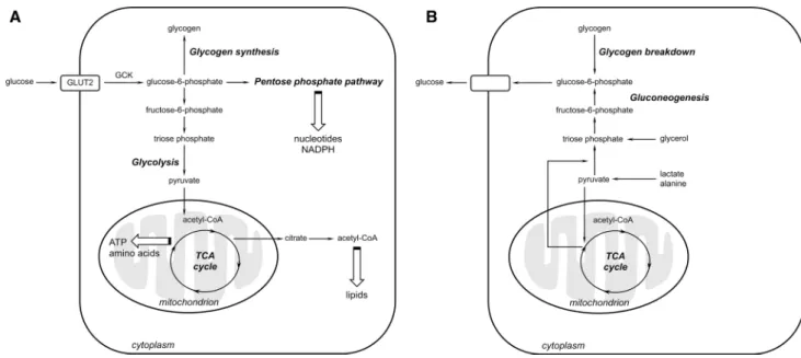
can be used as a substrate for lipid synthesis. These hepatic
glucose oxidation and storage pathways are summarized in
Fig.
1
a.
Hepatic glucose uptake and metabolism decrease as
soon as the intestinal absorption of glucose is completed.
During this period, which is often referred to as the
post-absorptive phase, most tissues reduce their glucose
con-sumption by switching to alternate energy sources.
endog-enous glucose production by the liver now represents the
major route of glucose supply to the bloodstream. The
maintenance of glucose homeostasis is particularly
impor-tant for cells that partly or fully rely on glucose as
ener-getic substrate such as neurons and erythrocytes. The liver
contributes to endogenous glucose production via two
G6P-generating pathways. In the initial postabsorptive
phase, hepatic G6P is derived from glycogen breakdown
while gluconeogenesis becomes the major source of G6P
after prolonged fasting. G6P generated through glycogen
breakdown and gluconeogenesis is first translocated from
the cytosol into the endoplasmic reticulum by the
glucose-6-phosphate transporter (G6Pt; also known as SLC37A4),
and subsequently dephosphorylated into glucose by
glu-cose-6-phosphatase (G6Pc). Glucose is finally released
into the bloodstream, presumably through the concerted
action of GLUT2 and a membrane traffic-based
mecha-nism [
13
,
14
,
23
]. These glucose-production pathways in
the liver are summarized in Fig.
1
b.
Postprandial glucose sensing in the liver
when blood glucose concentrations rise, hepatic glucose
sensors induce adaptive responses to shift the balance
toward hepatic glucose consumption and storage. GLUT2
is a high-capacity glucose transporter that allows glucose
to flow into hepatocytes in response to increasing glycemia
[
11
]. However, its activity does not appear to be critical for
postprandial glucose sensing in the liver, as was recently
reported [
13
]. In this study, hepatic GLUT2 deficiency did
not result in major perturbations in hepatic glucose
metabo-lism in fed and refed mice, suggesting that alternate
mecha-nisms compensate for the reduction in glucose transport.
GCK, on the contrary, is a major component of the hepatic
glucose-sensing system. By converting glucose into G6P,
GCK catalyzes the first step of intrahepatic glucose
metab-olism [
17
]. In contrast to hexokinases (HKs) I-II, GCK
exhibits low affinity for glucose, is not feedback-inhibited
by its product G6P [
17
,
24
], and its activity increases
sig-moidal with increasing glycemia [
18
,
25
]. High glucose
concentrations furthermore inhibit the interaction of GCK
with its regulatory protein (GCKR), hence promoting the
translocation of free GCK to the cytoplasm where it can
access glucose and convert it into G6P [
26
]. GCK
conse-quently acts as a glucose-sensitive enzyme that remains
active over a wide range of glucose concentrations and
enables hepatocytes to efficiently trap glucose in response
to glycemic fluctuations. Lack of hepatic GCK expression
in mice perturbs intrahepatic glucose metabolism [
27
,
28
]
while overexpression of GCK, but not HK-I, markedly
Fig. 1 Pathways of hepatic glucose metabolism. a Simplified scheme depicting the major biochemical pathways activated during postpran-dial glucose consumption and storage. b Simplified scheme depict-ing the major biochemical pathways activated durdepict-ing postabsorptive
glucose production. Glycerol, lactate, and alanine are used as gluco-neogenic substrates upon their conversion into triose phosphate and pyruvate. Acetyl-CoA acetyl-coenzyme A, GCK glucokinase, GLUT2 glucose transporter 2, TCA tricarboxylic acid
induces glycogen storage and glycolysis in hepatocytes
[
29
,
30
]. These fundamental differences of GCK versus
HK-mediated G6P synthesis illustrate the unique role of
hepatocytes as compared to other cells.
Further downstream metabolism of G6P generates
metabolites that act as signaling molecules to regulate
the activity of enzymes within seconds to minutes after
hepatic glucose exposure [
19
,
20
,
31
–
35
].
Glucose-medi-ated control of gene transcription in hepatocytes translates
into adaptive responses on longer timescales, i.e., within
a timeframe of minutes to hours [
36
–
38
]. The expression
of many glucose-sensitive genes is regulated by the
car-bohydrate response element binding protein (ChReBP;
also known as Mondo B or Max-like protein X
interact-ing protein-like, MLXIPL) [
39
,
40
], a transcription
fac-tor that recognizes conserved carbohydrate response
ele-ments (ChoRes) in gene promoters [
41
,
42
]. ChReBP is a
member of the Mondo family, which forms heterodimers
with Max-like protein X (Mlx) to induce transcriptional
responses [
43
–
48
]. Mondo-Mlx-dependent glucose
sens-ing is evolutionary conserved among worms, flies, and
vertebrates [
49
–
55
]. ChReBP has been identified as the
major mediator of ChoRe-dependent gene transcription
in the liver [
40
,
48
], while its paralog MondoA (or Mlx
interacting protein, MLXIP) has been proposed to act
predominantly in extrahepatic tissue [
45
,
56
]. However,
a recent study showed that MondoA also regulates
tran-scription of specific glucose-responsive genes in
hepato-cytes [
49
]. ChReBP is best-known for its effects on the
expression of enzymes involved in glycolysis and fatty
acid synthesis [
57
]. In addition, ChReBP suppresses
sir-tuin 1, thereby likely reducing PGC-1α-dependent
glu-coneogenesis under glucose abundant conditions [
58
].
Somewhat counter-intuitively, ChReBP also induces
G6Pc expression, a response that may serve to maintain
the intracellular G6P homeostasis [
59
]. ChIP-seq
analy-sis indicated that ChReBP not only regulates metabolism,
but also targets genes related to transport, development,
and cell motility [
39
].
Several studies have shown that hepatic ChReBP
activation requires GCK-dependent glucose
metabo-lism [
28
,
60
]. early work showed that the pentose
phos-phate pathway intermediate xylulose-5-phosphos-phate (X5P)
induces ChReBP dephosphorylation, thereby promoting
its nuclear translocation and transcriptional activity [
61
].
However, this model has been challenged, based on the
finding that pentose phosphate pathway inhibition leads
to a decrease rather than an increase in ChReBP
activ-ity [
62
,
63
]. Instead, G6P was suggested to be the major
signaling metabolite responsible for ChReBP activation
[
62
,
63
]. Finally, fructose-2,6-bisphosphate (F2,6bisP),
another glucose metabolite, has also been proposed to
induce ChReBP-mediated transcription in hepatocytes
[
49
,
64
]. The mechanisms through which these three
glucose derivatives act remain to be resolved, but likely
involve changes in allosteric regulation and
post-trans-lational modifications [
53
,
65
,
66
]. In this respect, it
should be noted that ChReBP activity is increased by
acetylation and O-linked β-N-acetylglucosaminylation
(O-GlcNAcylation) [
67
,
68
], two enzyme-catalyzed
post-translational modifications that use glucose metabolites as
substrates, as will be discussed in more detail below [
69
–
71
]. The fact that several independent glucose metabolites
(X5P, G6P, F2,6bisP, acetyl-CoA, and O-GlcNAc)
acti-vate hepatic ChReBP illustrates the unique
glucose-sens-ing ability of this transcription factor in hepatocytes [
57
].
A recent study furthermore showed that glucose promotes
the binding of full-length ChReBP-α to a ChoRe located
in an alternative promoter region of the Chrebp gene
thereby inducing transcription of a potent, short ChReBP
isoform (ChReBP-β) [
72
]. Future work should
iden-tify the specific glucose-dependent pathways that induce
and activate these different isoforms in hepatocytes, and
reveal whether ChReBP-α and ChReBP-β regulate
differ-ent target genes.
Regulation of the central hepatic glucose‑sensing axis
The GCK–ChReBP axis can be considered as the central
glucose-sensing system in the liver. Because GCK acts as
a gatekeeper for hepatic glucose metabolism and ChReBP
activation [
60
,
73
], regulation of its expression and activity
will significantly impact hepatic glucose sensing.
Interest-ingly, glucose increases GCKR expression while it
inhib-its GCK transcription in cultured hepatocytes [
59
].
How-ever, in vivo GCK expression is induced in response to an
oral glucose load [
60
]. Because insulin is a major
regula-tor of GCK expression in the liver [
31
], the discrepancy
between these findings can be explained by the lack of a
concomitant insulin-mediated GCK transcription under
in vitro conditions [
74
]. The mechanistic basis of
insulin-dependent GCK induction is incompletely understood [
31
,
75
]. Several transcription factors, i.e., hepatocyte nuclear
factor 4 (HNF-4), hypoxia-inducible factor 1 (HIF-1),
sterol regulatory binding protein-1c (SReBP-1c), liver x
receptor (LXR), peroxisome proliferator activated
recep-tor gamma (PPARγ), Kruppel-like facrecep-tor 6 (KLF-6) and
transcription factor e3 (TCFe3) have been shown to
con-trol hepatic GCK transcription [
60
,
76
–
82
]. Studies from
our laboratory have indicated that the nuclear receptor liver
receptor homolog 1 (LRH-1) coordinates multiple aspects
of hepatic intermediary metabolism by regulating
GCK-dependent G6P synthesis [
60
,
83
]. while initially
identi-fied as a transcriptional regulator of cholesterol and bile
salt homeostasis [
84
,
85
], LRH-1 has recently emerged as
a key integrator of hepatic glucose and fatty acid
metabo-lism [
60
,
83
,
86
,
87
]. LRH-1 contributes to basal GCK
expression under fed and fasted conditions and its activity
is not dependent on glucose. This was based on the
find-ing that ectopic LRH-1 expression is sufficient to induce
Gck
expression in hepatoma cells, and that increasing
glycemia fails to amplify LRH-1-mediated transcription
[
60
]. Hepatic LRH-1 deficiency significantly perturbed
the hepatic response to feeding, as illustrated by delayed
glycogen synthesis, as well as reduced ChReBP
expres-sion and activity, which resulted in a strong attenuation of
glycolysis and de novo fatty acid synthesis upon refeeding
[
60
]. Importantly, these perturbations occurred secondary
to reduced GCK activity, as GCK reconstitution restored
ChReBP target gene expression in hepatocyte-specific
LRH-1 knockout mice [
60
]. LRH-1-dependent glucose
sensing in the liver also affected systemic glucose
home-ostasis. In liver-specific LRH-1 knockout mice impaired
GCK-mediated glucose consumption triggered the
pan-creas to release more insulin, leading to elevated insulin
levels and increased glucose disposal [
60
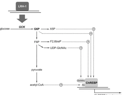
]. These findings
place LRH-1 upstream of the central glucose-sensing
sys-tem in the liver (Fig.
2
).
Similar functions have been attributed to LXR. Although
LXR has been identified as a transcriptional regulator of
both GCK and ChReBP [
76
,
88
–
92
], its deficiency does
not impair the hepatic response to carbohydrate refeeding
or ChReBP activity [
93
,
94
]. Further work will be
neces-sary to establish whether LXR is essentially required for
postprandial glucose sensing in the liver.
Glucose‑sensitive modifications as potential glucose
sensors in the liver
Post-translational modifications of regulatory proteins
allow for adaptive responses to a variety of metabolic
cues [
95
,
96
]. Interestingly, some post-translational
modi-fications are closely linked to glucose metabolism and
target metabolic enzymes, components of cellular signal
transduction pathways as well as transcription factors and
their co-regulators (reviewed in [
97
,
98
]). These
modifica-tions are typically enzyme-catalyzed, but can also occur
through non-enzymatic interaction between metabolites
and proteins. Although enzyme-mediated transfer of
glu-cose metabolites has been investigated most intensively,
a very recent study has identified a glucose-sensitive and
enzyme-independent post-translational modification that
controls hepatocyte function [
99
]. It is now also
increas-ingly recognized that glucose metabolism can induce
epigenetic changes through glucose-dependent
post-translational modification of histone proteins (reviewed
in [
96
,
100
–
102
]). Because the composition of the
his-tone code determines the degree of chromatin
condensa-tion, glucose-dependent modification of histones may
alter the accessibility for transcription factors and
regula-tory enzymes that may ultimately translate into changes
in transcriptional activity. In this section, we will discus
two enzyme-mediated glucose-sensitive post-translational
modifications that target regulatory proteins and
epig-enome, and may as such contribute to glucose sensing in
the liver. The metabolic origins, enzymatics, and hepatic
Fig. 2 LRH-1 is an upstream regulator of the central glucose-sensing system in the liver. ChReBP activation requires GCK-dependent synthesis of glucose metabolites. Because LRH-1 is a transcriptional regulator of GCK, it impacts postprandial G6P synthesis and ChReBP activity. Acetyl-CoA acetyl-coenzyme A, ChREBP carbohydrate response element binding protein, F2,6bisP fructose-2,6-bisphosphate, F6P fructose-6-phosphate, G6P glucose-6-phosphate, GCK glucokinase, LRH-1 liver recep-tor homolog 1, UDP-GlcNAc UDP-N-acetylglucosamine, X5P xylulose-5-phosphate
targets of these post-translational modifications are
sum-marized in Fig.
3
.
O
-GlcNAcylation of serine and threonine residues is
a modification that occurs in the cytoplasm, nucleus, and
mitochondria [
103
]. The substrate,
UDP-N-acetylglucosa-mine (UDP-GlcNAc), is generated by the hexosaUDP-N-acetylglucosa-mine
biosynthesis pathway, a branch of hepatic glucose
metab-olism that uses F6P, glutamine, acetyl-CoA, and uridine
triphosphate (UTP) [
104
]. The addition and removal of
UDP-GlcNAc is catalyzed by two enzymes. O-GlcNAc
transferase (OGT) mediates the addition of UDP-GlcNAc
to target proteins while O-GlcNAcase (OGA) catalyzes its
removal [
105
,
106
]. Both OGT and OGA are encoded by
single genes that are alternatively spliced in mammals, and
the different isoforms are located in separate subcellular
compartments [
105
,
107
–
110
]. Their activities are
regu-lated by protein–protein interactions and post-translational
modifications including O-GlcNAcylation, however this
domain is as yet largely unexplored [
111
].
O-GlcNAcyla-tion is considered as a unique glucose-sensitive
post-trans-lational modification [
112
] and has wide-ranging effects
on transcription, protein activity, and stability as well as on
epigenetic and genomic imprinting (reviewed in [
113
]). In
hepatocytes, O-GlcNAcylation has mainly been studied in
relation to its role in metabolism. Recent work has shown
that hepatic OGT is required to maintain circadian control
of glucose homeostasis by regulating the clock system in
the liver [
114
,
115
]. OGT also targets metabolic
transcrip-tional regulators such as LXR [
90
] and cAMP-regulated
transcriptional co-activator 2 (CRTC2), a coregulator of
the gluconeogenic transcription factor cyclic
AMP-respon-sive element binding protein (CReB) [
116
]. Moreover,
the activity of two other key gluconeogenic regulators,
i.e., forkhead box protein O (FOXO1) and peroxisome
proliferator-activated receptor gamma coactivator 1-alpha
(PGC-1α), is regulated by O-GlcNAcylation [
117
–
119
].
Another key finding is that OGT modifies multiple nodes
of the insulin signaling pathway [
108
,
120
]. Interestingly,
under normoglycemic conditions O-GlcNAcylation
con-tributes to insulin signaling [
121
], while it induces
insu-lin resistance when chronically activated [
120
]. Although
these studies point to a general role for O-GlcNAcylation
in regulating glucose homeostasis, strong evidence for a
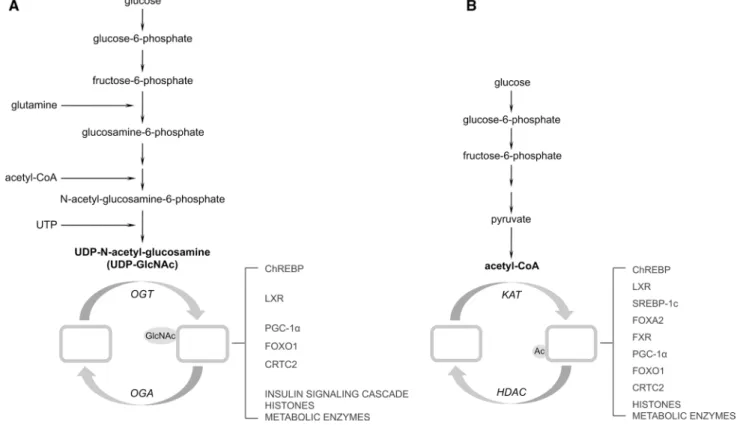
more specific role in glucose-sensing stems from the fact
Fig. 3 working model depicting the metabolic origins, enzymatics, and targets of glucose-sensitive post-translational modifications in the liver. a The hexosamine biosynthesis pathway uses F6P, glutamine, and UTP for O-linked β-N-acetylglucosaminylation. b Glycolysis can link glucose metabolism to acetylation. Acetyl-CoA acetyl-coenzyme A, ChREBP carbohydrate response element binding protein, CRTC2 cAMP-regulated transcriptional co-activator 2, FOXA2 forkhead box
protein A2, FOXO1 forkhead box protein O1, F6P fructose-6-phos-phate, FXR farnesoid x receptor, HDAC histone deacetylase, KAT lysine acetyltransferases, LXR liver x receptor, OGA O-GlcNAcase, OGT O-GlcNAc transferase, PGC-1α peroxisome proliferator-acti-vated receptor gamma coactivator 1-alpha, SREBP-1c sterol regula-tory element binding protein-1c, TCA tricarboxylic acid, UTP uridine triphosphate
that O-GlcNAcylation activates hepatic ChReBP [
68
].
Finally, it should be mentioned that despite the fact that
O
-GlcNAcylation is emerging as a histone-modifying
post-translational modification [
122
], there is currently no
evi-dence that O-GlcNAcylation also contributes to hepatic
glucose sensing via epigenetic regulation. As
methodolo-gies for high-throughput O-GlcNAc profiling are emerging
[
123
,
124
], more insight into the hepatic targets of
O-Glc-NAcylation and its potential contribution to hepatic glucose
sensing is expected in the near future.
Acetylation is another post-translational modification
that potentially reflects glucose availability. This
modifi-cation involves the enzymatic transfer of acetyl-CoA, and
is facilitated by lysine acetyltransferases (KATs) [
125
].
These enzymes act on the lysine residues of both histones
and non-histone proteins in different cellular
compart-ments. The reverse reaction is mediated by deacetylases,
which can be divided into four classes. Class I, II, and Iv
deacetylases are considered as the classical histone
dea-cetylases (HDACs). Class III deadea-cetylases, better known
as sirtuins, are structurally unrelated to HDACs. HDACs
and sirtuins are localized in the mitochondria or cytoplasm,
and are able to shuttle between the nucleus and the cytosol
[
126
–
129
]. High-throughput analysis of human liver
biop-sies and liver cells has shown that many metabolic enzymes
are acetylated [
69
,
130
], either to modulate their
activi-ties or to direct them towards proteosomal or lysosomal
degradation [
131
,
132
]. Moreover, the activity of several
transcriptional regulators of hepatic metabolism including
ChReBP, LXR, CRTC2, PGC-1α, FOXO1, SReBP-1c,
forkhead box protein A2 (FOXA2), and farnesoid x
recep-tor (FXR), is known to be modified by acetylation [
67
,
133
–
140
], in some cases in coordination with
phosphoryla-tion [
134
,
141
]. Studies in yeast and mammalian cell
cul-tures have shown that histone acetylation is dependent on
subcellular acetyl-CoA concentrations [
142
–
146
]. Notably,
glucose was shown to promote histone acetylation via ATP
citrate lyase (ACL), the enzyme that generates acetyl-CoA
from TCA-derived citrate in mammalian cell lines [
142
].
The existence of a similar mechanism in hepatocytes
chal-lenged with glucose would point to a glucose-sensing role
of histone acetylation in liver but needs to be confirmed.
The observation that both histones [
147
] and non-histone
proteins [
130
] are dynamically acetylated in response to
feeding/fasting cycles is also suggestive of
glucose-depend-ent acetylation in liver. Moreover, it has been reported
that hepatic acetyl-CoA levels increase upon short-term
refeeding as compared to fasted conditions [
148
]. It should
however be noted that besides being produced by
decar-boxylation of glycolytic pyruvate, hepatic acetyl-CoA can
also be derived from fatty acid oxidation and amino acid
metabolism. A dedicated analysis of acetylation profiles
in glucose-challenged hepatocytes is therefore warranted
to establish the impact of glucose metabolism on protein
acetylation, as well as the potential contribution of protein
acetylation to glucose sensing in the liver.
Metabolic liver diseases associated with aberrant
glucose sensing
Glucose sensors enable the liver to respond to dynamic
changes in glucose availability. However, when these
sen-sors are chronically activated, they may predispose to the
development of liver diseases.
During poorly controlled diabetes, the liver is
fre-quently exposed to hyperglycemic episodes. In type 2
dia-betes (T2D), GCK is constitutively active and GCK flux
is increased secondary to elevated glucose concentrations
[
149
,
150
]. This leads to sustained activation of glucose
sensors in the liver. For example, the hexosamine
biosyn-thesis pathway normally accounts for less than 5 % of the
hepatic glucose flux, yet its activity is markedly increased
by hyperglycemia [
151
,
152
]. Aberrant glucose sensing in
T2D results in triglyceride accumulation and excessive
glu-cose production in the liver [
116
,
153
]. while triglyceride
accumulation contributes to the development of liver
stea-tosis, increased hepatic glucose output leads to a further
increase in glycemia.
A clear association exists between hepatic steatosis and
the pathogenesis of T2D, cardiovascular disease, and
stea-tohepatitis [
154
]. It is, however, increasingly recognized
that, up to a certain threshold, the accumulation of
triglyc-erides may serve as a buffering system that would
actu-ally protect the liver against metabolic dysfunction [
154
].
In mice, ChReBP plays a key role in the development of
hepatic steatosis in T2D [
153
], and hepatic ChReBP
func-tion is perturbed in obese and (pre-)diabetic subjects [
155
,
156
]. Interestingly, a recent study revealed that ChReBP
overexpression protects against diet-induced glucose
intol-erance and insulin resistance [
157
]. This finding indicates
that under conditions of dietary fat overload,
ChReBP-mediated lipogenesis likely contributes to a metabolically
benign state by promoting mono-unsaturated fatty acid
syn-thesis [
154
,
157
]. It was furthermore shown that diabetic
steatosis is associated with ChReBP hyperacetylation [
67
]
and that hepatic lipid accumulation can be prevented when
ChReBP O-GlcNAcylation is reduced [
68
]. Increased
O
-GlcNAcylation also contributes to uncontrolled hepatic
glucose production under diabetic conditions. T2D is
asso-ciated with increased O-GlcNAcylation levels of the
glu-coneogenic co-regulator CRTC2 and removal of
O-Glc-NAc from CRTC2 normalizes glycemia in diabetic mice
[
116
]. whether sustained CRTC2 acetylation levels also
promote hepatic glucose production in diabetics remains
to be established. Likewise, it is as yet unknown whether
aberrant acetylation and O-GlcNAcylation of the
gluconeo-genic regulators FOXO1 and PGC-1α [
117
–
119
,
135
,
158
]
directly contribute to hyperglycemia in T2D.
Glucose sensors may also become deregulated by
inherited loss-of-function mutations in enzymes that
regulate intrahepatic glucose metabolism. An example of
such an “inborn error of metabolism” is Glycogen
Stor-age Disease type 1 (GSD-1) [
159
] which is caused by
loss of either G6Pc or G6Pt activity [
160
–
162
]. The
pri-mary consequences of perturbed hepatic G6Pase activity
in GSD-1 are hypoglycemia and the accumulation of G6P
in the liver [
163
–
165
]. In addition, GSD-1 is
character-ized by excessive glycogen and lipid storage in the liver
[
163
,
165
–
167
] as well as hyperlipidemia [
165
,
167
–
170
].
Interestingly, GSD-1 is associated with a
ChReBP-dependent increase in de novo fatty acid synthesis [
163
,
167
,
170
]. Combined, these observations indicate that
sustained activation of hepatic glucose sensors by
extra-hepatic (diabetes) or intraextra-hepatic (GSD-1) changes in
glu-cose homeostasis predisposes to development of hepatic
steatosis [
171
].
Another consequence of both T2D and GSD-1 is the
increased incidence of liver tumorigenesis [
172
–
175
].
Although steatosis has been proposed as a predisposing
factor for liver cancer [
176
,
177
], altered metabolism
may be the actual driving force for tumor development.
It is well known that tumors require specific metabolic
adaptations to support the bioenergetic and biosynthetic
demands of growth and proliferation [
178
]. More
spe-cifically, a switch to non-oxidative glucose metabolism
combined with a predominant anabolic role of the TCA
cycle are considered as major hallmarks of cancer
metab-olism [
179
]. T2D and GSD-1 are characterized by a high
flux from hepatic G6P towards glycolysis and lipid- and
nucleotide biosynthesis. The exact mechanisms by which
these metabolic adaptations confer a preneoplastic status
to hepatocytes and direct them towards tumorigenesis
are incompletely understood [
165
,
172
,
179
]. Glucose
sensors likely play an important role here. In support of
this hypothesis, ChReBP mediates the switch towards
pro-oncogenic metabolism in proliferating cells [
180
].
Moreover, ChReBP functionally interacts with the
pro-oncogenic transcription factor c-Myc, which is critical for
ChReBP-dependent glucose sensing in the liver [
45
,
181
,
182
]. Because mouse models of T2D and GSD-1 exhibit
increased hepatic ChReBP activity [
153
,
163
], ChReBP
may play a key role in the pathophysiology of liver tumor
development in these diseased states. The potential
exist-ence of such a mechanism urges for the exploration of
a potential oncogenic role of hepatic LRH-1, a potent
upstream regulator of the GCK–ChReBP axis in the liver
([
60
] and Fig.
2
) and a key player in the development
of colorectal, breast and pancreatic cancers [
183
–
185
].
Finally, acetylation and O-GlcNAcylation have recently
emerged as critical modifiers of the activity of metabolic
enzymes as well as of oncogenes and tumor suppressors
in cancer cells [
123
,
186
–
190
]. These glucose-sensing
post-translational modifications may therefore direct
hepatocytes towards a pro-oncogenic state under
condi-tions of excessive hepatic glucose metabolism [
142
,
191
,
192
].
Conclusions and future directions
Hepatic glucose sensing is critical for an adequate
post-prandial response and the maintenance of glycemic control.
However, it may also contribute to liver pathology under
conditions of excessive intrahepatic glucose metabolism.
Research in the past years has identified GCK–ChReBP
as the central glucose-sensing system in the liver. Further
exploration of the mechanisms by which different glucose
metabolites activate hepatic ChReBP, and the function of
the different ChReBP isoforms are required to unravel the
mechanistic basis of the glucose-sensing axis. In addition,
it remains to be established whether ChReBP’s paralog
MondoA, which can be activated by G6P and F2,6bisP [
49
,
56
], also contributes to postprandial glucose sensing in the
liver.
Because LRH-1 has recently emerged as a potent
upstream regulator of the GCK–ChReBP axis, modulation
of its activity may provide opportunities for the treatment
of diseases that are characterized by aberrant hepatic
glu-cose sensing. LRH-1 transcriptional activity can be
modi-fied by post-translational modifications or by agonists/
antagonist binding, and depends on its interaction with
co-regulators [
83
,
183
,
193
–
204
]. Detailed insight into these
processes is therefore needed to define strategies that target
LRH-1-dependent glucose sensing.
Finally, dedicated studies are required to uncover the
exact role of O-GlcNAcylation and acetylation in hepatic
glucose sensing. Systematic analysis of
glucose-depend-ent responses in the absence of OGT or KAT activity will
establish to what extent, and via which mechanisms these
post-translational modifications contribute to
glucose-sensing system in the liver. Moreover, there is extensive
crosstalk between post-translational modifications, and
different combinations of post-translational modifications
on a single target may lead to distinct biological outcomes
[
112
,
205
]. Future research will likely uncover novel
inter-plays between GlcNAcylation/acetylation and other
post-translational modifications including protein ubiquitination
and methylation (reviewed in [
206
,
207
]). Such crosstalk
may in turn unveil unexpected functions and consequences
of chronically activated hepatic glucose sensors that go
beyond metabolism.
Acknowledgments we thank Albert K. Groen for reading and com-menting on the manuscript. The work in the laboratory of the authors is supported by the ecole Polytechnique Fédérale de Lausanne (ePFL), the Swiss National Science Foundation, the Swiss Cancer League and the University Medical Center Groningen (UMCG).
References
1. Towle HC (2005) Glucose as a regulator of eukaryotic gene transcription. Trends endocrinol Metab 16(10):489–494. doi:10.1016/j.tem.2005.10.003
2. Daly Me, vale C, walker M, Littlefield A, Alberti KG, Mathers JC (1998) Acute effects on insulin sensitivity and diurnal met-abolic profiles of a high-sucrose compared with a high-starch diet. Am J Clin Nutr 67(6):1186–1196
3. Klover PJ, Mooney RA (2004) Hepatocytes: critical for glucose homeostasis. Int J Biochem Cell Biol 36(5):753–758
4. Mithieux G (2010) Brain, liver, intestine: a triumvirate to coordinate insulin sensitivity of endogenous glucose produc-tion. Diabetes Metab 36(Suppl 3):S50–S53. doi:10.1016/ S1262-3636(10)70467-5
5. Taniguchi CM, emanuelli B, Kahn CR (2006) Critical nodes in signalling pathways: insights into insulin action. Nat Rev Mol Cell Biol 7(2):85–96. doi:10.1038/nrm1837
6. Ramnanan CJ, edgerton DS, Kraft G, Cherrington AD (2011) Physiologic action of glucagon on liver glucose metabolism. Diabetes Obes Metab 13(Suppl 1):118–125. doi:10.1111/j.1463-1326.2011.01454.x
7. Capaldo B, Gastaldelli A, Antoniello S, Auletta M, Pardo F, Ciociaro D, Guida R, Ferrannini e, Sacca L (1999) Splanchnic and leg substrate exchange after ingestion of a natural mixed meal in humans. Diabetes 48(5):958–966
8. Ferrannini e, Bjorkman O, Reichard GA Jr, Pilo A, Olsson M, wahren J, DeFronzo RA (1985) The disposal of an oral glu-cose load in healthy subjects. A quantitative study. Diabetes 34(6):580–588
9. woerle HJ, Meyer C, Dostou JM, Gosmanov NR, Islam N, Popa e, wittlin SD, welle SL, Gerich Je (2003) Pathways for glucose disposal after meal ingestion in humans. Am J Phys-iol endocrinol Metab 284(4):e716–e725. doi:10.1152/ajpe ndo.00365.2002
10. Moore MC, Pagliassotti MJ, Swift LL, Asher J, Murrell J, Neal D, Cherrington AD (1994) Disposition of a mixed meal by the conscious dog. Am J Physiol 266(4 Pt 1):e666–e675
11. Mueckler M, Thorens B (2013) The SLC2 (GLUT) family of membrane transporters. Mol Aspects Med 34(2–3):121–138. doi:10.1016/j.mam.2012.07.001
12. Aschenbach JR, Steglich K, Gabel G, Honscha KU (2009) expression of mRNA for glucose transport proteins in jejunum, liver, kidney and skeletal muscle of pigs. J Physiol Biochem 65(3):251–266. doi:10.1007/BF03180578
13. Seyer P, vallois D, Poitry-Yamate C, Schutz F, Metref S, Tarus-sio D, Maechler P, Staels B, Lanz B, Grueter R, Decaris J, Turner S, da Costa A, Preitner F, Minehira K, Foretz M, Thorens B (2013) Hepatic glucose sensing is required to preserve beta cell glucose competence. J Clin Investig. doi:10.1172/JCI65538
14. Burcelin R, del Carmen Munoz M, Guillam MT, Thorens B (2000) Liver hyperplasia and paradoxical regulation of gly-cogen metabolism and glucose-sensitive gene expression in GLUT2-null hepatocytes. Further evidence for the existence of a membrane-based glucose release pathway. J Biol Chem 275(15):10930–10936
15. Burcelin R, Dolci w, Thorens B (2000) Glucose sensing by the hepatoportal sensor is GLUT2-dependent: in vivo analysis in GLUT2-null mice. Diabetes 49(10):1643–1648
16. williams TF, exton JH, Park CR, Regen DM (1968) Stereospe-cific transport of glucose in the perfused rat liver. Am J Physiol 215(5):1200–1209
17. wilson Je (2003) Isozymes of mammalian hexokinase: struc-ture, subcellular localization and metabolic function. J exp Biol 206(Pt 12):2049–2057
18. Cardenas ML, Cornish-Bowden A, Ureta T (1998) evolution and regulatory role of the hexokinases. Biochim Biophys Acta 1401(3):242–264
19. wera S, Bollen M, Moens L, Stalmans w (1996) Time-depend-ent pseudo-activation of hepatic glycogen synthase b by glucose 6-phosphate without involvement of protein phosphatases. Bio-chem J 315(Pt 1):91–96
20. Aiston S, Green A, Mukhtar M, Agius L (2004) Glucose 6-phosphate causes translocation of phosphorylase in hepato-cytes and inactivates the enzyme synergistically with glucose. Biochem J 377(Pt 1):195–204. doi:10.1042/BJ20031191
21. von wilamowitz-Moellendorff A, Hunter Rw, Garcia-Rocha M, Kang L, Lopez-Soldado I, Lantier L, Patel K, Peggie Mw, Martinez-Pons C, voss M, Calbo J, Cohen PT, wasserman DH, Guinovart JJ, Sakamoto K (2013) Glucose-6-phosphate-medi-ated activation of liver glycogen synthase plays a key role in hepatic glycogen synthesis. Diabetes. doi:10.2337/db13-0880
22. Owen Oe, Kalhan SC, Hanson Rw (2002) The key role of ana-plerosis and cataana-plerosis for citric acid cycle function. J Biol Chem 277(34):30409–30412. doi:10.1074/jbc.R200006200
23. Guillam MT, Burcelin R, Thorens B (1998) Normal hepatic glu-cose production in the absence of GLUT2 reveals an alternative pathway for glucose release from hepatocytes. Proc Natl Acad Sci USA 95(21):12317–12321
24. van Schaftingen e, Detheux M, veiga da Cunha M (1994) Short-term control of glucokinase activity: role of a regulatory protein. FASeB J 8(6):414–419
25. Heredia vv, Thomson J, Nettleton D, Sun S (2006) Glucose-induced conformational changes in glucokinase mediate allosteric regulation: transient kinetic analysis. Biochemistry 45(24):7553–7562. doi:10.1021/bi060253q
26. Iynedjian PB, Marie S, Gjinovci A, Genin B, Deng SP, Buhler L, Morel P, Mentha G (1995) Glucokinase and cytosolic phos-phoenolpyruvate carboxykinase (GTP) in the human liver. Reg-ulation of gene expression in cultured hepatocytes. J Clin Invest 95(5):1966–1973. doi:10.1172/JCI117880
27. Postic C, Niswender KD, Decaux JF, Parsa R, Shelton KD, Gouhot B, Pettepher CC, Granner DK, Girard J, Magnuson MA (1995) Cloning and characterization of the mouse glucokinase gene locus and identification of distal liver-specific DNase I hypersensitive sites. Genomics 29(3):740–750. doi:10.1006/g eno.1995.9943
28. Dentin R, Pegorier JP, Benhamed F, Foufelle F, Ferre P, Fau-veau v, Magnuson MA, Girard J, Postic C (2004) Hepatic glu-cokinase is required for the synergistic action of ChReBP and SReBP-1c on glycolytic and lipogenic gene expression. J Biol Chem 279(19):20314–20326. doi:10.1074/jbc.M312475200
29. O’Doherty RM, Lehman DL, Seoane J, Gomez-Foix AM, Guinovart JJ, Newgard CB (1996) Differential metabolic effects of adenovirus-mediated glucokinase and hexokinase I overexpression in rat primary hepatocytes. J Biol Chem 271(34):20524–20530
30. Seoane J, Gomez-Foix AM, O’Doherty RM, Gomez-Ara C, Newgard CB, Guinovart JJ (1996) Glucose 6-phosphate produced by glucokinase, but not hexokinase I, promotes the activation of hepatic glycogen synthase. J Biol Chem 271(39):23756–23760
31. Iynedjian PB (2009) Molecular physiology of mammalian glucokinase. Cell Mol Life Sci 66(1):27–42. doi:10.1007/ s00018-008-8322-9
32. Sommercorn J, Steward T, Freedland RA (1984) Activation of phosphofructokinase from rat tissues by 6-phosphogluco-nate and fructose 2,6-bisphosphate. Arch Biochem Biophys 232(2):579–584
33. Sawada M, Mitsui Y, Sugiya H, Furuyama S (2000) Ribose 1,5-bisphosphate is a putative regulator of fructose 6-phosphate/ fructose 1,6-bisphosphate cycle in liver. Int J Biochem Cell Biol 32(4):447–454
34. wallace JC, Jitrapakdee S, Chapman-Smith A (1998) Pyruvate carboxylase. Int J Biochem Cell Biol 30(1):1–5
35. Schrenk DF, Bisswanger H (1984) Measurements of electron spin resonance with the pyruvate dehydrogenase complex from Escherichia coli. Studies on the allosteric binding site of acetyl-coenzyme A. eur J Biochem 143(3):561–566
36. Desvergne B, Michalik L, wahli w (2006) Transcriptional reg-ulation of metabolism. Physiol Rev 86(2):465–514
37. Francis GA, Fayard e, Picard F, Auwerx J (2003) Nuclear receptors and the control of metabolism. Annu Rev Physiol 65:261–311
38. McKenna NJ, Cooney AJ, DeMayo FJ, Downes M, Glass CK, Lanz RB, Lazar MA, Mangelsdorf DJ, Moore DD, Qin J, Stef-fen DL, Tsai MJ, Tsai SY, Yu R, Margolis RN, evans RM, O’Malley Bw (2009) Minireview: evolution of NURSA, the nuclear receptor signaling atlas. Mol endocrinol 23(6):740– 746. doi:10.1210/me.2009-0135
39. Jeong YS, Kim D, Lee YS, Kim HJ, Han JY, Im SS, Chong HK, Kwon JK, Cho YH, Kim wK, Osborne TF, Horton JD, Jun HS, Ahn YH, Ahn SM, Cha JY (2011) Integrated expression profil-ing and genome-wide analysis of ChReBP targets reveals the dual role for ChReBP in glucose-regulated gene expression. PLoS One 6(7):e22544. doi:10.1371/journal.pone.0022544
40. Ma L, Robinson LN, Towle HC (2006) ChReBP*Mlx is the principal mediator of glucose-induced gene expression in the liver. J Biol Chem 281(39):28721–28730. doi:10.1074/jbc. M601576200
41. Thompson KS, Towle HC (1991) Localization of the carbohy-drate response element of the rat L-type pyruvate kinase gene. J Biol Chem 266(14):8679–8682
42. Shih HM, Liu Z, Towle HC (1995) Two CACGTG motifs with proper spacing dictate the carbohydrate regulation of hepatic gene transcription. J Biol Chem 270(37):21991–21997
43. Yamashita H, Takenoshita M, Sakurai M, Bruick RK, Henzel wJ, Shillinglaw w, Arnot D, Uyeda K (2001) A glucose-respon-sive transcription factor that regulates carbohydrate metabolism in the liver. Proc Natl Acad Sci USA 98(16):9116–9121 44. Billin AN, eilers AL, Coulter KL, Logan JS, Ayer De (2000)
MondoA, a novel basic helix-loop-helix-leucine zipper tran-scriptional activator that constitutes a positive branch of a max-like network. Mol Cell Biol 20(23):8845–8854
45. Peterson Cw, Ayer De (2011) An extended Myc network con-tributes to glucose homeostasis in cancer and diabetes. Front Biosci 16:2206–2223
46. Billin AN, eilers AL, Queva C, Ayer De (1999) Mlx, a novel Max-like BHLHZip protein that interacts with the Max network of transcription factors. J Biol Chem 274(51):36344–36350 47. Stoeckman AK, Ma L, Towle HC (2004) Mlx is the functional
heteromeric partner of the carbohydrate response element-binding protein in glucose regulation of lipogenic enzyme genes. J Biol Chem 279(15):15662–15669. doi:10.1074/jbc. M311301200
48. Ma L, Tsatsos NG, Towle HC (2005) Direct role of ChReBP.Mlx in regulating hepatic glucose-responsive genes. J Biol Chem 280(12):12019–12027. doi:10.1074/jbc.M413063200
49. Petrie JL, Al-Oanzi ZH, Arden C, Tudhope SJ, Mann J, Kieswich J, Yaqoob MM, Towle HC, Agius L (2013) Glu-cose induces protein targeting to glycogen in hepatocytes by
fructose 2,6-bisphosphate-mediated recruitment of MondoA to the promoter. Mol Cell Biol 33(4):725–738. doi:10.1128/ MCB.01576-12
50. McFerrin LG, Atchley wR (2011) evolution of the Max and Mlx networks in animals. Genome Biol evol 3:915–937. doi:10 .1093/gbe/evr082
51. Yuan J, Tirabassi RS, Bush AB, Cole MD (1998) The C. ele-gans MDL-1 and MXL-1 proteins can functionally substitute for vertebrate MAD and MAX. Oncogene 17(9):1109–1118. doi:10.1038/sj.onc.1202036
52. McFerrin LG, Atchley wR (2012) A novel N-terminal domain may dictate the glucose response of Mondo proteins. PLoS One 7(4):e34803. doi:10.1371/journal.pone.0034803
53. Li Mv, Chang B, Imamura M, Poungvarin N, Chan L (2006) Glucose-dependent transcriptional regulation by an evo-lutionarily conserved glucose-sensing module. Diabetes 55(5):1179–1189
54. Sassu eD, McDermott Je, Keys BJ, esmaeili M, Keene AC, Birnbaum MJ, DiAngelo JR (2012) Mio/dChReBP coordi-nately increases fat mass by regulating lipid synthesis and feed-ing behavior in Drosophila. Biochem Biophys Res Commun 426(1):43–48. doi:10.1016/j.bbrc.2012.08.028
55. Havula e, Teesalu M, Hyotylainen T, Seppala H, Hasygar K, Auvinen P, Oresic M, Sandmann T, Hietakangas v (2013) Mondo/ChReBP-Mlx-regulated transcriptional network is essential for dietary sugar tolerance in Drosophila. PLoS Genet 9(4):e1003438. doi:10.1371/journal.pgen.1003438
56. Stoltzman CA, Peterson Cw, Breen KT, Muoio DM, Billin AN, Ayer De (2008) Glucose sensing by MondoA:Mlx com-plexes: a role for hexokinases and direct regulation of thiore-doxin-interacting protein expression. Proc Natl Acad Sci USA 105(19):6912–6917. doi:10.1073/pnas.0712199105
57. Filhoulaud G, Guilmeau S, Dentin R, Girard J, Postic C (2013) Novel insights into ChReBP regulation and function. Trends endocrinol Metab. doi:10.1016/j.tem.2013.01.003
58. Noriega LG, Feige JN, Canto C, Yamamoto H, Yu J, Her-man MA, Mataki C, Kahn BB, Auwerx J (2011) CReB and ChReBP oppositely regulate SIRT1 expression in response to energy availability. eMBO Rep 12(10):1069–1076. doi:10.1038 /embor.2011.151
59. Arden C, Petrie JL, Tudhope SJ, Al-Oanzi Z, Claydon AJ, Bey-non RJ, Towle HC, Agius L (2011) elevated glucose represses liver glucokinase and induces its regulatory protein to safeguard hepatic phosphate homeostasis. Diabetes 60(12):3110–3120. doi:10.2337/db11-0061
60. Oosterveer MH, Mataki C, Yamamoto H, Harach T, Moullan N, van Dijk TH, Ayuso e, Bosch F, Postic C, Groen AK, Auw-erx J, Schoonjans K (2012) LRH-1-dependent glucose sens-ing determines intermediary metabolism in liver. J Clin Invest 122(8):2817–2826. doi:10.1172/JCI62368
61. Kabashima T, Kawaguchi T, wadzinski Be, Uyeda K (2003) Xylulose 5-phosphate mediates glucose-induced lipogenesis by xylulose 5-phosphate-activated protein phosphatase in rat liver. Proc Natl Acad Sci USA 100(9):5107–5112
62. Li Mv, Chen w, Harmancey RN, Nuotio-Antar AM, Imamura M, Saha P, Taegtmeyer H, Chan L (2010) Glucose-6-phosphate mediates activation of the carbohydrate responsive binding pro-tein (ChReBP). Biochem Biophys Res Commun 395(3):395– 400. doi:10.1016/j.bbrc.2010.04.028
63. Dentin R, Tomas-Cobos L, Foufelle F, Leopold J, Girard J, Postic C, Ferre P (2012) Glucose 6-phosphate, rather than xylu-lose 5-phosphate, is required for the activation of ChReBP in response to glucose in the liver. J Hepatol 56(1):199–209. doi:10.1016/j.jhep.2011.07.019
64. Arden C, Tudhope SJ, Petrie JL, Al-Oanzi ZH, Cullen KS, Lange AJ, Towle HC, Agius L (2012) Fructose 2,6-bisphosphate
is essential for regulated gene transcription of glucose-6-phosphatase and other ChReBP target genes in hepatocytes. Biochem J 443(1):111–123. doi:10.1042/BJ20111280
65. Sakiyama H, wynn RM, Lee wR, Fukasawa M, Mizuguchi H, Gardner KH, Repa JJ, Uyeda K (2008) Regulation of nuclear import/export of carbohydrate response element-binding pro-tein (ChReBP): interaction of an alpha-helix of ChReBP with the 14-3-3 proteins and regulation by phosphorylation. J Biol Chem 283(36):24899–24908. doi:10.1074/jbc.M804308200
66. Davies MN, O’Callaghan BL, Towle HC (2008) Glucose acti-vates ChReBP by increasing its rate of nuclear entry and relieving repression of its transcriptional activity. J Biol Chem 283(35):24029–24038. doi:10.1074/jbc.M801539200
67. Bricambert J, Miranda J, Benhamed F, Girard J, Postic C, Dentin R (2010) Salt-inducible kinase 2 links transcrip-tional coactivator p300 phosphorylation to the prevention of ChReBP-dependent hepatic steatosis in mice. J Clin Invest 120(12):4316–4331. doi:10.1172/JCI41624
68. Guinez C, Filhoulaud G, Rayah-Benhamed F, Marmier S, Dubuquoy C, Dentin R, Moldes M, Burnol AF, Yang X, Lefe-bvre T, Girard J, Postic C (2011) O-GlcNAcylation increases ChReBP protein content and transcriptional activity in the liver. Diabetes 60(5):1399–1413. doi:10.2337/db10-0452
69. Zhao S, Xu w, Jiang w, Yu w, Lin Y, Zhang T, Yao J, Zhou L, Zeng Y, Li H, Li Y, Shi J, An w, Hancock SM, He F, Qin L, Chin J, Yang P, Chen X, Lei Q, Xiong Y, Guan KL (2010) Regu-lation of cellular metabolism by protein lysine acetyRegu-lation. Sci-ence 327(5968):1000–1004. doi:10.1126/science.1179689
70. Hanover JA, Cohen CK, willingham MC, Park MK (1987) O-linked N-acetylglucosamine is attached to proteins of the nuclear pore. evidence for cytoplasmic and nucleoplasmic gly-coproteins. J Biol Chem 262(20):9887–9894
71. Holt GD, Hart Gw (1986) The subcellular distribution of ter-minal N-acetylglucosamine moieties. Localization of a novel protein-saccharide linkage, O-linked GlcNAc. J Biol Chem 261(17):8049–8057
72. Herman MA, Peroni OD, villoria J, Schon MR, Abumrad NA, Bluher M, Klein S, Kahn BB (2012) A novel ChReBP isoform in adipose tissue regulates systemic glucose metabolism. Nature 484(7394):333–338. doi:10.1038/nature10986
73. Postic C, Shiota M, Niswender KD, Jetton TL, Chen Y, Moates JM, Shelton KD, Lindner J, Cherrington AD, Mag-nuson MA (1999) Dual roles for glucokinase in glucose homeostasis as determined by liver and pancreatic beta cell-specific gene knock-outs using Cre recombinase. J Biol Chem 274(1):305–315
74. Iynedjian PB (1993) Mammalian glucokinase and its gene. Bio-chem J 293(Pt 1):1–13
75. Nelson JD, LeBoeuf RC, Bomsztyk K (2011) Direct recruit-ment of insulin receptor and eRK signaling cascade to insu-lin-inducible gene loci. Diabetes 60(1):127–137. doi:10.2337/ db09-1806
76. Kim TH, Kim H, Park JM, Im SS, Bae JS, Kim MY, Yoon HG, Cha JY, Kim KS, Ahn YH (2009) Interrelationship between liver X receptor alpha, sterol regulatory element-binding pro-tein-1c, peroxisome proliferator-activated receptor gamma, and small heterodimer partner in the transcriptional regula-tion of glucokinase gene expression in liver. J Biol Chem 284(22):15071–15083. doi:10.1074/jbc.M109.006742
77. Kim MY, Jo SH, Park JM, Kim TH, Im SS, Ahn YH (2013) Adenovirus-mediated overexpression of Tcfe3 ameliorates hyperglycaemia in a mouse model of diabetes by upregulat-ing glucokinase in the liver. Diabetologia 56(3):635–643. doi:10.1007/s00125-012-2807-7
78. Bechmann LP, Gastaldelli A, vetter D, Patman GL, Pascoe L, Hannivoort RA, Lee Ue, Fiel I, Munoz U, Ciociaro D, Lee YM,
Buzzigoli e, Miele L, Hui KY, Bugianesi e, Burt AD, Day CP, Mari A, Agius L, walker M, Friedman SL, Reeves HL (2012) Glucokinase links Kruppel-like factor 6 to the regulation of hepatic insulin sensitivity in nonalcoholic fatty liver disease. Hepatology 55(4):1083–1093. doi:10.1002/hep.24793
79. Roth U, Jungermann K, Kietzmann T (2004) Modulation of glucokinase expression by hypoxia-inducible factor 1 and upstream stimulatory factor 2 in primary rat hepatocytes. Biol Chem 385(3–4):239–247. doi:10.1515/BC.2004.018
80. Roth U, Curth K, Unterman TG, Kietzmann T (2004) The tran-scription factors HIF-1 and HNF-4 and the coactivator p300 are involved in insulin-regulated glucokinase gene expression via the phosphatidylinositol 3-kinase/protein kinase B pathway. J Biol Chem 279(4):2623–2631. doi:10.1074/jbc.M308391200
81. Roth U, Jungermann K, Kietzmann T (2002) Activation of glu-cokinase gene expression by hepatic nuclear factor 4alpha in primary hepatocytes. Biochem J 365(Pt 1):223–228. doi:10.10 42/BJ20020340
82. Ganjam GK, Dimova eY, Unterman TG, Kietzmann T (2009) FoxO1 and HNF-4 are involved in regulation of hepatic glucokinase gene expression by resveratrol. J Biol Chem 284(45):30783–30797. doi:10.1074/jbc.M109.045260
83. Lee JM, Lee YK, Mamrosh JL, Busby SA, Griffin PR, Pathak MC, Ortlund eA, Moore DD (2011) A nuclear-receptor-dependent phosphatidylcholine pathway with antidiabetic effects. Nature 474(7352):506–510. doi:10.1038/nature10111
84. Mataki C, Magnier BC, Houten SM, Annicotte JS, Argmann C, Thomas C, Overmars H, Kulik w, Metzger D, Auwerx J, Schoonjans K (2007) Compromised intestinal lipid absorp-tion in mice with a liver-specific deficiency of liver receptor homolog 1. Mol Cell Biol 27(23):8330–8339. doi:10.1128/ MCB.00852-07
85. Lee YK, Schmidt DR, Cummins CL, Choi M, Peng L, Zhang Y, Goodwin B, Hammer Re, Mangelsdorf DJ, Kliewer SA (2008) Liver receptor homolog-1 regulates bile acid homeostasis but is not essential for feedback regulation of bile acid synthesis. Mol endocrinol 22(6):1345–1356. doi:10.1210/me.2007-0565
86. Matsukuma Ke, wang L, Bennett MK, Osborne TF (2007) A key role for orphan nuclear receptor liver receptor homo-logue-1 in activation of fatty acid synthase promoter by liver X receptor. J Biol Chem 282(28):20164–20171. doi:10.1074/jbc. M702895200
87. Chong HK, Biesinger J, Seo YK, Xie X, Osborne TF (2012) Genome-wide analysis of hepatic LRH-1 reveals a promoter binding preference and suggests a role in regulating genes of lipid metabolism in concert with FXR. BMC Genomics 13:51. doi:10.1186/1471-2164-13-51
88. Mitro N, Mak PA, vargas L, Godio C, Hampton e, Molteni v, Kreusch A, Saez e (2007) The nuclear receptor LXR is a glu-cose sensor. Nature 445(7124):219–223
89. Cha JY, Repa JJ (2007) The liver X receptor (LXR) and hepatic lipogenesis. The carbohydrate-response element-binding pro-tein is a target gene of LXR. J Biol Chem 282(1):743–751 90. Anthonisen eH, Berven L, Holm S, Nygard M, Nebb HI,
Gronning-wang LM (2010) Nuclear receptor liver X receptor is O-GlcNAc-modified in response to glucose. J Biol Chem 285(3):1607–1615. doi:10.1074/jbc.M109.082685
91. Gauthier K, Billon C, Bissler M, Beylot M, Lobaccaro JM, vanacker JM, Samarut J (2010) Thyroid hormone receptor beta (TRbeta) and liver X receptor (LXR) regulate carbohydrate-response element-binding protein (ChReBP) expression in a tissue-selective manner. J Biol Chem 285(36):28156–28163. doi:10.1074/jbc.M110.146241
92. Beaven Sw, Matveyenko A, wroblewski K, Chao L, wilpitz D, Hsu Tw, Lentz J, Drew B, Hevener AL, Tontonoz P (2013) Reciprocal regulation of hepatic and adipose lipogenesis by
liver x receptors in obesity and insulin resistance. Cell Metab 18(1):106–117. doi:10.1016/j.cmet.2013.04.021
93. Denechaud PD, Bossard P, Lobaccaro JM, Millatt L, Staels B, Girard J, Postic C (2008) ChReBP, but not LXRs, is required for the induction of glucose-regulated genes in mouse liver. J Clin Invest 118(3):956–964. doi:10.1172/JCI34314
94. Oosterveer MH, van Dijk TH, Grefhorst A, Bloks vw, Havinga R, Kuipers F, Reijngoud DJ (2008) Lxralpha deficiency ham-pers the hepatic adaptive response to fasting in mice. J Biol Chem 283(37):25437–25445. doi:10.1074/jbc.M801922200
95. walsh CT, Garneau-Tsodikova S, Gatto GJ Jr (2005) Protein posttranslational modifications: the chemistry of proteome diversifications. Angew Chem Int ed engl 44(45):7342–7372. doi:10.1002/anie.200501023
96. Kaelin wG Jr, McKnight SL (2013) Influence of metabolism on epigenetics and disease. Cell 153(1):56–69. doi:10.1016/j. cell.2013.03.004
97. wellen Ke, Thompson CB (2012) A two-way street: reciprocal regulation of metabolism and signalling. Nat Rev Mol Cell Biol 13(4):270–276. doi:10.1038/nrm3305
98. Ruan HB, Singh JP, Li MD, wu J, Yang X (2013) Cracking the O-GlcNAc code in metabolism. Trends endocrinol Metab 24(6):301–309. doi:10.1016/j.tem.2013.02.002
99. Moellering Re, Cravatt BF (2013) Functional lysine modifica-tion by an intrinsically reactive primary glycolytic metabolite. Science 341(6145):549–553. doi:10.1126/science.1238327
100. Lu C, Thompson CB (2012) Metabolic regulation of epigenet-ics. Cell Metab 16(1):9–17. doi:10.1016/j.cmet.2012.06.001
101. Donohoe DR, Bultman SJ (2012) Metaboloepigenetics: inter-relationships between energy metabolism and epigenetic con-trol of gene expression. J Cell Physiol 227(9):3169–3177. doi:10.1002/jcp.24054
102. Katada S, Imhof A, Sassone-Corsi P (2012) Connecting threads: epigenetics and metabolism. Cell 148(1–2):24–28. doi:10.1016/j.cell.2012.01.001
103. Hanover JA, Yu S, Lubas wB, Shin SH, Ragano-Caracciola M, Kochran J, Love DC (2003) Mitochondrial and nucleocy-toplasmic isoforms of O-linked GlcNAc transferase encoded by a single mammalian gene. Arch Biochem Biophys 409(2): 287–297
104. Hanover JA, Krause Mw, Love DC (2010) The hex-osamine signaling pathway: O-GlcNAc cycling in feast or famine. Biochim Biophys Acta 1800(2):80–95. doi:10.1016/j.bbagen.2009.07.017
105. Kreppel LK, Blomberg MA, Hart Gw (1997) Dynamic glyco-sylation of nuclear and cytosolic proteins. Cloning and char-acterization of a unique O-GlcNAc transferase with multiple tetratricopeptide repeats. J Biol Chem 272(14):9308–9315 106. Gao Y, wells L, Comer FI, Parker GJ, Hart Gw (2001) Dynamic
O-glycosylation of nuclear and cytosolic proteins: cloning and characterization of a neutral, cytosolic beta-N-acetylglucosa-minidase from human brain. J Biol Chem 276(13):9838–9845. doi:10.1074/jbc.M010420200
107. Kreppel LK, Hart Gw (1999) Regulation of a cytosolic and nuclear O-GlcNAc transferase. Role of the tetratricopeptide repeats. J Biol Chem 274(45):32015–32022
108. Lubas wA, Hanover JA (2000) Functional expression of O-linked GlcNAc transferase. Domain structure and substrate specificity. J Biol Chem 275(15):10983–10988
109. Keembiyehetty CN, Krzeslak A, Love DC, Hanover JA (2011) A lipid-droplet-targeted O-GlcNAcase isoform is a key regula-tor of the proteasome. J Cell Sci 124(Pt 16):2851–2860. doi:10. 1242/jcs.083287
110. Gloster TM, vocadlo DJ (2010) Mechanism, structure, and inhi-bition of O-GlcNAc processing enzymes. Curr Signal Transduct Ther 5(1):74–91
111. vocadlo DJ (2012) O-GlcNAc processing enzymes: cata-lytic mechanisms, substrate specificity, and enzyme regulation. Curr Opin Chem Biol 16(5–6):488–497. doi:10.1016/j.cbpa.2012.10.021
112. Hart Gw, Slawson C, Ramirez-Correa G, Lagerlof O (2011) Cross talk between O-GlcNAcylation and phosphorylation: roles in signaling, transcription, and chronic disease. Annu Rev Biochem 80:825–858. doi:10.1146/annurev-biochem-060608-102511
113. Bond MR, Hanover JA (2013) O-GlcNAc cycling: a link between metabolism and chronic disease. Annu Rev Nutr. doi:10.1146/annurev-nutr-071812-161240
114. Li MD, Ruan HB, Hughes Me, Lee JS, Singh JP, Jones SP, Nitabach MN, Yang X (2013) O-GlcNAc signaling entrains the circadian clock by inhibiting BMAL1/CLOCK ubiquitination. Cell Metab 17(2):303–310. doi:10.1016/j.cmet.2012.12.015
115. Kaasik K, Kivimae S, Allen JJ, Chalkley RJ, Huang Y, Baer K, Kissel H, Burlingame AL, Shokat KM, Ptacek LJ, Fu YH (2013) Glucose sensor O-GlcNAcylation coordinates with phosphorylation to regulate circadian clock. Cell Metab 17(2):291–302. doi:10.1016/j.cmet.2012.12.017
116. Dentin R, Hedrick S, Xie J, Yates J 3rd, Montminy M (2008) Hepatic glucose sensing via the CReB coactivator CRTC2. Sci-ence 319(5868):1402–1405. doi:10.1126/science.1151363
117. Housley MP, Udeshi ND, Rodgers JT, Shabanowitz J, Puig-server P, Hunt DF, Hart Gw (2009) A PGC-1alpha-O-GlcNAc transferase complex regulates FoxO transcription factor activ-ity in response to glucose. J Biol Chem 284(8):5148–5157. doi:10.1074/jbc.M808890200
118. Housley MP, Rodgers JT, Udeshi ND, Kelly TJ, Shabanowitz J, Hunt DF, Puigserver P, Hart Gw (2008) O-GlcNAc regu-lates FoxO activation in response to glucose. J Biol Chem 283(24):16283–16292. doi:10.1074/jbc.M802240200
119. Ruan HB, Han X, Li MD, Singh JP, Qian K, Azarhoush S, Zhao L, Bennett AM, Samuel vT, wu J, Yates JR 3rd, Yang X (2012) O-GlcNAc transferase/host cell factor C1 complex regulates gluconeogenesis by modulating PGC-1alpha stability. Cell Metab 16(2):226–237. doi:10.1016/j.cmet.2012.07.006
120. Yang X, Ongusaha PP, Miles PD, Havstad JC, Zhang F, So wv, Kudlow Je, Michell RH, Olefsky JM, Field SJ, evans RM (2008) Phosphoinositide signalling links O-GlcNAc transferase to insulin resistance. Nature 451(7181):964–969. doi:10.1038/ nature06668
121. Soesanto YA, Luo B, Jones D, Taylor R, Gabrielsen JS, Parker G, McClain DA (2008) Regulation of Akt signaling by O-GlcNAc in euglycemia. Am J Physiol endocrinol Metab 295(4):e974–e980. doi:10.1152/ajpendo.90366.2008
122. Sakabe K, wang Z, Hart Gw (2010) Beta-N-acetylglucosamine (O-GlcNAc) is part of the histone code. Proc Natl Acad Sci USA 107(46):19915–19920. doi:10.1073/pnas.1009023107
123. Hahne H, Sobotzki N, Nyberg T, Helm D, Borodkin vS, van Aalten DM, Agnew B, Kuster B (2013) Proteome wide puri-fication and identipuri-fication of O-GlcNAc-modified proteins using click chemistry and mass spectrometry. J Proteome Res 12(2):927–936. doi:10.1021/pr300967y
124. Teo CF, Ingale S, wolfert MA, elsayed GA, Not LG, Chatham JC, wells L, Boons GJ (2010) Glycopeptide-specific monoclo-nal antibodies suggest new roles for O-GlcNAc. Nat Chem Biol 6(5):338–343. doi:10.1038/nchembio.338
125. Allis CD, Berger SL, Cote J, Dent S, Jenuwien T, Kouzarides T, Pillus L, Reinberg D, Shi Y, Shiekhattar R, Shilatifard A, workman J, Zhang Y (2007) New nomenclature for chroma-tin-modifying enzymes. Cell 131(4):633–636. doi:10.1016/j. cell.2007.10.039
126. Blander G, Guarente L (2004) The Sir2 family of protein deacet-ylases. Annu Rev Biochem 73:417–435. doi:10.1146/annurev. biochem.73.011303.073651