R E S E A R C H A R T I C L E
Open Access
Dynamic Notch signalling regulates neural
stem cell state progression in the
Drosophila
optic lobe
Esteban G. Contreras
1†, Boris Egger
1,2†, Katrina S. Gold
1and Andrea H. Brand
1*Abstract
Background: Neural stem cells generate all of the neurons and glial cells in the central nervous system, both during development and in the adult to maintain homeostasis. In the Drosophila optic lobe, neuroepithelial cells progress through two transient progenitor states, PI and PII, before transforming into neuroblasts. Here we analyse the role of Notch signalling in the transition from neuroepithelial cells to neuroblasts.
Results: We observed dynamic regulation of Notch signalling: strong activity in PI progenitors, low signalling in PII progenitors, and increased activity after neuroblast transformation. Ectopic expression of the Notch ligand Delta induced the formation of ectopic PI progenitors. Interestingly, we show that the E3 ubiquitin ligase, Neuralized, regulates Delta levels and Notch signalling activity at the transition zone. We demonstrate that the proneural transcription factor, Lethal of scute, is essential to induce expression of Neuralized and promote the transition from the PI progenitor to the PII progenitor state.
Conclusions: Our results show dynamic regulation of Notch signalling activity in the transition from neuroepithelial cells to neuroblasts. We propose a model in which Lethal of scute activates Notch signalling in a non-cell autonomous manner by regulating the expression of Neuralized, thereby promoting the progression between different neural stem cell states.
Keywords: Notch, Neuralized, Optic lobe, Neural stem cell Background
Throughout nervous system development, multipotent neural stem cells (NSCs) generate the vast diversity of neurons and glial cells present in the adult brain. In the mammalian brain, NSCs are a highly heterogeneous popu-lation that can alternate between active proliferative and quiescent states. Identifying the mechanisms that control NSC heterogeneity is essential for comprehending neuro-genesis and brain regeneration.
The Drosophila optic lobe, which shares many of the features of neurogenesis in the mammalian cerebral cor-tex [1], is a simple model for understanding NSC diver-sity. Drosophila and vertebrate neuroepithelial (NE) cells
exhibit states of amplification and differentiation [2–4], as well as interkinetic nuclear migration [5]. The optic lobe develops from neuroepithelial cells that divide sym-metrically, increasing their number, and then transform into neuroblasts (NBs) at a region called the ‘transition
zone’ (Fig. 1B). Asymmetrically dividing neuroblasts
self-renew and generate ganglion mother cells (GMCs) that divide once more to generate postmitotic neurons and/or glial cells [3, 4,6]. The optic lobe transition zone is characterised by the progressive change of NSC states from neuroepithelial cells into neuroblasts, via two inter-mediate types of neuronal progenitors: PI and PII. PI progenitors express low levels of the neuroblast marker, Deadpan (Dpn), while PII progenitors are defined by the expression of the proneural gene, Lethal of scute (L’sc) [6–8]. In order to generate the optic lobe retinotopic map, a strict regulation of neuroepithelial cell amplifica-tion and state progression is necessary. The transiamplifica-tion zone requires the action of several signalling pathways
* Correspondence:[email protected]
†Esteban G. Contreras and Boris Egger contributed equally to this work. 1The Gurdon Institute and Department of Physiology, Development and
Neuroscience, University of Cambridge, Tennis Court Road, Cambridge CB2 1QN, UK
Full list of author information is available at the end of the article
© The Author(s). 2018 Open Access This article is distributed under the terms of the Creative Commons Attribution 4.0 International License (http://creativecommons.org/licenses/by/4.0/), which permits unrestricted use, distribution, and reproduction in any medium, provided you give appropriate credit to the original author(s) and the source, provide a link to the Creative Commons license, and indicate if changes were made. The Creative Commons Public Domain Dedication waiver (http://creativecommons.org/publicdomain/zero/1.0/) applies to the data made available in this article, unless otherwise stated.
to regulate the expression of L’sc in a dynamic pattern described as a proneural wave [6–9] (Fig.1a). These sig-nalling pathways control NSC state progression, how-ever, how they are precisely integrated is not well understood.
The Notch signalling pathway is a key regulator of cell-cell communication required for stem cell self-re-newal and differentiation [10]. When either Delta or Ser-rate binds to Notch on a neighbouring cell, the Notch intracellular domain (NICD) is cleaved and translocated to the nucleus, promoting the expression of target genes [11]. Several studies indicate that Notch signalling is key for NSC maintenance in the developing and adult brain [10,12,13], however, Notch signalling can promote both NSC proliferation and quiescence depending on the sig-nalling context [14]. In the Drosophila optic lobe, Notch signalling regulates neuroepithelial cell amplification and fate maintenance in a manner similar to vertebrate NSCs. Notch signalling is activated across the entire neuroepithelium and loss of Notch function induces pre-mature transformation of neuroepithelial cells into neuro-blasts [7, 15–21]. Furthermore, ectopic activation of Notch signalling is sufficient to delay the transformation of neuroepithelial cells into neuroblasts [7, 19]. Although Notch function is required to maintain neuroepithelial cell fate, its signalling is essential for neuroblast proliferation
[22, 23]. How this dual role of Notch signalling is
regulated to allow the progressive change from neuroepi-thelial cells into neuroblasts is not completely understood. Here we show that the ligand Delta (Dl) and the E3 ubi-quitin ligase Neuralized (Neur) have key roles in the neuroepithelial cell to neuroblast transition. Dl and Neur are required for Notch signalling at the transition zone. We find that L’sc is sufficient to induce neur expression and the formation of ectopic transition zones. We propose a backward relay model in which L’sc controls cell autono-mous as well as cell non-autonoautono-mous mechanisms to drive the neuroepithelial to neuroblast transition.
Methods Drosophila lines
The following fly genotypes were used: E(spl)mγ-GFP [24], neur-lacZ/TM6B [25], UAS-Dl [26], UAS- NFL[27], UAS-NICD [28], hs-Flp; UAS-L’sc [29]. Flip-out clones were used for misexpression and they were generated using yw, hs-Flp; tub > Stop > GAL4, UAS-nls-lacZ/Cyo, Dfd-EYFP or Act5c > Stop > GAL4, UAS-GFP; neur-lacZ/ TM6B. Mutant clones were generated using hsFlp;;
FRT82B, Ubi-RFP/TM6B and FRT82B, Dlrev10/TM6B
[30] or FRT82B, neur1/TM6B [31].
Generation of mutant and misexpression clones
Flip-out clones and mutant clones were induced 24 h after larva hatching (ALH) and brains were dissected
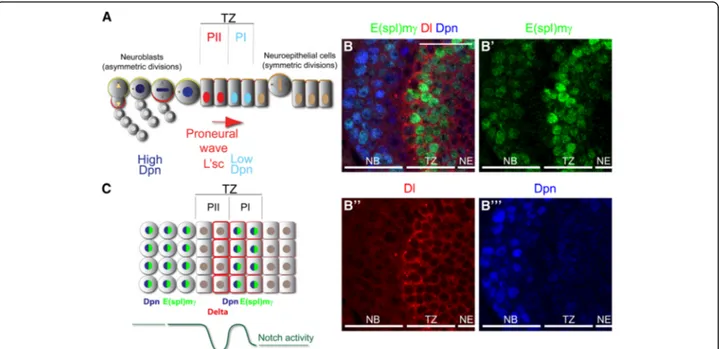
Fig. 1 E(spl)mγ expression reports Notch signalling at the transition zone. (a) Schematic model of the optic lobe transition (TZ) between NE cells into NBs. NE cells divide symmetrically to amplify their pool and transform into PI progenitors, expressing low levels of nuclear Dpn (blue). PI progenitors transform into PII progenitors, characterised by the expression of L’sc (red), and PII progenitors transform into NBs that divides asymmetrically and generate differentiated progeny. Modified from [8]. (b-b”’) Immunostaining of the optic lobe transition zone expressing the Notch reporter (b’) E(spl)mγ-GFP (green) and stained for (b”) Dl (red) and (b”’) Dpn (blue). (c) Schematic model of Notch signalling activation at the optic lobe transition zone, showing two peaks of Notch signalling activation in PI progenitors and in NBs. Scale bars are 20μm
and stained 78 h ALH. Flip-out clones were induced for 10 min at 37 °C, whereas for mutant clone generation larvae were heat-shocked for 30 min at 37 °C. Larvae were kept at 25 °C.
Immunofluorescence
Larval brains were fixed and stained as previously de-scribed [32]. The following primary antibodies were used: rabbit anti-Ase (1:1000 from Y.N. Jan), chicken anti-β-gal (1:100 abcam), mouse anti-Dl (1:100, C594.9B Develop-mental Studies Hybridoma Bank, DSHB), guinea pig anti-Dpn (1:5000, from J. Skeath), chicken and rabbit anti-GFP (1:2000 abcam), rat anti-L’sc (1:5000) and anti-Notch (1:50, C17.9C6 DSHB). Alexa Fluor conjugated secondary antibodies were diluted 1:200 (Molecular Probes, Invitrogen). Primary and secondary antibodies were incubated at 4 °C overnight.
In situ hybridisation
Probes were generated by PCR amplification from a em-bryonic cDNA library. Reverse primers contained the T7 polymerase promoter. Neur probe were generated using the following primers: Fw 5′- ACTCGCAATCAAACCT ACTAAAGC-3′ and Rv 5′- CAGTAATACGACTCACT ATTA AAGTGTAATTTAAAATGCGGCTTC-3′. For tom probe we used: Fw 5′- AAATCTCAACAATCCTCA ACACAA-3′ and Rv 5′- CAGTAATACGACTCACTAT
TA TACGAAGACCCTAACAAACAAACA-3′ [16].
in situ hybridisation was performed using a standard protocol. Briefly, third instar larval brains were fixed in 4% Formaldehyde in 1X PBS, washed with PBT (1X PBS,
0.1% Tween-20) and permeabilised using 50μg/mL
Proteinase K. Probes were hybridised at 55 °C, brains were blocked 30 min using 10% normal goat serum and incubated with anti-digoxigenin AP (1:2,000 Roche) for 2 h. Staining was performed using NBT/BCIP.
Imaging
Images were acquired using a Leica SP5 confocal microscope or a Zeiss Axioplasm microscope with a Leica DFC420C camera. Images, diagrams and figures were assembled using Fiji, adobe Photoshop CS2 and Illustrator CS3.
Results
E(spl)mγ reports Notch signalling in the optic lobe transition zone
Notch signalling is necessary to maintain both neuroepi-thelial cell and neuroblast fates. To understand the regula-tion of Notch signalling during the transiregula-tion of neuroepithelial cells to neuroblasts, we searched for a Notch reporter that precisely reflects the activation of the pathway. Several Notch reporters have been characterised as expressed in neuroepithelial cells and neuroblasts,
however, most of these express GFP or lacZ under the control of a Notch target gene promoter. Due to the sta-bility of GFP and β-galactosidase, these reporters do not reflect rapid changes in Notch signalling. To overcome this, we used the E(spl)mγ-GFP reporter (hereinafter re-ferred as E(spl)mγ) that contains the E(spl)mγ promoter and coding sequence fused to GFP, reflecting the dynam-ics of E(spl)mγ protein half-life and turnover [24].
E(spl)mγ was expressed at high levels at the transition zone (Fig. 1b-b”’). Interestingly, E(spl)mγ expression was
completely downregulated before neuroblast formation and then reexpressed in neuroblasts (high Dpn-positive cells, see Fig.1b’,b”’). Notch signalling downregulation
cor-related with high levels of Dl (Fig.1b,b”). This expression
pattern suggests that Notch signalling is highly active in PI progenitors, blocked after PII induction and restored upon neuroblast transformation (Fig.1b).
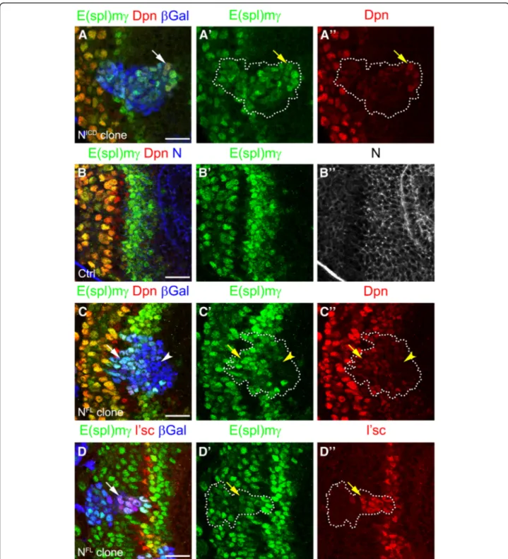
In order to confirm that E(spl)mγ expression was regu-lated by Notch signalling, we generated clones
misexpres-sing the intracellular domain of Notch (NICD), which
activates Notch signalling in a cell autonomous manner.
NICD clones marked with β-gal expressed high levels of
E(spl)mγ, confirming that the reporter was activated by Notch signalling. Furthermore, NICDclones also expressed low levels of Dpn suggesting that PI progenitor fate is in-duced by Notch signalling (see arrow in Fig.2a-a”).
There-fore, E(spl)mγ expression reflects the dynamic activity of Notch signalling at the transition zone.
Notch levels control signalling activity at the transition zone The expression of E(spl)mγ suggested a precise regula-tion of Notch signalling. Notch signalling was quickly blocked in one or two cells before neuroblast transform-ation and activated again in neuroblasts. Given that E(spl)mγ-negative cells were in direct contact with Dl-positive cells (Fig. 1b), we hypothesised that Notch signalling was regulated by the levels of receptor. We analysed the expression of Notch receptor at the transi-tion zone (Fig. 2b,b”). Although Notch was expressed in
all neuroepithelial cells, the E(spl)mγ reporter was acti-vated only at the transition zone (Fig. 2b,b”).
Interest-ingly, Notch and E(spl)mγ levels were reduced together at the end of the transition zone (see arrow Fig. 2b-b”)
and increased after neuroblast transformation, suggest-ing that Notch signallsuggest-ing is regulated by the levels of ex-pression of Notch.
To assess whether downregulation of Notch is the main mechanisms for blocking Notch signalling at the transi-tion zone, we generated clones expressing a full length form of Notch (NFL). NFL clones activated the E(spl)mγ reporter only at the transition zone, while no E(spl)mγ ex-pression was observed in clones in the middle of the neuroepithelium, where Dl is not expressed (Fig. 2c-c”).
maintained expression of E(spl)mγ and low levels of Dpn, suggesting that Notch signalling was active and induced PI progenitor fate (see arrow in Fig. 2c-c”). Additionally,
NFL
clones that crossed the transition zone maintained L’sc expression, delaying the transformation into neuro-blasts (see arrow in Fig.2d-d”). These results suggest that
Fig. 2 Notch signalling regulates PI progenitor fate and prevents PII progenitor conversion into neuroblasts. (a-Aa”) Staining of clone misexpressing the NICDin the optic lobe transition zone. Clone was marked byβ-gal expression (blue) and marked by dotted lines; E(spl)mγ expression in green, and Dpn in red. (b-b”) Wild-type brain transition zone stained for E(spl)mγ in green, Dpn in red and Notch receptor in blue (b) or grey (b”). Arrows indicate the end of Notch receptor and Notch signalling activation (c-d”) Staining of clones misexpressing a full length Notch receptor (NFL) for (c - d”) E(spl)mγ in green, Dpn in red (c, c”) and L’sc in red (d, d”). Arrows indicate E(spl)mγ activation after PI progenitor formation and (d-d”) a delay in PII progenitor transformation into NBs. Arrowheads show cells in the clone that do not activate Notch signalling (c-c”). Scale bars are 20 μm
Notch expression is rapidly downregulated in order to block its signalling, which is necessary to allow the precise transition from PII progenitors into neuroblasts.
Delta activates Notch signalling inducing the formation of PI progenitor state
To understand the role of Dl at the transition zone, we
generated Dl misexpression clones and assessed
E(spl)mγ expression. Dl misexpression blocked E(spl)mγ expression at the transition zone (Fig. 3a-a”), but
acti-vated E(spl)mγ expression and induced low levels of Dpn in neighbouring wild-type cells (see arrowheads in
Fig. 3b-b”). This result suggests that Dl can activate
Notch signalling, inducing PI progenitor fate in a non-cell autonomous manner, but that high levels of Dl block Notch signalling in a cell autonomous manner. However, we did not observe high levels of E(spl)mγ and Dpn surrounding the clones, suggesting that ectopic PI progenitors generated by Dl misexpression might not be competent to transform into neuroblasts.
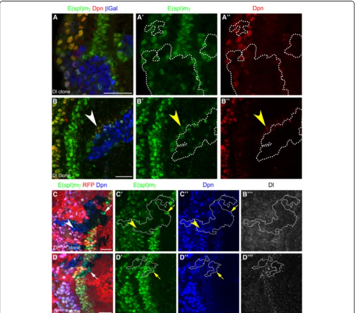
To characterise further the role of Dl in Notch signal-ling, we analysed Dl mutant clones. Mutant clones for a null allele of Dl (Dlrev10) [30] were generated by mitotic recombination and marked by the absence of RFP
Fig. 3 Delta necessary ans sufficient for Notch signalling inducing PI progenitor formation. (a-b”) Immunostaining of Dl misexpressing clones, E(spl)mγ in green, and Dpn in red. Clones were marked by β-gal staining in blue and dotted line. Arrowheads show E(spl)mγ activation in clone neighbouring cells. (c-d”’) Dlrev10mutant clones stained for E(spl)mγ in green, Dpn in blue, and Dl in gray. Clones were marked by the absence of
RFP expression and dotted lines. Arrows show E(spl)mγ expression inside mutant cells that were in contact with wild-type cells. Arrowheads show NBs not expressing E(spl)mγ. Scale bars are 20 μm
expression. These clones had no detectable Dl (Fig.3c-c") and the levels of the E(spl)mγ reporter and Dpn were decreased (see arrow in Fig.3c-c”), suggesting that Dl is
necessary for Notch signalling and PI progenitor induc-tion at the transiinduc-tion zone. Interestingly, E(spl)mγ ex-pression was also downregulated in mutant neuroblasts
(see arrowhead in Fig. 3c-c’). Small Dl mutant clones
were not affected and showed normal E(spl)mγ expres-sion, suggesting that wild-type cells can rescue Notch signalling in a non-cell autonomous manner (Fig.3d-d”’).
Non-cell autonomous activation could also be observed in mutant cells of larger clones, which were adjacent to Dl expressing wild-type cells (see arrows in Fig. 3d-d”’).
Together these results strongly suggest that Dl is the major ligand for Notch activation and PI progenitor state induction at the transition from neuroepithelial cells to neuroblasts.
Neuralized is required for Notch signalling at the transition zone
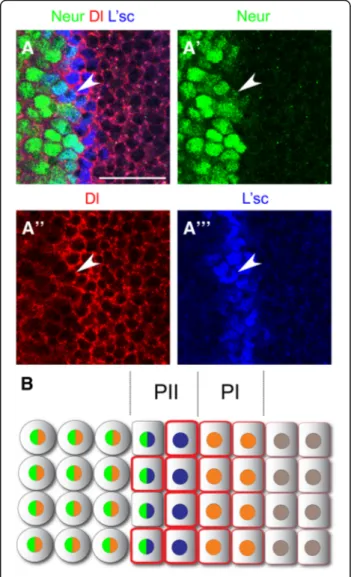
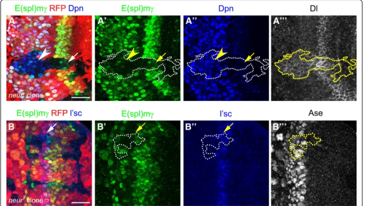
The E3 ubiquitin ligase neuralized (neur) [33–35] pro-motes endocytosis of the Dl ligand, activating Notch sig-nalling in neighbouring cells [36, 37]. As Neur function has not been assessed during optic lobe development, we decided to investigate whether it participates in the regulation of Notch signalling at the transition zone. We used a lacZ insertion in the neur locus (neur-lacZ) as an expression reporter during the transition from neuroepi-thelial cells into neuroblasts. [25]. neur-lacZ expression was observed at the end of the transition zone and in optic lobe neuroblasts. Neur is initiated in the second of the L’sc expressing PII progenitors, just prior to their transformation into Dpn positive neuroblasts (Fig. 4a). These medial PII progenitors also expressed Dl (see arrowhead in Fig. 4a-a”’), but at lower levels than the
most lateral PII progenitor. We observed high levels of neur mRNA at the transition zone, in a pattern comple-mentary to twin of m4 (tom) expression, a Notch target
gene expressed across the neuroepithelium [16]
(Add-itional file1: Figure S1). Therefore, neur is expressed in medial PII progenitors and in optic lobe neuroblasts (Fig.4b).
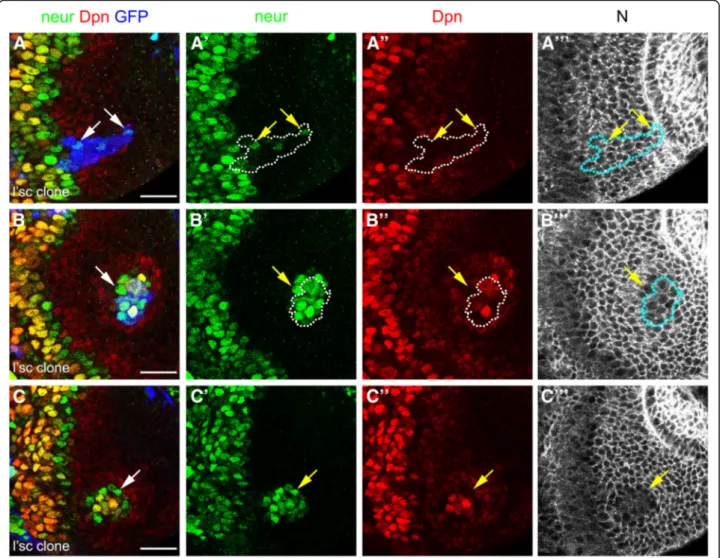
To assess Neur function, we generated neur mutant clones using a null allele (neur1) [31]. Mutant clones showed a reduction in E(spl)mγ expression in a cell
au-tonomous manner (Fig. 5a-a”’) resembling Dl mutant
clones (compare to Fig.3a-a”’). The reduction in E(spl)mγ
expression was observed in optic lobe neuroblasts (69.7% of clones, 23/33, see arrowhead in Fig.5a-a”) and also in
PI progenitors (52.0% of clones, 13/25), in which Dpn
levels were also reduced (see arrow in Fig. 5a”). In
addition, while L’sc levels were normal in neur mutant cells, Dl levels were upregulated in clones at the transition zone and in Dpn-positive neuroblasts (Fig.5a”’ and 5b”).
This suggests that neur is required to activate
Dl-mediated Notch signalling and to induce PI progenitor state in the neuroepithelial to neuroblasts transition zone. Lethal of scute is sufficient to induce neuralized
expression and to generate ectopic transition zones neur was expressed preferentially in the L’sc-positive PII progenitors closest to neuroblasts (Fig.5). PII progenitor fate is defined by the expression of L’sc [7], hence we hypothesised that L’sc regulates neur expression in order to activate Notch signalling and induce PI progenitor fate. To test this, we generated L’sc misexpression clones outside the transition zone. L’sc misexpression was suffi-cient to induce neur expression in neuroepithelial cells
Fig. 4 neuralized is expressed in PII progenitors and in optic lobe neuroblasts. (a) Immunostaining of neur-lacZ larval brains for β-gal/ neur in green, Dl in red and L’sc in blue. Arrowheads show PII progenitor expressing neur, Dl and L’sc. (b) Schematic representation of neur expression during the transition between NE cells into NBs. Scale bars are 20μm
(see arrows in Fig. 6a-a”’). Remarkably, L‘sc
misexpres-sion generated ectopic transition zones in the
neuroepi-thelium (Fig. 6b). These clones showed high levels of
Dpn and Neur, and a decrease in Notch receptor levels, demonstrating that ectopic neuroblasts were generated by L’sc misexpression (Fig.6b-b”’).
Interestingly, neur expression was also observed out-side the clones (see arrow in Fig.6b-b”) as were PI
pro-genitors (low Dpn-positive cells; Fig.6b”). In some cases,
L’sc misexpressing cells were found deep inside the optic lobe (see blue clone in Additional file2: Figure S2), sug-gesting that the L’sc misexpressing cells initiated the ec-topic transition zone and then delaminated from the neuroepithelium after neuroblast transformation. The ectopic transition zones remained in the neuroepithe-lium after the clones had delaminated (note lack of GFP
expression in Fig. 6c). These ectopic transition zones
contained Dpn-positive PI progenitors, Neur-positive PII progenitors and Dpn-positive/Neur-positive neuroblasts.
We conclude that the induction of L’sc within the
neuroepithelium is sufficient to induce neur expression and to generate ectopic transition zones containing PI and PII progenitor states in a non-cell autonomous manner. Remarkably these ectopic transition zones are maintained and continue to generate neuroblasts.
Discussion
Notch signalling acitivity is dynamically regulated in the
transition zone. The E(spl)mγ reporter is highly
expressed in PI progenitor cells, downregulated in PII progenitor cells and upregulated again in neuroblasts [8,
38]. Here, we demonstrate that the ligand Delta and the E3 Ubiquitin ligase Neur are required in PII progenitor cells to activate Notch signalling in neighbouring PI pro-genitors. We also show that Neur expression is induced by the proneural factor L’sc, which is able to induce the entire transition zone.
A switch from serrate to Delta mediates Notch signalling in the progression of neural stem cell states
Notch mutant clones are extruded from the neuroepi-thelium and prematurely transform into neuroblasts at ectopic positions [16]. Interestingly, Dl mutant clones in the lateral neuroepithelium do not phenocopy these Notch null mutant clones [7]. This suggests that Dl is not required for Notch signalling in more lateral prolif-erating neuroepithelial cells and that Notch is activated by a different ligand. Indeed, Perez-Gomez et al. [15] showed that glial cells adjacent to the neuroepithelium activate Notch signalling via the ligand Serrate (Ser). Ser is necessary for neuroepithelial cell proliferation and for
Fig. 5 Notch signalling activation requires Neuralized function at the transition zone. (a-b”’) neur1mutant clones stained for E(spl)mγ in green, (a,a”) Dpn in blue, (b,b”) L’sc in blue, (a”’) Dl in gray and (b”’) Asense (Ase), as a neuroblast marker, in gray. Clones were marked by the absence of RFP expression and dotted lines. (a-a”) Arrows show decrease in E(spl)mγ staining in PI progenitors and arrowheads in NBs. (b-b”) Arrows pointed L’sc-positive PII progenitor inside neur mutant clone. Scale bars are 20 μm
preventing PII progenitor formation [15]. Hence, we favour a model in which Notch signalling induce by Ser-rate maintains neuroepithelial cells in a proliferating state, while Notch signalling induced by Delta initiates PI progenitor formation and the neuroepithelial cell to neuroblast transition.
The differential expression of Notch signalling modu-lators, such as the protein Canoe (Cno), may explain preferential binding for one of the two ligands. Canoe stabilises the Notch receptor at adherens junctions and
promotes binding to Ser from glial cells [15]. The E3
ubiquitin ligase, Mind bomb, is required for the activa-tion of Ser while Neur controls the activity of Delta [39]. We show that neur expression is restricted to PII pro-genitors cells closest to the neuroblasts (Fig.4). However, the loss of neur affects cells that are not immediate neigh-bours, the PI progenitors, implying that Delta-Notch sig-nalling may work over a distance. Membrane protrusions
may allow Dl to activate N signalling at a distance, as has
been described during bristle development [40, 41]
(Fig.7a). Alternatively, it has been shown that Notch sig-nalling promotes Dl expression [20]. This positive feed-back loop may allow the initial Neur activity to propagate in a non-cell autonomous manner, generating a gradient of Notch signalling (Fig.7b).
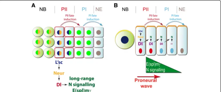
A backward relay mechanism controls changes in neural stem cell states
Neuroepithelial cells progress through two transient pro-genitor states prior to transforming into medulla neuro-blasts [6–8]. Here we show that PII progenitors can be further subdivided into L’sc, high Delta and L’sc, Neur expressing cells (Fig. 7). Two opposing signalling path-ways control the medial to lateral progression of the proneural wave that initiates the neuroepithelial cell to neuroblast transition. EGF signalling drives the wave
Fig. 6 Lethal of scute regulates neutralized expression and generates ectopic transition zone in a cell non-autonomous manner. (a-c”’) Immunostaining of L’sc misexpressing clones in neur-lacZ larval brain for β-gal/neur in green, Dpn in and Notch in gray. Clones were marked by GFP expression in blue and dotted lines. Arrows show ectopic activation of neur expression (a-a”’) inside and (b-c”’) outside L’sc misexpressing clones. Note that in (c-c”’) there is no NE cell misexpressing L’sc (no GFP expression, blue). Scale bars are 20 μm
forward, while JAK/STAT signalling slows the progres-sion of the wave [7, 9, 42] and prevents ectopic neuro-blast formation in the epithelium [43]. PII progenitor cells secrete the EGF ligand, Spitz, which activates the EGFR pathway in neighbouring lateral PI progenitor cells. These neuroepithelial cells are positive for the EGFR downstream target gene pointed P1 (pntP1). pnt or spitz loss-of-function mutant clones do not upregu-late L’sc, indicating that both the induction of the pro-neural wave, and its progression, are downstream of EGFR signalling [7]. Moreover, the EGF signalling con-trols the levels of Dl ligand, regulating Notch signalling and the progression of the proneural wave [7,44].
L’sc acts in a backward relay mechanism to induce the PI progenitor state. It induces the expression of Neur in PII progenitors and thus activates Delta-Notch signalling to induce PI. As a result neighbouring PI progenitors upregulate the Notch target gene E(spl)mγ. One role of high Notch signalling activity in PI is to induce cell cycle arrest in PI progenitor cells [19]. Hence, the backward relay mechanism controls the sequential and timely ac-quisition of progenitor states.
In order for neuroepithelial cells to transform into neuroblasts, Notch signalling must be blocked. Binding of Dl to Notch in the same cell can inhibit Notch signal-ling through a mechanism called‘cis-inhibition’ [45]. We observed high Dl levels in PII progenitor cells where
E(spl)mγ levels are low. Furthermore, we show that Dl
misexpression clones show no Notch signalling activity. Therefore, it is plausible that Dl activates Notch in trans,
inducing the PI progenitor state, while inhibiting Notch in cis to enable the progression from PII progenitors to neuroblasts.
Notch signalling regulates stem cell heterogeneity from flies to vertebrates
The Notch signalling pathway regulates stem cell main-tenance, proliferation and differentiation in different tis-sues, contributing to vertebrate development and organ regeneration. However, the effect of Notch signalling is highly dependent on the biological context [10]. During development and adult neurogenesis, NSCs are a highly heterogeneous population. NSCs can be found in prolif-erative or quiescent states. Furthermore, adult NSCs generate intermediate progenitor states with different potency before differentiation into neurons or glial cells
[46]. Notch signalling preserves NSC maintenance and
proliferation [47–49] and can also induce the quiescence state [14, 50–52]. The context of Notch signalling in NSCs determines the outcome. For example in zebrafish, whereas the Notch3 receptor induces a quiescence state in NSCs, Notch1b is required for NSC population
main-tenance [53]. This phenomenon resembles the different
responses to Notch signalling in neuroepithelial cells in the Drosophila optic lobe.
Notch signalling interaction with other pathways also regulates NSC behaviour in the vertebrate brain. EGFR
signalling in neural progenitors non-autonomously
blocks Notch signalling in NSCs, reducing NSC prolifer-ation at the adult subventricular zone [54]. Interestingly,
Fig. 7 Working models of Notch signalling during the transition of neural stem cell states. Two models showing the progression of the transition between NE cells into NBs. a Long-range activation of Notch signalling in PI progenitors can be controlled by L’sc in PII progenitors. L’sc regulates neur expression that activates Dl function. b Activation of Notch signalling is regulated by L’sc-positive/Neur-positive/Dl-positive PII progenitors inducing Dl expression in the closer neighbour and generating a gradient of E(spl)mγ expression in PI progenitors. In both models, PII progenitors are able to induce the PII fate in PI progenitor, while PI progenitors promote NE cells transformation intro PI state. When PII progenitors convert into NB, PI progenitors replace PII progenitors and NE cells convert into PI progenitors, promoting the progression of the proneural wave
EGFR is a downstream target of Notch signalling in
NSCs [48], suggesting that Notch promotes both NSC
maintenance and the formation of neural progenitors. Conclusions
Our study proposes a model of dynamic Notch signal-ling in the transition from neuroepithelial cells into neu-roblasts. During Drosophila optic lobe development, Notch signalling regulates NSC amplification and main-tenance in a similar manner to vertebrate NSCs. Notch signalling also induces the progression into PI/PII pro-genitor states. Understanding the dynamic regulation of Notch signalling during NSC state transitions in the optic lobe may yield new insights into the mechanisms that control adult neurogenesis and brain regeneration.
Aknowledgements We would like to thank Sarah Bray,
François Schweisguth, Eugenia Piddini, Pat Simpson, Yuh Nung Jan, Jim Skeath and DSHB for antibodies and fly stocks. We thank Takumi Suzuki and Carlos Oliva for comments on the manusctript.
Additional files
Additional file 1:Figure S1. neur is expressed in the transition zone. in situ hybridisation of larval brains using specific antisense probes against (A) tom and (B) neur. Note that neur and tom are expressed in a complementary pattern. (PSD 81600 kb)
Additional file 2:Figure S2. L’sc misexpression clones delaminate and induce ectopic transition zones in wild-type neuroepithelial cells. (A-A”) L’sc misexpressing clone that has delaminated from the neuroepithelium induces an ectopic transition zone. Staining for (A,A’) β-gal/neur in green, (A,A”) Dpn in red and (A) Notch receptor in gray. Clone is marked by GFP expression in blue. Note that the clone has delaminated inside the optic lobe and the ectopic transition zone has continued generating NBs. (PSD 12135 kb)
Abbreviations
Ase:Asense; Dl: Delta; Dpn: Deadpan; L’sc: Lethal of scute; N: Notch; NB: Neuroblast; NE: Neuroepithelial; Neur: Neuralized; NICD: Notch intracellular domain; NSC: Neural stem cell
Funding
This work was funded by the Royal Society Darwin Trust Research Professorship, Wellcome Trust Senior Investigator Award 103792 and Wellcome Trust Programme grant 092545 to A.H.B., Wellcome Trust 4-year PhD Studentships to E.G.C. and K.S.G. and a Fellowship from the Swiss National Foundation to B.E. A.H.B acknowledges core funding to the Gurdon Institute from the Wellcome Trust (092096) and CRUK (C6946/A14492).
Availability of data and materials All data is available upon request.
Authors’ contributions
EGC, BE and AHB designed the experiments, analysed the data and wrote the manuscript. EGC, BE and KSG performed the experiments. All authors read and approved the final manuscript.
Ethics approval and consent to participate Not applicable.
Consent for publication Not applicable.
Competing interests
The authors declare that they have no competing interests.
Publisher’s Note
Springer Nature remains neutral with regard to jurisdictional claims in published maps and institutional affiliations.
Author details
1
The Gurdon Institute and Department of Physiology, Development and Neuroscience, University of Cambridge, Tennis Court Road, Cambridge CB2 1QN, UK.2Present Address: Department of Biology, Zoology, University of
Fribourg, Chemin du Musée 10, CH-1700 Fribourg, Switzerland.
Received: 11 September 2018 Accepted: 13 November 2018
References
1. Brand AH, Livesey FJ. Neural stem cell biology in vertebrates and invertebrates: more alike than different? Neuron. 2011;70(4):719–29. 2. Lanet E, Gould A, Maurange C. Protection of neuronal diversity at the
expense of neuronal numbers during nutrient restriction in the Drosophila visual system. Cell Rep. 2013;3(3):587–94.
3. Egger B, Boone JQ, Stevens NR, Brand AH, Doe CQ. Regulation of spindle orientation and neural stem cell fate in the Drosophila optic lobe. Neural Dev. 2007;2:1.
4. Hofbauer A, Campos-Ortega J. Proliferation pattern and early differentiation of the optic lobes in Drosophila melanogaster. Roux Arch Dev Biol. 1990; 198(5):264–74.
5. Rujano MA, Sanchez-Pulido L, Pennetier C, le Dez G, Basto R. The microcephaly protein Asp regulates neuroepithelium morphogenesis by controlling the spatial distribution of myosin II. Nat Cell Biol. 2013;15(11): 1294–306.
6. Sato M, Suzuki T, Nakai Y. Waves of differentiation in the fly visual system. Dev Biol. 2013;380(1):1–11.
7. Yasugi T, Sugie A, Umetsu D, Tabata T. Coordinated sequential action of EGFR and Notch signaling pathways regulates proneural wave progression in the Drosophila optic lobe. Development. 2010;137(19):1–11.
8. Egger B, Gold KS, Brand AH. Regulating the balance between symmetric and asymmetric stem cell division in the developing brain. Fly. 2011;5(3): 237–41.
9. Yasugi T, Umetsu D, Murakami S, Sato M, Tabata T. Drosophila optic lobe neuroblasts triggered by a wave of proneural gene expression that is negatively regulated by JAK/STAT. Development. 2008;135(8):1471–80. 10. Koch U, Lehal R, Radtke F. Stem cells living with a Notch. Development.
2013;140(4):689–704.
11. Bray SJ. Notch signalling: a simple pathway becomes complex. Nat Rev Mol Cell Biol. 2006;7(9):678–89.
12. Pierfelice T, Alberi L, Gaiano N. Notch in the vertebrate nervous system: an old dog with new tricks. Neuron. 2011;69(5):840–55.
13. Ables JL, Breunig JJ, Eisch AJ, Rakic P. Not(ch) just development: Notch signalling in the adult brain. Nat Rev Neurosci. 2011;12(5):269–83. 14. Giachino C, Taylor V. Notching up neural stem cell homogeneity in
homeostasis and disease. Front Neurosci. 2014;8:32.
15. Perez-Gomez R, Slovakova J, Rives-Quinto N, Krejci A, Carmena A. A Serrate-Notch-Canoe complex mediates essential interactions between glia and neuroepithelial cells during Drosophila optic lobe development. J Cell Sci. 2013;126(Pt 21):4873–84.
16. Egger B, Gold KS, Brand AH. Notch regulates the switch from symmetric to asymmetric neural stem cell division in the Drosophila optic lobe. Development. 2010;137(18):2981–7.
17. Ngo KT, Wang J, Junker M, Kriz S, Vo G, Asem B, Olson JM, Banerjee U, Hartenstein V. Concomitant requirement for Notch and Jak/Stat signaling during neuro-epithelial differentiation in the Drosophila optic lobe. Dev Biol. 2010;346(2):284–95.
18. Orihara-Ono M, Toriya M, Nakao K, Okano H. Downregulation of Notch mediates the seamless transition of individual Drosophila neuroepithelial progenitors into optic medullar neuroblasts during prolonged G1. Dev Biol. 2011;351(1):163–75.
19. Wang W, Liu W, Wang Y, Zhou L, Tang X, Luo H. Notch signaling regulates neuroepithelial stem cell maintenance and neuroblast formation in Drosophila optic lobe development. Dev Biol. 2011;350(2):414–28. 20. Reddy BV, Rauskolb C, Irvine KD. Influence of fat-hippo and notch signaling
on the proliferation and differentiation of Drosophila optic neuroepithelia. Development. 2010;137(14):2397–408.
21. Green P, Hartenstein AY, Hartenstein V. The embryonic development of the Drosophila visual system. Cell Tissue Res. 1993;273(3):583–98.
22. Zacharioudaki E, Magadi SS, Delidakis C. bHLH-O proteins are crucial for Drosophila neuroblast self-renewal and mediate Notch-induced overproliferation. Development. 2012;139(7):1258–69.
23. Zhu S, Wildonger J, Barshow S, Younger S, Huang Y, Lee T. The bHLH repressor deadpan regulates the self-renewal and specification of Drosophila larval neural stem cells independently of Notch. PLoS One. 2012; 7(10):e46724–15.
24. Almeida MS, Bray SJ. Regulation of post-embryonic neuroblasts by Drosophila Grainyhead. Mech Dev. 2005;122(12):1282–93.
25. Phillips RG, Roberts IJ, Ingham PW, Whittle JR. The Drosophila segment polarity gene patched is involved in a position-signalling mechanism in imaginal discs. Development. 1990;110(1):105–14.
26. Doherty D, Feger G, Younger-Shepherd S, Jan LY, Jan YN. Delta is a ventral to dorsal signal complementary to serrate, another Notch ligand, in Drosophila wing formation. Genes Dev. 1996;10(4):421–34.
27. Zecchini V, Brennan K, Martinez-Arias A. An activity of Notch regulates JNK signalling and affects dorsal closure in Drosophila. Curr Biol. 1999;9(9):460–9. 28. Cooper MT, Bray SJ. R7 photoreceptor specification requires Notch activity.
Curr Biol. 2000;10(23):1507–10.
29. Carmena A, Bate M, Jimenez F. Lethal of Scute, a proneural gene, participates in the specification of muscle progenitors during Drosophila embryogenesis. Genes Dev. 1995;9(19):2373–83.
30. Haenlin M, Kramatschek B, Campos-Ortega JA. The pattern of transcription of the neurogenic gene Delta of Drosophila melanogaster. Development. 1990;110(3):905–14.
31. Lehmann R, Jiménez F, Dietrich U, Campos-Ortega J. On the phenotype and development of mutants of early neurogenesis inDrosophila melanogaster. Wilehm Roux Arch Dev Biol. 1983;192(2):62–74.
32. Van Vactor DL Jr, Cagan RL, Kramer H, Zipursky SL. Induction in the developing compound eye of Drosophila: multiple mechanisms restrict R7 induction to a single retinal precursor cell. Cell. 1991;67(6):1145–55. 33. Yeh E, Dermer M, Commisso C, Zhou L, McGlade CJ, Boulianne GL. Neuralized functions as an E3 ubiquitin ligase during Drosophila development. Curr Biol. 2001;11(21):1675–9.
34. Lai EC, Deblandre GA, Kintner C, Rubin GM. Drosophila neuralized is a ubiquitin ligase that promotes the internalization and degradation of delta. Dev Cell. 2001;1(6):783–94.
35. Deblandre GA, Lai EC, Kintner C. Xenopus neuralized is a ubiquitin ligase that interacts with XDelta1 and regulates Notch signaling. Dev Cell. 2001; 1(6):795–806.
36. Le Borgne R, Schweisguth F. Unequal segregation of Neuralized biases Notch activation during asymmetric cell division. Dev Cell. 2003;5(1):139–48. 37. Pavlopoulos E, Pitsouli C, Klueg KM, Muskavitch MA, Moschonas NK,
Delidakis C. neuralized encodes a peripheral membrane protein involved in delta signaling and endocytosis. Dev Cell. 2001;1(6):807–16.
38. Weng M, Haenfler J, Lee C. Changes in Notch signaling coordinates maintenance and differentiation of the Drosophila larval optic lobe neuroepithelia. Dev Neurobiol. 2012;72(11):1376–90.
39. Le Borgne R, Remaud S, Hamel S, Schweisguth F. Two distinct E3 ubiquitin ligases have complementary functions in the regulation of delta and serrate signaling in Drosophila. PLoS Biol. 2005;3(4):e96.
40. De Joussineau C, Soule J, Martin M, Anguille C, Montcourrier P, Alexandre D. Delta-promoted filopodia mediate long-range lateral inhibition in Drosophila. Nature. 2003;426(6966):555–9.
41. Cohen M, Georgiou M, Stevenson NL, Miodownik M, Baum B. Dynamic filopodia transmit intermittent Delta-Notch signaling to drive pattern refinement during lateral inhibition. Dev Cell. 2010;19(1):78–89. 42. Wang W, Li Y, Zhou L, Yue H, Luo H. Role of JAK/STAT signaling in
neuroepithelial stem cell maintenance and proliferation in the Drosophila optic lobe. Biochem Biophys Res Commun. 2011;410(4):714–20.
43. Tanaka Y, Yasugi T, Nagayama M, Sato M, Ei SI. JAK/STAT guarantees robust neural stem cell differentiation by shutting off biological noise. Sci Rep. 2018;8(1):12484.
44. Sato M, Yasugi T, Minami Y, Miura T, Nagayama M. Notch-mediated lateral inhibition regulates proneural wave propagation when combined with EGF-mediated reaction diffusion. Proc Natl Acad Sci U S A. 2016;113(35):E5153–62. 45. del Alamo D, Rouault H, Schweisguth F. Mechanism and significance of
cis-inhibition in Notch signalling. Curr Biol. 2011;21(1):R40–7.
46. Farkas LM, Farkas LM, Huttner WB, Huttner WB. The cell biology of neural stem and progenitor cells and its significance for their proliferation versus differentiation during mammalian brain development. Curr Opin Cell Biol. 2008;20(6):707–15.
47. Imayoshi I, Sakamoto M, Yamaguchi M, Mori K, Kageyama R. Essential roles of Notch signaling in maintenance of neural stem cells in developing and adult brains. J Neurosci. 2010;30(9):3489–98.
48. Andreu-Agullo C, Morante-Redolat JM, Delgado AC, Farinas I. Vascular niche factor PEDF modulates Notch-dependent stemness in the adult
subependymal zone. Nat Neurosci. 2009;12(12):1514–23.
49. Ehm O, Goritz C, Covic M, Schaffner I, Schwarz TJ, Karaca E, Kempkes B, Kremmer E, Pfrieger FW, Espinosa L, et al. RBPJkappa-dependent signaling is essential for long-term maintenance of neural stem cells in the adult hippocampus. J Neurosci. 2010;30(41):13794–807.
50. Carlen M, Meletis K, Goritz C, Darsalia V, Evergren E, Tanigaki K, Amendola M, Barnabe-Heider F, Yeung MS, Naldini L, et al. Forebrain ependymal cells are Notch-dependent and generate neuroblasts and astrocytes after stroke. Nat Neurosci. 2009;12(3):259–67.
51. Chapouton P, Skupien P, Hesl B, Coolen M, Moore JC, Madelaine R, Kremmer E, Faus-Kessler T, Blader P, Lawson ND, et al. Notch activity levels control the balance between quiescence and recruitment of adult neural stem cells. J Neurosci. 2010;30(23):7961–74.
52. Katz S, Cussigh D, Urban N, Blomfield I, Guillemot F, Bally-Cuif L, Coolen M. A nuclear role for miR-9 and Argonaute proteins in balancing quiescent and activated neural stem cell states. Cell Rep. 2016;17(5):1383–98.
53. Alunni A, Krecsmarik M, Bosco A, Galant S, Pan L, Moens CB, Bally-Cuif L. Notch3 signaling gates cell cycle entry and limits neural stem cell amplification in the adult pallium. Development. 2013;140(16):3335–47. 54. Aguirre A, Rubio ME, Gallo V. Notch and EGFR pathway interaction regulates