HAL Id: hal-02296685
https://hal.archives-ouvertes.fr/hal-02296685
Submitted on 25 Feb 2021HAL is a multi-disciplinary open access archive for the deposit and dissemination of sci-entific research documents, whether they are pub-lished or not. The documents may come from teaching and research institutions in France or abroad, or from public or private research centers.
L’archive ouverte pluridisciplinaire HAL, est destinée au dépôt et à la diffusion de documents scientifiques de niveau recherche, publiés ou non, émanant des établissements d’enseignement et de recherche français ou étrangers, des laboratoires publics ou privés.
Neutron-based computed microtomography: Pliobates
cataloniae and Barberapithecus huerzeleri as a test-case
study
Alessandro Urciuoli, Clément Zanolli, Josep Fortuny, Sergio Almécija,
Burkhard Schillinger, Salvador Moyà-Solà, David Alba
To cite this version:
Alessandro Urciuoli, Clément Zanolli, Josep Fortuny, Sergio Almécija, Burkhard Schillinger, et al.. Neutron-based computed microtomography: Pliobates cataloniae and Barberapithecus huerzeleri as a test-case study. American Journal of Physical Anthropology, Wiley, 2018, 166 (4), pp.987-993. �10.1002/ajpa.23467�. �hal-02296685�
1
Neutron-based computed microtomography: Pliobates cataloniae and
1
Barberapithecus huerzeleri as a test-case study
2 3
Alessandro Urciuoli1 | Clément Zanolli2 | Josep Fortuny3,1 | Sergio Almécija4,1 | 4
Burkhard Schillinger5 | Salvador Moyà-Solà1,6,7 | David M. Alba1 5
6
1Institut Català de Paleontologia Miquel Crusafont, Universitat Autònoma de Barcelona, 7
Edifici ICTA-ICP, c/ Columnes s/n, Campus de la UAB, 08193 Cerdanyola del Vallès,
8
Barcelona, Spain
9
2Laboratoire AMIS, UMR 5288 CNRS, Université Toulouse III Paul Sabatier, Toulouse, 10
France
11
3Centre de Recherches en Paléobiodiversité et Paléoenvironnements, Muséum National 12
d’Histoire Naturelle, Bâtiment de Paléontologie, CP38, 8 rue Buffon, 75005 Paris, France
13
4Center for the Advanced Study of Human Paleobiology, Department of Anthropology, The 14
George Washington University, Washington, DC 20052, USA
15
5Technische Universität München, Fakultat für Physik E21, James-Franck-Str.1, D-85747 16
Garching, Germany
17
6Institució Catalana de Recerca i Estudis Avançats, Pg. Lluís Companys 23, 08010, 18
Barcelona, Spain
19
7Unitat d’Antropologia (Departament de Biologia Animal, Biologia Vegetal i Ecologia), 20
Universitat Autònoma de Barcelona, 08193 Cerdanyola del Vallès, Barcelona,Spain
21 22
Number of text pages: 15
23
Number of figures: 3
24
Number of tables: 1
25
Abbreviated title: Neutron-µCT in paleoanthropology
2
KEYWORDS
27
X-rays, neutron radiation, neutron imaging, fossil catarrhines
28 29
Correspondence: David M. Alba, Institut Català de Paleontologia Miquel Crusafont,
30
Universitat Autònoma de Barcelona, Edifici ICTA-ICP, c/ Columnes s/n, Campus de la
31
UAB, 08193 Cerdanyola del Vallès, Barcelona, Spain. +34 5868304. Email:
32
Funding information: Spanish MINECO/FEDER EU, Project number: CGL2014-54373-P;
34
Spanish AEI/FEDER EU, Project number: CGL2016-76431-P; Generalitat de Catalunya,
35 CERCA Programme. 36 37 Abstract 38
Objectives: High-resolution imaging of fossils with X-ray computed microtomography
39
(μCT) has become a very powerful tool in paleontological research. However, fossilized
40
bone, embedding matrix, and dental tissues do not always provide a distinct structural
41
signal with rays. Here we report on neutron radiation as an alternative to standard
X-42
rays for the μCT of ‘problematic’ fossils.
43
Materials and Methods: We compare neutron with X-ray μCT scans of fossils from two
44
Miocene catarrhines from the Vallès-Penedès Basin: the cranium (IPS58443.1, holotype)
45
of the putative stem hominoid Pliobates cataloniae, to discriminate between bone and
46
matrix; and two lower molars (IPS1724n,o, holotype) of Barberapithecus huerzeleri, to
47
discriminate among dental tissues.
48
Results: X-ray μCT scans of these specimens fail to retrieve any contrast between
49
matrix/bone and enamel/dentine, whereas neutron μCT scans deliver high-contrast
50
images, enabling a proper evaluation of the specimens’ internal anatomy.
3
Discussion: Low bone/matrix intensity difference with X-ray μCT scans in IPS58443.1 is
52
due to the extreme similarity in chemical composition between the matrix and the fossilized
53
tissues, and the presence of high-density elements. In IPS1724, it is attributable to the
54
convergence of enamel and dentine compositions during fossilization. On the contrary,
55
neutron radiation returns very different contrasts for different isotopes of the same element
56
and easily penetrates most metals. Neutron-based μCT scans therefore enable a correct
57
definition of the bone/sediment and enamel/dentine interfaces, and hence a better
58
segmentation of the images stack. We conclude that neutron radiation represents a
59
successful alternative for high-resolution µCT of small-sized fossils that are problematic
60
with standard X-rays.
61 62
1 | INTRODUCTION
63 64
The use of computed microtomography (μCT) in paleontological research has dramatically
65
increased during the last decade, in parallel to concomitant enhancements in image
66
detectors and computing power. The success of computed tomography in paleontology is
67
due to the fact that it enables the non-destructive study of internal anatomy, the virtual
68
extraction of sediment-embedded fossils, the virtual reconstruction of damaged specimens
69
(including the mirroring of antimeres), and even the retrodeformation of plastically
70
deformed fossils (Macchiarelli et al., 2004; Olejniczak & Grine, 2006; Olejniczak,
71
Tafforeau, Temming, Smith, & Hublin, 2007; Abel, Rettondini Laurini, & Richter, 2012;
72
Benazzi, Kullmer, Schulz, Gruppioni, & Weber, 2013; Faulwetter, Vasileiadou, Kouratoras,
73
Dailianis, & Arvanitidis, 2013; Macchiarelli, Bayle, Bondioli, Mazurier, & Zanolli, 2013;
74
Benazzi, Gruppioni, Strait, & Hublin, 2014; Cunningham, Rahman, Lautenschlager,
75
Rayfield, & Donoghue, 2014; Lautenschlager, 2016). Modern X-ray μCT (especially based
76
on synchrotron radiation) can reach a very high spatial resolution (up to less than 1 μm;
4
Gren et al., 2016), and the size of the specimens that can be scanned with a single
78
acquisition has recently increased considerably (currently in the order of some decimeters;
79
e.g., Tuniz et al., 2013).
80
Depending on the taphonomic processes occurring during fossilization, not all
81
vertebrate fossil remains are amenable to internal anatomy analysis based on standard
X-82
ray μCT. The latter may fail to retrieve an adequate contrast between the fossil bone and
83
the matrix or between different dental tissues because of two main problems: (1) the
84
embedding matrix and the fossilized tissues have a very similar composition, due to
85
element exchange resulting from permineralization (Zanolli, Grine, Kullmer, Schrenk, &
86
Macchiarelli, 2015; Beaudet et al., 2016); (2) a considerable amount of high-density
87
elements is present in the embedding sediment or in the fossil itself (Spoor, Zonneveld &
88
Macho, 1993; Abel et al., 2012). Either of these problems hinders and might even entirely
89
preclude the segmentation of the fossil, thus preventing the extraction of essential
90
paleobiological evidence (e.g., Schwarz, Vontobel, Lehmann, Meyer, & Bongartz, 2005;
91
Smith et al., 2009; Zanolli et al., 2017a). For these reasons, an alternative to X-ray µCT is
92
needed. Neutron radiography and tomography, respectively developed in the 1950s and
93
1970s (Kardjilov et al., 2003; Schwarz et al., 2005; Winkler, 2006), constitute a potential
94
alternative. However, so far neutron-based µCT (n-μCT) has only sporadically been used
95
in paleontological and paleoanthropological research (Schwarz et al., 2005; Sutton, 2008;
96
Zanolli et al., 2013, 2017a; Beaudet et al., 2016; Laaß & Kaestner, 2017; Schillinger,
97
2017)., Although the reliability of n-μCT for investigating the internal anatomy of fossils has
98
previously been demonstrated by previous researchers (Schwarz et al., 2005; Beaudet et
99
al., 2016; Zanolli et al., 2017a), here we test further the applicability of this method to
100
specimens that cannot be properly analyzed by means of X-ray µCT. In particular, by
101
focusing on the fossil remains of two European Miocene catarrhines, we address two
102
common problems in CT-based paleoprimatological research: difficulties in discriminating
5
fossilized cranial bone from the surrounding or embedding matrix (as exemplified by the
104
putative stem hominoid Pliobates); and the inability to discriminate well between enamel
105
and dentine in fossil teeth (exemplified by the pliopithecoid Barberapithecus).
106 107
2 | MATERIALS AND METHODS
108 109
2.1 | Studied sample
110
The remains of two Miocene catarrhines from the Vallès-Penedès Basin (NE Iberian
111
Peninsula), housed at the Institut Català de Paleontologia Miquel Crusafont (Sabadell,
112
Spain; ICP), were investigated: (1) the partial cranium of Pliobates cataloniae
113
(IPS58443.1, holotype), a putative stem hominoid from the stratigraphic series of
114
Abocador de Can Mata (ACM) locality ACM/C8-A4 (Alba et al., 2015: Figs. 1, 4), with an
115
estimated age of 11.6 Ma (middle to late Miocene boundary; Alba, Casanovas-Vilar,
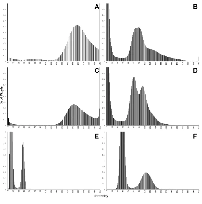
116
Garcés, & Robles, 2017); and (2) two lower molars (right M2 and left M3) of a single 117
individual of Barberapithecus huerzeleri (respectively IPS1724n,o, holotype), a
118
pliopithecoid from Castell de Barberà (Alba & Moyà-Solà, 2012: Figs. 4F–G, 5D, 10D–I,
119
12F, 13C), with an estimated age of 11.2–10.3 Ma (late Miocene; Casanovas-Vilar et al.,
120
2016). These specimens are part of the holotypes of their respective species, that of
121
Pliobates consisting of a partial skeleton, and that of Barberapithecus consisting of
122
associated upper and lower teeth of a single individual. The former is thus far the only
123
known individual of this species, which represents the only small-bodied ape currently
124
known from the Miocene of Europe (Alba et al., 2015), whereas the hypodigm of
125
Barberapithecus is restricted to two additional isolated teeth and a fragment o radius from
126
its type locality (Alba & Moyà-Solà, 2012; Moyà-Solà, Alba, & Almécija, 2013). The
127
cranium of Pliobates consists of two main parts that are very crushed but not plastically
128
deformed, enabling the virtual reconstruction of its external appearance based on X-ray
6
µCT scanning. However, the poor discrimination between cranial bone and the embedding
130
matrix precludes a clear ascertainment of the morphology of inner cranial structures such
131
as the carotid canal, which in Pliobates apparently displays an orientation uniquely shared
132
with extant hylobatids (Alba et al., 2015). In turn, the teeth of Barberapithecus are well
133
preserved; however, unlike for other Vallès-Penedès pliopithecoids (Zanolli et al., 2017b),
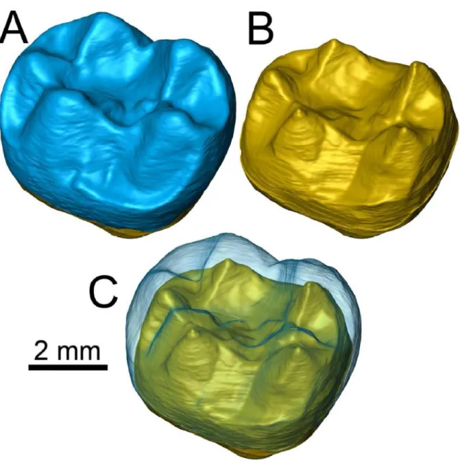
134
standard X-ray µCt do not enable to adequately discriminate between enamel and dentine.
135
This precludes ascertaining the endostructural dental morphology—in particular, the
136
enamel-dentin junction (EDJ) morphology—or to compute 3D relative enamel thickness in
137
this taxon, with potential implications for further clarifying its taxonomic/phylogenetic
138
affinities as well as its paleodietary adaptations, respectively.
139 140
2.2 | Neutron-based computed microtomography
141
Radiographic contrast is generated using the attenuation and scattering of a beam that
142
passes through an object. The main difference between X-ray and neutron-based
143
tomography lies on the particles that interact with matter (photons produced by the kinetic
144
variation of the electrons in the former, and neutrons in the latter). X-rays and synchrotron
145
radiation interact with the electron cloud that surrounds the atoms, and thus are more
146
scattered or attenuated by elements possessing a large number of electrons. Being
147
uncharged, neutrons only interact with the nuclei via very short-range forces (Schwarz et
148
al., 2005). The probability of absorption depends on the number of nucleons, thus showing
149
major differences between neighboring elements or even isotopes of the same element
150
(Schillinger, 2017). The attenuation coefficient of X-rays rises monotonously with the
151
number of protons of the elements, while neutrons show a decreasing trend (Schillinger,
152
2017). This allows high penetration power for heavy mineral elements, which are
153
commonly present in paleontological specimens (Beaudet et al., 2016). On the other hand,
154
neutrons are strongly scattered by hydrogen and some other light elements, such that
7
hydrogen-rich materials (i.e., organic materials, glues and resins) are easily detected
156
(Schwarz et al., 2005; Winkler, 2006; Schillinger, 2017).
157
Most samples become activated if irradiated with neutrons. The standard decay time for
158
the radioactivity ranges between some days and few weeks, after which the specimens
159
can be released (Schillinger, 2017; Schulz et al., 2017). In addition, specific elements
160
(such as europium and cobalt) may achieve hazardous levels of radioactivity if activated
161
by the neutron beam (Sutton, 2008). However, this issue can be easily avoided by running
162
preliminary tests of short-time irradiation and consecutive gamma scan (Schillinger, 2017).
163
Even if both X-ray and neutron-based radiography produce a shadow image of the
164
sample, the beams used for the analysis differ considerably. X-ray tubes generate a cone
165
beam that magnifies the projection of the sample, while a neutron beam is approximately
166
parallel and does not magnify (Schillinger, 2017). The quality of the image thus depends
167
on the collimation ratio and on the distance between the detector and the sample, while
168
the limit for the resolution of a parallel beam is constrained by the detector’s resolution.
169
Using thinned detector screens (5–20 μm), a resolution of 10–20 μm is achieved
170
(Schillinger, 2017; Schulz et al., 2017).
171 172
2.3 | Scanning settings
173
The dimensions and scanning parameters for the studied samples have bee reported in
174
Table 1. They were scanned with n-μCT at the imaging facility ANTARES, which is located
175
at the cold neutron beam port of the reactor of the Forschungs-Neutronenquelle Heinz
176
Maier-Leibnitz (FRM II; Garching bei München, Germany). It allows different detector
177
positions and two different chambers, according to the requirements of the sample size,
178
beam size, neutron flux and spatial resolution (for the specifics of the facility, see Schulz et
179
al., 2017). Specimens were placed in chamber two, on a XY-Phi-table with an additional
180
high precision 5-axes HUBER table. Measurements were carried out using the parallel
8
neutron beam originated from the cold source of the FRM II reactor with an energy range
182
of 3–25 meV and a collimation ratio of 500. Three different scans were obtained for
183
IPS58443.1: a general one (877 projections for 2,400 slices) with a final isotropic voxel
184
size of 19.90 μm; and two close-ups, for the temporal (1,105 projections for 2,518 slices)
185
and maxillary (876 projections for 2,486 slices) areas, with a voxel size of 14.22 μm. In
186
turn, due to the reduced size of the specimens (< 1 cm), IPS1724n,o were scanned with a
187
single cumulative acquisition (2,221 slices) that yielded a final isotropic voxel size of 17.98
188
μm. The histograms of the images stacks were computed with Fiji (Schindelin et al., 2012).
189
X-ray μCT were performed for the same specimens. IPS58443.1 was scanned at the
190
American Museum of Natural History (New York, USA) using a Phoenix v|tome|x s180
191
system, using 160 kV voltage, 1.4 mA current, 0.2 mm Cu filter, and magnification of 2.10,
192
obtaining 1,600 slices (virtual cross-sectional images) of 0.2 mm in thickness and a pixel
193
size of 95.23 μm. In turn, IPS1724n,o were scanned in a single acquisition at the TomoLab
194
of the Multidisciplinary Laboratory of the International Centre for Theoretical Physics
195
(Trieste; ICTP), with 13 kV voltage, 72 μA current, and a 1 mm Al filter, obtaining 1800
196
slices and a voxel size of 7.56 μm.
197 198
3 | RESULTS
199
The resulting images of X-rays and neutron radiography are very different. Standard
200
radiography provides sharper air-specimen boundaries and uniform dark background,
201
whereas neutron radiography shows more diffuse background noise, especially for
202
IPS58443.1, probably due to the presence of the acrylic resin (Paraloid® B72, 5% diluted 203
in acetone) and nitrocellulosic glue (Imedio® Banda Azul) used in the preparation process. 204
However, the contrast provided by X-ray μCT for IPS58443.1 is insufficient to perform an
205
accurate analysis of the internal anatomy.
9
The partial cranium of Pliobates is filled with a mudstone matrix that is firmly stuck to the
207
fossilized tissues and was only partially removed during the preparation process.
Electron-208
dense elements fill some of the fractures along the specimen or are found dispersed in the
209
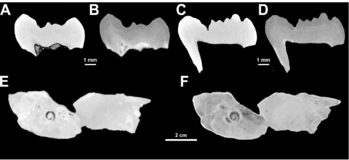
sediment, resulting in bright spots on the images stack that led to the underexposure of the
210
radiographies. The histograms calculated for the whole images stack show a low-shifted
211
distribution of the intensity curve with two extremely steep peaks (Fig. 2E). The lowest one
212
belongs to the air background, while the other includes both sediment and fossilized bone.
213
Due to the spot-like accumulation of the denser minerals, their peak is diluted within the
214
histogram and becomes visible only when cropping the image to the exact region of the
215
bright spots. Even if contrast is very low, segmentation between outer sediment and
216
fossilized tissues is possible (Alba et al., 2015). However, contrast is insufficient to
217
properly discern the inner matrix from internal bone boundaries of cranial cavities (Fig.
218
1E), especially close to the bright spots. In contrast, n-μCT produced more balanced
219
images (Fig. 1F). This is clearly visible in the distribution of the intensities in the
220
histograms (Fig. 2F), where the second peak (corresponding to the
embedding-221
sediment/fossilized tissues compound) is lower and more distributed along the intensity
222
axis. The fossilized tissues appear in lighter gray, as the permineralization process is likely
223
not complete (or affects different areas of the fossil in a differential way) and their mineral
224
composition still differs from the embedding matrix, which appears darker and richer in
225
heavy elements (Fig. 1F). This enables the distinction and manual segmentation of the
226
borders of the inner cavities, such as the bony labyrinth, the inner ear nerves, or the
227
carotid canal, inter alia.
228
The two molars of Barberapithecus (IPS1724n,o) appear as a uniform gray-to-white
229
mass in the X-ray scans, such that there is no discernible EDJ (Fig. 1A,C). Probably this is
230
the result of deep mineralization of the dentine during the fossilization process, which
231
caused it to converge in chemical composition with the enamel. The histograms calculated
10
for each tooth, on the whole slices stack (Fig. 2A,C), show that there is a shift of the
233
intensity curve towards higher intensity values for the standard X-ray μCT images stacks.
234
The right end of the curve shows a peak that corresponds to the brighter areas of the
235
dentine, in which elements with a high attenuation coefficient have penetrated. The steep
236
black background peak occupies the lowest intensity range. These two peaks flank a lower
237
one (ranging from ca. 155 to 240 of the grayscale) that corresponds to the enamel-dentine
238
compound, visible as an indistinct gray to white mass (Fig. 1A,C). In contrast, n-μCT
239
images stacks have more balanced histograms, in which the dental tissues (i.e., dentine
240
and enamel) are better differentiated (Fig. 2B,D). Apart from broader lower peak, due to a
241
lighter background, the curves displays a different intensity for the enamel (darker) and the
242
dentine (lighter). Inside the pulp cavity, the presence of lighter and hydrogen-rich minerals
243
in the two molars (Fig. 1B,D) locally produces some noise (clearly visible in the histogram
244
as an anomalous peak at the very end of the color map). However, it does not affect
245
contrast in the EDJ, enabling the manual segmentation of enamel and dentine, and
246
therefore a correct identification of the former (Fig. 3).
247 248
4 | DISCUSSION
249
Our results indicate that n-µCT provides a higher anatomical resolution for two specimens,
250
whose internal anatomy could not be adequately segmented by means of X-ray µCT
251
scans, due to several problems: an extreme similarity between the chemical composition
252
of the matrix and that of the fossilized tissues (in the cranium of Pliobates); a high similarity
253
between the chemical composition of different fossilized tissues (in the molars of
254
Barberapithecus); and the presence of high-density elements (in the cranium of Pliobates).
255
Standard X-ray and synchrotron radiation μCT are preferable because of the greater
256
sharpness of the images, the lack of activation, and the availability of the facilities in the
257
case of the former. However, n-µCT provides a better contrast of different isotopes of the
11
same element and more easily penetrates metals than X-ray µCT. These advantages
259
provide a better definition of the bone/sediment boundary and between different dental
260
tissues, thereby enabling a better segmentation of the images stack in fossils that are
261
problematic with standard µCT. The complementarity of the contrasts obtained by n-µCT
262
and its enhanced penetration power are particularly indicated for thick and heavy-element
263
rich fossil material—two conditions that are commonly found in paleontological specimens.
264
We therefore conclude that neutron radiation represents an accessible and successful
265
alternative to X-rays for the µCT of fossil specimens when the latter fail to obtain the
266 desired outcome. 267 268 ACKNOWLEDGEMENTS 269
We thank Marta S. March and Jordi Galindo for assistance in collection managing, and J.
270
Thostenson and M. Hill for assistance with using the Microscopy and Imaging Facility of
271
the American Museum of Natural History. This work has been supported by the Spanish
272
Ministerio de Economía, Industria y Competitividad and the European Regional
273
Development Fund of the European Union (MINECO/FEDER EU, project
CGL2014-274
54373-P), the Spanish Agencia Estatal de Investigación and the European Regional
275
Development Fund of the European Union (AEI/FEDER EU, project CGL2016-76431-P),
276
and the Generalitat de Catalunya (CERCA Programme). We are also grateful to an
277
annonymous reviewer for constructive comments that helped us to improve a previous
278
version of this paper.
279 280
REFERENCES
281
Abel, R. L., Rettondini Laurini, C., & Richter, M. (2012). A palaeobiologist’s guide to
282
‘virtual’ micro-CT preparation. Palaeontologia Electronica, 15, 15.2.6T.
12
Alba, D. M., & Moyà-Solà, S. (2012). A new pliopithecid genus (Primates: Pliopithecoidea)
284
from Castell de Barberà (Vallès-Penedès Basin, Catalonia, Spain). American Journal
285
of Physical Anthropology, 147, 88–112.
286
Alba, D. M., Almécija, S., DeMiguel, D., Fortuny, J., Pérez de los Ríos, M., Pina, M.,
287
Robles, J. M., & Moyà-Solà S. (2015). Miocene small-bodied ape from Eurasia sheds
288
light on hominoid evolution. Science, 350, aab2625.
289
Alba, D. M., Casanovas-Vilar, I., Garcés, M., & Robles, J. M. (2017). Ten years in the
290
dump: An updated review of the Miocene primate-bearing localities from Abocador de
291
Can Mata (NE Iberian Peninsula). Journal of Human Evolution, 102, 12–20.
292
Beaudet, A., Braga, J., de Beer, F., Schillinger, B., Steininger, C., Vodopivec, V., & Zanolli,
293
C. (2016). Neutron microtomography-based virtual extraction and analysis of a
294
cercopithecoid partial cranium (STS 1039) embedded in a breccia fragment from
295
Sterkfontein Member 4 (South Africa). American Journal of Physical Anthropology,
296
159, 737–745.
297
Benazzi, S., Kullmer, O., Schulz, D., Gruppioni, G., & Weber, G. W. (2013). Technical
298
note: Individual tooth macrowear pattern guides the reconstruction of Sts 52
299
(Australopithecus africanus) dental arches. American Journal of Physical
300
Anthropology, 150, 324–329.
301
Benazzi, S., Gruppioni, G., Strait, D. S., & Hublin, J. J. (2014). Technical note: Virtual
302
reconstruction of KNM-ER 1813 Homo habilis cranium. American Journal of Physical
303
Anthropology, 153, 154–160.
304
Casanovas-Vilar, I., Garcés, M., Van Dam, J., García-Paredes, I., Robles, J. M., & Alba, D.
305
M. (2016). An updated biostratigraphy for the late Aragonian and the Vallesian of the
306
Vallès-Penedès Basin (Catalonia). Geologica Acta, 14, 195–217.
13
Cunningham, J. A., Rahman, I. A., Lautenschlager, S., Rayfield, E. J., & Donoghue, P. C.
308
J. (2014). A virtual world of paleontology. Trends in Ecology and Evolution, 29, 347–
309
357.
310
Faulwetter, S., Vasileiadou, A., Kouratoras, M., Dailianis, T., & Arvanitidis, C. (2013).
311
Micro-computed tomography: Introducing new dimensions to taxonomy. Zookeys,
312
263, 1–45.
313
Gren, J. A., Sjövall, P., Eriksson, M. E., Sylvestersen, R. L., Marone, F., Sigfridsson
314
Clauss, K. G. V., Taylor, G. J., Carlson, S., Uvdal, P., & Lindgren, J. (2016).
315
Molecular and microstructural inventory of an isolated fossil bird feather from the
316
Eocene Fur Formation of Denmark. Palaeontology, 60, 73–90.
317
Kardjilov, N., Baechler, S., Bastürk, M., Dierick, M., Jolie, J., Lehmann, E., Materna, T.,
318
Schillinger, B., & Vontobel, P. (2003). New features in cold neutron radiography and
319
tomography Part II: applied energy-selective neutron radiography and tomography.
320
Nuclear Instruments and Methods in Physics Research Section A: Accelerators,
321
Spectrometers, Detectors and Associated Equipment, 501, 536–546.
322
Laaß, M., & Kaestner, A. (2017). Evidence for convergent evolution of a neocortex-like
323
structure in a late Permian therapsid. Journal of Moprhology, 278, 1033–1057.
324
Lautenschlager, S. (2016). Reconstructing the past: methods and techniques for the digital
325
restoration of fossils. Royal Society Open Science, 3, 160342.
326
Macchiarelli, R., Bondioli, L., Falk, D., Faupl, P., Illerhaus, B., Kullmer, O., Richter, W.,
327
Hasen, S., Sandrock, O., Schäfer, K., Urbanek, C., Viola, B. T., Weber, G. W., &
328
Seidler, H. (2004). Early Pliocene hominid tooth from Galili, Somali Region, Ethiopia.
329
Collegium Antropologicum, 28 Suppl. 2, 65–76.
330
Macchiarelli, R., Bayle, P., Bondioli, L., Mazurier, A., & Zanolli, C. (2013). From outer to
331
inner structural morphology in dental anthropology: integration of the third dimension
332
in the visualization and quantitative analysis of fossil remains. In G. R. Scott & J. D.
14
Irish (Eds.), Anthropological perspectives on tooth morphology: Genetics, evolution,
334
variation (pp. 250–277). Cambridge: Cambridge University Press.
335
Moyà-Solà, S., Alba, D. M., & Almécija, S. (2013). A proximal radius of Barberapithecus
336
huerzeleri (Primates, Pliopithecidae) from the Miocene site of Castell de Barberà (NE
337
Iberian Peninsula). Journal of Vertebrate Paleontology, 33 Suppl. 2, 182.
338
Olejniczak, A. J., & Grine, F. E. (2006). Assessment of the accuracy of dental enamel
339
thickness measurements using microfocal X-ray computed tomography. Anatomical
340
Record A, 288, 263–275.
341
Olejniczak, A. J., Tafforeau, P., Temming, H., Smith, T. M., & Hublin, J.-J. (2007).
342
Technical note: compatibility of microtomographic imaging systems for dental
343
measurements. American Journal of Physical Anthropology, 134, 130–134.
344
Schindelin, J., Arganda-Carreras, I., Frise, E., Kaynig, V., Longair, M., Pietzsch, T.,
345
Preibisch, S., Rueden, C., Saalfeld, S., Schmid, B., Tinevez, J., White, D. J.,
346
Hartenstein, V., Eliceiri, K., Tomancak, P., & Cardona, A. (2012). Fiji: an open-source
347
platform for biological-image analysis. Nature Methods, 9, 676–682.
348
Schillinger, B. (2017). Why use neutrons? Restaurierung und Archäologie, 8, 1–7.
349
Schulz, M., Schillinger, B., Calzada, E., Bausenwein, D., Schmakat, P., Reimann, T., &
350
Böni, P. (2017). Die neue Anlage ANTARES für Neutronenbildgebung am FRM II.
351
Restaurierung und Archäologie, 8, 9–14.
352
Schwarz, D., Vontobel, P., Lehmann, E. H., Meyer, C. A., & Bongartz, G. (2005). Neutron
353
tomography of internal structures of vertebrate remains: A comparison with X-ray
354
computed tomography. Palaeontologia Electronica, 8, 8.2.30A.
355
Smith, T. M., Olejniczak, A. J., Kupczik, K., Lazzari, V., de Vos, J., Kullmer, O., Schrenk,
356
F., Hublin, J. J., Jacob, T., & Tafforeau, P. (2009). Taxonomic assessment of the
357
Trinil molars using nondestructive 3D structural and development analysis.
358
PaleoAnthropology, 2009, 117–129.
15
Spoor, C. F., Zonneveld, F. W., & Macho, G. A. (1993). Linear measurements of cortical
360
bone and dental enamel by computed tomography: applications and problems.
361
American Journal of Physical Anthropology, 91, 469–484.
362
Sutton, M. D. (2008). Tomographic techniques for the study of exceptionally preserved
363
fossils. Proceedings of the Royal Society B, 275, 1587–1593.
364
Tuniz, C., Bernardini, F., Cicuttin, A., Crespo, M. L., Dreossi, D., Gianoncelli, A., Macini, L.,
365
Mendoza Cuevas, A., Sodini, N., Tromba, G., Zanini, F., & Zanolli, C. (2013). The
366
ICTP-Elettra X-ray laboratory for cultural heritage and archaeology. Nuclear
367
Instruments and Methods in Physics Research Section A: Accelerators,
368
Spectrometers, Detectors and Associated Equipment, 711, 106–110.
369
Winkler, B. (2006). Applications of neutron radiography and neutron tomography. Reviews
370
in Mineralogy and Geochemistry, 63, 459–471.
371
Zanolli, C., Grine, F.E., Kullmer, O., Schrenk, F., & Macchiarelli, R. (2015). The Early
372
Pleistocene deciduous hominid molar FS-72 from the Sangiran Dome of Java,
373
Indonesia: A taxonomic reappraisal based on its comparative endostructural
374
characterization. American Journal of Physical Anthropology, 157, 666–674.
375
Zanolli, C., Mancini, L., Kullmer, O., Macchiarelli, R., Rook, L., Schillinger, B., Schrenk, F.,
376
Tuniz, C., & Vodopivec, V. (2013). Problems and limitations of X-ray
377
microtomography for the endostructural characterization of fossil tooth tissues. In B.
378
Schillinger (Ed.), NINMACH 2013. 1st International Conference on Neutron Imaging
379
and Neutron Methods in Archaeology and Cultural Heritage Research. Abstract
380
booklet (pp. 56–57). Garching bei München: Technische Universität München.
381
Zanolli, C., Schillinger, B., Beaudet, A., Kullmer, O., Macchiarelli, O., Mancini, L., Schrenk,
382
F., Tuniz, C., & Vodopivec V. (2017a). Exploring hominin and non-hominin primate
383
dental fossil remains with neutron microtomography. Physics Procedia, 88, 109–115.
16
Zanolli, C., Alba, D. M., Dean, M. C., Fortuny, J., Macchiarelli, R., & Rook, L. (2017b).
385
Oreopithecus bambolii is still an “enigmatic anthropoid”. American Journal of Physical
386
Anthropology, 162 S64, 420.
17 388
18 389
FIGURE 1 Selected images of X-ray and neutron microcomputed tomography (μCT) of two
390
Barberapithecus molars (IPS1724n,o) and the Pliobates cranium (IPS58443.1). (A-B)
391
Cross-section of IPS1724n through the metaconid and hypoconulid, based on X-ray (A)
392
and neutron (B) µCT. (C-D) Cross-section of IPS1724o through the protoconid and
393
hypoconulid, based on X-ray (C) and neutron (D) µCT. (E-F) Cross-section of IPS58443.1
394
through the petrosal bone, based on X-ray (E) µCT and neutron (F) µCT. Note: the
395
sections compared differ slightly due to different slice thickness.
396 397
19 398
FIGURE 2 Whole images stack histograms from different sources: (A) X-ray
399
microcomputed tomography (µCT? of IPS1724n; (B) neutron µCT of IPS1724n; (C) X-ray
400
µCT of IPS1724o; (D) neutron µCT of IPS1724o; (E) X-ray µCT of IPS58443.1; (F) neutron
401
µCT of IPS58443.1.
402 403
20 404
FIGURE 3 Virtually reconstructed right M2 of Barberapithecus huerzeleri (IPS1724n) 405
based on neutron microcomputed tomography (µCT) scans, in oblique (semiocclusal and
406
mesiobuccal) view: (A) external morphology (enamel surface); (B) inner morphology
407
(enamel-dentine junction, EDJ); (C) enamel surface superimposed in semitransparency to
408
the EDJ.
409 410