HAL Id: hal-02135114
https://hal.archives-ouvertes.fr/hal-02135114
Submitted on 21 May 2019
HAL is a multi-disciplinary open access
archive for the deposit and dissemination of
sci-entific research documents, whether they are
pub-lished or not. The documents may come from
teaching and research institutions in France or
abroad, or from public or private research centers.
L’archive ouverte pluridisciplinaire HAL, est
destinée au dépôt et à la diffusion de documents
scientifiques de niveau recherche, publiés ou non,
émanant des établissements d’enseignement et de
recherche français ou étrangers, des laboratoires
publics ou privés.
Periprosthetic shoulder infection: an overview
Nicolas Bonnevialle, Florence Dauzères, Julien Toulemonde, Fanny Elia,
Jean-Michel Laffosse, Pierre Mansat
To cite this version:
Nicolas Bonnevialle, Florence Dauzères, Julien Toulemonde, Fanny Elia, Jean-Michel Laffosse, et al..
Periprosthetic shoulder infection: an overview. EFORT Open Reviews, British Editorial Society of
Bone & Joint Surgery, 2017, 2 (4), pp.104-109. �10.1302/2058-5241.2.160023�. �hal-02135114�
O
pen
A
rchive
T
oulouse
A
rchive
O
uverte
(OATAO)
OATAO is an open access repository that collects the work of some Toulouse
researchers and makes it freely available over the web where possible.
This is
an author'sversion published in:
https://oatao.univ-toulouse.fr/23071Official URL :
https://doi.org/10.1302/2058-5241.2.160023
To cite this version :
Any correspondence concerning this service should be sent to the repository administrator:
tech-oatao@listes-diff.inp-toulouse.fr
Bonnevialle, Nicolas and Dauzères, Florence and Toulemonde, Julien and Elia, Fanny and
Laffosse, Jean-Michel and Mansat, Pierre Periprosthetic shoulder infection: an overview.
(2017) EFORT Open Reviews, 2 (4). 104-109. ISSN 2396-7544
OATAO
Periprosthetic shoulder infection: an overview
Nicolas Bonnevialle 1 Florence Dauzères2 Julien Toulemonde2 Fanny Elia2 Jean-Michel Laffosse3 Pierre Mansat1•
Periprosthetic shoulder infection (PSI) is rare butpoten-tially devastating. The rate of PSI is increased in cases of revision procedures, reverse shoulder implants and
co-morbidities. One specific type of PSI is the occurrence of
low-grade infections caused by non-suppurative bacteria
such as Propionibacterium acnes or Staphy/ococcus
epide-mermidis.
•
Success of treatment depends on micro-organismiden-tification, appropriate surgical procedures and antibiotic
administration efficiency. Post-operative early PSI can
be treated with simple debridement, while chronic PSI
requires a one- or two-stage revision procedure.
Indica-tion for one-time exchange is based on pre-operative
identification of a causative agent. Resection arthroplasty remains an option for low-demand patients or recalcitrant infection.
Keywords: infection; arthroplasty; shoulder; Propionibacte-rium acnes; revision
In
t
r
o
du
ct
i
o
n
While more than 66000 prosthetic shoulder procedures were performed in 2011 in the United States, the rate of post-operative infection seems to remain stable with 0.98% of cases.1·3 However, when infection occurs, this
complication is always devastating with significant clinical and socioeconomic consequences.2 The rate is higher after revision surgery than after a primary procedure and reaches close to 5% in cases of reverse shoulder arthro-plasty (RSA).4•5 Patients undergoing primary RSA are
found to have a six times greater risk of infection com-pared with patients having primary unconstrained total shoulder arthroplasty.6 Arthroplasties for trauma are more
at risk of infection than those from other causes.6
Comor-bidities such as coagulopathy, renal failure, diabetes, lupus erythematosus, rheumatoid arthritis, intra-articular steroid injections and corticosteroid therapy increase the risk of periprosthetic shoulder infection (PSl).7 PSI is the major cause for revis ion within the first two post-operative years after an arthroplasty.8
The aim of this review is to investigate PSI from diagno-sis to prevention and to report the main results of different therapeutic options.
M
ic
r
o
bi
o
l
ogy
The micro-organisms most commonly isolated in cases of PSI are Staphylococcus (S.) aureus, S. epidermidis and Propi-onibacterium (P.) acnes.9 P. acnes is a gram-positive
anaer-obic bacillus, much more concentrated in the axilla than in the knee or hip.10 Different studies have identified a presence of P. acnes in the operative field during primary surgery, despite rigorous skin preparation and timely administered prophylactic antibiotics.11 -13 lt colonises the
pilosebaceous follicles and there is a likelihood of finding
it in the dermal layer, in male patients and after corticoid
injection.12-14 Hudek et al14 identified a positive culture twofold greater for the anterolateral approach than for the deltopectoral approach. Surprisingly, it is even thought to be involved in the pathogenesis of glenohumeral osteoar-thritis.11 lt is often responsible for low-grade infection, with mild clinical symptoms, so that many classic clinical patterns do not strictly apply. The mean duration for cul-ture incubation is between seven and > 21 days.
Prevention
Antibiotic prophylaxis is not specific to shoulder arthro-plasty compared with other arthroplasties. lntravenous cephalosporine (2 g) administration is mandatory, given 30 minutes before the skin incision in many countries. However, some authors recommend a single 160 mg of
gentamicin by intra-articular injection at the end of the procedure to reduce the risk of PSI.15
Saltzman et al16 have shown that pre-operative
prepa-ration of the surgical site with ‘ChloraPrep’ 2% (chlorhex-idine gluconate and 70% isopropyl alcohol; ChloraPrep, Leawood, Kansas) was more effective than ‘DuraPrep’ (iodine povacrylex and 74% isopropyl alcohol; 3M, Min-neapolis, Minnesota) and povidone-iodine (Purdue Pharma, Stamford, Connecticut) at eliminating overall bacteria, and that ChloraPrep and Duraprep were more effective than povidone-iodine regarding coagulase- negative Staphylococcus.
Hair removal is commonly performed before orthopae-dic procedures and the use of razors is classically discour-aged because micro-abrasions are created by shaving. However, removal of axillary hairs for shoulder surgery did not prove to have any effect on the cell-count of P. acnes before surgical preparation.17
Diagnosis
Classically, post-operative infections can be classified into acute infection (one to three months), subacute infection (four to 12 months) and late infection (> 12 months), depending on the time of diagnosis after the surgery.18
However, some authors advocate the distinguishing of early and late infection (cutoff at six weeks), so that there is a real chance to save the index prosthesis by avoiding any delay.19
One specific type of PSI is low-grade infections by non-suppurative bacteria such as P. acnes or S. epidemermidis.20,21
Diagnosis remains a challenge because P. acnes does not usually cause swelling, erythema, fever, purulent dis-charge or increasing level of biological parameters such as C-reactive protein (CRP), white blood cell count (WBC) and interleukin (IL)-6. Aspiration of synovial fluid is man-datory if a distinct fluid collection is identified (with positive serum levels giving positive findings of WBC > 3000/mm3
and > 80% for polymorphonuclear neutrophils). Also, deep biopsies that contact the prosthesis are needed.22-24
Performing five to six independent samples in order to optimise the sensitivity and specificity for the diagnosis of a PSI has been advocated. The cutoff for a definite diag-nostic of infection should be three or more samples
positive to the same micro-organism.24 The concern is that
the growth duration from intra- operative samples is long for P. acnes, and every laboratory should be aware that they should not discard the culture samples early. How-ever, early positive cultures seem to be more predictive of a true infection than a late growth culture, which can be a false-positive result.20
On the other hand, in the case of a virulent micro-organism, patients with PSI can also present classical symptoms including a painful shoulder with local and/or systemic physical signs. The blood tests reveal the same non-specific inflammatory markers that can be identified in a knee or hip prosthetic infection. Obviously, erythro-cyte sedimentation, as well as CRP, have a poor sensitivity for the diagnosis of an infected arthroplasty in the post-operative setting, even if the CRP is normalised two weeks after an uneventful procedure.25
As proposed by a workgroup from the Musculoskeletal Infection Society focused on lower limb arthroplasty infec-tions, the diagnosis should be based on a combination of biological and clinical criteria (Table 1).26
Treatment
Superficial wound infections are usually diagnosed in post-operative care and require local measures and anti-biotic therapy. However, it is always necessary to suspect a deep infection and the patient should be treated as such until proven otherwise. In this potentially serious situa-tion, success depends on early identification of micro-organisms, appropriate surgical procedures and efficient antibiotic administration. Different therapeutic options are available: debridement, simple resection arthroplasty, removal of the prosthesis and replacement with a cement spacer (spacer), single-stage revision, two-stage revision, arthrodesis, chronic antibiotic administration and even amputation. The outcomes of some of these options are described below.
Debridement
Debridement, irrigation and multiple deep samples may be proposed in cases of acute infection in order to save the prosthesis. Coste et al27 treated eight cases of acute infection
with debridement and succeeded when it was performed
Table 1. Criteria of Musculoskeletal Infection Society (MSIS) for retaining the prosthesis in periprosthetic infection26
There is a sinus tract communicating with the prosthesis; or
A pathogen is isolated by culture from at least two separate tissue or fluid samples obtained from the affected prosthetic joint; or
Four of the following criteria exist:
1. Elevated serum erythrocyte sedimentation rate and serum C-reactive protein concentration, 2. Elevated synovial leukocyte count,
3. Elevated synovial neutrophil percentage, 4. Presence of pus in the affected joint,
5. Isolation of a micro-organism in one culture of periprosthetic tissue or fluid, or
6. Greater than five neutrophils per high-power field in five high-power fields observed from histological analysis of peri-prosthetic tissue at ×400 magnification.
within eight days after the diagnosis. They concluded that the earlier the debridement is done, the more effective it is in eradicating the infection. This procedure can be repeated, based on the patient’s response.28 Moreover, mobile parts
of the prosthesis may be exchanged during the procedure especially in case of RSA (glenosphere, polyethylene liner) providing better access for debridement. Then, an appropri-ate antibiotics regime is required for a minimum of four weeks.21,28,29 However, the rate of success reported in the
literature is only in the range of 50% to 95%.22,27-29
Cement spacer
An antibiotic-loaded cement spacer can be used either per-manently or as the first step of a two-stage revision proce-dure. In this case, it maintains the space and soft-tissue tension for re-implantation and theoretically releases anti-biotics to decrease the growth of microorganisms. Anti-biotic mean concentration peak is reached at day 1 and dramatically decreases during the following seven days.30
Levy et al31 described a ‘functional cement spacer’
model, which is made of a hemi-arthroplasty coated with cement. In their series of 14 patients initially chosen for a two-stage procedure, nine did not undergo a prosthesis re-implantation because of satisfactory clinical outcomes. On the other hand, verhelst et al32 did not prove any
differ-ence between patients with a cement spacer and resection arthroplasty regarding infection control and clinical out-comes. Complications of the cement-spacer such as break-age, glenoid erosion or dislocation have been reported.32
One-stage revision arthroplasty
Based on the experience of knee and hip infection man-agement, a single-stage exchange is proposed as a rea-sonable option when the infecting micro-organism is satisfactiorily identified. The advantages are a reduced hospital stay, costs, period of antibiotic administration and the best clinical outcomes (Table 2).9,22,27,29,33-37
Klatte et al33 reported the outcomes of the largest
single- centre series of 35 patients with a mean follow-up of 4.7 years. The authors excised infected tissues,
thoroughly irrigated the wound using pulsatile lavage with polyhexanide before re-implantation and delivered specific intravenous antibiotherapy for an average of two weeks post-operatively. The success rate was more than 90%. No recurrence was observed in the series of Coste et al,27 Ince et al34 and Cuff et al.35 The presence of a
pro-ductive fistula seems not to be a contra-indication for many authors.33,36 Beekman et al36 performed a one-stage
revision in 11 cases of RSAs, among which eight had a fis-tula, and achieved a success rate of 90%.
Two-stage revision arthroplasty
In a medically stable patient with a high demand, a two-stage revision procedure is generally accepted (Table 3).22,27,29,33,38-40 It is highly recommended when the
micro-organism responsible for the infection is unknown. The first step consists of infection eradication after prosthetic removal: an antibiotic-loaded cement spacer is often implanted and general antibiotics are administrated, sec-ondarily adapted to the micro-organism(s) identified. Anti-biotics are generally continued for six to eight weeks. Markers such as CRP or IL-6 have been shown to be valua-ble to predict the eradication of infection and, so, the time of re-implantation.41,42 However, IL-6 seems to be
normal-ised faster than CRP and allows earlier revision for better out-comes.41 An iterative irrigation and debridement could be
proposed in case of persistent infection. For re- implantation, RSA has been gaining ground in recent years as the implant of choice. First, it allows a larger debridement at the first stage with less concern for soft-tissue preservation. Sec-ondly, it offers the possibility of addressing the glenoid bone defect with or without bone graft. Shirwaiker et al43
reported that there is still uncertainty whether two-stage revision is superior to one-stage (Fig. 1).
Resection arthroplasty
Shoulder resection should remain a salvage procedure for frail or low-demand patients, and recalcitrant infec-tion. It offers the option of a single definitive procedure
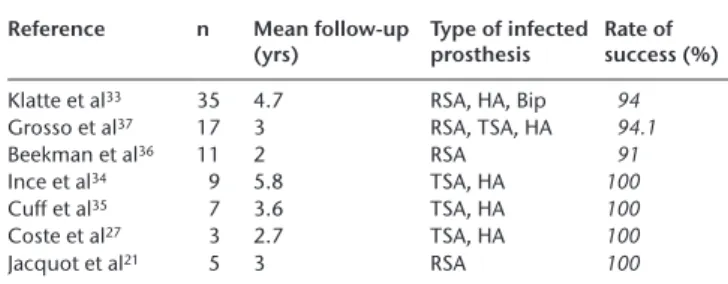
Table 2. One-stage revision arthroplasty (RSA, reverse shoulder arthro-plasty; HA, hemiarthroarthro-plasty; TSA, total shoulder arthroarthro-plasty; Bip, bipolar arthroplasty)
Reference n Mean follow-up
(yrs) Type of infected prosthesis Rate of success (%)
Klatte et al33 35 4.7 RSA, HA, Bip 94
Grosso et al37 17 3 RSA, TSA, HA 94.1
Beekman et al36 11 2 RSA 91
Ince et al34 9 5.8 TSA, HA 100
Cuff et al35 7 3.6 TSA, HA 100
Coste et al27 3 2.7 TSA, HA 100
Jacquot et al21 5 3 RSA 100
Table 3. Two-stage revision prosthesis (RSA, reverse shoulder arthro-plasty; HA, hemiarthroarthro-plasty; TSA, total shoulder arthroplasty)
Reference n Mean follow-up
(yrs) Type of infected prosthesis Rate of success (%)
Strickland et al38 19 2.9 HA, TSA 63
Romanò et al29 17 3.8 RSA, HA 100
Sabesan et al40 17 3.8 RSA, TSA, HA 94
Jacquot et al21 14 3 RSA 64
Ortmaier et al39 12 6.1 RSA 75
Coste et al27 10 2.6 TSA, HA 60
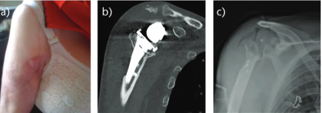
Fig. 2 Radiographs (a and b) of an 86-year-old woman, with a loose implant secondary to chronic periprosthetic shoulder infection.
c) Because of numbers of co-morbidities and huge bone loss on glenoid side, a simple resection arthroplasty was performed.
for infection eradication (Fig. 2). It has been shown that functional results are poor, but pain relief is achieved in more than 50% of cases.9,44 Rispoli et al44 reported a mean
active elevation of 70° at long-term follow-up after ‘ana-tomical’ shoulder arthroplasty removal. verhelst et al32
demonstrated that preservation of the tuberosities is a predictive factor for better results, because it can avoid antero- superior subluxation of the humerus. In cases of RSA, Jacquot et al21 did not improve functional outcomes
after removal of the implant and identified a high rate of post-operative complications. Bone loss and soft-tissue impairment after such constrained prostheses could partly explain these findings. Despite Jacquot21 and Coste’s27
studies, the literature reports a high rate of infection eradi-cation, reaching more than 90% of cases.9,27,29,32,44,45
Conclusions
PSIs are a rare but remain a devastating complication in terms of functional outcomes. Acute infection (less than two months post-operatively) requires prosthetic washout
as soon as possible, and the exchange of the mobile part of the implant. However, the important point in manag-ing PSI is the high rate of low-grade infections that must be suspected in cases with abnormal clinical outcomes including pain and stiffness. It is often considered as sub-acute or chronic infection because of a ‘wait and see’ approach by the practitioner.46 In this situation, S.
epider-midis or P. acnes are frequently involved and a simple
debridement is too late. One-stage revision is possible if the micro-organism is identified pre-operatively, other-wise a two-stage procedure is recommended. Resection arthroplasty remains the option for low-demand patients. A multidisciplinary collaboration is nowadays recom-mended to optimise the antibiotic treatment and the sur-gical procedure.
Fig. 1 a) Radiograph of a 73-year-old man with a chronic periprosthetic shoulder infection of a reverse shoulder arthroplasty (RSA).
b) A two-stage revision was decided with a cement spacer implantation for eight weeks. c) Propionibacterium acnes was identified on peri-operative samples taken from the back of the glenosphere. d) After four weeks free of antibiotics, a new RSA was implanted with a proximal humeral allograft.
Author InformAtIon
1Orthopaedic Department and Biomechanics Department, IMFT CNRS URM
ICmJE ConflICt of IntErEst stAtEmEnt
NB reports he is a consultant for Wright, Smith & Nephew and Stryker. PM reports that he receives personal fees from Wright, Smith & Nephew and Synthes. All other authors declare no conflict of interest.
fundIng stAtEmEnt
No benefits in any form have been received or will be received from a commercial party related directly or indirectly to the subject of this article.
lICEnCE
© 2017 The author(s)
This article is distributed under the terms of the Creative Commons Attribution-Non Commercial 4.0 International (CC BY-NC 4.0) licence (https://creativecommons.org/ licenses/by-nc/4.0/) which permits non-commercial use, reproduction and distribu-tion of the work without further permission provided the original work is attributed.
rEfErEnCEs
1. schairer WW, nwachukwu Bu, lyman s, Craig EV, gulotta lV. National
utilization of reverse total shoulder arthroplasty in the United States. J Shoulder Elbow Surg 2015;24:91-97.
2. Padegimas Em, maltenfort m, ramsey ml, et al. Periprosthetic shoulder
infection in the United States: incidence and economic burden. J Shoulder Elbow Surg 2015;24:741-746.
3. day Js, lau E, ong Kl, et al. Prevalence and projections of total shoulder and elbow
arthroplasty in the United States to 2015. J Shoulder Elbow Surg 2010;19:1115-1120.
4. Zumstein mA, Pinedo m, old J, Boileau P. Problems, complications,
reoperations, and revisions in reverse total shoulder arthroplasty: a systematic review.
J Shoulder Elbow Surg 2011;20:146-157.
5. morris BJ, o’Connor dP, torres d, et al. Risk factors for periprosthetic infection
after reverse shoulder arthroplasty. J Shoulder Elbow Surg 2015;24:161-166.
6. richards J, Inacio mC, Beckett m, et al. Patient and procedure-specific risk factors
for deep infection after primary shoulder arthroplasty. Clin Orthop Relat Res 2014;472:2809-2815.
7. smucny m, menendez mE, ring d, feeley Bt, Zhang Al. Inpatient surgical
site infection after shoulder arthroplasty. J Shoulder Elbow Surg 2015;24:747-753.
8. Portillo mE, salvadó m, Alier A, et al. Prosthesis failure within 2 years of
implantation is highly predictive of infection. Clin Orthop Relat Res 2013;471:3672-3678.
9. saltzman md, marecek gs, Edwards sl, Kalainov dm. Infection after shoulder
surgery. J Am Acad Orthop Surg 2011;19:208-218.
10. Patel A, Calfee rP, Plante m, fischer sA, green A. Propionibacterium acnes
colonization of the human shoulder. J Shoulder Elbow Surg 2009;18:897-902.
11. levy o, Iyer s, Atoun E, et al. Propionibacterium acnes: an underestimated
etiology in the pathogenesis of osteoarthritis? J Shoulder Elbow Surg 2013;22:505-511.
12. lee mJ, Pottinger Ps, Butler-Wu s, et al. Propionibacterium persists in the skin
despite standard surgical preparation. J Bone Joint Surg [Am] 2014;96:1447-1450.
13. maccioni CB, Woodbridge AB, Balestro JC, et al. Low rate of Propionibacterium
acnes in arthritic shoulders undergoing primary total shoulder replacement surgery using a strict specimen collection technique. J Shoulder Elbow Surg 2015;24:1206-1211.
14. hudek r, sommer f, Kerwat m, et al. Propionibacterium acnes in shoulder
surgery: true infection, contamination, or commensal of the deep tissue? J Shoulder Elbow
Surg 2014;23:1763-1771.
15. lovallo J, helming J, Jafari sm, et al. Intraoperative intra-articular injection of
gentamicin: will it decrease the risk of infection in total shoulder arthroplasty? J Shoulder
Elbow Surg 2014;23:1272-1276.
16. saltzman md, nuber gW, gryzlo sm, marecek gs, Koh Jl. Efficacy of surgical
preparation solutions in shoulder surgery. J Bone Joint Surg [Am] 2009;91-A:1949-1953.
17. marecek gs, Weatherford Bm, fuller EB, saltzman md. The effect of axillary
hair on surgical antisepsis around the shoulder. J Shoulder Elbow Surg 2015;24:804-808.
18. franceschini V, Chillemi C. Periprosthetic shoulder infection. Open Orthop J
2013;7:243-249.
19. härle A. Infection management in total hip replacement. Arch Orthop Trauma Surg
1989;108:63-71.
20. frangiamore sJ, saleh A, grosso mJ, et al. Early versus late culture growth
of Propionibacterium acnes in revision shoulder arthroplasty. J Bone Joint Surg [Am] 2015;97:1149-1158.
21. Jacquot A, sirveaux f, roche o, et al. Surgical management of the infected
reversed shoulder arthroplasty: a French multicenter study of reoperation in 32 patients.
J Shoulder Elbow Surg 2015;24:1713-1722.
22. sperling JW, Kozak tK, hanssen Ad, Cofield rh. Infection after shoulder
arthroplasty. Clin Orthop Relat Res 2001;382:206-216.
23. dilisio mf, miller lr, Warner JJ, higgins ld. Arthroscopic tissue culture for the
evaluation of periprosthetic shoulder infection. J Bone Joint Surg [Am] 2014;96:1952-1958.
24. Zhang Al, feeley Bt, schwartz Bs, Chung tt, ma CB. Management of deep
postoperative shoulder infections: is there a role for open biopsy during staged treatment?
J Shoulder Elbow Surg 2015;24:e15-e20.
25. niskanen ro, Korkala o, Pammo h. Serum C-reactive protein levels after total
hip and knee arthroplasty. J Bone Joint Surg [Br] 1996;78-B:431-433.
26. Parvizi J, Zmistowski B, Berbari Ef, et al. New definition for periprosthetic
joint infection: from the Workgroup of the Musculoskeletal Infection Society. Clin Orthop
Relat Res 2011;469:2992-2994.
27. Coste Js, reig s, trojani C, et al. The management of infection in arthroplasty of
the shoulder. J Bone Joint Surg [Br] 2004;86-B:65-69.
28. duncan sf, sperling JW. Treatment of primary isolated shoulder sepsis in the
adult patient. Clin Orthop Relat Res 2008;466:1392-1396.
29. romanò Cl, Borens o, monti l, meani E, stuyck J. What treatment for
periprosthetic shoulder infection? Results from a multicentre retrospective series. Int Orthop 2012;36:1011-1017.
30. Anagnostakos K, Wilmes P, schmitt E, Kelm J. Elution of gentamicin and
vancomycin from polymethylmethacrylate beads and hip spacers in vivo. Acta Orthop 2009;80:193-197.
31. levy JC, triplet J, Everding n. Use of a functional antibiotic spacer in treating
infected shoulder arthroplasty. Orthopedics 2015;38:e512-e519. 2Orthopaedic Department, Riquet Hospital, University Centre, Place Baylac,
31059 Toulouse, France.
3Orthopaedic Department, CRIOAC and Biomechanics Department, IMFT CNRS,
Riquet Hospital, University Centre, Place Baylac, 31059 Toulouse, France. Correspondence should be sent to: Dr Nicolas Bonnevialle, Orthopaedic Department, Riquet Hospital, University Centre, Place Baylac, 31059 Toulouse, France.
32. Verhelst l, stuyck J, Bellemans J, debeer P. Resection arthroplasty of the
shoulder as a salvage procedure for deep shoulder infection: does the use of a cement spacer improve outcome? J Shoulder Elbow Surg 2011;20:1224-1233.
33. Klatte to, Junghans K, Al-Khateeb h, et al. Single-stage revision for
peri-prosthetic shoulder infection: outcomes and results. Bone Joint J 2013;95-B:391-395.
34. Ince A, seemann K, frommelt l, Katzer A, loehr Jf. One-stage exchange
shoulder arthroplasty for peri-prosthetic infection. J Bone Joint Surg [Br] 2005;87-B: 814-818.
35. Cuff dJ, Virani nA, levy J, et al. The treatment of deep shoulder infection and
glenohumeral instability with debridement, reverse shoulder arthroplasty and postoperative antibiotics. J Bone Joint Surg [Br] 2008;90-B:336-342.
36. Beekman Pd, Katusic d, Berghs Bm, Karelse A, de Wilde l. One-stage
revision for patients with a chronically infected reverse total shoulder replacement. J Bone
Joint Surg [Br] 2010;92-B:817-822.
37. grosso mJ, sabesan VJ, ho JC, ricchetti Et, Iannotti JP. Reinfection
rates after 1-stage revision shoulder arthroplasty for patients with unexpected positive intraoperative cultures. J Shoulder Elbow Surg 2012;21:754-758.
38. strickland JP, sperling JW, Cofield rh. The results of two-stage
re-implantation for infected shoulder replacement. J Bone Joint Surg [Br] 2008;90-B: 460-465.
39. ortmaier r, resch h, hitzl W, et al. Treatment strategies for infection after
reverse shoulder arthroplasty. Eur J Orthop Surg Traumatol 2014;24:723-731.
40. sabesan VJ, ho JC, Kovacevic d, Iannotti JP. Two-stage reimplantation for
treating prosthetic shoulder infections. Clin Orthop Relat Res 2011;469:2538-2543.
41. Coffey mJ, Ely EE, Crosby lA. Treatment of glenohumeral sepsis with a commercially
produced antibiotic-impregnated cement spacer. J Shoulder Elbow Surg 2010;19:868-873.
42. Villacis d, merriman JA, Yalamanchili r, et al. Serum interleukin-6 as a
marker of periprosthetic shoulder infection. J Bone Joint Surg [Am] 2014;96:41-45.
43. shirwaiker rA, springer Bd, spangehl mJ, et al. A clinical perspective on
musculoskeletal infection treatment strategies and challenges. J Am Acad Orthop Surg 2015;23:S44-S54.
44. rispoli dm, sperling JW, Athwal gs, schleck Cd, Cofield rh. Pain relief and
functional results after resection arthroplasty of the shoulder. J Bone Joint Surg [Br] 2007;89-B:1184-1187.
45. Weber P, utzschneider s, sadoghi P, et al. Management of the infected
shoulder prosthesis: a retrospective analysis and review of the literature. Int Orthop 2011;35:365-373.
46. Atkins Bl, Athanasou n, deeks JJ, et al. Prospective evaluation of criteria for
microbiological diagnosis of prosthetic-joint infection at revision arthroplasty. The OSIRIS Collaborative Study Group J Clin Microbiol 1998;36:2932-2939.